____________________________________________________________________________________________________
*Corresponding author: Email: [email protected], [email protected]; www.sciencedomain.org
An Obturation Technique for Closure of Body Wall
Defects
David Y. Johnson
1, Mark A. Franke
1, Nancy J. Phillips
2and Frank E. Johnson
1,3*1Departments of Surgery, Saint Louis University Medical Center, 3635 Vista Avenue, P.O.Box 15250,
Saint Louis, Missouri 63110-0250, USA. 2
Departments of Pathology, Saint Louis University Medical Center, 3635 Vista Avenue, P.O.Box 15250, Saint Louis, Missouri 63110-0250, USA. 3Department of Surgery, Saint Louis Veterans Affairs Medical Center, 915 North Grand Boulevard,
Saint Louis, Missouri 63106, USA.
Authors’ contributions
Authors DJ and MF contributed literature reviews, author NP contributed pathologic examination, author FJ contributed clinical details of patient’s course and performed the surgery.
Article Information
DOI: 10.9734/BJMMR/2015/16100 Editor(s): (1) Gaetano Santulli, College of Physicians and Surgeons Columbia University Medical Center, New York, USA. Reviewers: (1)Anonymous, Turkey.
(2)Anonymous, USA. (3)Anonymous, Japan. Complete Peer review History:http://www.sciencedomain.org/review-history.php?iid=952&id=12&aid=8865
Received 7th January 2015 Accepted 24th March 2015 Published 17th April 2015
ABSTRACT
Aims: In the vast majority of instances, closure of abdominal wall defects relies on the tensile strength of transposed native tissue and/or prosthetic material. The purpose of this report is to alert clinicians to a different strategy for closure that we have used successfully on several occasions.
Presentation of Case: A 72 year old man had a bulky inguinal nodal metastasis from cutaneous squamous cell carcinoma. He had an extended radical groin dissection including full thickness abdominal wall resection, with primary closure, followed by external beam radiotherapy. After 30 months, he developed an abdominal wall hernia and enterocutaneous fistula at the surgical site. Direct closure and local vascularized flaps were not feasible. Obturation of the defect by omentum was employed, taking advantage of its relative incompressibility rather than its minimal tensile strength. The wound was subsequently covered by a skin graft. The patient survived 10 years with an intact hernia repair and died of unrelated causes.
Discussion: The technique has yielded good results.
Conclusions: This surgical option is valuable for situations in which the abdominal wall defect to be closed is fibrotic, has been radiated, is infected, or is otherwise not suitable for conventional techniques.
Keywords: Hernia; surgery; omentum; obturation; abdominal wall defect.
1. INTRODUCTION
Closure of abdominal wall defects is a common indication for surgery. Standard techniques are adequate for most situations but inadequate for others. These include full-thickness abdominal wall defects, those with marked fibrosis, those in which radiation therapy has been administered, and those which are infected at the time of attempted closure. In some of these situations, the clinician can mitigate these risk factors before operating. In others, urgent surgery is required. Here we describe a technique that relies on obturation of the defect.
2. CASE REPORT
The patient was a 72-year-old male with bulky inguinal metastases from a previously resected squamous cell carcinoma of the intergluteal skin. He had severe atherosclerotic vascular disease. He received an extended radical groin dissection, including full-thickness abdominal wall resection. The defect was closed primarily and followed by external-beam radiation therapy. The patient did well for thirty months before developing an abdominal wall hernia and an enterocutaneous fistula at the surgical site. Radiation-related dense tissue fibrosis, local infection, and ulceration of the skin were present at the fistula site. Through a midline incision, we mobilized the herniated small bowel, resected the fistula, and restored intestinal continuity, leaving a 4 cm. full-thickness defect in the abdominal wall. Direct suture closure was impossible due to the fibrotic nature of the local tissues. A vascularized local flap was not feasible because relevant local arteries had been divided during the groin dissection. Utilizing plastic or biologic prosthetic material was not feasible because the previously skeletonized iliac and femoral arteries comprised one of the edges of the abdominal wall defect. To close the defect, we used pedicled greater omentum [Fig. 1.] based on the right gastroepiploic vessels. The omental pedicle was folded into several layers, placed en masse into the abdominal wall defect, and sutured to its fibrotic edges [Figs. 2 and 3]. The skin over the hernia site was not closed primarily, which allowed us to inspect the omentum easily.
Fig. 1. Vascular supply of the greater omentum. There are numerous anastomoses
among the branches of the main gastroepiploic vessels. This permits the creation of long pedicles, when necessary. Sobotta: Atlas der Anatomie des Menschen.
Elsevier GmbH, Urban & Fischer Verlag München
Fig. 2. Lateral view of folded omentum obturating a full-thickness abdominal wall defect. It is being held in place with synthetic
Fig. 3. View from the peritoneal surface of folded omentum obturating an abdominal wall defect
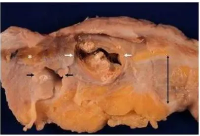
Vacuum-assisted wound management was used and seven weeks later a skin graft was applied. All wounds healed primarily. The patient lived independently until he died of a bowel obstruction 10 years later. Our repair of the abdominal wall defect remained intact. Autopsy showed that the pedicled tissue had retained its size and position. A portion of the relevant anatomy is shown [Fig. 4]. A film of the procedure we describe is available at the American College of Surgeons online video library (A radiated, infected, full-thickness abdominal wall defect; http://cine-med.net/).
Fig. 4. Gross photograph showing the skin graft surface at the top, mature scar created by the free graft (double-headed black arrow),
omentum (white asterisk), and external iliac artery (white arrows) and vein (single-headed
black arrows), approximate magnification 1.5 X. The external iliac artery is markedly
atherosclerotic. Sutures are present superficial to the vein
We carried out computer searches with PubMed, Ovid, and MEDLINE to determine if the obturation closure technique had previously been described. Searching with PubMed, we used the MeSH search terms “omentum,” “surgical procedures,” “operative,” and “abdominal wall,” which yielded 16 results. Searching with Ovid, we used the search terms “omentum,” “surgical procedure,” and “abdominal hernia,” which yielded four results. Searching with MEDLINE, we used the search terms “omentum,” “surgical procedure,” and “abdominal wall,” which yielded 18 results. Our computer search disclosed one article which described spontaneous obturation of hernias by omentum [1] [Fig. 5]. Another described the use of transposed omentum to close a large inguinal defect, but omentum was not used to obturate the defect, as in our example [2]. Several concerned the history of surgical procedures utilizing omentum [3-9]. We considered the Mathes textbook to provide the most comprehensive literature review and reference source [10]. Mathes and Hansen have summarized the classification of flaps and their applications [11].
3. DISCUSSION
around an intestinal anastomosis. By 1896 greater omentum had been used to close a gastric perforation [6] and to improve portal-systemic blood flow in a patient with cirrhosis [5]. There are a number of uses of greater omentum when employed as a pedicled flap based on one of the gastroepiploic vessels. In 1963, Kiricuta described the use of such a flap to repair a chest wall defect created by resection of a locally invasive breast cancer [7]. Goldsmith devised many other applications, including several outside the abdomen [3]. Based on one of the gastroepiploic arteries and use of appropriate lengthening maneuvers, an omental pedicle can be transferred as far as the head or below the knee to deal with a range of extraperitoneal disorders, wounds, and other problems such as lymphedema and radiation injury [4,8]. Mathes and Hansen have detailed the many described applications of omental pedicle flaps but do not mention management of high-risk full-thickness abdominal wall defects [11]. Free tissue transfer using microvascular techniques might be valuable in some situations. Free tissue transfer without revascularization, as described by Senn [9], is predictably successful and might be required for obturation closure of a body wall defect if the omentum is small. Because the volume of nonvascularized transferred omentum typically decreases by 20-40% within weeks, a proportionately larger volume is generally used [13].
Fig. 5. Line drawing illustrating obturation closure of umbilical hernia by greater omentum. From Br Med J 1:76-8; 1906 [1]
During abdominal surgery performed years after a previous operation, we have noticed instances in which omentum has spontaneously obturated iatrogenic abdominal wall defects such as prior laparoscopy port sites, sites where retention
sutures had cut through the abdominal wall muscles and fascia, peritoneal dialysis sites, and the like. Morison noted this as well in 1906: “In both inguinal and femoral regions I have found a cork of omentum blocking the hernial openings so firmly that radical cure was affected by its means” [1]. The authors believe the method described here may be useful in situations such as routine stoma takedown, management of infected parastomal hernias, closure of infected Richter’s hernias, and abdominal wall defects located so close to a major vessel that repairs with prosthetic material would risk erosion into the vessel. There are likely to be other situations in which closure with transposed omentum may be valuable.
4. CONCLUSION
Conventional closure of a hernia relies on suturing sturdy native tissue to prosthetic mesh with sturdy suture material. The technique we describe here relies not on the tensile strength of the omental tissue, but rather on its incompressibility. The ideal abdominal wall defect for this method is one surrounded by rather rigid tissue and not very large. We place the center of the omental pedicled graft (rather than an edge) over the defect to be repaired. The omentum should extend well past the edge of the abdominal wall defect and it should have considerable bulk. Bulky omentum may not need to be folded; thin omentum may need to be folded several times [Figs. 2 and 3]. Methods of holding the tissue in place will vary according to the circumstances. The obturation concept requires attention to the relevant pressure gradient. Unless the abdomen is open, intra-abdominal pressure is always greater than atmospheric pressure so the bulky obturating omental tissue should be placed on the peritoneal surface. The technique should be adaptable to laparoscopic and/or robotic approaches.
CONSENT
All authors declare that written informed consent was obtained from the patient for publication of this case report and accompanying images.
ETHICAL APPROVAL
COMPETING INTERESTS
Authors have declared that no competing interests exist.
REFERENCES
1. Morison R. Remarks on some functions of the omentum. Br Med J. 1906;1:76-8. 2. Watkins RM, Thomas JM. The role of
greater omentum in reconstructing skin and soft tissue defects of the groin and axilla. Br J Surg. 1985;72:925-926.
3. Goldsmith HS. The omentum: research and clinical applications. New York, Springer-Verlag; 1990.
4. Williams RJLL, White H. Transposition of the greater omentum in the prevention and treatment of radiation injury. Neth J Surg. 1991;43:161-166.
5. Drummond D, Morison RA. Case of ascites due to cirrhosis of the liver cured by operation. Br Med J. 1896;2:728-729. 6. Bennett WH. A case of perforating gastric
ulcer in which the opening, being otherwise intractable, was closed by means of an omental plug; recovery. Lancet. 1896;148:310-311.
7. Kiricuta I. L’emploi du grand épiploondans la chirurgie du seincancéreux. (Use of the greater omentum in cancer surgery.) Presse Med. 1963;71:15-17. French. 8. Bostwick J, Hill HL, Nahai F. Repairs in the
lower abdomen, groin, or perineum with myocutaneous or omental flaps. Plast Reconstr Surg. 1979;63:186-194.
9. Senn N, cited by Bothe FA. The fate of the free omental graft in abdominal surgery. Ann Surg. 1929;89:886-901.
10. Mathes SJ. Plastic surgery. 2nd ed. Philadelphia, Saunders-Elsevier. 2006. 11. Mathes SJ, Hansen SL. Flap classification
and applications. In: Mathes SJ (ed) Plastic surgery, 2nd ed. Philadelphia, Saunders-Elsevier; 2006;365-481.
12. Liebermann-Meffert D. Historical images and ideas about the greater omentum. In: Goldsmith HS (ed) The omentum: research and clinical applications. New York, Springer-Verlag. 1990.5-17.
13. Stevenson TR, Whetzel TP. Repair and grafting of dermis, fat, and fascia. In: Mathes SJ (ed) Plastic surgery, 2nd ed. Philadelphia, Saunders-Elsevier. 2006; 569-589.
© 2015 Johnson et al.; This is an Open Access article distributed under the terms of the Creative Commons Attribution License (http://creativecommons.org/licenses/by/4.0), which permits unrestricted use, distribution, and reproduction in any medium, provided the original work is properly cited.
Peer-review history: