www.impactjournals.com/oncotarget/ Oncotarget, 2017, Vol. 8, (No. 46), pp: 81538-81557
Intersecting transcriptomic profiling technologies and long
non-coding RNA function in lung adenocarcinoma: discovery,
mechanisms, and therapeutic applications
Jonathan Castillo
1,2,3, Theresa R. Stueve
2and Crystal N. Marconett
1,2,31 Department of Surgery, Keck School of Medicine, University of Southern California, Los Angeles, CA, USA.
2 Department of Biochemistry and Molecular Medicine, Keck School of Medicine, University of Southern California, Los
Angeles, CA, USA
3 Department of Norris Comprehensive Cancer Center, Keck School of Medicine, University of Southern California, Los
Angeles, CA, USA
Correspondence to: Crystal N. Marconett, email: [email protected]
Keywords: LncRNA biology, transcriptomic analysis, lung adenocarcinoma, cancer, RNA biology Received: October 19, 2016 Accepted: March 13, 2017 Published: June 09, 2017
Copyright: Castillo et al. This is an open-access article distributed under the terms of the Creative Commons Attribution License 3.0 (CC BY 3.0), which permits unrestricted use, distribution, and reproduction in any medium, provided the original author and source are credited.
ABSTRACT
Previously thought of as junk transcripts and pseudogene remnants, long non-coding RNAs (lncRNAs) have come into their own over the last decade as an essential component of cellular activity, regulating a plethora of functions within multicellular organisms. lncRNAs are now known to participate in development, cellular homeostasis, immunological processes, and the development of disease. With the advent of next generation sequencing technology, hundreds of thousands of lncRNAs have been identified. However, movement beyond mere discovery to the understanding of molecular processes has been stymied by the complicated genomic structure, tissue-restricted expression, and diverse regulatory roles lncRNAs play. In this review, we will focus on lncRNAs involved in lung cancer, the most common cause of cancer-related death in the United States and worldwide. We will summarize their various methods of discovery, provide consensus rankings of deregulated lncRNAs in lung cancer, and describe in detail the limited functional analysis that has been undertaken so far.
RECOGNITION FOR THE DIVERSITY OF
LNCRNAS AND THEIR INVOLVEMENT
IN CANCER
The first lncRNA was discovered decades ago
during the characterization of X-chromosome inactivation
[1]. Following that early discovery, several lncRNAs
were inadvertently uncovered and characterized as
anomalous molecules before the community recognized
that lncRNAs represent a distinct class of regulatory
RNAs. With the completion of the human genome project
in 2003 and subsequent characterization the genomic
landscape, attempts at bioinformatics prediction of mRNA
genes was found to be cluttered with many fold higher
predicted transcripts than were experimentally verified
as the precursors of proteins [2-5]. What these programs
revealed was a glut of predicted transcripts, genes with
hallmarks of transcription but no discernable protein
coding function. In addition, these were thought to have
no practical biological function because they had little
evolutionary conservation [6]. Initially, these unverified
genes were dismissed as programming artifacts to be
eliminated [7-9]. However, it was quickly realized the lack
of evolutionary conservation did not rule out function [10].
It is now accepted that the human genome contains many
thousands of lncRNA transcripts. Functional implications
of this discovery have yet to be fully elucidated. To date
lncRNAs have been detected throughout development and
in every cell type tested thus far.
One field that has been particularly active in lncRNA
discovery is cancer biology. Due to the pressing need
for development of novel therapeutics and diagnostics,
many newly emergent fields have been focused on cancer
research. These include the discovery of microRNAs,
targeted immunotherapy, and most recently circulating
tumor cells. Added to this ever-growing list are lncRNAs.
Their implication in a diverse array of regulatory roles has
heightened interest in these molecules as functional players
in the development and heterogeneity of cancer [11].
Recently, the pace of discovery and functional validation
for lncRNAs has been increasing exponentially with the
advent of sequencing technologies (Figure 1). But due to
the rapid pace of discovery little headway has been made
in functionally characterizing the bulk of these
newly-discovered genes.
One cancer type with particularly high mortality is
lung cancer. The overall mortality rate (all stages) remains
at ~85% [12], which is comparable to stage IV breast
cancer. The landmark National Lung cancer Screening
Trial (NLST) study identified Spiral CT as an effective
detection tool that reduced overall mortality, however the
study was only applicable to lifelong smokers and has a
false positive rate of 92% [13]. One issue complicating
the development of early detection strategies is that lung
cancer is composed of several distinct subtypes, each with
their own etiology and clinical outcomes. Lung cancer is
loosely broken into two subcategories, small cell (SCLC,
~13% of cases) and non-small cell lung cancer (NSCLC,
~87% of cases). Lung adenocarcinoma (LUAD) is the
most common subtype of NSCLC, and arises from the
distal alveolar epithelium (Figure 2). This cancer has
been linked to mutations in EGFR for never-smokers and
mutations in kRAS for smokers [14]. In addition, dozens
of other oncogenic mutations, copy number variations,
and epigenetic alterations have been described in LUAD
[15, 16]. Several oncogenic mutations in protein coding
genes have been exploited for the development of targeted
therapeutics. Notably among them are Erlotinib and
Gefintinib, both EGFR inhibitors, and Crizotinib, an ALK/
ROS1/MET kinase inhibitor [17-20]. While Erlotinib and
Gefitinib are in use clinically, each is associated with a
high rate of relapse in patients due to further molecular
alterations that develop, such as the 790M mutation to
EGFR, which renders the cancer resistant [21]. Therefore,
there is a pressing need to both define molecular hallmarks
that distinguish LUAD from other lung cancers and
normal tissues, and to specifically target those cancerous
cells while leaving lung function intact.
In this review, we focus on lncRNAs with
characteristics indicating they could be exploited
in improved efficacy of LUAD detection, clinical
management, and outcome prediction. We first outline the
current state of molecular characterization for lncRNAs
with known involvement in LUAD etiology. Then, we
utilize multiple high-throughput analysis recently made
publicly available to define a subset of high-interest
candidate lncRNAs. Of these, we provide a synopsis on
what is currently known about the predicted candidates.
We end with discussion of ways in which knowledge of
dysregulated lncRNAs in LUAD can be leveraged in the
clinic.
KNOWN LNCRNAS INVOLVED IN LUAD
The biological significance of lncRNAs is under
intense investigation. Because lncRNAs were grouped into
a broad category of any non-coding RNA longer than 200
nucleotides, this class of RNAs represents a heterogeneous
group in terms of mechanism and function. lncRNAs are
implicated in transcriptional regulation, cellular signaling,
chromatin remodeling, splicing, and a host of other
processes [22-25]. Mechanistically, lncRNAs can regulate
transcriptional activity at the endogenous locus through
antisense activity and in trans through the regulation of
epigenetic structure [26, 27]. At the post-transcriptional
level, lncRNAs regulate splicing, micro-RNA targeting,
and through RNA-protein interactions, can influence their
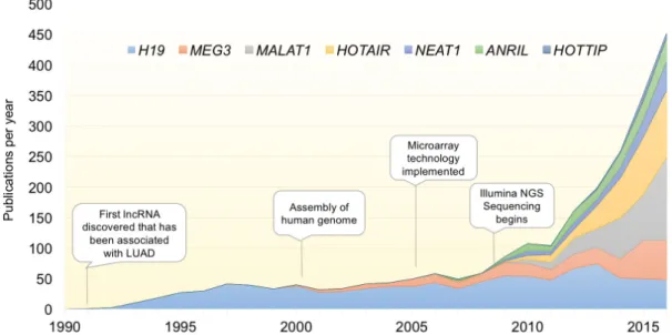
Figure 1: Exponential discovery of lncRNAs with the adoption of transcriptome-wide gene expression technologies.
Graph indicates the total number of publications per year for select lncRNAs with known involvement in LUAD. With the advent of transcriptomic profiling, the pace of lncRNA discovery and papers characterizing their function has increased exponentially over the last decade. lncRNAs included are those described in Table 1.binding partner function, localization, and activity [28-30].
In terms of biological processes, lncRNAs are involved
in regulation of the cell cycle, apoptosis, differentiation,
and immunological response [31-34]. Despite the large
repertoire of lncRNAs expressed in lung, only a handful
have been functionally linked to LUAD development.
Some exhibit hallmarks of tumor suppression, such as
MEG3 [35], while others, such as HOTAIR, behave as
oncogenes through increased proliferation and reduced
survival [36]. Table 1 highlights some of the known
lncRNAs involved in LUAD and their cellular mechanism
of action. However, for most lncRNAs, a defined
molecular mechanism has yet to be discovered.
Pan-cancer lncRNAs
It is important to note that, while the lncRNAs in
Table 1 play a role in LUAD development, they are all
implicated in the development of multiple cancer types,
and therefore do not confer specificity to any given cancer.
Because multiple types of cancers depend on similar
pathways for sustained growth, it is not surprising that
a subset of lncRNAs have been linked to suppression of
p53, Wnt signaling activation, epithelial to mesenchymal
transition (EMT), and similar early steps in the process
of oncogenesis. Here, we discuss examples of lncRNAs
that not only promote LUAD but are also involved in
tumorigenesis in a variety of cancers.
H19
The maternally expressed and imprinted gene
H19 is elevated in numerous cancers. Overexpression
occurs through the loss of epigenetic repression at the
paternal allele [37-39]. More recently, H19 was found
to be upregulated in NSCLC tissue and correlated with
poor prognosis [40]. Many genes involved in embryonic
growth and implicated in cancer lie within the H19 locus
and are cis regulated by H19 [41, 42]. In addition, H19
upregulation has been linked to MYC oncogene activation
[43]. Therefore, disrupted paternal imprinting on H19
acts as an oncogenic driver in several cancers, including
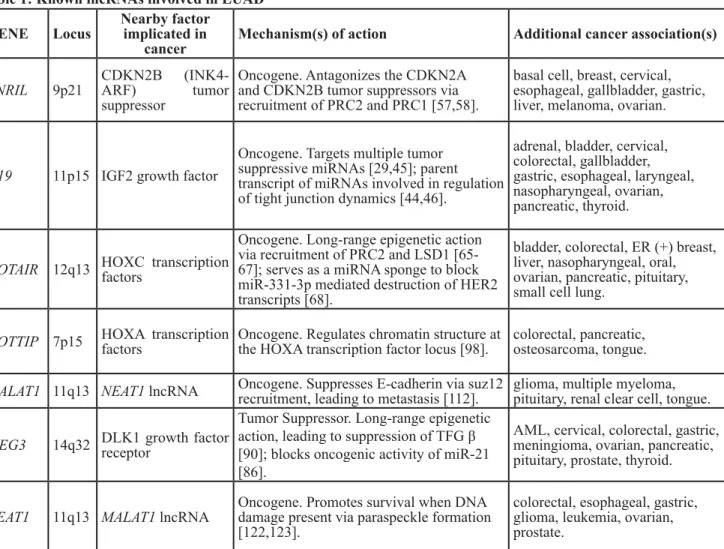
Table 1: Known lncRNAs involved in LUAD
GENE
Locus
Nearby factor
implicated in
cancer
Mechanism(s) of action
Additional cancer association(s)
ANRIL
9p21
CDKN2B (INK4-
ARF)
tumor
suppressor
Oncogene. Antagonizes the CDKN2A
and CDKN2B tumor suppressors via
recruitment of PRC2 and PRC1 [57,58].
basal cell, breast, cervical,
esophageal, gallbladder, gastric,
liver, melanoma, ovarian.
H19
11p15 IGF2 growth factor
Oncogene. Targets multiple tumor
suppressive miRNAs [29,45]; parent
transcript of miRNAs involved in regulation
of tight junction dynamics [44,46].
adrenal, bladder, cervical,
colorectal, gallbladder,
gastric, esophageal, laryngeal,
nasopharyngeal, ovarian,
pancreatic, thyroid.
HOTAIR 12q13 HOXC transcription
factors
Oncogene. Long-range epigenetic action
via recruitment of PRC2 and LSD1
[65-67]; serves as a miRNA sponge to block
miR-331-3p mediated destruction of HER2
transcripts [68].
bladder, colorectal, ER (+) breast,
liver, nasopharyngeal, oral,
ovarian, pancreatic, pituitary,
small cell lung.
HOTTIP 7p15 HOXA transcription
factors
Oncogene. Regulates chromatin structure at
the HOXA transcription factor locus [98].
colorectal, pancreatic,
osteosarcoma, tongue.
MALAT1 11q13 NEAT1 lncRNA
Oncogene. Suppresses E-cadherin via suz12
recruitment, leading to metastasis [112].
glioma, multiple myeloma,
pituitary, renal clear cell, tongue.
MEG3
14q32 DLK1 growth factor
receptor
Tumor Suppressor. Long-range epigenetic
action, leading to suppression of TFG β
[90]; blocks oncogenic activity of miR-21
[86].
AML, cervical, colorectal, gastric,
meningioma, ovarian, pancreatic,
pituitary, prostate, thyroid.
NEAT1
11q13 MALAT1 lncRNA
Oncogene. Promotes survival when DNA
damage present via paraspeckle formation
[122,123].
colorectal, esophageal, gastric,
glioma, leukemia, ovarian,
prostate.
The RefSeq gene name annotation, alongside the hg19 chromosomal location are listed. In addition, the established mechanism
of action is listed, as well as other cancers where the lncRNA has demonstrated effects on tumor initiation, promotion,
progression, and/or patient survival outcomes.
NSCLC.
Mechanistically, H19 serves as the precursor
miR-675, which is processed from the first exon of H19 and
in its mature miRNA form mediates degradation of
ZO-1 and E-Cadherin mRNA, disrupting tight junction
formation, which in turn disrupts epithelial architecture
and leads to increased invasion [44, 45]. In addition to
being a precursor to miRNAs, H19 can suppress several
miRNAs, including let-7 [46], 138, and
miR-200a [29] by serving as a competing endogenous RNA
(ceRNA). Suppression of both miR-138 and miR-200a
via H19 was shown to re-activate expression of the
mesenchymal marker genes ZEB1, ZEB2, and Vimentin,
resulting in EMT progression in bladder cancer [29]. In
addition, H19 was recently shown to suppress miR-141
and miR-22, both of which function as antagonists of Wnt
signaling [47, 48]. This H19-mediated suppression lead
to the activation of the Wnt/β-catenin pathway during
osteoblast differentiation [49]. Their role in Wnt/B-catenin
signaling might suggest an alternative means by which
H19 can promote tumorigenesis. In addition, H19 has
been linked with Wnt-mediated tumorigenesis via PRC2/
EZH2 recruitment to the Wnt antagonist gene NKD1
[50]. The convergent activity of H19 on different parts
of the Wnt signaling pathway is interesting from both a
mechanistic and evolutionary standpoint, revealing that
individual lncRNAs can perform multiple functional roles
simultaneously to effect intracellular signaling cascades.
H19 has also been shown to negatively regulate p53
signaling. Ectopic expression of H19 can cause increased
cell growth and decreased p53 transcriptional activity [51].
This was attributed to a physical interaction between H19
and the p53 protein. However, the mechanism by which
this interaction mediates the inactivation of p53 remains
ambiguous and more investigation is needed to fully
evaluate the effect of H19 on p53-mediated cellular arrest
and apoptosis.
ANRIL (antisense non-coding RNA in the INK4 locus)
This gene lies within the 9p21.3 gene cluster,
consisting of the p14
ARF, p15
INK4b, and p16
INK4atumor
suppressor genes. Within this locus, ANRIL is the natural
antisense transcript of the p16
INK4agene. p14
ARFis involved
in stabilizing p53 levels by negatively regulating MDM2
[52], whereas both p15
INK4band p16
INK4aare critical
regulators of the cell cycle [53]. Their deactivation
promotes an increase in cellular proliferation and is seen
in several cancers [54, 55]. In addition, the deactivation
of the 9p21.3 gene cluster often occurs in conjunction
with LUAD driven by mutationally-activated kRAS [56].
The proximity of ANRIL within the gene cluster allows
for a cis-mediated suppression of p16
INK4athat occurs
through recruitment of PRC2 complex which compacts
chromatin and subsequently deactivates gene expression
[57, 58]. A more recent study demonstrated that ANRIL is
overexpressed in NSCLC, correlating with poor prognosis
[59].
HOTAIR (HOX transcript antisense RNA)
This well-characterized lncRNA has been the sole
subject of previous reviews [36, 60-63], and acts by
binding and promoting chromatin compaction through
association with GA-rich DNA sequence motifs that
subsequently recruit PRC2 [64-66]. This results in
genome-wide epigenetic regulation of differentiation
and cancer development [67]. HOTAIR can also regulate
miRNA by acting as a competing endogenous RNA
(ceRNA) to deplete cells of miR-331-3p, enhancing
Figure 2: Molecular origins of LUAD.
Lung adenocarcinoma (LUAD) arises in the distal alveolar epithelium from progenitor alveolar epithelial cells. LUAD develops from these precursor cells though oncogenic activation (and deactivation of tumor suppressors) by induced mutations to the DNA, amplification and fusion events, as well as epigenomic alterations. Genes listed were taken from TCGA analysis of LUAD (15). Added to this is the newly-emergent appreciation for altered lncRNA regulation of cellular processes as an oncogenic event.expression of the HER2 receptor tyrosine kinase and
thereby promoting oncogenesis [68]. Indeed, HOTAIR
displays all the canonical behaviors of an oncogene,
including poor prognosis when present [69, 70],
chemoresistance [71], reduced overall survival [72-75],
and increased metastasis [76-80]. This occurs in a number
of cancers, including both SCLC [81] and NSCLC [82,
83].
MEG3 (maternally expressed gene 3)
One of many maternally imprinted lncRNAs [84],
MEG3 exhibits the hallmarks of a tumor suppressor,
namely inhibition of proliferation and induction of
apoptosis [35, 85-87] in numerous cancers. Multiple
functions for MEG3 in cancer have been described [87,
88], Locally, expression is inversely correlated with the
nearby tumor suppressor DLK1, which it may regulate
[89]. MEG3 can also act throughout the genome as an
epigenomic regulator of TGFβ-responsive distal regulatory
elements. It does so by forming RNA:DNA triplex helix
structures at GA-rich sequence recognition sites, which
bring EZH2 to target loci, effectively condensing local
chromatin regions to disrupt enhancer activity and block
TGFβ-induced proliferation [90]. Additionally, MEG3
can mediate the destruction of miR-21, blocking this
microRNAs oncogenic potential [86].
It is interesting to note that both HOTAIR and MEG3
bind GA-rich sequence elements that facilitate recruitment
of PRC2 complex and condense the local chromatin
environment, yet HOTAIR functions as an oncogene while
MEG3 functions as a tumor suppressor. Considering that
HOTAIR oncogenic activity is seen in multiple cancers and
MEG3 tumor suppressor activity is also observed across
cancers, the simple explanation of differing mechanisms
in differing tumors does not seem applicable. Instead,
follow up studies on the genomic distribution of the two
lncRNAs, their relative expression to each other, and any
mechanistic interactions they may have are warranted to
address this question.
HOTTIP (HOXA transcript at the distal TIP)
HOTTIP is another lncRNA transcribed from the
HOXA locus which exhibits the oncogenic properties
of increased proliferation, expression in advanced
pathological stages alongside distant metastasis, inhibition
of apoptosis, and association with overall poor prognosis
in multiple cancers [91-96]. While there are observed
correlations between HOTTIP and vitamin D receptor
signaling [97] as well as p21 silencing [91], the main
role of HOTTIP described in cancer progression is its
ability to utilize three-dimensional chromatin looping
structures. These allow HOTTIP to regulate cis-members
of the HOXA cluster by recruitment of WDR5 to drive
H3K4me3 deposition into chromatin, activating target
gene expression [98].
Figure 3: Overlap of deregulated lncRNAs in LUAD between multiple large-scale bioinformatic studies. Results from
microarray analysis, de novo RNA-seq transcriptome assembly of TCGA LUAD datasets from the Maher study on LCALs (supplementary data file 5 from their study which includes LUAD-specific lncRNAs), and robust statistical analysis of multiple lung cancer datasets were overlapped in the hg19 UCSC genome browser to determine a unifying set of lncRNAs deregulated in all three studies. These forty deregulated lncRNAs fall broadly into two categories, 15 were antisense transcripts and the remaining 25 were intergenic genes.The MEN (MALAT1-NEAT1) locus
The MEN locus is located on chromosome 11 at
p13.1 and harbors both the MALAT1 and NEAT1 lncRNA
genes. NEAT1 is about 53kb upstream of the 5’ end of
MALAT1, and both transcripts are deregulated in LUAD
[99, 100].
MALAT1 (metastasis-associated lung
adenocarcinoma transcript-1; multiple endocrine
neoplasia-alpha)
This is a single exon gene originally identified
as expressed specifically in lung cancer. Because it is
associated with poor prognosis and distant metastasis in
NSCLC [101-103], along with other cancers [102,
104-107], much of the emphasis in studying this gene is to
utilize it as a prognostic biomarker [108, 109]. Identified
over two decades ago, this functional RNA has been
the sole subject of previous reviews [110, 111] and was
initially implicated in RNA splicing through extensive
studies in vitro.
Functionally, MALAT1 interacts with Suz12
resulting in decreased expression of E-cadherin, a cell
adhesion molecule essential in maintaining epithelial
architecture. The loss of E-cadherin is a commonly
observed phenomenon in cancers of epithelial origin, and
co-occurs with upregulation of N-cadherin and fibronectin.
This ultimately leads to metastasis, as reported in bladder
cancer [112]. In addition, knockout studies of MALAT1
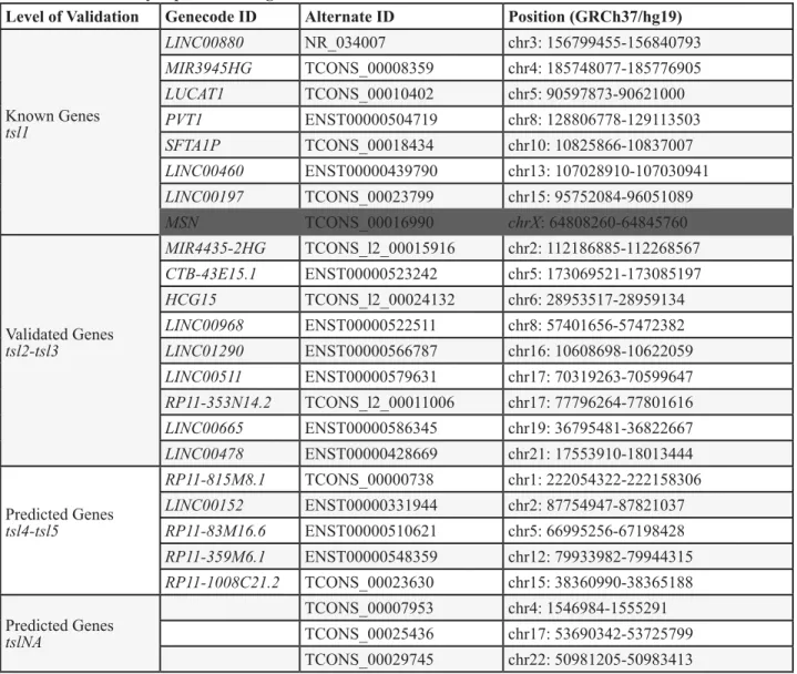
Table 2: Differentially expressed intergenic lncRNAs
Level of Validation
Genecode ID
Alternate ID
Position (GRCh37/hg19)
Known Genes
tsl1
LINC00880
NR_034007
chr3: 156799455-156840793
MIR3945HG
TCONS_00008359
chr4: 185748077-185776905
LUCAT1
TCONS_00010402
chr5: 90597873-90621000
PVT1
ENST00000504719
chr8: 128806778-129113503
SFTA1P
TCONS_00018434
chr10: 10825866-10837007
LINC00460
ENST00000439790
chr13: 107028910-107030941
LINC00197
TCONS_00023799
chr15: 95752084-96051089
MSN
TCONS_00016990
chrX: 64808260-64845760
Validated Genes
tsl2-tsl3
MIR4435-2HG
TCONS_l2_00015916
chr2: 112186885-112268567
CTB-43E15.1
ENST00000523242
chr5: 173069521-173085197
HCG15
TCONS_l2_00024132
chr6: 28953517-28959134
LINC00968
ENST00000522511
chr8: 57401656-57472382
LINC01290
ENST00000566787
chr16: 10608698-10622059
LINC00511
ENST00000579631
chr17: 70319263-70599647
RP11-353N14.2
TCONS_l2_00011006
chr17: 77796264-77801616
LINC00665
ENST00000586345
chr19: 36795481-36822667
LINC00478
ENST00000428669
chr21: 17553910-18013444
Predicted Genes
tsl4-tsl5
RP11-815M8.1
TCONS_00000738
chr1: 222054322-222158306
LINC00152
ENST00000331944
chr2: 87754947-87821037
RP11-83M16.6
ENST00000510621
chr5: 66995256-67198428
RP11-359M6.1
ENST00000548359
chr12: 79933982-79944315
RP11-1008C21.2
TCONS_00023630
chr15: 38360990-38365188
Predicted Genes
tslNA
TCONS_00007953
chr4: 1546984-1555291
TCONS_00025436
chr17: 53690342-53725799
TCONS_00029745
chr22: 50981205-50983413
LncRNAs that were deregulated in three studies (from Figure 3) that also occupy chromosomal regions in between mRNAs.
LncRNAs were segregated by their Gencode transcript confidence level (tsl1 = highest quality, full transcript is validated;
tsl2-3, one or many spliced ESTs are validated; tsl4-5 = one or none ESTs support the validity of the transcript, and those ESTs
are suspect.) Those without a tsl ranking do not have a representative transcript in Gencode. All coordinates span the entire
transcript length and are hg19 genome-based. The MSN transcript overlaps in the same orientation the MSN mRNA, therefore
there is a high probability that this is not truly a lncRNA (and thus is greyed out in figure).
have demonstrated that in lung cancer, MALAT1 can also
directly regulate the expression of pro-metastatic genes
[99]. These observations tie together the poor prognosis
and increased metastatic behaviors observed when
MALAT1 is over expressed in tumors.
NEAT1 (nuclear paraspeckle assembly transcript 1;
Nuclear Enriched Abundant Transcript-1)
This is another single exon gene that is transcribed
from the same locus as MALAT1. It exhibits many similar
characteristics with MALAT1, including tumor recurrence
[113], poor prognosis [114], and metastasis [115, 116]. Of
special note, the close proximity of NEAT1 to MALAT1,
and their similar roles as oncogenes in multiple cancers
suggests that the entire locus may be subject to aberrant
regulation in cancer [117]. Indeed, several studies have
demonstrated a correlation between MALAT1 and NEAT1
expression [115]. While NEAT1 acts as part of chromatin
remodeling complexes [118], less is understood about
a direct functional role for NEAT1 in carcinogenesis.
NEAT1 serves as a scaffold for nuclear paraspeckle
formation [119-121], which accumulate in response to
DNA-damaged induced genotoxic stress [122, 123]. It
is possible that NEAT1 acts to promote carcinogenesis
directly by abrogating the stressors placed on the genomes
of cancer cells. Further research is needed to determine if
NEAT1 plays a direct role in oncogenesis, or if instead the
MALAT1/NEAT1 locus is under mutual regulation, with
NEAT1 upregulation in cancer being a byproduct of its
genomic proximity to MALAT1.
Unclear mechanism(s) affecting cancer
progression
Highlighting the need for further research, there
are still several lncRNAs which are deregulated across
cancer types, yet have not undergone in-depth functional
characterization. This lack of mechanistic understanding
hinders further investigation into the application of
targeted therapeutics toward these deregulated lncRNAs,
for concerns regarding off-target effects. One such
transcript is CCAT2. It was originally identified as having
LUAD-specific expression [124], but is now implicated
in a host of cancers [125, 126] and associated with
smoking [127]. It contains rs6983267, a single nucleotide
polymorphism (SNP) identified through genome-wide
association studies (GWAS) as conferring an increased
risk of prostate and colorectal cancer [128-130]. This gene
lies within the 8q24 ‘gene desert’ hotspot that is home
to the MYC oncogene and is associated with numerous
cancers [131-134], highlighting the significance of
lncRNAs in genetic predisposition to cancer. However,
rs6983267 has not been associated with LUAD risk in
the numerous LUAD GWAS studies performed to date.
Instead, it appears that copy number alterations in 8q24
occur frequently in lung cancers, suggesting an alternate
mechanism other than SNP regulation of the CCAT2
transcript in lung cancer pathogenesis. Mechanistically,
CCAT2 can alter cancer metabolism depending on the
allele transcribed through altered binding affinity to
pre-Table 3: Differentially expressed anti-sense lncRNAs
Genecode
Alternate IDs
Anti-Sense Gene Multi-exonic
Position (GRCh37/hg19)
LOC101928370 RP4-575N6.1
S1PR1
YES
chr1: 101701238-101702084
LINC00883
DUBR
LINC00882
YES
chr3: 106959538-107045811
LINC00312
LINC00312
LMCD1
NO
chr3: 8613467-8634810
LHFPL3-AS2
RP11-325F22.5
LHFPL3
YES
chr7: 104558006-104567077
FEZF1-AS1
FEZF1-AS1
FEZF1
YES
chr7: 121945003-121945871
HSPC324
RP11-251M1.1
EGFL7
YES
chr9: 139541826-139554873
LOC105369340 RP11-783K16.5
PPP1R14B
YES
chr11: 64014525-64015649
LOC101929340 RP11-677M14.3 ESAM
YES
chr11: 124632326-124635257
SBK1-AS1
RP11-57A19.2
SBK1
YES
chr16: 28270020-28303385
FENDRR
FENDRR
FOXF1
YES
chr16: 86508050-86542705
TBX2-AS1
RP11-332H18.5
TBX2
YES
chr17: 59470732-59477096
RP11-720L2.4
COLEC2
YES
chr18: 314886-319165
GATA6-AS1
GATA6-AS1
GATA6
YES
chr18: 19746858-19748929
LINC01271
RP11-290F20.2
LINC01270
YES
chr20: 48909256-48937879
LINC00649
LINC00649
ATP5O
YES
chr21: 35295736-35351160
The Gencode annotation for each lncRNA is indicated, along with the genomic coordinates of the lncRNA and the mRNA
that is transcribed in the antisense orientation. All but LINC00312 are multi-exonic, indicative of splicing. The hg19-based
lncRNA coordinates are listed.
mRNA cleavage (CFIm) splicing factors [135]. However,
little is known regarding how this altered splicing affects
other cellular processes, or whether the differing alleles of
CCAT2 target the splicing complex to different chromatin
locations to affect cancer development. This is but one
example of the many lncRNAs that have been identified
as deregulated in LUAD. Below, we highlight recent
methods that have taken a more systematic approach to
identifying the extent of lncRNA deregulation in LUAD,
and what, if anything, is known about these genes.
GENOME-WIDE ANALYSIS OF LNCRNAS
IN LUAD
Original attempts to characterize the lncRNA
landscape in LUAD were performed using microarray
technology. While these arrays were designed to target
mRNAs, many unaccounted-for exons that were later
classified as lncRNAs were included in several platforms,
notably the Affymetrix Human Exon 1.0 ST Array.
Illumina-developed arrays contained less information
on lncRNAs due to their design emphasis on 3’UTR
targeting, however several lncRNAs were included under
the ‘LOC_” definition. Using this probe-based approach
several studies were able to identify lncRNA expression
profiles in lung cancer and perform preliminary analysis
[136-139], the results of which have been conveniently
collated by lnc2cancer [140]. However, the discernable
drawbacks of such techniques include the lack of discovery
and low-expression levels of lncRNAs, thwarting
detection efforts. With the advent and widespread adoption
of RNA sequencing technology, the ability to detect novel
transcripts had increased exponentially. Indeed, the rate at
which the non-coding RNA transcriptome expanded has
rapidly outpaced the identification of mRNA genes over
the last five years.
Adding to the discovery landscape was The Cancer
Genome Atlas (TCGA). LUAD samples from TCGA
[15] underwent whole section RNA-seq analysis using
Illumina TruSeq technology. This allows for polyA
selection to minimize genomic contamination; however,
it eliminates any non-polyadenyated signal from the final
sequence alignments, therefore expression of only the
poly adenylated lncRNAs were captured with this method.
While the main purpose of RNA-seq analysis performed
by TCGA was to quantify mRNA expression, several
groups have utilized this dataset for dual interrogation
of lncRNA transcriptome changes. LncRNAtor [141],
MiTranscriptome [142], and The Atlas of Noncoding
RNA In Cancer [TANRIC] [143] have each performed
re-analysis of RNA-seq data from TCGA data to detect
lncRNAs, with differing results based on the reference
genomes utilized, filtering criteria, lncRNA references
databases, and incorporation of secondary data sets. Their
differing results highlight how alternate bioinformatic
approaches can vastly affect the results of an analysis.
LncRNAtor showcases a re-analysis of several
NGS datasets. They constructed a reference lncRNA
library that included sequence from the EMSEMBL,
lncRNAdb, HGNC and MBI datasets. They then
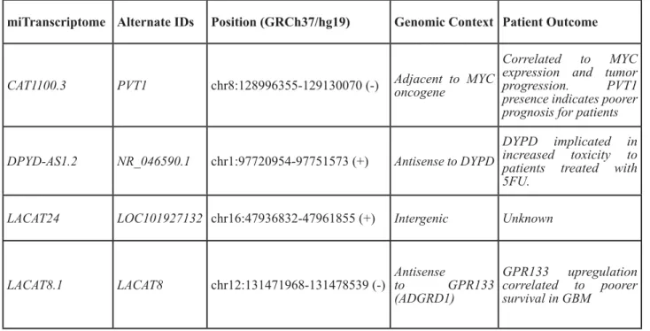
Table 4: miTranscriptome-defined LUAD lineage-specific lncRNAs
miTranscriptome Alternate IDs Position (GRCh37/hg19)
Genomic Context Patient Outcome
CAT1100.3
PVT1
chr8:128996355-129130070 (-) Adjacent to MYC
oncogene
Correlated to MYC
expression and tumor
progression. PVT1
presence indicates poorer
prognosis for patients
DPYD-AS1.2
NR_046590.1
chr1:97720954-97751573 (+)
Antisense to DYPD
DYPD implicated in
increased toxicity to
patients treated with
5FU.
LACAT24
LOC101927132 chr16:47936832-47961855 (+)
Intergenic
Unknown
LACAT8.1
LACAT8
chr12:131471968-131478539 (-)
Antisense
to GPR133
(ADGRD1)
GPR133 upregulation
correlated to poorer
survival in GBM
analyzed each transcript for phylogenetic conservation
and filtered transcripts against protein coding potential to
arrive at a consensus reference set. Against this pipeline
they re-aligned over 200 large-scale NGS datasets from
23 different cancers, including from GEO, ENCODE,
and TCGA. Of these, only TCGA LUAD and LUSC
dataset were specific for lung cancers. Their reanalysis
identified 860 lncRNAs significantly differentially
expressed between LUAD tumors and non-paired
normal lung (adjusted p-value <0.01). However, many
of the transcripts annotated as lncRNAs included known
protein coding genes, such as MMP12 and UHRF1,
suggesting that inadequate filtering for coding potential
may have inadvertently included protein coding genes. By
comparison, lncRNAtor computed that 15,331 mRNAs
were differentially expressed between LUAD tumors
and normal tissue. The vast difference in the magnitude
of changes (10-fold greater number of differentially
expressed mRNAs) indicates that their analysis showed
more variability in mRNAs then in lncRNAs. However,
follow up work will be needed to determine if this is a
reproducible phenomenon across cancers and sample sets.
MiTranscriptome was a re-analysis of data generated
by TCGA, the Michigan Center for Translational
Pathology, and ENCODE, developed to discover novel
lncRNAs involved in cancer. Utilizing these sample sets,
58,674 lncRNA genes were identified, 1,150 of which
were differentially expressed in LUAD (and obtainable
through their portal linked to the UCSC genome browser).
While their dataset has been made accessible via the
BETA portal, as of July 2016 it does not contain the entire
statistical analysis presented in their paper.
TANRIC contains the systematic re-analysis of
TCGA data from over 20 cancer types, one of which
was LUAD. Included in their analysis were 488 LUAD
tumors and 58 unmatched normal lung sections from
TCGA, along with re-analysis of RNA-seq data derived
from the SEO study [16] of Korean LUAD cancer patients
(83 LUAD and 77 matched normal samples). However,
TANRIC focused on tumor heterogeneity and correlation
with clinical covariants rather than simple tumor/normal
comparisons, which can be influenced by issues such as
tumor purity, degree of necrosis and other confounders.
Another study by the Mather group took this
analysis a step further [144]. After initially performing
tumor-normal differential analysis and finding 592 altered
lncRNAs, they then subtracted differentially expressed
lncRNAs in multiple cancers to arrive at a lung cancer
specific deregulated lncRNA class (including LUAD
and LUSC). Their analysis also included re-analysis of
the SEO dataset and a smaller subset of TCGA LUAD
samples as performed by TANRIC (55 LUAD tumors and
matched normals, with another 243 unmatched tumors).
For the purposes of this review, the differentially expressed
lncRNAs released by miTranscriptome, the Mather
group, and microarray data [136] were compared (Figure
3). In this way, we referenced the newest technologies
as well as compared them to the older microarray data,
to provide a more refined review of the current state of
LUAD-associated lncRNA discovery. Forty transcripts
were differentially expressed between tumor and normal
in all three data sets. We have segregated these forty
differentially-expressed lncRNAs into stand-alone genes,
heretofore labeled as intergenic lncRNAs (Table 2),
and those that occur antisense to a protein-coding gene
(Table 3). We have highlighted for further discussion
several of these with a high degree of validation and prior
mechanistic work, but all are potential candidate driver
genes for LUAD.
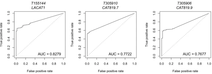
Figure 4: Lineage-specific lncRNAs identified in LUAD by miTranscriptome.
Data on lineage and cancer-specific LUAD lncRNAs was obtained from miTranscriptome. The top three lncRNAs with specificity to LUAD are shown. AUC = Area under the curve. False positive rate and false negative rate were generated using miTranscriptome-calculated expression levels for primary LUAD and normal lung tissue samples generated by TCGA. ROC curves were generated using the ROCR package in R.INTERGENIC DISCOVERY-BASED
LNCRNAS AND THEIR FUNCTIONAL
IMPLICATIONS
Intergenic lncRNAs are herein defined as those
lncRNA genes located in the space between protein coding
genes. Prior evidence has shown that lncRNAs occurring
in cis with protein coding genes can loop back and affect
the nearby mRNA [98]. However, for intergenic lncRNA
genes, their distal location makes functional predictions
difficult. Instead, intergenic lncRNA can mediate their
function in trans through a variety of mechanisms, such as
their involvement with chromatin remodeling complexes
[145]. Here, we focus on four lncRNAs that came from
our review of LUAD transcriptomic profiling (Table 2).
LUCAT1: (lung cancer associated transcript-1, also
known as smoke and cancer-associated lncRNA-1,
SCAL1)
is a multi-exonic lncRNA located deep within
the gene desert of chromosome 5q14. This lncRNA
is upregulated by cigarette smoke in vivo and in vitro
through activation of the NRF-2 transcription factor
[146]. NRF-2 (also known as NFE2L2) protects cells
from oxidative stress and cigarette smoke toxicity [147],
but its overexpression in LUAD cell lines results in drug
resistance [148]. Consistent with dichotomous nature of
the NRF-2 response [149], downregulation of LUCAT1
also results in smoke-mediated cell death [146] suggesting
LUCAT1 may in part mediate the response of NRF-2 to
oxidative stress. In addition, LUCAT1 is upregulated
in cisplatin-resistant ovarian cancer [150]. It remains
unclear whether LUCAT1 targets novel downstream genes
involved in oxidative stress or whether it aids NRF-2 in
activating NRF-2-dependent genes. Further mechanistic
research on LUCAT1 can elucidate if the oxidative stress
response is related to or independent from LUCAT1
upregulation by chemotherapy.
PVT1 (plasmacytoma variant transcript 1)
is transcribed ~60kb downstream of the MYC
oncogene, and both reside within the 8q24 locus which
undergoes copy number amplification in several cancers
[151, 152]. Although MYC is an established oncogene,
PVT1 is also emerging as a prominent player in cancer. A
recent study illustrated that some MYC-driven cancers are
dependent on PVT1 activity, as PVT1 could stabilize MYC
protein levels by preventing MYC phosphorylation [153].
In addition, silencing of PVT1 in PVT1/MYC amplified
cancers resulted in apoptosis, whereas MYC silencing
had no effect, implying PVT1 has a MYC-independent
role in blocking apoptosis. The inhibition of apoptosis
due to PVT1 overexpression may be partially due to its
role in silencing the LATS2 gene via recruitment of EZH2
to its locus, inducing chromatin remodeling and gene
silencing [154]. The LATS2 tumor suppressor is involved
in a variety of functions, including induction of apoptosis
and cell cycle control [155, 156]. Inhibition of LATS2 was
previously observed in NSCLC, and PVT1 overexpression
was found to correlate with poorer overall prognosis [154].
The PVT1 locus also contains multiple miRNA
genes, including 1204, 1205, 1206,
miR-1207-3p, miR-1207-5p, and miR-1208 [157]. Of interest,
both miR-1204 and miR-1207-5p have demonstrated
tumor suppressive properties [158, 159]. Surprisingly, p53
mediates the transcriptional expression of both PVT1 and
1204 [158]. In addition, ectopic expression of
miR-1204 induced p53-mediated growth inhibition in HCT116
cells. Therefore, induction of transcripts from the 8q24
locus results in lncRNAs that promote oncogenesis, and
paradoxically, miRNAs that inhibit tumor promotion via
p53. This may seem contradictory, but there have been
reports of p53 mediating pro-survival pathways during
DNA repair [160], such as p53 activation of p21/NRF2
signaling [161]. How this tight balance between
pro-survival during DNA repair and apoptosis/cell death is
disrupted in cancer will require further research in the
downstream targets of the PVT1 locus transcripts. The
significance of other miRNAs inhabiting the 8q24 locus
in p53-mediated signaling is unknown at the time of this
writing.
Targeted therapy against 8q24 amplified cancers has
remained challenging due to MYC being essential and in
high abundance across normal tissue [162]. Because PVT1
is less abundant in across normal tissue and possesses a
protective role for MYC protein, PVT1 appears to be a
promising target for 8q24 amplified cancers.
SFTA1P (surfactant associated 1 pseudogene)
Surfactant signaling is the distinguishing hallmark
of alveolar epithelial type 2 (AT2) cells, a purported cell of
origin for LUAD [163]. SFTA1P expression is correlated
with other components of the surfactant machinery [164],
and elevated SFTA1P levels indicate a better prognostic
outcome for LUAD cancer patients (cox p-value = 0.009)
[143]. This indicates that SFTA1P may hold potential as a
biomarker of outcome prediction. However, because this
gene is co-expressed with markers of differentiated AT2
cells, the loss of SFTA1P in a subset of LUAD cancers
may be reflective of the overall differentiation state of the
tumors. Moreover, the SFTA1P pseudogene is not located
within genomic proximity to any of the other
surfactant-protein producing genes, and the mechanisms (if any) by
which SFTA1P functions remains unknown.
LINC00460
This lncRNA is a multi-exonic, intergenic lncRNA
over 100kb from the nearest mRNA gene, EFNB2. In
addition to being found overexpressed in LUAD in
the above studies, LINC00460 is upregulated in head
and neck squamous cell carcinoma, kidney carcinoma,
and pancreatic cancer [165, 166]. While expression of
p=4.35e-28(143)), little research into the function or
application of this lncRNA has been performed. The
neighboring gene, EFNB2, encodes for EphrinB, one of
many ligands for the Ephrin tyrosine kinase receptor.
Much has been done implicating EFNB2 and the EphrinB
receptor in development and progression of lung cancer
[167-169]. However, the role LINC00460 plays in this
process, if any, has yet to be determined.
LUAD ANTISENSE TRANSCRIPTS
AND THEIR RELATION TO NEARBY
PROTEIN CODING GENES
Antisense transcription has been observed at the
transcription start site of numerous protein coding genes
[170, 171]. This class of antisense transcripts range from
siRNA [172] to antisense lncRNAs, such as HOTAIR
[173]. Many have documented antagonistic activity,
from epigenetic regulation [174] to direct disruption of
the transcriptional machinery [175]. Here, we highlight a
few antisense lncRNAs identified through bioinformatics
analysis to be involved in LUAD, while the entire list is
summarized in Table 3.
FENDRR (FoxF1 adjacent non-coding developmental
RNA)
This gene is transcribed in the antisense direction
from the adjacent FOXF1 transcription factor. As
expected, expression of FENDRR is highly correlated
to FOXF1 (R=0.816, p value =1.52e-85 [143]). The
FOXF1 transcription factor is implicated in mesoderm
development, and similarly FENDRR is implicated in
embryogenic mesoderm formation, specifically heart
development [176, 177]. FOXF1 is overexpressed in
LUAD and plays a central role in regulating
epithelial-to-mesenchymal transition by promoting tumorigenesis
of adenomas toward adenocarcinomas [178, 179].
Mechanistically, FENDRR is proposed to affect the
extracellular matrix due to its inverse correlation with
fibronectin1 expression in gastric cancer cell lines [180].
Disrupting fibronectin1 is associated with tumor migration
and metastasis [181]. Adding extra weight to the argument
that FENDRR may promote EMT and metastasis, Xu et
al., found that lower FENDRR expression correlates with
higher metastatic potential and poorer outcomes in LUAD
patients [180]. Similar to HOTAIR and MEG3, FENDRR
appears to form RNA:DNA triplexes to recruit PRC2
complex during embryonic mesoderm patterning, which
when disrupted leads to deformation of the heart and
embryo death [182].
FEZF1-AS1 (FEZ family zinc finger 1- antisense 1)
FEZF1 (also known as ZNF312b) is a zinc finger
transcriptional repressor that is an epigenetically-regulated
oncogene in gastric cancer [183]. This protein promotes
proliferation via kRAS-oncogene activation [184].
FEZF1-AS1 positively regulates expression of FEZF1
mRNA expression in vitro as well as in the TANRIC
analysis of TCGA LUAD data (p=2.85e-104). FEZF1-AS1
is upregulated in human primary colorectal carcinoma, and
affects colorectal cancer cell proliferation, metastasis, and
invasion [185]. However, it remains to be determined how
FEZF1-AS1 and FEZF1 interact mechanistically.
SBK1-AS1 (SH3 domain binding kinase 1 antisense-1;
RP11-57A19.2)
SBK1 is a serine/threonine kinase family member
implicated in ovarian serous adenocarcinoma cell
survival [186]. Several serine threonine kinase family
members exhibit oncogenic behavior, such as PIM1 and
BRAF. These are attractive therapeutic targets, as small
molecule inhibitors have proven effective in halting cancer
progression [187, 188]. RP11-57A19.2 is transcribed in
an antisense direction from the SBK1 promoter, and the
two have correlated expression in multiple cancer types
(LUAD R=0.597, OVR=0.701,
TCGA-BRCA=0.671, p=3.95e-108 [143]), however little to
nothing is known about the expression, function, and
regulation of RP11-57A19.2.
GATA6-AS1 (GATA-binding protein-6 antisense-1)
GATA6 is an important regulatory transcription
factor in alveolar epithelial cell biology [189, 190]. The
antisense GATA6-AS1 is correlated to GATA6 expression
during lung development, albeit with more cell-type
specific restriction than the GATA6 transcription factor
[191], as well as in TCGA LUAD datasets (R=0.772,
p=3.53e-71 [143]). Overexpression of BM742401, an
expressed sequence tag which corresponds to
GATA6-AS1, reduced cancer metastasis and decreased secretion
levels of MMP9, though the mechanism by which
GATA6-AS1 mediated these effects remains unknown.
Interestingly, these authors investigated whether
GATA6-AS1 overexpression affected the expression of GATA6,
but found no change (this data not reported) [192].
Expression of GATA6 in LUAD is associated with a more
differentiated state, and reflective of that, with better
overall patient survival [193]. Whether the expression of
GATA6-AS1 functions to maintain the differentiated state,
or is merely a passive reflection of differentiation, remains
to be determined.
CLINICAL RAMIFICATIONS
Lung cancer remains the leading cause of
cancer-related death in the United States. Although improvements
in surgical treatment and chemotherapies have shown
some progress, the 5-year survival rate lingers at ~15%
[12]. In addition, NSCLC is composed of several differing
subtypes, each with their own set of heterogenic factors
that result in cancer. The array of molecular mechanisms
implicated in the genesis of this disease underscores the
need more accurate prognostic markers, to implement
therapies targeted to the specific pathways disrupted in
each disease subtype and inform clinicians on predicted
patient outcomes.
Non-coding RNAs hold promise as biomarkers
for a variety of cancers. For instance, increased
miR-486 levels showed efficacy as a blood-based biomarker
for early detection of NSCLC, and lower levels of
miR-486 post-surgical resection was an effective predictor
of recurrence-free survival of NSCLC patients [194].
As mentioned above, MALAT1 and LUCAT1 also hold
promise as prognostic biomarkers as their elevated
expression is linked with poorer overall survival [195].
However, enthusiasm surrounding MALAT1’s applicability
to early detection was tempered by research describing
its overall sensitivity of detection at only 56%, meaning
almost half of true positive LUAD cases would be missed
using this biomarker alone [196]. To circumvent this
issue, MALAT1 was included in a panel of lncRNAs to
improve the detection sensitivity while maintaining the
specificity for LUAD. The other lncRNAs included in the
panel were: ENST00000540136, NR034174, uc001gzl.3,
uc004bbl.1, and ENST00000434223. When combined, this
panel outperformed any individual lncRNA in the training
set. The testing set reached an AUC of 0.978 for tumor
identification, with 92% sensitivity and 98% specificity
[138], reinforcing the notion that the combining the
inherent cell-type specificity of many lncRNAs with the
sensitivity of others can aide in the development of early
detection tools.
Separate from this panel of biomarkers,
miTranscriptome has also undertaken the task of
calculating the specificity of expression within each
individual cancer for the entire lncRNA transcriptome.
They identified 25 lncRNAs that demonstrated statistically
robust specificity for LUAD [142], only five of which
were previously annotated. These genes included PVT1
(mentioned above as adjacent to the MYC oncogene),
DYPD-AS1, LACAT24, and LACAT8 (Table 4). It is
interesting to note that this analysis included 23 cancerous
tissues alongside 12 normal tissues, a step not typically
undertaken when assessing the feasibility of early
detection tools. As no single study can yet address the
wide diversity of cell types present in the human body
[197], including as many cell types as is available can
nevertheless decrease the risk of investing heavily in the
development of a biomarker, only to see it fail in clinical
trials due to off-target effects.
In the future, testing for lncRNA expression could
also yield gains in personalized medicine. For instance,
overexpression of HOTAIR in LUAD results in
chemo-resistance towards cisplatin [198]. HOTAIR-induced drug
resistance was attributed in part to downregulation of p21.
Although the mechanism by which HOTAIR regulates p21
remains unknown, previous studies have reported EZH2 is
involved in p21 suppression [199]. Considering the known
involvement of HOTAIR with the PRC2/EZH2 complex,
HOTAIRs role in mediating p21 suppression and cisplatin
resistance might be due to epigenic modifications.
Therefore, testing for HOTAIR expression after resection
may insulate a subset of the patient population from
having to undergo chemotherapy and instead direct
them toward promising EZH2 inhibitors, such as E7438,
currently under evaluation in clinical trials [200].
A lncRNA with oncogenic activity and expression
restricted to a specific cancer would be an ideal therapeutic
target. To that end, anti-RNA treatments are currently
being developed to diversify the options available to
clinicians. However, many obstacles have arisen, including
a lack of efficient delivery methods, RNA degradation,
and aberrant immune system activation [201]. In addition,
some lncRNAs have high turnover and low transcriptional
expression, making them difficult to target effectively. In
such cases, knowledge of pathway involvement is needed
to develop effective treatments that target downstream
signaling molecules affected by aberrant lncRNA activity.
CONCLUSIONS
Over the last decade, lncRNAs have been
recognized as a diverse class of macromolecules in
terms of function and mechanism. Many lncRNAs have
demonstrated functional activity in a wide range of
cancers. While the lncRNA transcriptome is far from
complete, we now have an appreciation of their diversity
due to cell-type specificity. This characteristic can aid in
the development of early detection methods and targeted
therapies for multiple cancer types. Alongside these
immediate applications, understanding the mechanism(s)
by which these transcripts are regulated will shed light on
the etiology of cancer development, allowing clinicians to
implement better treatment strategies and improve overall
survival rates.
While research in this field is still in preliminary
stages, multiple large-cohort patient studies such
as TCGA, ENCODE, and SEO alongside previous
microarray studies have delineated a host of lncRNAs
with potential for novel therapeutic strategies. As such,
they have provided framework for the interrogation of
molecular mechanisms that lncRNAs utilize in LUAD
initiation, promotion, and progression. While in this
review we focused on lncRNAs validated by three
different studies, it is by no means exhaustive. It remains
to be seen if other case-controlled, multi-ethnic cohorts
will unveil an entirely new set of lncRNAs with implicit
utility in the diagnosis and treatment of LUAD. It will
be interesting to follow what discoveries that work will
yield in the development of novel therapeutics and early
detection strategies in the years to come.
ACKNOWLEDGMENTS
Laird-Offringa on the topics discussed, and artwork generated by
Pat Marconett. Funding was provided by the Department
of Surgery, Keck School of Medicine, USC.
CONFLICTS OF INTEREST
There is no conflict of interest.
REFERENCES
1. Brown CJ, Ballabio A, Rupert JL, Lafreniere RG, Grompe M, Tonlorenzi R, Willard HF. A gene from the region of the human X inactivation centre is expressed exclusively from the inactive X chromosome. Nature. 1991;349:38-44. 2. Borodovsky M, Lomsadze A, Ivanov N, Mills R.
Eukaryotic gene prediction using GeneMark.hmm. Curr Protoc Bioinformatics. 2003;Chapter 4:Unit4.6.
3. Besemer J, Borodovsky M. GeneMark: web software for gene finding in prokaryotes, eukaryotes and viruses. Nucleic Acids Res. 2005;33:W451-4.
4. Lukashin AV, Borodovsky M. GeneMark.hmm: new solutions for gene finding. Nucleic Acids Res. 1998;26:1107-15.
5. van Baren MJ, Koebbe BC, Brent MR. Using N-SCAN or TWINSCAN to predict gene structures in genomic DNA sequences. Curr Protoc Bioinformatics. 2007;Chapter 4:Unit 4.8.
6. Wang J, Zhang J, Zheng H, Li J, Liu D, Li H, Samudrala R, Yu J, Wong GK. Mouse transcriptome: neutral evolution of ‘non-coding’ complementary DNAs. Nature. 2004;431:1 p following 757; discussion following
7. Gross SS, Brent MR. Using multiple alignments to improve gene prediction. J Comput Biol. 2006;13:379-93.
8. Korf I, Flicek P, Duan D, Brent MR. Integrating genomic homology into gene structure prediction. Bioinformatics. 2001 (Suppl 1); 17:S140–48.
9. van Baren MJ, Brent MR. Iterative gene prediction and pseudogene removal improves genome annotation. Genome Res. 2006;16:678-85.
10. Pang KC, Frith MC, Mattick JS. Rapid evolution of noncoding RNAs: lack of conservation does not mean lack of function. Trends Genet. 2006;22:1-5.
11. Chen Z, Fillmore CM, Hammerman PS, Kim CF, Wong KK. Non-small-cell lung cancers: a heterogeneous set of diseases. Nat Rev Cancer. 2014;14:535-46.
12. Siegel RL, Miller KD, Jemal A. Cancer statistics, 2016. CA Cancer J Clin. 2016;66:7-30.
13. Aberle DR, Adams AM, Berg CD, Black WC, Clapp JD, Fagerstrom RM, Gareen IF, Gatsonis C, Marcus PM, Sicks JD, and National Lung Screening Trial Research Team. Reduced lung-cancer mortality with low-dose computed tomographic screening. N Engl J Med. 2011;365:395-409. 14. Sun S, Schiller JH, Gazdar AF. Lung cancer in never
smokers—a different disease. Nat Rev Cancer.
2007;7:778-90.
15. Network CG, and Cancer Genome Atlas Research Network. Comprehensive molecular profiling of lung adenocarcinoma. Nature. 2014;511:543-50.
16. Seo JS, Ju YS, Lee WC, Shin JY, Lee JK, Bleazard T, Lee J, Jung YJ, Kim JO, Shin JY, Yu SB, Kim J, Lee ER, et al. The transcriptional landscape and mutational profile of lung adenocarcinoma. Genome Res. 2012;22:2109-19.
17. Pao W, Miller V, Zakowski M, Doherty J, Politi K, Sarkaria I, Singh B, Heelan R, Rusch V, Fulton L, Mardis E, Kupfer D, Wilson R, et al. EGF receptor gene mutations are common in lung cancers from “never smokers” and are associated with sensitivity of tumors to gefitinib and erlotinib. Proc Natl Acad Sci USA. 2004;101:13306-11. 18. Camidge DR, Bang YJ, Kwak EL, Iafrate AJ,
Varella-Garcia M, Fox SB, Riely GJ, Solomon B, Ou SH, Kim DW, Salgia R, Fidias P, Engelman JA, et al. Activity and safety of crizotinib in patients with ALK-positive non-small-cell lung cancer: updated results from a phase 1 study. Lancet Oncol. 2012;13:1011-9.
19. Solomon BJ, Mok T, Kim DW, Wu YL, Nakagawa K, Mekhail T, Felip E, Cappuzzo F, Paolini J, Usari T, Iyer S, Reisman A, Wilner KD, et al, and PROFILE 1014 Investigators. First-line crizotinib versus chemotherapy in ALK-positive lung cancer. N Engl J Med. 2014;371:2167-77.
20. Christensen JG, Zou HY, Arango ME, Li Q, Lee JH, McDonnell SR, Yamazaki S, Alton GR, Mroczkowski B, Los G. Cytoreductive antitumor activity of PF-2341066, a novel inhibitor of anaplastic lymphoma kinase and c-Met, in experimental models of anaplastic large-cell lymphoma. Mol Cancer Ther. 2007;6:3314-22.
21. Pao W, Miller VA, Politi KA, Riely GJ, Somwar R, Zakowski MF, Kris MG, Varmus H. Acquired resistance of lung adenocarcinomas to gefitinib or erlotinib is associated with a second mutation in the EGFR kinase domain. PLoS Med. 2005;2:e73.
22. Sun M, Kraus WL. Minireview: Long noncoding RNAs: new “links” between gene expression and cellular outcomes in endocrinology. Mol Endocrinol. 2013;27:1390-402. 23. Fu X, Li H, Liu C, Hu B, Li T, Wang Y. Long noncoding
RNA AK126698 inhibits proliferation and migration of non-small cell lung cancer cells by targeting Frizzled-8 and suppressing Wnt/-catenin signaling pathway. Onco Targets Ther. 2016;9:3815-27.
24. Bernstein E, Allis CD. RNA meets chromatin. Genes Dev. 2005;19:1635-55.
25. Shahryari A, Jazi MS, Samaei NM, Mowla SJ. Long non-coding RNA SOX2OT: expression signature, splicing patterns, and emerging roles in pluripotency and tumorigenesis. Front Genet. 2015;6:196.
26. Guil S, Esteller M. Cis-acting noncoding RNAs: friends and foes. Nat Struct Mol Biol. 2012;19:1068-75.