Lehrstuhl f¨ur Computerunterst¨utzte Klinische Medizin der Medizinischen Fakult¨at Mannheim, Universit¨at Heidelberg
(Direktor: Prof. Dr. rer. nat. Lothar R. Schad)
Methods for three-dimensional Registration of
Multimodal Abdominal Image Data
Inauguraldissertation zur Erlangung des
Doctor scientiarum humanarum (Dr. sc. hum.) der
Medizinischen Fakult¨at Mannheim der Ruprecht-Karls-Universit¨at
zu Heidelberg
vorgelegt von
Barbara Ingeborg Waldkirch
aus
Ludwigshafen am Rhein
Dean: Prof. Dr. med. Sergij Goerdt
Abstract
Multimodal image registration benefits the diagnosis, treatment planning and the performance of image-guided procedures in the liver, since it enables the fusion of complementary information provided by pre- and intrainterventional data about tumor localization and access. Although there exist various registration methods, approaches which are specifically optimized for the registration of multimodal abdominal scans are only scarcely available. The work presented in this thesis aims to tackle this problem by focusing on the development, optimization and evaluation of registration methods specifically for the registration of multimodal liver scans.
The contributions to the research field of medical image registration include the development of a registration evaluation methodology that enables the comparison and optimization of linear and non-linear registration algorithms using a point-based accuracy measure. This methodology has been used to benchmark standard registration methods as well as novel approaches that were developed within the frame of this thesis. The results of the methodology showed that the employed similarity measure used during the registration has a major impact on the registration accuracy of the method.
Due to this influence, two alternative similarity metrics bearing the potential to be used on mul-timodal image data are proposed and evaluated. The first metric relies on the use of gradient information in form of Histograms of Oriented Gradients (HOG) whereas the second metric em-ploys a siamese neural network to learn a similarity measure directly on the image data. The evaluation showed, that both metrics could compete with state of the art similarity measures in terms of registration accuracy. The HOG-metric offers the advantage that it does not require ground truth data to learn a similarity estimation, but instead it is applicable to various data sets with the sole requirement of distinct gradients. However, the Siamese metric is characterized by a higher robustness for large rotations than the HOG-metric. To train such a network, registered ground truth data is required which may be critical for multimodal image data. Yet, the results show that it is possible to apply models trained on registered synthetic data on real patient data. The last part of this thesis focuses on methods to learn an entire registration process using neu-ral networks, thereby offering the advantage to replace the traditional, time-consuming iterative registration procedure. Within the frame of this thesis, the so-called VoxelMorph network which was originally proposed for monomodal, non-linear registration learning is extended for affine and multimodal registration learning tasks. This extension includes the consideration of an image mask during metric evaluation as well as loss functions for multimodal data, such as the pretrained Siamese metric and a loss relying on the comparison of deformation fields. Based on the devel-oped registration evaluation methodology, the performance of the original network as well as the extended variants are evaluated for monomodal and multimodal registration tasks using multiple data sets. With the extended network variants, it is possible to learn an entire multimodal registra-tion process for the correcregistra-tion of large image displacements. As for the Siamese metric, the results imply a general transferability of models trained with synthetic data to registration tasks including real patient data. Due to the lack of multimodal ground truth data, this transfer represents an important step towards making Deep Learning based registration procedures clinically usable.
Zusammenfassung
Multimodale Bildregistrierung ist ein wichtiges Forschungsgebiet in der medizinischen Bildverar-beitung, da sie die Fusion von komplement¨aren Informationen erm¨oglicht, die von verschiedenen bildgebenden Verfahren geliefert werden. Obwohl es eine Vielzahl an Registrierungsmethoden gibt, sind Ans¨atze, die speziell f¨ur die Registrierung von multimodalen Bildaufnahmen des Abdomens optimiert wurden, kaum verf¨ugbar. Das Ziel dieser Arbeit ist deshalb die Entwicklung, Optimierung und Evaluation von Verfahren f¨ur die Registrierung multimodaler Bilddaten des Abdomens. Die vorgeschlagenen Beitr¨age zum Forschungsgebiet der medizinischen Bildregistrierung umfassen die Entwicklung einer Evaluationsmethodik f¨ur Registrierungsverfahren, die den Vergleich und die Optimierung von linearen und nichtlinearen Registrierungsmethoden mittels eines punktbasierten Genauigkeitsmaßes erm¨oglicht. Diese Methodik wurde sowohl f¨ur die Bewertung und Optimierung von Standardregistrierungsmethoden als auch f¨ur neuartige Ans¨atze, die in dieser Arbeit entwickelt wurden, verwendet. Die Ergebnisse zeigen, dass vor allem die f¨ur die Registrierung verwendete
¨
Ahnlichkeitsmetrik einen großen Einfluss auf die Registrierungsgenauigkeit der Methode hat. Daher wurden im Rahmen dieser Arbeit zwei alternative ¨Ahnlichkeitsmetriken f¨ur den Vergleich von multimodale Bilddaten entwickelt und evaluiert. Die erste Metrik beruht auf der Verwendung von Gradienteninformation in Form von Histogrammen Orientierter Gradienten (HOG), w¨ahrend die zweite Metrik ein sog. Siamesisches neuronales Netz verwendet, um ein ¨Ahnlichkeitsmaß direkt auf den vorliegenden Bilddaten zu erlernen. Die Auswertung zeigt, dass beide Metriken in Bezug auf die erreichbare Registrierungsgenauigkeit mit traditionellen ¨Ahnlichkeitsmaßen konkurrieren k¨onnen. Die HOG-Metrik bietet den Vorteil, dass sie zum Erlernen einer ¨Ahnlichkeitssch¨atzung keine Ground Truth-Daten ben¨otigt, sondern auf verschiedensten Datens¨atzen anwendbar ist. Aller-dings zeichnet sich die siamesische Metrik durch eine h¨ohere Robustheit f¨ur große Rotationen aus. Nachteil eines siamesischen Netzwerks ist der Bedarf an registrierten Ground Truth-Daten um das Netz trainieren zu k¨onnen. Die Ergebnisse weisen jedoch auf eine allgemeine Anwendbarkeit der mit synthetischen Daten trainierten Modelle auf realen Patientendaten hin.
Der letzte Teil dieser Arbeit konzentriert sich auf die Verwendung von neuronalen Netzen, um einen kompletten Registrierungsprozess zu erlernen. In dieser Arbeit wurde das sog. VoxelMorph -Netzwerk, das urspr¨unglich f¨ur das Erlernen eines monomodalen, nichtlinearen Registrierungs-prozesses vorgestellt wurde, f¨ur affine und multimodale Registrierungsaufgaben erweitert. Diese Erweiterung beinhaltet die Ber¨ucksichtigung einer Bildmaske bei der Metrikberechnung, sowie die Integration alternativer Verlustfunktionen die auf multimodalen Daten anwendbar sind. Diese Funktionen umfassen die vortrainierte Siamesische Metrik, sowie eine Verlustfunktion, die auf dem Vergleich von Deformationsfeldern beruht. Basierend auf der entwickelten Evaluationsmethodik wurde die Registrierungsgenauigkeit des urspr¨unglichen Netzes sowie der erweiterten Varianten f¨ur monomodale und multimodale Registrierungen bewertet. Die Ergebnisse zeigen, dass es mit den erweiterten Netzvarianten m¨oglich ist, einen Registrierungsprozess f¨ur die Korrektur großer Bild-verschiebungen zu erlernen. Des Weiteren zeigen die Resultate auch hier eine ¨Ubertragbarkeit der mit synthetischen Daten trainierten Modelle auf die Registrierung realer Patientendaten. Aufgrund des Mangels multimodaler Ground Truth-Daten, repr¨asentiert dieser Transfer einen ersten Schritt um auf Deep Learning basierende Registrierungsverfahren klinisch nutzbar zu machen.
List of Abbreviations
AMMI Advanced Mattes Mutual Information CBCT Cone-Beam Computed Tomography CNN Convolutional Neural Network
CT Computed Tomography
DL Deep Learning
DFL Deformation Field Loss
DOF Degrees Of Freedom
DSC Dice Similarity Coefficient FCN Fully Convolutional Network FCSS Fully Convolutional Self-Similarity FLE Fiducial Localization Error
FOV Field of View
FRE Fiducial Registration Error GAN Generative Adversarial Network GPU Graphics Processing Unit
GT Ground Truth
HOG Histogram of Oriented Gradients
ITK Insight Segmentation and Registration Toolkit M2OLIE Mannheim Molecular Intervention Environment
MI Mutual Information
MIND Modality Independent Neighborhood Descriptor MITK Medical Imaging Interaction Toolkit
MRI Magnetic Resonance Imaging
MSE Mean-Squared Error
NGF Normalized Gradient Field NMI Normalized Mutual Information OMD Oligometastatic Disease
PET Positron Emission Tomography
ROI Region Of Interest
RF Radio Frequency
SDM Siamese Deep Metric
SIFT Scale-Invariant Feature Transform SSD Sum of Squared Differences STN Spatial Transformer Network SURF Speeded Up Robust Features TACE Transarterial Chemoembolization TRE Target Registration Error
2D Two-dimensional
Contents
List of Abbreviations v 1 Introduction 1 1.1 Motivation . . . 1 1.2 Objectives . . . 2 1.3 Thesis Structure . . . 4 2 Theoretical Background 5 2.1 Medical Background . . . 52.2 Medical Image Acquisition . . . 8
2.2.1 X-ray Computed Tomography . . . 8
2.2.2 Magnetic Resonance Imaging . . . 11
2.3 Fundamentals of Image Registration . . . 15
2.3.1 Classification of Image Registration Methods . . . 16
2.3.2 Geometric Transformation . . . 19
2.3.3 Interpolator . . . 22
2.3.4 Similarity Metrics . . . 25
2.3.5 Optimizer . . . 29
2.3.6 Challenges and Limitations . . . 30
2.4 Fundamentals of Deep Learning . . . 31
2.4.1 Artificial Neural Networks . . . 31
2.4.2 Training a Neural Network . . . 33
2.4.3 Convolutional Neural Networks . . . 34
2.4.4 Encoder-Decoder Architecture . . . 36
3 State of the Art 39 3.1 Image Registration Evaluation . . . 39
3.2 Novel Multimodal Similarity Metrics . . . 44
3.3 End-to-End Registration Learning using Neural Networks . . . 50
4 Materials and Methods 53 4.1 Toolkits and Hardware . . . 54
4.2 Evaluation Methodology for Medical Image Registration . . . 55
4.2.1 Data Pre-processing and Generation of Ground Truth Data . . . 56
4.2.2 Registration Evaluation Methodology for Multimodal Abdominal Data . . . . 63
4.2.3 Experimental Setup . . . 66
4.3 Novel Similarity Metrics . . . 71
4.3.1 Metric based onHistograms of Oriented Gradients . . . 72
4.3.2 Deep Metric Learning based on a Siamese Neural Network . . . 75
4.4 End-to-End Image Registration Learning . . . 80
4.4.1 Extension of the Classical VoxelMorph Network Architecture . . . 81
4.4.2 Training VoxelMorph Models . . . 84
4.4.3 Experiments . . . 86
5 Results 89 5.1 Evaluation of Linear Image Registration . . . 89
5.1.1 Initialization . . . 89
5.1.2 Similarity Metrics . . . 91
5.1.3 Application of Masks . . . 93
5.1.4 Number of Resolution Levels . . . 93
5.1.5 Rigid vs. Affine Registration . . . 94
5.2 Evaluation of Nonlinear Image Registration . . . 95
5.2.1 Physical Grid Spacing . . . 96
5.2.2 Similarity Metrics . . . 97
5.2.3 Application of Masks . . . 97
5.2.4 Number of Resolution Levels . . . 98
5.3 Novel Similarity Metrics . . . 99
5.3.1 Similarity Metric based on Histograms of Oriented Gradients . . . 99
5.3.2 Similarity Metric based on a Siamese Neural Network . . . 105
5.4 End-to-End Image Registration Learning . . . 119
5.4.1 Monomodal Registration Learning . . . 119
5.4.2 Multimodal Registration Learning . . . 122
5.4.3 Transfer to Patient Data . . . 125
6 Discussion 127 6.1 Evaluation of Linear and Nonlinear Image Registration . . . 127
6.2 Novel Similarity Metrics . . . 129
6.2.1 Similarity Metric based on Histograms of Oriented Gradients . . . 129
6.2.2 Similarity Metric based on a Siamese Neural Network . . . 130
6.3 End-to-End Image Registration Learning . . . 132
7 Summary and Outlook 137
Bibliography 141
Appendix 163
List of Publications 169
Curriculum Vitae 171
1
Introduction
1.1
Motivation
Multimodal image registration is an important research field in medical image processing, since it enables the fusion of complementary information provided by different imaging modalities. This fusion can benefit various applications in the clinical context, ranging from an improved diagnosis, to treatment planning and navigation during image-guided procedures. Image registration can even be relevant for treatment monitoring, since it e.g. enables the control of tumor shrinkage after treatment, by registration of pre- and post-procedural image data.
Preoperative imaging data is used for the visualization and localization of tumor tissue and anatom-ical landmarks and therefore builds the basis for interventional and surganatom-ical planning. It is often useful to acquire image data of more than one modality, since different image modalities provide different information. However, the precise localization of target anatomy with respect to planning data during an intervention or surgery can be challenging due to differences in patient positioning, anatomical deformations and the intervention itself. Therefore, intra-interventional image data is additionally acquired in order to visualize contrasted vessels, interventional tools like a catheter or the patients anatomy for navigation. Image registration then aims to incorporate the intraoperative information as a real-time update in the preoperative surgical planning. Thus, image registration is not only an important means for preoperative interventional/surgical planning, but can also benefit other applications such as automatized tool positioning and tracking during the intervention. Exemplary use-cases for such a scenario are image-guided procedures in the liver, such as e.g. biopsies or treatment of liver cancer by means of a transarterial chemoembolization. The im-provement of the whole treatment cycle of oligometastatic liver cancer, including the performance of image-guided procedures in the liver, is subject of the research campus “Mannheim Molecular Intervention Environment” (M2OLIE). Following the intended workflow of M2OLIE, the pre- and post-interventional data is commonly acquired using three-dimensional computed tomography (CT) and magnetic resonance imaging (MRI), whereas the intraoperative data corresponds to projective X-ray fluoroscopy or three-dimensional cone-beam computed tomography (CBCT). The aim is to fuse all available morphological, functional and molecular information about the tumor provided by the different imaging modalities applied prior to the intervention into a multimodal data set. This data set then builds the basis for an individualized treatment and is ultimately transferred in
the intervention room and registered to the interventional image data to improve surgical planning and the guidance of a robotic assistance system. Hence, such a scenario is based on the use of multimodal image registration methods. There are various requirements for registration methods in this context ranging from the use of an appropriate similarity metric which is able to compare the image data of different modalities, to fast computation times to provide a time-efficient registration of pre- and intraoperational data. Moreover, registration methods for interventional procedures are required to yield a high registration accuracy that results in an optimal overlap of corresponding structures in the images.
In general, the quality of the registration can have a high impact on the interventional or surgical outcome, since a clear definition of the tumor margins is essential for its localization during an in-tervention. Especially for soft tissues, this task can be very challenging due to tissue deformation. The registration of the liver represents a particular difficult task, since the liver tissue is deformed by respiratory as well as digestive motion. Yet, there exists no universal solution to image regis-tration and regisregis-tration methods have to be optimized for a specific task. Due to the challenges of registering the liver, methods, especially multimodal registration methods, which are specifically designed for this task are only scarcely available [1, 2].
Therefore, the work presented in this thesis focuses on the development, evaluation and optimization of image registration approaches for three-dimensional multimodal scans of the liver with regard to interventional applications.
1.2
Objectives
As a result of the limited availability of specified registration methods for abdominal image data, general-purpose registration methods, which were often designed and optimized for other body parts (mostly the brain), are transferred to be used for the registration of abdominal scans [3]. This potentially leads to suboptimal results for a number of reasons, including i.e. different degrees of organ displacement, different image resolutions depending on the utilized imaging techniques, but foremost different intensity distributions in the images which are to be registered. Additionally, the task of registering the liver entails specific challenges, due to the homogeneity of its tissue displaying less structure than i.e. the brain. Therefore, the choice and parametrization of the registration method for abdominal scans reveals itself to be a challenging task and a performance characterization of different methods to attain an accurate and robust registration result can be necessary.
The general aim of this thesis is the development of novel multimodal registration methods for abdominal data, as well as the optimization of existing methods for this task. The focus is set on use-cases related to image-guided procedures of the liver, and therefore, the methods presented in this work focus on the registration of three-dimensional (3D) CT, MRI and CBCT data.
Yet, to optimize a registration method and characterize its performance, an evaluation standard has to be defined. This is a critical point in the field of image registration, since there exists no standardized evaluation methodology to compare and benchmark registration methods until today.
This is mainly caused by the diversity of different registration types [4] and the lack of appropriate ground truth data that corresponds to optimally registered data for inter-subject or multimodal registration.
Therefore, the first part of this thesis aims to develop an evaluation methodology that enables a comparison and optimization of linear and non-linear multimodal registration methods based on the estimation of registration accuracy. In general, the development of an evaluation strategy requires a set of registered ground truth data as reference for an optimal image alignment. However, these data sets are only scarcely available, especially for multimodal data, due to differences in the acquisition procedures of different imaging modalities. Therefore, the development of an evaluation methodology also includes the generation of multimodal ground truth data sets. Thus, different approaches for the generation of these data sets are investigated, including manual preprocessing of actual patient data as well as the generation of synthetic ground truth data using a neural network. The idea is to implement an evaluation methodology in form of a framework that allows the integration of any available registration approach. This methodology can then be used to evaluate and optimize the performance of state of the art registration methods as well as novel approaches for the multimodal registration of abdominal scans.
The most challenging part in multimodal image registration is represented by the choice of an appropriate similarity measure to estimate the alignment of corresponding structures in images of different modality. The main difficulty is represented by the fact, that different image acquisition techniques may result in very dissimilar grey value distributions in the images, so that the same anatomical features appear differently in these images. This potentially leads to the generation of statistical correlations between image structures that do not correspond to the same anatomical structures, thus violating the main assumption of most intensity-based similarity measures. Up to now, there exists only a limited number of multimodal similarity measures that mostly rely on the concept of mutual information [5]. To investigate further alternatives, the second part of the thesis focuses on the development and evaluation of similarity measures that bear the potential to be applicable on multimodal image data.
The last part of the thesis aims to investigate approaches to perform end-to-end registration learn-ing uslearn-ing a neural network. The rise of Deep Learnlearn-ing methods in the field of medical image processing benefits image registration not only in terms of novel similarity estimations but also in the development of neural networks that are able to learn an entire registration process. Since traditional registration methods correspond to iterative optimization procedures, an advantage of novel approaches using Deep Learning is the fast computation time of the registration process, once such a network is trained. A widely used network architecture for end-to-end registration learning is the so-called VoxelMorph network [6]. However, as most of these registration learn-ing networks, the VoxelMorph network is currently restricted to monomodal nonlinear registration learning. Therefore, the last part of this thesis aims at the extension of the VoxelMorph network for the application on multimodal image data as well as for affine registration to enable the correction of larger image displacements.
In summary, this thesis aims to contribute to three different research areas in the field of medical image registration:
Image registration evaluation and generation of ground truth data,
novel similarity measures,
and Deep Learning in medical image registration.
1.3
Thesis Structure
This thesis is composed of seven parts. After this introductorychapter 1explaining the motivation for the work presented in this thesis, all theoretical basics which are relevant to this work will be explained in chapter 2. This includes a short presentation of the medical background of image-guided procedures in the liver. Since this work focuses on the image registration of multimodal data, chapter 2 also includes an explanation of the physical principals for the image contrast generation in CT, CBCT and MRI. Next, the fundamentals of medical image registration such as the different types of registration, the main components of an registration algorithm as well as the challenges and limitations for registration methods are explained. Since this work not only relies on traditional image processing but also on novel approaches using artificial neural networks, chapter 2 also includes a general overview of the fundamentals of Deep Learning. This work proposes contributions to three different research areas in the field of medical image registration: image registration evaluation, novel feature-based similarity metrics and Deep Learning in medical image registration. The current state of the art of these three topics and a brief description of the advancements proposed in this thesis are presented in chapter 3. In chapter 4, the approches proposed in this work are presented in detail. This includes the presentation of the ground truth data used for the experiments in this thesis and the developed evaluation methodology for linear and non-linear registration methods. Moreover, the basics and implementation details of two alternative similarity metrics relying on traditional HOG features and a siamese network are presented as well as the extensions integrated in the VoxelMorph network to enable affine and multimodal end-to-end registration learning. The results obtained for the evaluation of various registration methods, including registrations based on the alternative similarity measures as well as the extended VoxelMorph network, using the novel evaluation methodology, are presented in chapter 5and discussed in detail inchapter 6. The thesis concludes with a summary of the work presented in this thesis and an outlook to future work concerning the proposed approaches given inchapter 7.
2
Theoretical Background
In the following chapter, the medical background and the clinical use-cases of this work are de-scribed. Moreover, the fundamentals of medical image acquisition as well as medical image reg-istration are presented. The content and characteristics of medical images highly depend on the acquisition technique. Therefore, the following section is dedicated to create a deeper understand-ing of the fundamentals of medical image acquisition causunderstand-ing the different contrasts in different modalities.
Since the focus of this thesis is set on the development and evaluation of image registration methods, an overview over the general structure of image registration algorithms is presented. The basics for each algorithm are explained as well as the general challenges of image registration. To take into account the latest developments in the field of medical image processing, the fundamentals of deep neural networks and their influence on the field of medical image registration will also be presented.
2.1
Medical Background
This work aims to develop and optimize image registration algorithms specifically for the regis-tration of abdominal scans that are acquired for the diagnosis and treatment of liver cancer. To further understand the necessity of image registration in this context, the following section aims to described the anatomy of the human liver as well as common diseases and the treatment focusing on image-guided interventional procedures.
Anatomy of the Human Liver
The liver is the largest internal organ in the human body and occupies a multitude of important and complex functions concerning the entire human metabolism. It is located in the right upper part of the abdomen, partially covered by the ribcage right below the diaphragm. The human liver typically weights between 1.5 and 2 kg, making it the heaviest organ, and the largest gland in the body of vertebrates [7]. The anatomy of the liver is divided into two main sections, the right lobe (Lobus Dexter) and left lobe (Lobus sinister) in axial view, which are again subdivided in 8 subsegments according to theCouinaud system. This system relies relies on functional anatomy and divides the liver in 8 parts based on a transverse plane through the bifurcation of the portal vein
Figure 2.1: Schematic illustration of the human liver, displaying the vascular system and the liver segments according to the Couinaud system (image adapted from [9]).
(Porta hepatis) as shown in fig. 2.1. In the hepatic portal system, the liver receives a double blood supply: the portal vein carries venous blood from the gastrointestinal tract to the liver providing 70% of the total liver blood flow, while 30% comes from the right and left hepatic arteries which carry oxygenated blood to the liver [8]. The liver regulates a variety of different vital functions including detoxification, synthesis and storage. It i.a. filters and removes harmful substances and toxins from the body, assures the metabolism of carbohydrates, fats and proteins while producing bile which is essential for the digestion process and stores glucose, vitamins and iron.
Liver Diseases
In general, the liver is prone to many diseases. These range from diseases caused by viruses, such as hepatitis, to diseases caused by intoxication such as the fatty liver disease and cirrhosis, inherited diseases such as hemochromatosis or Wilson disease and liver cancer. In some cases, the development of liver cancer can be linked to cirrhosis, which describes an increase in the scarring of the liver tissue caused by a previous hepatitis infection or hemochromatosis [10], but the general causing effects for the development of a liver tumor are not known. Since this work mainly focuses on use-cases for the diagnosis and treatment of liver cancer, the different types of liver cancer are described in more detail in the following.
It is distinguished between primary and secondary liver cancer. Primary tumors grow at the organ where the tumor progression began whereas secondary tumors, so-called metastasis, are caused by the spread of cancer cells from a primary site to a secondary site within surrounding tissue or to a distant organ by intruding the circulatory or lymphathic system. Metastases are the major cause of cancer morbidity and mortality and it is estimated that they account for 90% of cancer deaths [11]. The most frequent sites for the spread of metastasis are lungs, liver, brain and bones [12]. The most common type of primary liver cancer in adults is the hepatocellular carcinoma (HCC), a malignant liver tumor which ranks as the second leading cause of cancer death in East Asia and sub-Saharan Africa and the sixth most common cause of cancer death in western countries [13]. Due the blood supply from the abdominal organs into the liver via portal vein, tumor cells can
spread from these organs into the liver parenchyma, thus making the liver also prone to be a site for metastatic (secondary) cancer. Liver metastases are often linked to colorectal cancer metastasis which is the second most common tumor type in Germany [14]. If the tumor metastasizes only to a limited number of sites and number of distinct metastases (typically between 1 and 5 metastases), the disease is referred to as oligometastatic disease (OMD). Although metastatic malignancies are generally associated with a poor treatment prognosis, the curability highly depends on the number and diameter of the metastases, thus increasing the possibility to successfully treat OMD [15].
Image-guided Procedures in the Liver
To determine the severity of the liver disease, the extraction and analysis of samples cells or tissues by means of a biopsy is an important measure for diagnosis and treatment monitoring [16]. A percutaneous liver biopsy involves the insertion of a thin biopsy needle through the patients abdomen to extract a small tissue sample which can then be analyzed on a molecular level. Other types of biopsy include the transjugular biopsy, during which the needle is inserted via catheter and a vein in the neck, or the laparoscopic biospy using a small abdominal incision to enter the needle and endoscope. The analysis of the biopsy then builds the basis for further treatment decisions. The main focus of treatment for liver metastases is systemic therapy, however local therapies provide an additional possibility to extend survival. These local treatments include surgical resection of liver tumors, which can be challenging due to the location or extend of disease. Additionally, they include liver directed therapies such as transarterial chemoembolization, radio embolization, radiofrequency ablation, microwave ablation and stereotactic body radiotherapy [17]. A short discussion of treatment procedures with respect to the staging of liver cancer is given in [18]. A uniting factor for the diagnostic biopsy as well as the liver directed therapies is that they all represent interventional procedures which are performed using image-guidance in the operation room. In general, image-guided interventions rely on computer-aided systems for the visualization of target and risk structures in the intervention room. The first step for this visualization is often represented by the preprocessing of the diagnostic image data that is acquired before the inter-vention to develop specific models that can be employed for patient-individual treatment planning as well as guidance during the intervention [19]. These models range from geometrical models en-hancing the morphological information (e.g. deformation fields or segmentation masks), functional models (e.g. perfusion maps) to interaction maps of the tissue with radiation or drugs [20]. The basis for most of these applications is the generation of a multimodal image map in which the same organ structures overlap and which displays the complementary information offered by different imaging modalities. The key technology for the generation of a fused data set is image registration. Image registration generally describes the determination of a geometric transforma-tion field which transfers two datasets in the same coordinate system in a way that same structures overlay in both datasets. To further understand the necessity of multimodal imaging, the funda-mental basics and differences in information content of standard imaging techniques will be further explained in the following section.
2.2
Medical Image Acquisition
Medical imaging refers to techniques and processes which enable a visualization of the interior of a body for diagnosis, treatment and monitoring of medical conditions. There exist various types, also called modalities, of medical imaging procedures, which use different technologies and therefore yield different specific information about the imaged body region. These can range from morphological to functional and sometimes even molecular information. Thus, it is often necessary to combine different imaging techniques to obtain the full understanding of a medical condition. The presented image acquisition techniques are restricted to the modalities which are in standard use for the diagnosis of liver diseases and the performance of liver interventions relying on a C-arm system to acquire interventional image data. The presented modalities comprise Computed Tomography (CT), Cone-Beam Computed Tomography (CBCT) and Magnetic Resonance Imaging (MRI).
2.2.1 X-ray Computed Tomography
In 1895, Wilhem Conrad Roentgen published a radiography of his wife’s hand representing the first published medical image [21]. The generation of this image relies on X-ray radiation, a form of high-energy electromagnetic radiation whose wavelength ranges between 0.01 to 10 nm and is therefore not included in the visible spectrum of electromagnetic waves.
There are two different physical processes causing the generation of X-rays: During the first process, a charged particle is decelerated due to the strong electric field near other charged particles, typically an electron which is decelerated by an atomic nuclei. The loss of kinetic energy of the moving particle is converted to electromagnetic radiation displaying a continuous energy spectrum, so called Bremsstrahlung. The second process occurs when an orbital electron is knocked out of an atom by a charged particle and the resulting vacancy is filled by an outer-shell electron. The change in energy during this electron transition is compensated by the emission of a quantized photon whose energy corresponds to the energy difference between the higher and the lower orbital level. Due to the discrete energy levels in an atom, this process results in a discrete energy spectrum which depends on the material of the target that is hit by the charged particle. Thus, this type of radiation is called Characteristic X-rays.
When X-rays penetrate matter, both the amplitude and the phase of the electromagnetic wave are affected due to different interaction processes taking place in the material. By detecting and analyzing the intensity profile of the outcoming X-rays, it is possible to draw conclusions about the object properties. This builds the basis of X-ray imaging.
In a homogenous object or body, the initial intensity I0 of monochromatic X-rays is decreased following the so-called Lambert-Beers-Law
I(x) =I0·e−µ(E,Z)·x , (2.1) whereas x represents the propagation distance in the body and µ(E, Z) corresponds to the
so-Patient
Detector X-ray Source
Figure 2.2: Setup for the acquisition of a radiography.
Patient
Detector X-ray Source
Figure 2.3: Modern Spiral-CT scanners are equipped with detector arrays surrounding the patient 360◦.
called linear attenuation coefficient which depends on the X-ray energy E and the atomic number
Z of the element that composes the material. The linear attenuation coefficient characterizes the penetrability of a medium.
The fact that X-ray radiation is attenuated in a body can be used for imaging purposes by aligning an X-ray source with a patient and a detector to capture the resulting intensity profile as shown in Figure 2.2. Different tissues are characterized by different attenuation coefficients which enables a visualization of the patients morphology. By shifting the patient in a plane perpendicular to plane containing the X-ray source and the detector array, a two-dimensional intensity profile can be determined. The resulting two-dimensional image is called a radiography. Highly absorbing structures, such as bones, appear as high intensity areas in X-ray images (since the image is digitally inverted due to historical reasons), whereas low absorbing structures, such as soft-tissue, result in low intensity values. Thus, X-ray imaging is characterized by a low soft tissue contrast, but a high image contrast for high density objects. This is exploited for various imaging techniques, such as e.g. angiography, where a highly absorbing contrast agent is injected to enhance certain features as e.g. vessels. In a context in which real-time moving images are acquired, the term fluoroscopy is used instead of radiography, which refers to fixed still images.
Computed Tomography
In the early 1970s, the development of Computed Tomography (CT) opened up various new pos-sibilities in the field of diagnostic radiology, since it enabled the generation of three-dimensional cross-sectional images of the body and thus the visualization of superimposing objects which could not be distinguished in 2D representations such as radiographies [22].
For the calculation of a three-dimensional CT scan, multiple two-dimensional X-ray projections are acquired from different angles whilst rotating the X-ray tube and the detector array around the patient. In modern generation CT scanners, this is done by placing the patient in a detector array which is arranged as an outer circle and rotating the X-ray source in a spiral around the patient as shown in Figure 2.3. By applying a geometric reconstruction algorithm to these projections, such as i.e. a filtered back projection [23] or iterative reconstruction [24], a three-dimensional volume of the patient’s anatomy is calculated from the measured intensity profiles. The grey values in a volume element (voxel) of a CT scan correspond to the mean attenuation coefficient of X-rays due
Figure 2.4: Artis Zeego C-arm system (Siemens Healthineers, Forchheim, Erlangen, Germany) implemented in the intervention
room of the M2OLIE research campus (image courtesy by Vanessa
Stachel, Fraunhofer IPA).
Patient
Detector X-ray Source
Figure 2.5: Setup of a Cone Beam CT.
to the absorption caused by the tissue covered in this voxel. To assure a standardized comparison between different CT images, the image intensity is measured in terms of Houndsfield Units. These units are defined such that the value of water corresponds to zero and can be estimated using the following relation
HU = µtissue−µH2O µH2O
·1000. (2.2)
The employment of Hounsfield Units allows to estimate electron densities for dose calculations. Due to their tomographic nature, CT scans enable the acquisition of detailed images of internal organs. The development of widened fan-beam X-ray sources as well as multi-array detectors corresponding to an increased width of the detector ring, allowed for a simultaneous acquisition of multiple images in scan plane during a single rotation. Since this so-called Multi-Slice CT (MSCT) grants a fast image acquisition in the range of a few seconds with a high spatial resolution. It can be used to record dynamic processes, such as the beating of the heart, by acquiring several CTs over time resulting in a 4D CT scan [25].
In general, CT is a very precise imaging technology, which offers a high speed of acquisition, and therefore a low risk of distortions due motion during the scan. Moreover, CT data usually provides a high spatial resolution and in comparison to other imaging modalities a pronounced dense tissue contrast. On the other hand, CT scans yield a low soft tissue contrast and the biggest disadvantage is the exposure of the patient to ionized radiation, which can cause damage to the patients DNA. Therefore a large field of research focuses on the development of image acquisition and reconstruction techniques which require less X-ray projections [26] and thus, less radiation dose.
Cone-Beam CT
Conventional CT devices do not support the acquisition of 2D projections or fluoroscopies which can pose a problem for interventional procedures that rely on real-time moving images to visualize e.g. vessels by observing the spreading of a contrast agent [27]. In addition, conventional CT devices
offer only a limited patient access and working space for the interventionalist. Therefore, open C-arm systems equipped with a Cone Beam CT source and a flat panel detector as exemplary shown in Figure 2.4 represent an important alternative, since they allow the acquisition of fluoroscopies as well as 3D volumes in a single orbit around the stationary positioned patient.
One of the main differences to a conventional CT is the employment of an X-ray cone beam geometry instead of a fan-shaped X-ray beam. Rather than spiraling the X-ray source around the patient by shifting of the patient table, the C-arm rotates the X-ray source and the detector around the stationary patient to acquire a three-dimensional image volume (shown in Figure 2.5). Since this enables the acquisition of a three-dimensional image volume with a single orbit, the acquisition time is reduced compared to a conventional CT that requires a spiral motion. Moreover, this acquisition leads to a reduction of radiation dose for the patient compared to the image acquisition using a conventional CT. However, the use of the cone beam geometry also results in a higher complexity concerning the image reconstruction to generate cross-sectional views. Due to the geometry of the image acquisition process, CBCTs are generally characterized by a circular shaped field of view in the image plane perpendicular to the rotation axis of the system and hexagonal shape in the planes parallel to this axis [28]. This limitation of the field of view is a major difference to other tomography techniques.
Another difference to conventional CT is the use of flat panel detectors instad of detector rings. On one hand, the employment of flat panel detectors offers the potential to use smaller pixel sizes which results in a high spatial resolution of the acquired image volumes. On the other hand, smaller detector pixel sizes lead to an increased level of Poisson noise due to a smaller number of photons that can be registered [29]. Moreover, the employment of a cone beam geometry instead of a fan beam combined with a flat panel detector results in an significant increase of scattered radiation in a CBCT [30]. The combination of both factors leads to a decreased image quality of a CBCT in comparison with a MSCT [31, 32]. Since C-arm systems are favorable to be used in an interventional environment as means for image-guided procedures, the image quality is often additionally affected by artefacts due to highly absorbing interventional tools. Another major difference in a CBCT compared to a CT is the dependence of the image value of a voxel of an organ on its position in the image volume [33, 34]. Thus, the values of a CBCT do not correspond to the HU values for similar structures in a conventional CT. A summary of typical image artefacts in CBCT data is given in [28].
2.2.2 Magnetic Resonance Imaging
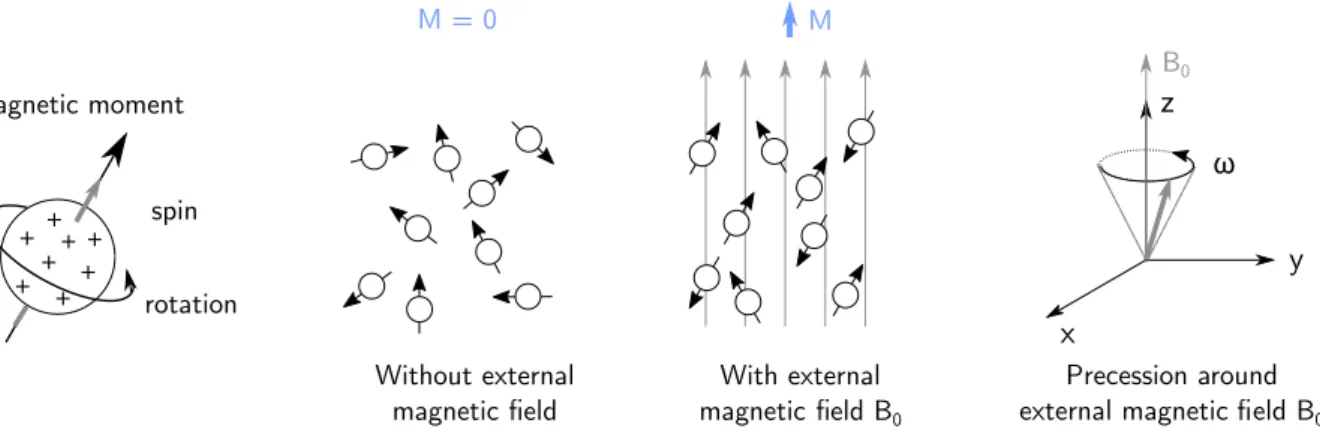
As an alternative to X-ray imaging, research on Magnetic Resonance Imaging (MRI) started in the early 1970s and the first MRI prototypes were tested in the 1980s [35]. MRI represents a tomography imaging technique which relies on the varying magnetic properties of atoms to produce an image. Thus, the imaging process does not involve the use of ionizing radiation which is associated with potential harmful effects and is therefore considered a non-invasive imaging method. Since water represents one of the main components of the human body, mainly the electromagnetic effects of the nuclei of hydrogen atoms, thus single protons, are used for imaging. In general, atoms with an odd number of protons or neutrons possess a nuclear angular momentum. These nuclei can be
M M = 0 Without external magnetic eld With external magnetic eld B0 + + + + + + + + magnetic moment rotation spin B0 ω Precession around external magnetic eld B0
Figure 2.6: Alignment of magnetic moments with and without external magnetic field B0. Without an external
magnetic field, the spins are randomly aligned. With B0 the spin of a hydrogen nucleus aligns either parallel or antiparallel to the external field and start to precess around its direction.
visualized as spinning charged spheres which possess a magnetic momentum as shown in Figure 2.6, referred to as spin. In the absence of an external magnetic field, these spins are oriented in random directions due to thermal motion and the summarized macroscopic magnetic moment M0 equals zero. The quantum model restricts the spin of a proton to align either parallel or anti-parallel to an external magnetic fieldB0 whereas the parallel alignment corresponds to a low-energy state and the anti-parallel alignment to a high-energy state. This results in an excess of parallel aligned spins creating a macroscopic magnetic moment M0 >0 when an external magnetic field is applied. The fact, that the nucleus has an angular momentum and therefore is subjected to momentum conservation is leading to a precession around the axis ofB0 (see figure 2.6). The frequency of this precession is called Lamor frequency ω0 and defined as
ω0=γ·B0. (2.3)
The factorγrefers to the gyromagnetic ratio, a known constant for each type of atom (for hydrogen:
γ/2π = 42.58 MHz/T). Conventionally, the direction of the applied magnetic field is defined as z-axis. Under this assumption, the transverse component M0,xy defined as the projection of M0 in the x-y-plane equals zero in equilibrium, since all contributions to M0,xy are dephased.
To obtain an MR signal, a radio frequency (RF) pulse is used to generate a secondary oscillating magnetic field B1 which is applied perpendicular to B0 using the resonance frequency matching
ω0. The spin precesses around the field direction of the superimposed magnetic field, causing the nuclear spin to spiral into a higher energetic state. Depending on the time that the oscillating field is active and its magnitude, the nuclear spin orientation is directed away from the z-axis, resulting in a transverse component M0,xy >0. If M0 is e.g. flipped completely to the x-y-plane, the corresponding radio frequency pulse is referred to as 90◦ pulse, a 180◦ pulse inverts the bulk magnetization. The RF excitement is followed by an exponential relaxation, during which the system is restoring its equilibrium state. It is possible to measure M0,xy during these processes
since it induces an electric voltage in another radio frequency coil. This signal builds the basis for the image formation in MRI.
longitudinal and transverse relaxation. The longitudinal relaxation refers to the increase of the longitudinal magnetization (parallel to B0) caused by an energy exchange between the spin system and the surrounding thermal reservoir referred to as ’lattice’ (spin-lattice relaxation):
M0,z(t) =M0
(︂
1−e−t/T1)︂. (2.4)
Here,T1 refers to the time constant of this relaxation process andtto the time since the application of the RF pulse. For t → ∞, the magnitude of the longitudinal magnetization approaches the magnitude of the initial magnetizationM0,z →M0. The transverse relaxation describes the decrease
of the transverse magnetization componentM0,xydue to interactions with neighboring atoms (
spin-spin relaxation) with time constantT2:
M0,xy =M0,xye−t/T2. (2.5)
Thus, for t → ∞, the transverse magnetization approaches zero M0,xy → 0. Generally T2 ≤ T1. However, the transverse relaxation is not only influenced by spin-spin interactions, but also by inhomogenities in the local magnetic field, which is taken into account by defining the effective transverse relaxation time T2∗.
T1 andT2 represent important quantities in MRI since they build the source of the image contrast. To obtain high quality relaxation signals, spin echo sequences are applied which describe a certain sequence of RF pulses to readout T1 and T2. A spin echo sequence consists of a 90◦ RF pulse followed by a 180◦ pulse. The 90◦ RF pulse tips the magnetization into the x-y-plane. M0,xy can
then be measured to defineT2∗, whereas the signal directly after a high frequency pulse is called Free Induction Decay. The spins start dephasing in the x-y-plane. By applying a 180◦ pulse, the spins flip in the opposite direction in the transversal plane and start to refocus resulting in a pronounced transverse magnetization signal, the so-called spin echo. Echo pulse sequences are characterized by the echo time TE which denotes the time between the 90◦ pulse and the spin echo signal. When
repetitive spin echo sequences are used, the time between the application is called repetition time
TR. It is possible to measure T1 by applying several 90◦ pulses with short TR, since it flips the
non-relaxed longitudinal magnetization in the x-y-plane where its signal can be measured. T2 can be measured by repetitively applying spin echo sequences measuring the amplitude decay of the spin echo signals.
To spatially encode the source of measured magnetization signal, a magnetic gradient field is superimposed to the constant external field B0 between the excitement with an RF pulse and the signal measurement. As a result, the effective Larmor frequency of the precession is depended on the local magnetic field ω0(z) = γ ·(B0+B(z)), and the previously aligned spins are artificially dephased. Thus, all spins precess in the same frequency but different phases, however, spins in the same row perpendicular to the gradient direction have the same phase. This is called phase encoding and translates to the phase of the measurement signal. This procedure can be extended by frequency encoding, which is also based on the use of a magnetic gradient field perpendicular to the gradient field for phase encoding. In contrast to phase encoding, this field is continuously applied during the signal measurement, causing the previously dephased spins to rotate with different
CT T1 MRI T2 MRI PET-CT CB-CT
a) b) c) d) e)
f) g) h) i) j)
Figure 2.7: Axial slices through an antrophomorphic phantom extracted at two different axial positions. The phantom is specifically designed to demonstrate the advantages of multimodal imaging, since the composition of the artificial lesions is chosen in such a way that the visibility varies in different image modalities. The phantom is measured under clinically relevant conditions using the different imaging devices: a) + f) Multislice CT; b) + g) T1-weighted MRI; c) + h) T2-weighted MRI; d) + i) PET-CT; e) + j) Cone Beam CT. The red arrows indicate the positions of the artificial lesions.
frequencies depending on the spatial position. By applying a reconstruction based on a Fourier transform and a complete sampling of the data matrix containing the raw MRI data, the so-called k-space, it is possible to generate a spatially resolved image based on the local magnitude of the transverse magnetization. This magnitude is proportional to the proton density of the tissue, and thus it is possible to deduce a proton weighted image. By varying TR during the application of a
series of 90◦ pulses, a T1-weighted image can be calculated, whereas the signal decreases with an increasing T1. Choosing long TR and TE when applying spin echo sequences allows to calculate T2-weighted images, in which the signal is proportional to T2. Thus, varying the time parameters during a measurement leads to different image contrasts.
This represents one of the main advantages of MRI, since it is possible to generate images with different contrasts using the same imaging device. Moreover, MRI offers a very pronounced soft tissue contrast, since the image generation is based on the spin of hydrogen nuclei, and even allows to perform functional imaging [36]. However, the application of measurement sequences takes up to several minutes, making it time-consuming and the images prone to motion artifacts due to patient movements. Additionally inhomogeneities in the magnetic field or effects such as chemical shift can lead to small distortions of the morphology [37, 38].
In summary, CT generally offers a stable spatial resolution, fast acquisition times and a pronounced contrast for high-density structures such as bones, whereas MRI as a non-invasive technique is fa-vorable for the imaging of soft tissue structures. Thus, it is often essential to employ multimodal imaging for diagnosis and treatment planning, to integrate all necessary information. The ad-vantages of multimodal imaging are demonstrated by the measurement of an antrophomorphic phantom (designed within the research campus M2OLIE) containing an artificial liver with three lesions: Figure 2.7 shows that some lesions are only visible in one modality, thus demonstrating the need for multimodal imaging. The material composition of the lesions has been specifically chosen to yield different contrast in different imaging devices.
Before registration After registration
u(x) v(y)
y = T(x)
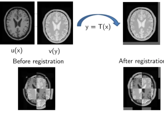
Figure 2.8: Optimization process of a geometrical transformation T so that corresponding features in the images
overlap.
2.3
Fundamentals of Image Registration
The main challenge in the context of multimodal imaging of a patient is the fact that images acquired on different devices do not occupy the same physical space. Moreover, the organs of the patient are subjected to deformation due to different positioning of the patient in different devices, organ motion or image artefacts, impeding the comparison between different scans. Here, image registration plays a major role to geometrically transform the images and enable the fusion of different modalities in to a multimodal data set.
Image registration generally describes the process of determining a geometrical transformation to align two images of the same object or scene and transfer them into the same coordinate system so that identical features overlap as shown in Figure 2.8. There are various situations in which this is necessary, including scenarios in which images of an object are acquired from different angles, with different devices or at different points in time. Depending on the intended application, image registration is used to either determine the geometrically transformed image data or the transformation parameters. Image registration builds the basis to compare images of the same object and is therefore important in many fields of application, ranging from computer vision and pattern recognition, to medical image processing or even geosciences, when it comes to compare data from satellites [39, 40]. In a clinical context, image registration is e.g. necessary to align images acquired on different imaging devices or for treatment monitoring of diseases over time.
In a registration scenario, one of the images is chosen as reference image which is stationary and is therefore calledtarget,reference orfixed image. The other image is referred to assource ormoving image, since it is the image which is going to be geometrically transformed to align with the target image. Image registration then aims to find a reasonable transformation so that the transformed version of the source image is similar to the reference image.
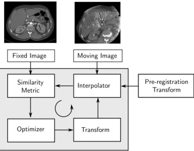
Most image registration methods are considered as iterative optimization processes which aim to find the optimal parameters of the geometric transformation to spatially align two images so that the similarity between both images increases. This process is typically composed of four basic elements as shown in Figure 2.9, namely:
Fixed Image Moving Image Similarity Metric Interpolator Optimizer Transform Pre-registration Transform
Figure 2.9: Visualization of the different components of a registration method and their interplay.
Atransformto geometrically distort the source image so that it aligns with the target image. The type of transformation also defines the number of transformation parameters which are optimized during the registration process.
Aninterpolatorto resample voxel intensities to the new coordinate system according to the geometric transformation.
Asimilarity metricwhich measures the (dis)similarity of images for the different geometric alignments during the registration process based on their intensities, geometric features or higher-level information, such es e.g. segmentation labels.
An optimizer which aims to find the optimum, i.e. minimum or maximum, of an energy function consisting of a similarity term or regularization penalty.
These components are often extended by a pre-registration transformation (or initialization) which prealigns the target and source image in terms of a higher image similarity. Initializations can be realized by overlaying the geometric image centers, by overlaying the intensity based centers of mass, by identifying and matching corresponding points [41] or features [42] in the images or by extracting and aligning segmentation masks [43].
All of these steps play a major role for the outcome of a registration method, and will therefore be explained in more detail in the following sections 2.3.2 – 2.3.5. Image registration approaches display a high diversity concerning the processed image information and structure of the methods. Therefore, a short overview of the different types of image registration algorithms will be given in the next section.
2.3.1 Classification of Image Registration Methods
There exist various different registration approaches, which can be sorted into different categories depending on various characteristics. According to Maintz et al. [4] these characteristics include:
Image dimensionality: 2D-2D, 3D-3D, 2D-3D or time series
Registration methods are divided according to the spatial dimensionality of the input images for which it is distinguished between 2D-2D, 3D-3D and 2D-3D registration methods. Most medical registration methods focus on 3D-3D registration methods to register tomographic data sets or 2D-2D registration methods to register slices of tomographic data. 2D-3D meth-ods are e.g. used for the registration of a preoperative 3D image to an intra-operative pro-jective image. In clinical applications, medical images are sometimes also acquired over time intervals to examine medical processes such as e.g. tumor growth, leading to the acquisition of 4D data sets. High image dimensionality corresponds to a high information density which needs to be processed by the registration algorithm and therefore generally increases the registration complexity as well as the computation time.
Nature of transformation: rigid, affine, deformable
In general, it is distinguished between rigid and non-rigid transformations. Rigid transforms are limited to translation and rotation and extended to affine methods by including scaling and shearing whereas non-rigid or deformable methods result in the generation of complex deformation fields. The type of the transformation determines the number of transform parameters which are optimized during registration and is therefore directly related to the complexity of the registration task. An extended overview over the different transformation models is given in section 2.3.2.
Domain of transformation: global vs. local
The domain of transformation describes the image area on which the transformation is ap-plied. A global transformation is applied on the entire image whereas a local transformation warps only a subsection, a so-called Region Of Interest (ROI). Registration methods are most commonly employed on a global basis, i.e. a global geometric transformation is applied to warp the source image to the target image.
Nature of registration basis: extrinsic vs. intrinsic
Extrinsic registration methods are based on the detection of foreign objects (markers), which are introduced to the image space and well visible in the image data. This allows a fast and easy registration by aligning the artificial object whereas the main drawbacks are the often invasive character of the object and the fact that provisions must be made before the image acquisition. Moreover, since extrinsic methods rely on the alignment of external objects, no patient information is included making it an insufficient method for tasks such as soft organ alignment.
In contrast, intrinsic methods rely on patient generated image content only. This content can be represented by salient image points (landmarks), by segmented binary structures such as object surfaces (segmentation based) or by the intensity distribution of the grey values in the image (voxel property based). In medical image processing, landmarks are points in the patients morphology which can be accurately detected and located. By identifying and matching these points on two data sets, it is possible to estimate a geometric transformation to align the images. In many applications, these points are interactively identified by a user which makes these approaches unsuitable for daily clinical routine. However, in some
cases where the landmarks are based on geometrical properties (corners, local curvature), an automatized identification is possible. A high density of such landmarks allows for an accurate registration including deformable tasks. Segmentation based methods extend this approach by identifying and aligning surfaces or volumes in different image data sets. In contrast, voxel property based methods rely directly on the image grey values without including prior knowledge as landmarks or segmentation based methods. To establish a similarity measure between the images, the grey value distributions in the images are analyzed via correlation metric, Fourier properties or other means of structural analysis. A brief overview over some of the most commonly employed intensity-based and approaches for feature-based metrics is given in chapter 2.3.4.
Interaction: interactive, semi-automatic, automatic
Concerning registration methods, three levels of user interaction are distinguished. Interactive processes include all methods in which a user performs the registration himself, but is assisted by software giving him visual or numerical feedback. Semi-automatic methods require either a user performed initialization of the image alignment or user generated input data such as e.g. segmentation labels or user feedback in form of a rejection of acceptance of the suggested registration hypotheses. Most approaches aim to realize an automatic registration method, which requires only the image data and limited information on the image acquisition by the user. Automatic methods provide a high comfort for the user making it suitable for daily clinical practice, however the method has to be very robust to limit potentially false registration results.
Optimization procedure: direct, indirect, multi-stage
Since image registration represents an iterative optimization procedure, the results highly depend on the choice of optimization. The required transform parameters can either be computed directly from the available data or searched for by finding the optimum of a cost function. A direct computation is often only feasible for sparse data (as e.g. shown in [44] or [45]). To search the optimal parameter setting a quasi-convex mathematical cost function has to be defined depending on the transformation parameters to quantify the similarity between the images. The optimization then aims to identify the optimum of this function. A summary of most commonly used optimization techniques used in image registration is given in chapter 2.3.5. Additional approaches to accelerate convergence of the optimization process include multi-resolution approaches during which the spatial image resolution is altered from coarse to fine during registration or multi-stage approaches, during which a rigid registration to roughly align the images is ensued by a deformable registration for fine alignment.
Involved image modalities: monomodal, multimodal, modality to model, patient to model Monomodal registration methods refer to the alignment of two or more images which were acquired using the same imaging device whereas multimodal registration involves the mapping of input images acquired on different devices, e.g. MRT and CT. The latter results in a highly increased difficulty for the registration method, since different imaging modalities are based on different physical principles and therefore often display dissimilar object structures as discussed in chapter 2.2. Thus, it represents a challenge to establish a relation between the
rigid a ne non-linear original
transformation
increasing number of degrees of freedom
Figure 2.10: Visualization of 2D transformations using the example of a rectangle.
input images to estimate and optimize an image similarity measure. Additional registration scenarios include the registration of an image modality to a mathematical model or the patient himself.
Registration subject: intra-subject, inter-subject, atlas
Intra-subject registration refers to tasks involving the image data of a single patient whereas inter-subject registration is accomplished when the data of different patients is registered. Atlas registration describes the task to register the image data of a patient to a constructed image from an image formation database, such as binary segmentation masks.
An extensive summary of these criteria is given in [4] and [46]. These categories show that there ex-ists a high diversity concerning image registration methods with profound differences in complexity and application possibilities. In this thesis, the focus is set to intra-subject mono- and multimodal 3D-3D registration techniques which provide a wide range of applications in clinical practice.
2.3.2 Geometric Transformation
A geometric transformation maps points from one image space to another. The choice of a geometric transformation model highly depends on the nature of the data to be registered and can be crucial for the success of a registration, since it defines the possibilities how an image can be warped. Some transformation types result in global transforms which are applied to the entire image whereas other transformation types yield local transforms which are only applied to a sub-region of the image and are therefore useful to correct small organ movement or deformations. In general, it is distinguished between linear and non-linear transformation types whereas the term ‘linear’ refers to the function which is used for the mapping of one vector space to the other. Both types will be briefly explained in the following. A visualization of the most commonly used geometric transformations is shown in Figure 2.10.
Rigid Transformation
The simplest type of linear transformations are rigid transformations. Rigid transformations com-prise geometric transformations which preserve the Euclidean distance between every pair of points in an image, which means any object will keep its shape and size after the application of a rigid transformation. Rigid transformations include translations and rotations as well as their combi-nations. They are very useful for the registration of rigid structures, such as e.g. the skull, or an
initial alignment of two images, but fail to compensate non-linear motions or organ deformations. However, due to its limited degrees of freedom (DOF), the employment of a rigid transformation allows for a fast and simple image registration since less parameters have to be optimized.
Mathematically, a rigid transformationT :R3 ↦→R3of the 3D pointx= (x, y, z) can be formulated
as
Trigid(x) =Rx+t (2.6)
Here,t = (tx, ty, tz) denotes the translation vector and R the rotation matrix. There are different
approaches to describe an arbitrary rotation either including Euler angles α1,α2,α3 or e.g. quater-nions. For 3D data, a rigid transformation is defined by the three components of the translation vector t and three parameters describing the rotation angles. This results in six DOF in 3D and four in 2D.
Affine Transformation
Affine transformations represent the simplest type of non-rigid transformations. They extend rigid transformations by shearing and scaling. Angles between lines or distances between points are no longer preserved, although the ratio of distances between points on a straight line are maintained as in the original image. Thus, they are also considered as type of linear transformation. A complex affine transformation Taffine can be represented by a sequence of basic transforms. This if often
described using homogenous coordinates which are a concept that stems from the mathematical field of projective geometry. They represent an extension of standart three-dimensional vectors and allow the simplification of various transforms and their computation. In homogenous coordinates, the sequence of basis transform to generate an affine transform can be expressed as a matrix multiplication such that
Taffine =Ttranslation·Trotation·Tshear·Tscaling = ⎛ ⎜ ⎜ ⎜ ⎜ ⎝ m1 m2 m3 m10 m4 m5 m6 m11 m7 m8 m9 m12 0 0 0 1 ⎞ ⎟ ⎟ ⎟ ⎟ ⎠ (2.7)
Here, m10 to m12 denote the translation parameters whereas all other parameters define scaling, shear and rotation. This results in 12 DOF in a 3D scenario, and six in a 2D scenario.
Nonlinear Transformation
The previously presented linear transformations mainly capture global image motion, but are not sensitive to model local structure deformations. This limitation is overcome by employing non-linear, or deformable, transformations which represent the most complex type of geometric trans-formations. They do not preserve straightness of lines nor parallelism, making them suitable to model complex tissue deformations. This requires a high number of DOF and is generally a sophis-ticated task.