R E V I E W
Open Access
Control of viral infections by
epigenetic-targeted therapy
Zeina Nehme
1,2, Sébastien Pasquereau
1and Georges Herbein
1,3*Abstract
Epigenetics is defined as the science that studies the modifications of gene expression that are not owed to mutations or changes in the genetic sequence. Recently, strong evidences are pinpointing toward a solid interplay between such epigenetic alterations and the outcome of human cytomegalovirus (HCMV) infection. Guided by the previous possibly promising experimental trials of human immunodeficiency virus (HIV) epigenetic reprogramming, the latter is paving the road toward two major approaches to control viral gene expression or latency. Reactivating HCMV from the latent phase (“shock and kill” paradigm) or alternatively repressing the virus lytic and reactivation
phases (“block and lock” paradigm) by epigenetic-targeted therapy represent encouraging options to overcome
latency and viral shedding or otherwise replication and infectivity, which could lead eventually to control the infection and its complications. Not limited to HIV and HCMV, this concept is similarly studied in the context of hepatitis B and C virus, herpes simplex virus, and Epstein-Barr virus. Therefore, epigenetic manipulations stand as a pioneering research area in modern biology and could constitute a curative methodology by potentially consenting the development of broad-spectrum antivirals to control viral infections in vivo.
Keywords:Virus, HIV, HCMV, Epigenetics, Treatment, Cancer
Background
Since its emergence for the first time in 1940, the epigen-etic field is witnessing a continuous surge over the last de-cades [1]. Although the epigenetic term is well thought out to be a large umbrella under which falls concepts re-lated to development, heredity, and evolution [2], recent technical advancements have narrowed the term’s defin-ition in the standpoint of molecular biology [3]. Hence, epigenetics could be defined as “the study of heritable changes in gene expression that are not due to changes in DNA sequence” [4]. Recent numerous literature is show-ing a correlation between epigenetic modifications and a wide array of human diseases including—but not limited to—cancer, neurological and psychiatric disorders (Alzhei-mer’s disease, schizophrenia), autoimmune disorders (rheumatoid arthritis, systemic lupus erythematosus), and others [5–7]. However, this association was converted and extended to the clinical level first in the cancerology field
with the FDA-approved DNA methyltransferase (DNMT) inhibitors (azacytidine, decitabine) and histone deacetylase (HDAC) inhibitors (vorinostat, romidepsin, belinostat, panobinostat) [8]. In fact, in addition to DNMT and HDAC, the epigenetic machinery entails composite com-plexes that each of which could constitute a valuable tar-get for the development of potential new epigenetic antiviral drugs [9]. This review examines and discusses the involvement and the role of various epigenetic players throughout the different viral life cycle stages and high-lights their potential implications in the clinical manage-ment of several viral infections, especially human immunodeficiency virus (HIV) and human cytomegalo-virus (HCMV), in addition to hepatitis cytomegalo-viruses, herpes simplex virus-1 (HSV-1), and Epstein-Barr virus (EBV).
HIV and epigenetics, a leading example as a proof of concept
Since the introduction of combination antiretroviral therapy (cART), survival and quality of life among HIV-infected patients significantly improved [10], with a more favorable outcome with therapy initiation in the setting of early asymptomatic infection [11]. This shifted
© The Author(s). 2019Open AccessThis article is distributed under the terms of the Creative Commons Attribution 4.0 International License (http://creativecommons.org/licenses/by/4.0/), which permits unrestricted use, distribution, and reproduction in any medium, provided you give appropriate credit to the original author(s) and the source, provide a link to the Creative Commons license, and indicate if changes were made. The Creative Commons Public Domain Dedication waiver (http://creativecommons.org/publicdomain/zero/1.0/) applies to the data made available in this article, unless otherwise stated. * Correspondence:[email protected]
1
Department Pathogens & Inflammation-EPILAB, UPRES EA4266, University of Franche-Comté, University of Bourgogne Franche-Comté, 16 route de Gray, F-25030 Besançon cedex, France
3Department of Virology, CHRU Besancon, F-25030 Besançon, France
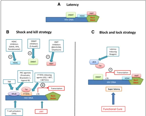
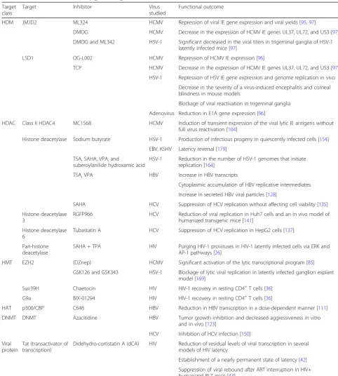
HIV conception from a non-curable devastating fatal illness to a possibly manageable chronic one. However, cART is yet not the ideal road map for HIV manage-ment, as physical and psychological burden are still im-posed by this therapy [12, 13], leading sometimes to a reduced compliance or even discontinuation [14]. Mark-edly, a major limitation associated with cART cessation is viral rebound [15]. This is due to the presence of HIV reservoirs, mainly in the latently infected resting CD4+ memory T cells and myeloid cells such as macrophages and microglia, that are difficult to be targeted by cART or immune effector mechanisms [16–18]. Interestingly, the integrated provirus in those cells is subjected to transcriptional silencing by host chromatin-modifying enzymes, comprising deacetylases, methyltransferases, and others [19]. This paved the road to the emergence of two new epigenetic therapeutic approaches, namely the “shock and kill” and the “block and lock” strategies [20–23]. Here, we present general points about these two anti-HIV therapeutic strategies (Fig.1, Table1).
Shock and kill strategy
This strategy is grounded on the concept that the latent HIV provirus could be switch on from latency (shock) into an active form prone to eradication (kill) through the humoral immune response, CD8+ T cells-mediated lysis, virus-induced apoptosis, or activation-induced cell death [24]. Several latency-reversing agents (LRA) or “shock” inducers have been proposed [25, 26] including histone deacetylase (HDAC), histone methyltransferase (HMT), and DNA methyltransferase (DNMT) inhibitors. Histone deacetylases family is composed of 18 enzymes that are gathered into four major groups: HDAC I–IV [27]. HDAC enzymes are responsible of removing acetyl groups from histones, which favors the formation of a compacted, tran-scriptionally repressed chromatin structure [28]. HDACs have gained an ascending attention after the FDA approval of HDACs inhibitors for cancer treatment [29], such as vorinostat or suberanilohydroxamic acid (SAHA) for the management of cutaneous T cell lymphoma [30] and panobinostat in relapsed multiple myeloma [31]. HDAC inhibitors like SAHA or DNMT inhibitors could be used alone to reactivate HIV gene expression along with effi-cient cART [32]. For instance, co-treatment with the HDAC inhibitor SAHA and the global T cell activator 12-O-tetradecanoylphorbol-13-acetate (TPA) revealed a significant synergistic effect on purging HIV-1 proviruses in HIV-1 latently infected cells [26]. Other activators of NF-kB such as prostratin have been also used in combin-ation with HDAC inhibitors to reactivate HIV, these former players reactivating HIV in the absence of immune activation [33]. The concomitant use of protein kinase C (PKC) agonists (prostratin, bryostatin-1, and ingenol-B), which are known to activate NF-κB signaling pathway as
well as the positive transcription elongation factor B (P-TEFb), used alone or in combination with P-TEFb-releasing agents (HMBA and Bromodomain and Extra-terminal (BET) inhibitors JQ1, I-BET and I-BET151) leads to synergistic HIV reactivation from latency [34]. More-over, sequential treatment with the DNMT inhibitor 5-aza-2′-deoxycytidine (5-AzadC) and HDAC inhibitors reactivates HIV-1 from latency [35]. In addition, it has been shown that the use of chaetocin and BIX-01294, specific inhibitors of HMT Suv39H1 and G9a respectively, resulted in HIV-1 recovery in resting CD4+ T cells in highly active antiretroviral therapy (HAART)-treated pa-tients with undetectable viral load [36]. Another thera-peutic approach could be considering tumor necrosis factor alpha (TNF)-based therapies, where combining HDAC inhibitors or HMT inhibitors with TNF, disrupts HIV-1 latency by triggering the activation of transcrip-tional activators like NF-κB and preventing the formation of heterochromatin, enhancing thus HIV-1 long terminal repeat (LTR) transcription and viral purge [37]. In fact, targeting several cellular proteins involved in the epigen-etic control of viral gene expression usually amplifies HIV-1 reactivation. Although this approach is facing sev-eral hurdles, including—but not limited to—reactivating and possibly eliminating only a small subset of the latent HIV genome, it constitutes however one tactic that could be used in parallel to other approaches to achieve a fully effective cure [38].
Block and lock strategy
HIV-specific viral protein with no cellular homolog, using dCA to “block and lock” HIV should not silence other regulatory pathways essential to fight other infec-tions. In addition, Akt activation favors HIV-1 reactiva-tion from resting CD4+ T cells and monocytes/ macrophages, the two major HIV-1 cellular reservoirs [46–48]. Thus, Akt inhibitors, but also HIV protease in-hibitors which display an anti-Akt activity [47], inhibit Akt activation in HIV-1 infected cells thereby favoring a “lock” stage, decreasing cell viability, and opening thus the door to the clearance of infected cells under Akt blockade. These results strongly encourage and open new insights to the possible addition of the “block and
lock” approach as an additional potential therapeutic management strategy.
Human cytomegalovirus and epigenetics
Human cytomegalovirus (HCMV) is a ubiquitous patho-gen also denoted as human herpesvirus 5 (HHV5). It is a member of betaherpesvirinae, a subfamily of the Herpes-viridae family [49]. HCMV infection is very common, as 40 to 95% of the population is seropositive [50]. However, the pathological outcomes depend on the host’s immune status, where infection in immunocompetent individuals rarely causes evident manifestations at the clinical level [51]. Conversely, HCMV infection significantly affects
A
B
C
morbidity and mortality in solid organ or stem cell trans-plantation recipients and immunocompromised individ-uals such HIV patients [52, 53], where infection could result in interstitial pneumonia, retinitis, gastrointestinal tract complications like gastroenteritis, hepatitis, and graft failure [54]. Added to the previously mentioned hosts, HCMV infection poses a real burden in congenitally
infected newborns with immature immune system, result-ing possibly in deafness and neurodevelopmental delay [55]. HCMV exhibits two modes of viral infection: a lytic and a latent one [56]. The lytic phase is a highly regulated stage that ensures the production and the release of the new viral progeny outside the infected cells. This is followed by latency, a state characterized by a lifelong Table 1Functional outcomes of epigenetic regulation in viral infections
Target class
Target Inhibitor Virus
studied
Functional outcome
HDM JMJD2 ML324 HCMV Repression of viral IE gene expression and viral yields [95,97]
DMOG HCMV Decrease in the expression of HCMV IE genes UL37, UL72, and US3 [97]
DMOG and ML342 HSV-1 Significant decreased in the viral titers in trigeminal ganglia of HSV-1 latently infected mice [97]
LSD1 OG-L002 HCMV Repression of HCMV IE expression [96]
TCP HCMV Decrease in the expression of HCMV IE genes UL37, UL72, and US3 [97]
HSV-1 Repression of HSV IE gene expression and genome replication in vivo
Decrease in the severity of a virus-induced encephalitis and corneal blindness in mouse models
Blockage of viral reactivation in trigeminal ganglia
Adenovirus Reduction in E1A gene expression [96]
HDAC Class II HDAC4 MC1568 HCMV Induction of transient expression of the viral lytic IE antigens without full virus reactivation [104]
Histone deacetylase Sodium butyrate HSV-1 Production of infectious progeny in quiescently infected cells [154]
EBV, KSHV Latency reversal [179]
TSA, SAHA, VPA, and
suberoylanilide hydroxamic acid
HSV-1 Reduction in the number of HSV-1 genomes that initiate replication [164]
TSA, VPA HBV Increase in HBV transcripts
Cytoplasmic accumulation of HBV replicative intermediates
Increase in secreted HBV viral particles [128]
SAHA HCV Suppression of HCV replication without affecting cell viability [135]
Histone deacetylase 3
RGFP966 HCV Reduction of viral replication in Huh7 cells and an in vivo model of humanized transgenic mice [141]
Histone deacetylase 6
Tubastatin A HCV Suppression of HCV replication in HepG2 cells [137]
Pan-histone deacetylase
SAHA + TPA HIV Purging HIV-1 proviruses in HIV-1 latently infected cells via ERK and AP-1 pathways [26]
HMT EZH2 (DZnep) HCMV Significant activation of the lytic transcriptional program [85]
GSK126 and GSK343 HSV-1 Blockage of lytic viral replication in latently infected ganglion explant model [169]
Suv39H Chaetocin HIV HIV-1 recovery in resting CD4+T cells [36]
G9a BIX-01294 HIV HIV-1 recovery in resting CD4+T cells [36]
HAT p300/CBP C646 HBV Reduction in HBV transcription in a dose-dependent manner [111]
DNMT DNMT Azacitidine HBV Tumor growth inhibition and decreased aggressiveness in vitro and in vivo [123]
HCV Inhibition of HCV infection [150]
Viral protein
Tat (transactivator of transcription)
Didehydro-cortistatin A (dCA) HIV Reduction of residual levels of viral transcription in several models of HIV latency
Establishment of a nearly permanent state of latency [42]
persistence in the host with the ability to reactivate under certain circumstances [53, 54]. During lytic infection, HCMV endures a well-regulated cascade of gene expres-sion that starts with the expresexpres-sion of the immediate early viral genes [57] via the interaction of various cellular factors with the major immediate-early promoter (MIEP) [58]. This is followed by the expression of the early viral genes that play a role in the cellular modulation to favor viral replica-tion [59] and later on by the late viral gene expression that ensures viral progeny assembly and release [60]. Recent studies have shown that epigenetic modifications play a role in the early productive infection events [61]. In fact, after viral entry, viral DNA rapidly becomes associated with his-tones, which makes it a vulnerable candidate to epigenetic modifications [62]. Such modifications usually result in a si-lent repressive state to viral gene expression as an intrinsic cellular defense mechanism. However, this repression is ul-timately overcome, allowing the sequential expression cas-cade of lytic viral genes mentioned previously [63].
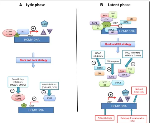
Although the few available antiviral drugs have granted chief advances in HCMV disease treatment and prophy-laxis, their clinical applicability and utility confront several barriers [64]. First, resistance to antivirals is documented after prolonged use [65]. Added to this is the poor oral bioavailability and the dose-limited hematologic and renal toxicities reported with their use [66]. Moreover, they do not target the latent viral form in the host, which leaves the door open for viral shed-ding and transmission in saliva, urine, milk, vaginal secretions, and other bodily fluids [67]. It is worth to mention that the current antivirals used in HCMV man-agement are ganciclovir, and its oral prodrug valganci-clovir, cidofovir, and foscavir that target the viral DNA polymerase, in addition to fomivirsen, an antisense anti-viral drug used in the treatment of CMV retinis [68], and the recently FDA-approved letermovir used to pre-vent viral infection following allogenic hematopoietic stem cell transplant [69]. Since those antivirals target the viral DNA replication step, the function and expression of the immediate early (IE) and early (E) HCMV genes during the early stages of infection are not blocked, pav-ing the road to immunopathology and raise the risk of graft rejection [70]. Thus, this mandates and sheds the light on the urgent necessity of developing new antiviral drugs with novel mechanisms of action based on new potential viral or cellular targets. This is particularly conceivable with the enhanced understanding of HCMV molecular biology and the epigenetic mechanisms involved with its regulation (Fig.2, Table1).
Reactivating HCMV from the latent phase the“shock and kill”paradigm
In contrast to the virus’wide tropism during productive infection [71], several laboratories have pinpointed the
against HCMV, associates with the proximal enhancer region of the MIEP and promotes heterochromatin condensation possibly through the recruitment of co-repressors that SPOC1 is known to interact with not-ably the previously mentioned KAP-1 and H3K9 methyltransferase [88]. On the other hand, Ets-2 re-pressor factor (ERF) is a cellular protein that physic-ally interacts with the HCMV MIEP and functions as a transcriptional repressor of the latter by suppressing IE gene expression [89]. GST fusion assays showed a strong interaction between ERF and the N-terminus of HDAC1, a result that was further confirmed in vivo, suggesting that the physical interaction
between ERF and HDAC1 could mediate repression of the MIEP [90]. Moreover, ying-yang 1 (YY1), a zinc finger DNA-binding protein and a multifunc-tional transcription factor [91], has been shown to re-press the HCMV MIEP [92] partly by indirectly recruiting HDACs to the promoter via the nuclear protein SAP30, a component of the human HDAC complex [93]. In fact, forcing HCMV out of latency along with the conventional antiviral drug use in an attempt to eradicate it and establish a sterilizing cure is in correspondence with the “shock and kill” con-cept currently studied in HIV. Nevertheless, a better understanding of the molecular mechanisms involved
A
B
in HCMV latency and reactivation could open future avenues for HCMV infection control.
Repressing both HCMV lytic and reactivation phases by epigenetic-targeted therapy,“blocking and locking”the virus
HCMV lytic cycle and reactivation from latency are under the control of several epigenetic mechanisms. For example, histone demethylases (HDMs), due to their ability to remove the repressive marks, will promote pro-ductive infection. HDMs constitute a large family of more than 20 demethylases that are divided into two functional enzymatic families: the Lys-specific demethy-lase (LSD), also known as KDM1A and the Jumonji C (JMJC) protein families [94]. Two HDMs were studied in the context of HCMV lytic infection: KDM4 (JMJD2) and KDM6 (UTX/JMJD3), which demethylase histone H3-lysine 9 and lysine 27, respectively [95]. By using the JMJD2 demethylase inhibitor ML324, viral IE gene ex-pression and viral yields were potently repressed, which could suggest that targeting these histone demethylases may potentially block viral gene expression and viral replication at a very early stage of infection and possibly abrogate it. In the same perspective, the LSD1 inhibitor OG-L002 repressed the expression of HCMV IE expres-sion in HCMV-infected MRC5 cells [96]. In addition, the use of another LSD1 inhibitor, tranylcypromine (TCP), or alternatively the JMJD2 inhibitors, dimethylox-alylglycine (DMOG) or the previously mentioned ML324, resulted in a decrease in the expression of HCMV IE genes UL37, UL72, and US3 with a noted po-tent inhibition of IE gene expression with ML324 [97]. Thus, HDMs inhibitors could provide a therapeutic tool to target the initiation of infection or the spontaneous reactivation by blocking the viral cycle at an early stage, as a mimic for the proposed HIV“block and lock” strat-egy. This could be highly beneficial in the context of HCMV infection, as the expression or functions of viral IE and E gene products has shown their potential ability to elicit immuno-inflammatory responses that can lead to tissue rejection [98]. Not limited to inflammatory damage, some IE gene products can significantly inter-fere with important oncogenic signaling pathways and exhibit oncomodulatory properties, such as in glioblast-oma cells [99]. In the context of oncomodulation, the PRC2 complex is associated with HCMV latency by the induction of transcriptional silencing [81–84]. HCMV-infected cells have been showed to exhibit enhanced ex-pression of cellular oncogenic pathways, including c-Myc, c-Fos, c-Jun, Akt, and NF-κB [100–103]. This transcriptional activation will in turn lead to an increased expression of EZH2, resulting in an auto-amplifying loop. This oncomodulatory effect of HCMV infection could be targeted by PRC2 inhibitors and
HDAC inhibitors that could both block cellular trans-formation and induce the activation of the viral lytic transcriptional program. This latter effect could allow through the expression of IE antigens the infected cells to be cleared by CMV-specific cytotoxic T cells (CTLs) [104].
Epigenetic therapy, a general approach to cure viral infections?
Besides infection with HIV and HCMV, any viral infec-tion might be potentially treated by new therapeutics targeting the epigenetic mechanisms (Table 1). We present below several examples of viral infections which could benefit from such new therapies.
Hepatitis B virus
resulting in a marked increase of H3K9me3 and a de-crease of H3K4me3 on cccDNA [110]. Moreover, treat-ment with the small molecule C646 that specifically inhibits p300/CBP, the histone acetyltransferases (HAT) for H3K27ac and H3K122ac reduced HBV transcription in a dose-dependent manner in the absence of measur-able toxicity [111]. Likewise, PRMT5, a protein arginine methyltransferase 5, restricted HBV transcription and replication partly through regulation of symmetric dimethylation of arginine 3 on H4 on cccDNA exclu-sively [109]. In the same context, PRMT1, another argin-ine methyltransferase, is directly recruited to cccDNA, where its overexpression results in a 60% inhibition of HBV transcription in HepG2 cells. It is worthy to men-tion that this transcripmen-tion inhibitory effect is limited to PRMT1 as PRMT3 overexpression did not affect tran-scription. Interestingly, PRMT1 was shown to interact with the regulatory hepatitis B virus X protein (HBx), which in turn inhibits PRMT1 methyltransferase activity [112]. HBx protein is a multifunctional regulatory pro-tein that enhances HBV replication in vitro and in vivo [113] and affects numerous cellular processes including apoptosis [114], DNA repair mechanism [115], mito-chondrial function [116], and cell signaling [117, 118]. Importantly, HBx could epigenetically influence cccDNA transcription through its recruitment onto the cccDNA minichromosome where it modulates the recruitment of chromatin-modifying enzymes such as the acetyltrans-ferase p300 and HDACs including Sirt1 and HDAC1 [119]. HBx could also induce epigenetic aberrations that may lead to HBV-related HCC [120]. Those abnormal-ities include hypermethylation of several tumor suppres-sor genes, including—but not limited to—IGFBP-3 by DNMT3A1 and DNMT3A2 [121] and the E-cadherin promoter by DNMT1 [122]. Treatment with the DNMT inhibitor AZA restored the expression of the HBx-mediated epigenetically repressed secreted frizzled-related protein 1 (SFRP1), resulting in tumor growth in-hibition and decreased aggressiveness in vitro and in vivo through negatively regulating the Wnt/β-catenin signaling pathway. This effect was further synergized by the use of the HDAC inhibitor trichostatin A (TSA) [123]. HBx-induced upregulation of SIRT2 expression promotes HBV replication in HepAD38 cells and en-hances cell migration and invasion in the human hepa-toma Huh7 cells, facilitating thus hepatocarcinogenesis [124]. The HBx protein upregulates the insulin-like growth factor 2 (IGF2) oncogene through hypomethyla-tion of its promoter resulting in a poorer clinical out-come for HBV-related HCC patients [125]. Further understanding of the exact mechanisms of HBx-induced epigenetic alterations is highly needed, as those modifi-cations could be used as biomarkers for the detection of early malignant transformation or as potential targets to
treat persistent infection or HBV-related HCC. Never-theless, for other epigenetic players such as DNMT, a critical balance in the context of HBV-related HCC should be maintained. Although DNMTs induce a de-crease in the viral gene expression and replication [126], it could also result in silencing of tumor suppressor genes through DNA methylation, contributing thus to hepatocarcinogenesis [127]. On the other hand, treat-ment of HBV-transfected HuH7 cells with class I/II HDAC inhibitors, valproic acid (VPA) and TSA resulted in an increase in HBV transcripts, cytoplasmic accumu-lation of HBV replicative intermediates, and an increase in secreted HBV viral particles [128]. This was on con-troversy to a study that showed that some HDAC inhibi-tors like TSA and apicidin, a class I HDAC-specific inhibitor, suppressed cccDNA transcription in a duck hepatitis B virus (DHBV)-transfected chicken hepatoma cell line [129], which could be possibly due to some specificities or differences at the cellular level between the avian and the human cell model or at the viral level between the human and the duck virus.
Hepatitis C virus
HCV by activating the Th1-type immune system. Cor-respondingly, tubastatin A, a selective inhibitor of HDAC6 suppressed HCV replication in HepG2 cells, along with α-tubulin hyperacetylation [137]. Although α-tubulin is known to be deacetylated by the histone deacetylase HDAC6 [138], the latter also controls the acetylation levels of other targets such as peroxire-doxins Prx1/2 [139] and the chaperone Hsp90 [140]. Thus, studying the effects of tubastatin A on those targets would be necessary to unveil the mecha-nism(s) by which this inhibitor is mediating its anti-viral activity. Similarly, the HDAC3 inhibitor RGFP966 reduced viral replication in Huh7 cells and in in vivo model of humanized transgenic mice [141] with a downregulation in Apo-A1 expression, an in-dispensable protein for HCV infectivity maintenance [142], leading possibly to HCV secretion suppression. On the other hand, it has been shown that HCV in-fection could result in the DNA hypermethylation of some epigenetic markers [143]. For example, the methylation of the suppressor of cytokine signaling 1 (SOCS1), a negative regulator of the JAK/STAT path-way regarded as a tumor suppressor gene [144], was found to be positively associated with HCV infection status [145]. In the same context, the promoter of the tumor suppressor gene GADD45 (growth arrest and DNA damage-inducible gene 45) was detected to be hypermethylated in the context of HCV infection in mice transgenic for the entire HCV open reading frame, notwithstanding that the exact HCV-altered methylation mechanisms during infection remain to be explored [146]. The hypermethylation of GADD45 promoter by HCV downregulates GADD45 gene ex-pression and interferes with its ability to block prolif-eration and tumorigenesis [147]. In addition, hepatitis C virus core protein has been linked to E-cadherin and p16 downregulation through upregulation of DNMT1 and DNMT3b [148, 149]. Thus DNMT in-hibitors could constitute a novel approach for the treatment HCV-associated HCC. In this context, 5-Aza-C and 5-Aza-dC, two well-known DNMT in-hibitors, significantly inhibited HCV infection. Inter-estingly, this effect is due not only to a decreased DNMT expression, but also through DNMT1 degrad-ation [150]. It is worthy to mention that DNMTs ex-pression could be varied between the different HCV genotypes 1b, 2a, 3a, 4h, and 5a. For example, DNMT3b mRNA is upregulated in genotype 1b HCV but not changed in genotypes 2a, 3a, 4h, and 5a [151]. Hence, the identification of HCV-induced epigen-etic regulation that may actively participate in tumorigen-esis and linking their prevalence to different HCV genotypes could possibly decipher new therapeutic targets for HCV infection and HCC management.
Herpes simplex virus
is possibly due to the HDACi-induced increase in the levels of some intrinsic immunity proteins know to exhibit antiviral immunity like promyelocytic leukemia (PML) bodies [166]. Same was shown with the nucleosome re-modeler chromodomain helicase DNA-binding 3 protein (CHD3) that mediates repression of HSV genome upon infection. The CHD3 protein conserves its ability to iden-tify and bind the repressive histone marks H3K27-trimethyl and H3K9-H3K27-trimethyl, promoting the formation of heterochromatin [167, 168]. Surprisingly, the EZH2/1 inhibitors GSK126 and GSK343 suppressed productive viral lytic phase and decreased viral yields instead of indu-cing activation in vitro and in vivo [169]. This was attrib-utable to the fact that treatment with those inhibitors enhanced cellular antiviral state by triggering antipathogen pathways. In the same context, other epigenetic players could negatively impact the reactivation of HSV-1 latent or quiescent infection. The JMJD2 inhibitors DMOG or ML342 significantly decreased the viral titers in trigeminal ganglia of HSV-1 latently infected mice through suppress-ing IE gene transcription and expression [97]. Attractively, the monoamine oxidase inhibitor (MAOi) tranylcypro-mine repressed HSV IE gene expression and genome rep-lication in vivo, decreased the severity of a virus-induced encephalitis and corneal blindness in mouse models, and blocked viral reactivation in trigeminal ganglia [170,171]. This is explained by the ability of MAOis to inhibit the LSD-1 mediated demethylation of lysine residues via a flavin-adenine-dinucleotide-dependent reaction [172,
173], resulting in the accumulation of repressive H3K9 chromatin marks at the IE promoters. Further studies are definitely needed to identify additional possible compo-nents and mechanisms involved in the epigenetic regula-tion of HSV-1 infecregula-tion.
Epstein-Barr virus
Epstein-Barr virus (EBV), or human herpesvirus 4 (HH4), is a DNA virus [174] that belongs to the Herpes-viridae family, subfamily Gammaherpesvirinae [175]. Like other herpes viruses, EBV persists predominantly in the latently infected B lymphocytes as a covalently closed circular episome [176]. It has been shown that re-activation from latency is coupled to and initiated by ex-pression of the viral BZLF1 gene [177]. During latency, BZLF1 promoter is silenced partly by the recruitment of repressive factors, such YY1 and the zinc finger E-box-binding factor (ZEB), that block the access of some transcriptional activators and ease the binding or function of repressive transcriptional co-factors like HDAC, maintaining thus a low levels of histone acetyl-ation [178, 179]. Therefore, it is not surprising that HDAC inhibitors, like sodium butyrate, can reverse la-tency [180]. Not limited to low acetylation level, BZLF1 gene proximal promoter Zp is also silenced due to
by the use of the HDAC inhibitor vorinostat, in a xeno-graft mouse model, resulting in cells sensitization to doxorubicin and cyclophosphamide, as evidenced by the increased survival rate [194].
Epigenetic players in viral infections: filling the gap between basic research and clinical
application
Powered by the supportive results in cell cultures and mouse models, epigenetic drug candidates are recently being clinically evaluated as potential antiviral drugs. In this section, some completed and ongoing clinical trials are cited, as an attempt to present preliminary data about the use of epigenetic drugs to manipulate viral
infections or viral infection-related malignancies (Table 2). As most trials are being published recently, analyzing toxicity, schedules, doses, and measuring clin-ical response stand up as the main aim. In the setting of HIV infection, several HDAC inhibitors were tested as a combination with antiretroviral therapy: panobinostat (NCT01680094), vorinostat (NCT01319383), and romi-depsin (NCT02092116, NCT01933594), in addition to VPA (NCT00289952). Although with varying degrees, all HDAC inhibitors showed an increase in viral transcrip-tion with no significant effect on the size of the HIV-1 functional reservoir, as no inhibitor has demonstrated complete clearance of latent infection [195–198]. This is possibly attributable to incomplete latency reversal or
Table 2Clinical trials of histone deacetylase inhibitors in viral infections and viral-associated malignancies
Drug Combination Indication Clinical result Study phase
and status
Trial* Reference
Panobinostat Antiretroviral therapy
HIV infection -Increase in unspliced HIV RNA -No reduction in integrated HIV DNA
-Safe, well tolerated
Phases I–II NCT01680094 [195]
-Antiretroviral therapy
-Interferon-alpha2a
HIV infection Ongoing Phases I–II NCT02471430
Vorinostat Antiretroviral therapy HIV infection -Increase in cell-associated HIV RNA with no effective depletion of persistent HIV reservoir -Safe, well tolerated
Phases I–II NCT01319383 [196]
-Antiretroviral therapy -Autologous dendritic cell vaccine (AGS 004)
HIV infection No published results yet Phase I NCT02707900
-Antiretroviral therapy -Disulfiram
HIV infection Suspended Phases I–II NCT03198559
Romidepsin Antiretroviral therapy HIV infection -Increase in cell-associated un-spliced HIV-1 RNA -No effect on the number of HIV-specific T cells -No severe adverse events
Phases I–II NTC02092116 [197]
Antiretroviral therapy HIV infection No published results yet Phases I–II NCT01933594 MVA.HIVconsv
vaccine
HIV infection No published results yet Phase I NCT02616874
Broadly neutralizing antibody (3BNC117)
HIV infection Ongoing Phase II NCT03041012
Valproic acid Antiretroviral therapy
HIV infection No significant reductions in the frequency of CD4+ T cells harboring replication-competent HIV
Phase II NCT00289952 [198]
Belinostat None Unresectable
hepatocellular carcinoma
-Tumor stabilization -Well tolerated
Phases I–II NCT00321594 [200]
Mocetinostat None Relapsed and refractory classical Hodgkin lymphoma
-Decrease in tumor measurements -Grade 3 and 4 adverse events (neutropenia and pneumonia)
Phase II NCT00358982 [201]
Tractinostat Valganciclovir EBV-associated lymphoid malignancies
Ongoing Phase Ib/II NCT03397706
insufficient clearance of latency-reactivated cells. It is suggested that the clearance of HIV latent reservoir could be enhanced by adding immune enhancement treatments, such as the immunomodulatory cytokine interferon-alpha2a with panobinostat (NCT02471430); the therapeutic vaccine MVA.HIVconsv (NCT02616874); the bNAb-based therapeutic HIV vaccine 3BNC117 (NCT03041012) with romidepsin; alternatively, the au-tologous dendritic cell vaccine AGS 004 (NCT02616874); or disulfiram (NCT03198559) with vorinostat, as disulfiram was shown to reactivate latent HIV-1 in a pri-mary CD4+ T cell model [199]. Importantly, caution should be engaged as the increased efficacy implicated by those combinations could be complemented with adverse effects not noted with the used of HDAC inhibitors alone. Not limited to HIV infection, some HDAC inhibitors have been tested in the context of viral-induced malignancies. Treatment with belinostat demonstrated tumor stabilization in unresectable hepatocellular carcinoma (NCT00321594) [200]. In addition, mocetinostat (MGCD0103) showed promising disease control in patients with relapsed classical Hodgkin lymphoma (NCT00358982) [201]. Tractinostat (VRx-3996) in combination with valganciclovir is currently under investigation in EBV-associated lymphoid malignan-cies (NCT03397706). Interestingly, HDAC inhibitors could induce EBV lytic-phase gene expression and act as sensitizers to antivirals for the treatment of EBV-associated lymphomas [202] as in the case of ar-ginine butyrate and ganciclovir [203]. Another epigen-etic player, the DNMT inhibitor azacitidine, was shown to reverse the dense CpG methylation and po-tentially triggering gene re-expression in patients with EBV-positive tumors [204]. Although those inhibitors might be future promising candidates for inclusion in the current therapeutic management, caution should be taken as they could reactivate some latent DNA vi-ruses like HBV or EBV in the setting of other condi-tions treatment [205]. For instance, the use of SAHA or TSA aggravated the severity of myocarditis induced by coxsackievirus B3 (CVB3) through CVB3-induced myocardial apoptosis [206]. Thus, a careful assessment before the use of HDAC inhibitors is highly needed.
Conclusion and future perspectives
Over the past years, epigenetic studies have revealed novel principles and profoundly broadened our knowledge about the interplay between viruses, cellular transcription factors, histones, and nonhistones modifying enzymes. As most of those modifications are reversible, manipulating this com-plex machinery could have a critical role in determining an active lytic or latent viral infection and subsequent viral re-activation from latency. This diverts the end goal to per-manently silence the virus in latent reservoirs so that the
possibility of reactivation is diminished, or to eradicate it through purging the viral reservoirs after reactivating it. Advantageously, and in contrast to the conventional antivi-rals, it is hypothesized that the emergence of resistant strains is minimized, as those chromatin modulation components target the host, rather than viral-encoded fac-tors. Furthermore, epigenetic therapy could exemplify the “two birds, one stone” concept in the scenario of viral co-infection, e.g., with HCMV-HSV, HIV-HCV, and HIV-HBV. However, this tremendous array for new targets is a double-edged sword. In fact, the available epigenetics therapies lack specificity, which raises questions about their cytotoxic side effects due to unintended global epigenetic modifications and complicates the achievement of a thera-peutic index within the acceptable toxicity levels. Intri-guingly, designing and testing target-specific inhibitors (specific HDAC inhibitors rather than pan-inhibitors for example) could improve therapeutic outcomes through dropping the off-target undesired effects. This could be partly achieved through studying structure-activity relation-ship (SAR) to select a potent and selective compound for further mechanistic studies. In addition, advancement in epigenetic analysis tools such as epigenome microarray and combining chromatin immune-precipitation (Chip) to next-generation sequencing (NGS) could provide a useful tool to decipher the multiprotein complexes involved in the epigenetic control of viral infections. In addition, addressing the role of the less studied post-translational modifications such as phosphorylation or sumoylation can shed light on new aspects of the dynamic host-viral interplay. Altogether, new therapeutic approaches are actively needed to fight viral infections and drugs targeting epigenetic players could lead to major therapeutic breakthroughs in the future.
Abbreviations
5-Aza:5-Azacytidine; BL: Burkitt’s lymphoma; cART: Combination antiretroviral therapy; cccDNA: Covalently closed circular DNA; CHB: Chronic hepatitis B; CTL: Cytotoxic T cells; CVB3: Coxsackievirus B3; DAA: Direct-acting antiviral; dCA: Didehydro-cortistatin A; DHBV: Duck hepatitis B virus;
DMOG: Dimethyloxalylglycine; DNMT: DNA methyltransferase; EBV: Epstein-Barr virus; EED: Embryonic ectoderm development; ERF: Ets-2 repressor factor; EZH2: Enhancer of zeste homolog 2; GADD45: Growth arrest and DNA damage-inducible gene 45; HAART: Highly active antiretroviral therapy; HAT: Histone acetyltransferases; HBV: Hepatitis B virus; HBx: Hepatitis B virus X protein; HCC: Hepatocellular carcinoma; HCF-1: Host cell factor-1;
cancer 1; SUZ12: Suppressor of zeste 12; Tat: Transactivator of transcription; TCP: Tranylcypromine; TNF: Tumor necrosis factor; TPA:
12-O-Tetradecanoylphorbol-13-acetate; TSG: Tumor suppressor gene; TSS: Transcription start site; UTR: Untranslated region; VPA: Valproic acid; YY1: Ying-yang 1
Acknowledgements
Not applicable.
Funding
This work was supported by grants from the University of Franche-Comté (UFC). Zeina Nehmeh is a recipient of a doctoral scholarship from the municipality of Habbouch.
Availability of data and materials
Not applicable.
Authors’contributions
ZN, SP, and GH wrote the paper. All authors read and approved the final manuscript.
Ethics approval and consent to participate
Not applicable.
Consent for publication
Not applicable.
Competing interests
The authors declare that they have no competing interests.
Publisher’s Note
Springer Nature remains neutral with regard to jurisdictional claims in published maps and institutional affiliations.
Author details
1
Department Pathogens & Inflammation-EPILAB, UPRES EA4266, University of Franche-Comté, University of Bourgogne Franche-Comté, 16 route de Gray, F-25030 Besançon cedex, France.2Université Libanaise, Beirut, Lebanon. 3Department of Virology, CHRU Besancon, F-25030 Besançon, France.
Received: 20 November 2018 Accepted: 13 March 2019
References
1. Deans C, Maggert KA. What do you mean,“epigenetic”? Genetics. 2015;199: 887–96.
2. Osborne A. The role of epigenetics in human evolution. Biosci Horiz. 2017;10:1–8.
3. Stotz K, Griffiths P. Epigenetics: ambiguities and implications. Hist Philos Life Sci. 2016;38:22.
4. Eccleston A, DeWitt N, Gunter C, Marte B, Nath D. Epigenetics. Nature. 2007; 447:395.
5. Shamsi MB, Firoz AS, Imam SN, Alzaman N, Samman MA. Epigenetics of human diseases and scope in future therapeutics. J Taibah Univ Med Sci. 2017;12:205–11.
6. Moosavi A, Ardekani AM. Role of epigenetics in biology and human diseases. Iran Biomed J. 2016;20:246.
7. Egger G, Liang G, Aparicio A, Jones PA. Epigenetics in human disease and prospects for epigenetic therapy. Nature. 2004;429:457–63.
8. Li J, Hao D, Wang L, Wang H, Wang Y, Zhao Z, et al. Epigenetic targeting drugs potentiate chemotherapeutic effects in solid tumor therapy. Sci Rep. 2017;7:4035.
9. Allis CD, Jenuwein T. The molecular hallmarks of epigenetic control. Nat Rev Genet. 2016;17:487–500.
10. Sainz B, LaMarca HL, Garry RF, Morris CA. Synergistic inhibition of human cytomegalovirus replication by interferon-alpha/beta and interferon-gamma. Virol J. 2005;2:14.
11. Panos G, Samonis G, Alexiou VG, Kavarnou GA, Charatsis G, Falagas ME. Mortality and morbidity of HIV infected patients receiving HAART: a cohort study. Curr HIV Res. 2008;6:257–60.
12. Lifson AR, Grund B, Gardner EM, Kaplan R, Denning E, Engen N, et al. Improved quality of life with immediate versus deferred initiation of antiretroviral therapy in early asymptomatic HIV infection. AIDS. 2017;31: 953–63.
13. Chen W-T, Shiu C-S, Yang JP, Simoni JM, Fredriksen-Goldsen KI, Lee TS-H, et al. Antiretroviral therapy (ART) side effect impacted on quality of life, and depressive symptomatology: a mixed-method study. J AIDS Clin Res. 2013;4:218. 14. Montessori V, Press N, Harris M, Akagi L, Montaner JSG. Adverse effects of
antiretroviral therapy for HIV infection. CMAJ. 2004;170:229–38.
15. d’Arminio Monforte A, Lepri AC, Rezza G, Pezzotti P, Antinori A, Phillips AN, et al. Insights into the reasons for discontinuation of the first highly active antiretroviral therapy (HAART) regimen in a cohort of antiretroviral naïve patients. I.CO.N.A. Study Group. Italian Cohort of Antiretroviral-Naïve Patients. AIDS. 2000;14:499–507.
16. Hamlyn E, Ewings FM, Porter K, Cooper DA, Tambussi G, Schechter M, et al. Plasma HIV viral rebound following protocol-indicated cessation of ART commenced in primary and chronic HIV infection. PLoS One. 2012;7:e43754. 17. Abbas W, Tariq M, Iqbal M, Kumar A, Herbein G. Eradication of HIV-1 from
the macrophage reservoir: an uncertain goal? Viruses. 2015;7:1578–98. 18. Le Douce V, Herbein G, Rohr O, Schwartz C. Molecular mechanisms of HIV-1
persistence in the monocyte-macrophage lineage. Retrovirology. 2010;7:32. 19. Churchill MJ, Deeks SG, Margolis DM, Siliciano RF, Swanstrom R. HIV
reservoirs: what, where and how to target them. Nat Rev Microbiol. 2016;14:55–60.
20. Mbonye U, Karn J. Transcriptional control of HIV latency: cellular signaling pathways, epigenetics, happenstance and the hope for a cure. Virology. 2014;454–455:328–39.
21. Darcis G, Van Driessche B, Van Lint C. HIV latency: should we shock or lock? Trends Immunol. 2017;38:217–28.
22. Kumar A, Darcis G, Van Lint C, Herbein G. Epigenetic control of HIV-1 post integration latency: implications for therapy. Clin Epigenetics. 2015;7:103. 23. Khan S, Iqbal M, Tariq M, Baig SM, Abbas W. Epigenetic regulation of HIV-1
latency: focus on polycomb group (PcG) proteins. Clin Epigenetics. 2018;10:14. 24. Archin NM, Margolis DM. Emerging strategies to deplete the HIV reservoir.
Curr Opin Infect Dis. 2014;27:29–35.
25. Deeks SG. HIV: shock and kill. Nature. 2012;487:439–40.
26. Archin NM, Liberty AL, Kashuba AD, Choudhary SK, Kuruc JD, Crooks AM, et al. Administration of vorinostat disrupts HIV-1 latency in patients on antiretroviral therapy. Nature. 2012;487:482–5.
27. Seto E, Yoshida M. Erasers of histone acetylation: the histone deacetylase enzymes. Cold Spring Harb Perspect Biol. 2014;6:a018713.
28. Delcuve GP, Khan DH, Davie JR. Roles of histone deacetylases in epigenetic regulation: emerging paradigms from studies with inhibitors. Clin Epigenetics. 2012;4:5.
29. West AC, Johnstone RW. New and emerging HDAC inhibitors for cancer treatment. J Clin Invest. 2014;124:30–9.
30. Bubna AK. Vorinostat—an overview. Indian J Dermatol. 2015;60:419. 31. Raedler LA. Farydak (Panobinostat): first HDAC inhibitor approved for
patients with relapsed multiple myeloma. Am Health Drug Benefits. 2016;9:84–7.
32. du Chéné I, Basyuk E, Lin Y-L, Triboulet R, Knezevich A, Chable-Bessia C, et al. Suv39H1 and HP1gamma are responsible for chromatin-mediated HIV-1 transcriptional silencing and post-integration latency. EMBO J. 2007;26:424–35.
33. Reuse S, Calao M, Kabeya K, Guiguen A, Gatot J-S, Quivy V, et al. Synergistic activation of HIV-1 expression by deacetylase inhibitors and prostratin: implications for treatment of latent infection. PLoS One. 2009;4:e6093. 34. Darcis G, Kula A, Bouchat S, Fujinaga K, Corazza F, Ait-Ammar A, et al. An
in-depth comparison of latency-reversing agent combinations in various in vitro and ex vivo HIV-1 latency models identified bryostatin-1+JQ1 and ingenol-B+JQ1 to potently reactivate viral gene expression. PLoS Pathog. 2015;11:e1005063.
35. Bouchat S, Delacourt N, Kula A, Darcis G, Van Driessche B, Corazza F, et al. Sequential treatment with 5-aza-2′-deoxycytidine and deacetylase inhibitors reactivates HIV-1. EMBO Mol Med. 2016;8:117–38.
36. Boehm D, Ott M. Host methyltransferases and demethylases: potential new epigenetic targets for HIV cure strategies and beyond. AIDS Res Hum Retrovir. 2017;33:S8–22.
38. Park SY, Kim K-C, Hong K-J, Kim SS, Choi B-S. Histone deactylase inhibitor SAHA induces a synergistic HIV-1 reactivation by
12-O-tetradecanoylphorbol-13-acetate in latently infected cells. Intervirology. 2013;56:242–8.
39. Pasquereau S, Kumar A, Herbein G. Targeting TNF and TNF receptor pathway in HIV-1 infection: from immune activation to viral reservoirs. Viruses. 2017;9:64.
40. Das AT, Harwig A, Berkhout B. The HIV-1 Tat protein has a versatile role in activating viral transcription. J Virol. 2011;85:9506–16.
41. Laspia MF, Rice AP, Mathews MB. HIV-1 Tat protein increases transcriptional initiation and stabilizes elongation. Cell. 1989;59:283–92.
42. Mousseau G, Clementz MA, Bakeman WN, Nagarsheth N, Cameron M, Shi J, et al. An analog of the natural steroidal alkaloid cortistatin a potently suppresses Tat-dependent HIV transcription. Cell Host Microbe. 2012;12:97–108. 43. Mousseau G, Kessing CF, Fromentin R, Trautmann L, Chomont N, Valente ST.
The Tat inhibitor didehydro-cortistatin A prevents HIV-1 reactivation from latency. MBio. 2015;6:e00465.
44. Chun T-W, Nickle DC, Justement JS, Large D, Semerjian A, Curlin ME, et al. HIV-infected individuals receiving effective antiviral therapy for extended periods of time continually replenish their viral reservoir. J Clin Invest. 2005; 115:3250–5.
45. Gandhi RT, Bosch RJ, Aga E, Albrecht M, Demeter LM, Dykes C, et al. No evidence for decay of the latent reservoir in HIV-1–infected patients receiving intensive enfuvirtide-containing antiretroviral therapy. J Infect Dis. 2010;201:293–6.
46. Kumar A, Abbas W, Bouchat S, Gatot J-S, Pasquereau S, Kabeya K, et al. Limited HIV-1 reactivation in resting CD4(+) T cells from aviremic patients under protease inhibitors. Sci Rep. 2016;6:38313.
47. Kumar A, Abbas W, Colin L, Khan KA, Bouchat S, Varin A, et al. Tuning of AKT-pathway by Nef and its blockade by protease inhibitors results in limited recovery in latently HIV infected T-cell line. Sci Rep. 2016;6:24090. 48. Pasquereau S, Kumar A, Abbas W, Herbein G. Counteracting Akt activation
by HIV protease inhibitors in monocytes/macrophages. Viruses. 2018;10:190. 49. Schottstedt V, Blümel J, Burger R, Drosten C, Gröner A, Gürtler L, et al. Human
cytomegalovirus (HCMV)–revised*. Transfus Med Hemother. 2010;37:365–75. 50. Mocarski ES, Shenk T, Griffiths PD, Pass RF. Cytomegaloviruses. In: Knipe DM,
Howley PM, editors. Fields virology. 6th ed. Philadelphia: Wolters Kluwer Lippincott Williams & Wilkins; 2013. p. 1960–2014.
51. Emery VC. Investigation of CMV disease in immunocompromised patients. J Clin Pathol. 2001;54:84–8.
52. Rubin RH. Impact of cytomegalovirus infection on organ transplant recipients. Rev Infect Dis. 1990;12(Suppl 7):S754–66.
53. Klatt EC, Shibata D. Cytomegalovirus infection in the acquired
immunodeficiency syndrome. Clinical and autopsy findings. Arch Pathol Lab Med. 1988;112:540–4.
54. Manicklal S, Emery VC, Lazzarotto T, Boppana SB, Gupta RK. The“silent” global burden of congenital cytomegalovirus. Clin Microbiol Rev. 2013; 26:86–102.
55. Ljungman P, Hakki M, Boeckh M. Cytomegalovirus in hematopoietic stem cell transplant recipients. Hematol Oncol Clin North Am. 2011;25:151–69. 56. Sinclair J, Sissons P. Latency and reactivation of human cytomegalovirus. J
Gen Virol. 2006;87:1763–79.
57. Wathen MW, Stinski MF. Temporal patterns of human cytomegalovirus transcription: mapping the viral RNAs synthesized at immediate early, early, and late times after infection. J Virol. 1982;41:462–77.
58. Torres L, Tang Q. Immediate-early (IE) gene regulation of cytomegalovirus: IE1- and pp71-mediated viral strategies against cellular defenses. Virol Sin. 2014;29:343–52.
59. Compton T, Feire A. Early events in human cytomegalovirus infection. In: Arvin A, Campadelli-Fiume G, Mocarski E, Moore PS, Roizman B, Whitley R, et al., editors. Human herpesviruses: biology, therapy, and
immunoprophylaxis. Cambridge: Cambridge University Press; 2007. 60. Anders DG, Kerry JA, Pari GS. DNA synthesis and late viral gene expression. In:
Arvin A, Campadelli-Fiume G, Mocarski E, Moore PS, Roizman B, Whitley R, et al. , editors. Human herpesviruses: biology, therapy, and immunoprophylaxis. Cambridge: Cambridge University Press; 2007.
61. Sinclair J. Chromatin structure regulates human cytomegalovirus gene expression during latency, reactivation and lytic infection. Biochim Biophys Acta. 2010;1799:286–95.
62. Reeves MB. Chromatin-mediated regulation of cytomegalovirus gene expression. Virus Res. 2011;157:134–43.
63. Woodhall DL, Groves IJ, Reeves MB, Wilkinson G, Sinclair JH. Human Daxx-mediated repression of human cytomegalovirus gene expression correlates with a repressive chromatin structure around the major immediate early promoter. J Biol Chem. 2006;281:37652–60.
64. Biron KK. Antiviral drugs for cytomegalovirus diseases. Antivir Res. 2006; 71:154–63.
65. Gilbert C, Boivin G. Human cytomegalovirus resistance to antiviral drugs. Antimicrob Agents Chemother. 2005;49:873–83.
66. Jacobsen T, Sifontis N. Drug interactions and toxicities associated with the antiviral management of cytomegalovirus infection. Am J Health Syst Pharm. 2010;67:1417–25.
67. Cannon MJ, Hyde TB, Schmid DS. Review of cytomegalovirus shedding in bodily fluids and relevance to congenital cytomegalovirus infection. Rev Med Virol. 2011;21:240–55.
68. Andrei G, De Clercq E, Snoeck R. Drug targets in cytomegalovirus infection. Infect Disord Drug Targets. 2009;9:201–22.
69. Marty FM, Ljungman P, Chemaly RF, Maertens J, Dadwal SS, Duarte RF, et al. Letermovir prophylaxis for cytomegalovirus in hematopoietic-cell transplantation. N Engl J Med. 2017;377:2433–44.
70. Carbone J. The immunology of posttransplant CMV infection: potential effect of CMV immunoglobulins on distinct components of the immune response to CMV. Transplantation. 2016;100:S11.
71. Sinzger C, Digel M, Jahn G. Cytomegalovirus cell tropism. In: Shenk TE, Stinski MF, editors. Human cytomegalovirus. Berlin-Heidelberg: Springer-Verlag; 2008. p. 63–83.
72. Poole E, Wills M, Sinclair J. Human cytomegalovirus latency: targeting differences in the latently infected cell with a view to clearing latent infection. New J Sci. 2014;2014:313761.
73. Dupont L, Reeves MB. Cytomegalovirus latency and reactivation: recent insights into an age old problem. Rev Med Virol. 2016;26:75–89.
74. Liu X, Wang X, Yan S, Zhang Z, Abecassis M, Hummel M. Epigenetic control of cytomegalovirus latency and reactivation. Viruses. 2013;5:1325–45. 75. Slobedman B, Cao JZ, Avdic S, Webster B, McAllery S, Cheung AK, et al.
Human cytomegalovirus latent infection and associated viral gene expression. Future Microbiol. 2010;5:883–900.
76. Kumar A, Herbein G. Epigenetic regulation of human cytomegalovirus latency: an update. Epigenomics. 2014;6:533–46.
77. Sourvinos G, Morou A, Sanidas I, Codruta I, Ezell SA, Doxaki C, et al. The downregulation of GFI1 by the EZH2-NDY1/KDM2B-JARID2 axis and by human cytomegalovirus (HCMV) associated factors allows the activation of the HCMV major IE promoter and the transition to productive infection. PLoS Pathog. 2014;10:e1004136.
78. Moritz LE, Trievel RC. Structure, mechanism, and regulation of polycomb-repressive complex 2. J Biol Chem. 2018;293:13805–14.
79. Rajasekhar VK, Begemann M. Concise review: roles of polycomb group proteins in development and disease: a stem cell perspective. Stem Cells. 2007;25:2498–510.
80. Chittock EC, Latwiel S, Miller TCR, Müller CW. Molecular architecture of polycomb repressive complexes. Biochem Soc Trans. 2017;45:193–205. 81. Margueron R, Reinberg D. The Polycomb complex PRC2 and its mark in life.
Nature. 2011;469:343–9.
82. Abraham CG, Kulesza CA. Polycomb repressive complex 2 targets murine cytomegalovirus chromatin for modification and associates with viral replication centers. PLoS One. 2012;7:e29410.
83. Kirmizis A, Bartley SM, Kuzmichev A, Margueron R, Reinberg D, Green R, et al. Silencing of human polycomb target genes is associated with methylation of histone H3 Lys 27. Genes Dev. 2004;18:1592–605. 84. Kuzmichev A, Nishioka K, Erdjument-Bromage H, Tempst P, Reinberg D.
Histone methyltransferase activity associated with a human multiprotein complex containing the enhancer of zeste protein. Genes Dev. 2002;16: 2893–905.
85. Abraham CG, Kulesza CA. Polycomb repressive complex 2 silences human cytomegalovirus transcription in quiescent infection models. J Virol. 2013;87:13193–205.
86. Rosenfeld MG, Lunyak VV, Glass CK. Sensors and signals: a coactivator/ corepressor/epigenetic code for integrating signal-dependent programs of transcriptional response. Genes Dev. 2006;20:1405–28.
88. Reichel A, Stilp A-C, Scherer M, Reuter N, Lukassen S, Kasmapour B, et al. Chromatin-remodeling factor SPOC1 acts as a cellular restriction factor against human cytomegalovirus by repressing the major immediate early promoter. J Virol. 2018;92(14):e00342–18.
89. Bain M, Mendelson M, Sinclair J. Ets-2 repressor factor (ERF) mediates repression of the human cytomegalovirus major immediate-early promoter in undifferentiated non-permissive cells. J Gen Virol. 2003;84:41–9. 90. Wright E, Bain M, Teague L, Murphy J, Sinclair J. Ets-2 repressor factor
recruits histone deacetylase to silence human cytomegalovirus immediate-early gene expression in non-permissive cells. J Gen Virol. 2005;86:535–44. 91. Gordon S, Akopyan G, Garban H, Bonavida B. Transcription factor YY1:
structure, function, and therapeutic implications in cancer biology. Oncogene. 2006;25:1125–42.
92. Liu R, Baillie J, Sissons JG, Sinclair JH. The transcription factor YY1 binds to negative regulatory elements in the human cytomegalovirus major immediate early enhancer/promoter and mediates repression in non-permissive cells. Nucleic Acids Res. 1994;22:2453–9.
93. Huang NE, Lin CH, Lin YS, Yu WCY. Modulation of YY1 activity by SAP30. Biochem Biophys Res Commun. 2003;306:267–75.
94. Kooistra SM, Helin K. Molecular mechanisms and potential functions of histone demethylases. Nat Rev Mol Cell Biol. 2012;13:297–311. 95. Gan X, Wang H, Yu Y, Yi W, Zhu S, Li E, et al. Epigenetically repressing
human cytomegalovirus lytic infection and reactivation from latency in THP-1 model by targeting H3K9 and H3K27 histone demethylases. PLoS One. 2017;12:e0175390.
96. Liang Y, Quenelle D, Vogel JL, Mascaro C, Ortega A, Kristie TM. A novel selective LSD1/KDM1A inhibitor epigenetically blocks herpes simplex virus lytic replication and reactivation from latency. MBio. 2013;4:e00558–12. 97. Liang Y, Vogel JL, Arbuckle J, Rai G, Jadhav A, Simeonov A, et al. Targeting
the JMJD2 histone demethylases to epigenetically control herpesvirus infection and reactivation from latency. Sci Transl Med. 2013;5:167ra5. 98. Craigen JL, Grundy JE. Cytomegalovirus induced up-regulation of LFA-3
(CD58) and ICAM-1 (CD54) is a direct viral effect that is not prevented by ganciclovir or foscarnet treatment. Transplantation. 1996;62:1102–8. 99. Cobbs CS, Soroceanu L, Denham S, Zhang W, Kraus MH. Modulation of
oncogenic phenotype in human glioma cells by cytomegalovirus IE1-mediated mitogenicity. Cancer Res. 2008;68:724–30.
100. Boldogh I, AbuBakar S, Deng CZ, Albrecht T. Transcriptional activation of cellular oncogenes fos, jun, and myc by human cytomegalovirus. J Virol. 1991;65:1568–71.
101. Khan KA, Coaquette A, Davrinche C, Herbein G. Bcl-3-regulated transcription from major immediate-early promoter of human cytomegalovirus in monocyte-derived macrophages. J Immunol. 2009;182:7784–94. 102. Kumar A, Tripathy MK, Pasquereau S, Al Moussawi F, Abbas W, Coquard L,
et al. The human cytomegalovirus strain DB activates oncogenic pathways in mammary epithelial cells. EBioMedicine. 2018;30:167–83.
103. Herbein G. The human cytomegalovirus, from oncomodulation to oncogenesis. Viruses. 2018;10(8):408.
104. Krishna BA, Lau B, Jackson SE, Wills MR, Sinclair JH, Poole E. Transient activation of human cytomegalovirus lytic gene expression during latency allows cytotoxic T cell killing of latently infected cells. Sci Rep. 2016;6:24674. 105. MacLachlan JH, Cowie BC. Hepatitis B virus epidemiology. Cold Spring Harb
Perspect Med. 2015;5:a021410.
106. Yuen M-F, Chen D-S, Dusheiko GM, Janssen HLA, Lau DTY, Locarnini SA, et al. Hepatitis B virus infection. Nat Rev Dis Primers. 2018;4:18035. 107. Brahmania M, Feld J, Arif A, Janssen HLA. New therapeutic agents for
chronic hepatitis B. Lancet Infect Dis. 2016;16:e10–21.
108. Nassal M. HBV cccDNA: viral persistence reservoir and key obstacle for a cure of chronic hepatitis B. Gut. 2015;64:1972–84.
109. Tropberger P, Mercier A, Robinson M, Zhong W, Ganem DE, Holdorf M. Mapping of histone modifications in episomal HBV cccDNA uncovers an unusual chromatin organization amenable to epigenetic manipulation. Proc Natl Acad Sci U S A. 2015;112:E5715–24.
110. Hensel KO, Rendon JC, Navas M-C, Rots MG, Postberg J. Virus-host interplay in hepatitis B virus infection and epigenetic treatment strategies. FEBS J. 2017;284:3550–72.
111. Ren J-H, Hu J-L, Cheng S-T, Yu H-B, Wong VKW, Law BYK, et al. SIRT3 restricts hepatitis B virus transcription and replication through epigenetic regulation of covalently closed circular DNA involving suppressor of variegation 3-9 homolog 1 and SET domain containing 1A histone methyltransferases. Hepatology. 2018;68:1260–76.
112. Benhenda S, Ducroux A, Rivière L, Sobhian B, Ward MD, Dion S, et al. Methyltransferase PRMT1 is a binding partner of HBx and a negative regulator of hepatitis B virus transcription. J Virol. 2013;87:4360–71. 113. Keasler VV, Hodgson AJ, Madden CR, Slagle BL. Enhancement of hepatitis B
virus replication by the regulatory X protein in vitro and in vivo. J Virol. 2007;81:2656–62.
114. Tang R-X, Kong F-Y, Fan B-F, Liu X-M, You H-J, Zhang P, et al. HBx activates FasL and mediates HepG2 cell apoptosis through MLK3-MKK7-JNKs signal module. World J Gastroenterol. 2012;18:1485–95.
115. Lee ATC, Ren J, Wong E-T, Ban KHK, Lee LA, Lee CGL. The hepatitis B virus X protein sensitizes HepG2 cells to UV light-induced DNA damage. J Biol Chem. 2005;280:33525–35.
116. Lee YI, Hwang JM, Im JH, Lee YI, Kim NS, Kim DG, et al. Human hepatitis B virus-X protein alters mitochondrial function and physiology in human liver cells. J Biol Chem. 2004;279:15460–71.
117. Li C, Lin C, Cong X, Jiang Y. PDK1-WNK1 signaling is affected by HBx and involved in the viability and metastasis of hepatic cells. Oncol Lett. 2018;15:5940–6.
118. Kongkavitoon P, Tangkijvanich P, Hirankarn N, Palaga T. Hepatitis B virus HBx activates notch signaling via delta-like 4/Notch1 in hepatocellular carcinoma. PLoS One. 2016;11:e0146696.
119. Belloni L, Pollicino T, De Nicola F, Guerrieri F, Raffa G, Fanciulli M, et al. Nuclear HBx binds the HBV minichromosome and modifies the epigenetic regulation of cccDNA function. Proc Natl Acad Sci U S A. 2009;106:19975–9. 120. Kgatle M. The role of HBx-mediated transcriptional activities and epigenetic alterations in hepatitis B virus induced hepatocellular carcinoma. J Emerg Dis Virol. 2017;3.
121. Park IY, Sohn BH, Yu E, Suh DJ, Chung Y-H, Lee J-H, et al. Aberrant epigenetic modifications in hepatocarcinogenesis induced by hepatitis B virus X protein. Gastroenterology. 2007;132:1476–94.
122. Lee J-O, Kwun HJ, Jung JK, Choi KH, Min DS, Jang KL. Hepatitis B virus X protein represses E-cadherin expression via activation of DNA methyltransferase 1. Oncogene. 2005;24:6617–25.
123. Xie Q, Chen L, Shan X, Shan X, Tang J, Zhou F, et al. Epigenetic silencing of SFRP1 and SFRP5 by hepatitis B virus X protein enhances hepatoma cell tumorigenicity through Wnt signaling pathway. Int J Cancer. 2014;135:635–46.
124. Cheng S-T, Ren J-H, Cai X-F, Jiang H, Chen J. HBx-elevated SIRT2 promotes HBV replication and hepatocarcinogenesis. Biochem Biophys Res Commun. 2018;496:904–10.
125. Tang S, Hu W, Hu J, Wu S, Li J, Luo Y, et al. Hepatitis B virus X protein promotes P3 transcript expression of the insulin-like growth factor 2 gene via inducing hypomethylation of P3 promoter in hepatocellular carcinoma. Liver Int. 2015;35:608–19.
126. Zhang W, Chen J, Wu M, Zhang X, Zhang M, Yue L, et al. PRMT5 restricts hepatitis B virus replication through epigenetic repression of covalently closed circular DNA transcription and interference with pregenomic RNA encapsidation. Hepatology. 2017;66:398–415.
127. Vivekanandan P, Daniel HD-J, Kannangai R, Martinez-Murillo F, Torbenson M. Hepatitis B virus replication induces methylation of both host and viral DNA. J Virol. 2010;84:4321–9.
128. Dong Y, Wang A. Aberrant DNA methylation in hepatocellular carcinoma tumor suppression (review). Oncol Lett. 2014;8:963–8.
129. Pollicino T, Belloni L, Raffa G, Pediconi N, Squadrito G, Raimondo G, et al. Hepatitis B virus replication is regulated by the acetylation status of hepatitis B virus cccDNA-bound H3 and H4 histones. Gastroenterology. 2006;130:823–37.
130. Shetty S, Kim S, Shimakami T, Lemon SM, Mihailescu M-R. Hepatitis C virus genomic RNA dimerization is mediated via a kissing complex intermediate. RNA. 2010;16:913–25.
131. Lauer GM, Walker BD. Hepatitis C virus infection. N Engl J Med. 2001;345:41–52. 132. Chen SL, Morgan TR. The natural history of hepatitis C virus (HCV) infection.
Int J Med Sci. 2006;3:47–52.
133. Feeney ER, Chung RT. Antiviral treatment of hepatitis C. BMJ. 2014; 348:g3308.
134. Chan A, Patel K, Naggie S. Genotype 3 infection: the last stand of hepatitis C virus. Drugs. 2017;77:131–44.
136. Ashkar S, Weber GF, Panoutsakopoulou V, Sanchirico ME, Jansson M, Zawaideh S, et al. Eta-1 (osteopontin): an early component of type-1 (cell-mediated) immunity. Science. 2000;287:860–4.
137. Kozlov MV, Kleymenova AA, Konduktorov KA, Malikova AZ, Kochetkov SN. Selective inhibitor of histone deacetylase 6 (tubastatin A) suppresses proliferation of hepatitis C virus replicon in culture of human hepatocytes. Biochemistry (Mosc). 2014;79:637–42.
138. Hubbert C, Guardiola A, Shao R, Kawaguchi Y, Ito A, Nixon A, et al. HDAC6 is a microtubule-associated deacetylase. Nature. 2002;417:455–8.
139. Parmigiani RB, Xu WS, Venta-Perez G, Erdjument-Bromage H, Yaneva M, Tempst P, et al. HDAC6 is a specific deacetylase of peroxiredoxins and is involved in redox regulation. Proc Natl Acad Sci U S A. 2008;105:9633. 140. Kovacs JJ, Murphy PJM, Gaillard S, Zhao X, Wu J-T, Nicchitta CV, et al.
HDAC6 regulates Hsp90 acetylation and chaperone-dependent activation of glucocorticoid receptor. Mol Cell. 2005;18:601–7.
141. Zhou Y, Wang Q, Yang Q, Tang J, Xu C, Gai D, et al. Histone deacetylase 3 inhibitor suppresses hepatitis C virus replication by regulating Apo-A1 and LEAP-1 expression. Virol Sin. 2018;33(5):418–28.
142. Mancone C, Steindler C, Santangelo L, Simonte G, Vlassi C, Longo MA, et al. Hepatitis C virus production requires apolipoprotein A-I and affects its association with nascent low-density lipoproteins. Gut. 2011;60:378–86. 143. Nishida N, Nagasaka T, Nishimura T, Ikai I, Boland CR, Goel A. Aberrant
methylation of multiple tumor suppressor genes in aging liver, chronic hepatitis, and hepatocellular carcinoma. Hepatology. 2008;47:908–18. 144. Zhang J, Li H, Yu J-P, Wang SE, Ren X-B. Role of SOCS1 in tumor
progression and therapeutic application. Int J Cancer. 2012;130:1971–80. 145. Ko E, Kim S-J, Joh J-W, Park C-K, Park J, Kim D-H. CpG island
hypermethylation of SOCS-1 gene is inversely associated with HBV infection in hepatocellular carcinoma. Cancer Lett. 2008;271:240–50.
146. Higgs MR, Lerat H, Pawlotsky J-M. Downregulation of Gadd45beta expression by hepatitis C virus leads to defective cell cycle arrest. Cancer Res. 2010;70:4901–11.
147. Michaelis KA, Knox AJ, Xu M, Kiseljak-Vassiliades K, Edwards MG, Geraci M, et al. Identification of growth arrest and DNA-damage-inducible gene beta (GADD45beta) as a novel tumor suppressor in pituitary gonadotrope tumors. Endocrinology. 2011;152:3603–13.
148. Arora P, Kim E-O, Jung JK, Jang KL. Hepatitis C virus core protein downregulates E-cadherin expression via activation of DNA methyltransferase 1 and 3b. Cancer Lett. 2008;261:244–52.
149. Park S-H, Lim JS, Lim S-Y, Tiwari I, Jang KL. Hepatitis C virus core protein stimulates cell growth by down-regulating p16 expression via DNA methylation. Cancer Lett. 2011;310:61–8.
150. Chen C, Pan D, Deng A-M, Huang F, Sun B-L, Yang R-G. DNA
methyltransferases 1 and 3B are required for hepatitis C virus infection in cell culture. Virology. 2013;441(1):57–65.
151. Benegiamo G, Vinciguerra M, Mazzoccoli G, Piepoli A, Andriulli A, Pazienza V. DNA methyltransferases 1 and 3b expression in Huh-7 cells expressing HCV Core protein of different genotypes. Dig Dis Sci. 2012;57:1598–603. 152. Lacasse JJ, Schang LM. During lytic infections, herpes simplex virus type 1
DNA is in complexes with the properties of unstable nucleosomes. J Virol. 2010;84:1920–33.
153. Wald A, Corey L. Persistence in the population: epidemiology, transmission. In: Arvin A, Campadelli-Fiume G, Mocarski E, Moore PS, Roizman B, Whitley R, et al., editors. Human herpesviruses: biology, therapy, and
immunoprophylaxis. Cambridge: Cambridge University Press; 2007. 154. Brown JC. Herpes simplex virus latency: the DNA repair-centered pathway.
Adv Virol. 2017;2017:7028194.
155. Kent JR, Zeng P-Y, Atanasiu D, Gardner J, Fraser NW, Berger SL. During lytic infection herpes simplex virus type 1 is associated with histones bearing modifications that correlate with active transcription. J Virol. 2004;78:10178–86.
156. Lang F, Li X, Vladimirova O, Hu B, Chen G, Xiao Y, et al. CTCF interacts with the lytic HSV-1 genome to promote viral transcription. Sci Rep. 2017;7:39861.
157. Huang J, Kent JR, Placek B, Whelan KA, Hollow CM, Zeng P-Y, et al. Trimethylation of histone H3 lysine 4 by Set1 in the lytic infection of human herpes simplex virus 1. J Virol. 2006;80:5740–6.
158. Narayanan A, Ruyechan WT, Kristie TM. The coactivator host cell factor-1 mediates Set1 and MLL1 H3K4 trimethylation at herpesvirus immediate early promoters for initiation of infection. Proc Natl Acad Sci U S A. 2007; 104:10835–40.
159. Cai Y, Jin J, Swanson SK, Cole MD, Choi SH, Florens L, et al. Subunit composition and substrate specificity of a MOF-containing histone acetyltransferase distinct from the male-specific lethal (MSL) complex. J Biol Chem. 2010;285:4268–72.
160. Guelman S, Suganuma T, Florens L, Swanson SK, Kiesecker CL, Kusch T, et al. Host cell factor and an uncharacterized SANT domain protein are stable components of ATAC, a novel dAda2A/dGcn5-containing histone acetyltransferase complex in Drosophila. Mol Cell Biol. 2006;26:871–82. 161. Kalamvoki M, Roizman B. Circadian CLOCK histone acetyl transferase
localizes at ND10 nuclear bodies and enables herpes simplex virus gene expression. Proc Natl Acad Sci U S A. 2010;107:17721–6.
162. Tyagi S, Chabes AL, Wysocka J, Herr W. E2F activation of S phase promoters via association with HCF-1 and the MLL family of histone H3K4
methyltransferases. Mol Cell. 2007;27:107–19.
163. Whitlow Z, Kristie TM. Recruitment of the transcriptional coactivator HCF-1 to viral immediate-early promoters during initiation of reactivation from latency of herpes simplex virus type 1. J Virol. 2009;83:9591–5.
164. Danaher RJ, Jacob RJ, Steiner MR, Allen WR, Hill JM, Miller CS. Histone deacetylase inhibitors induce reactivation of herpes simplex virus type 1 in a latency-associated transcript-independent manner in neuronal cells. J Neuro-Oncol. 2005;11:306–17.
165. Shapira L, Ralph M, Tomer E, Cohen S, Kobiler O. Histone deacetylase inhibitors reduce the number of herpes simplex virus-1 genomes initiating expression in individual cells. Front Microbiol. 2016;7:1970.
166. Everett RD, Rechter S, Papior P, Tavalai N, Stamminger T, Orr A. PML contributes to a cellular mechanism of repression of herpes simplex virus type 1 infection that is inactivated by ICP0. J Virol. 2006;80:7995–8005. 167. Tencer AH, Cox KL, Di L, Bridgers JB, Lyu J, Wang X, et al. Covalent
modifications of histone H3K9 promote binding of CHD3. Cell Rep. 2017;21:455–66.
168. Arbuckle JH, Kristie TM. Epigenetic repression of herpes simplex virus infection by the nucleosome remodeler CHD3. MBio. 2014;5:e01027–13. 169. Arbuckle JH, Gardina PJ, Gordon DN, Hickman HD, Yewdell JW, Pierson TC,
et al. Inhibitors of the histone methyltransferases EZH2/1 induce a potent antiviral state and suppress infection by diverse viral pathogens. MBio. 2017; 8(4):e01141–17.
170. Yao H-W, Lin P-H, Shen F-H, Perng G-C, Tung Y-Y, Hsu S-M, et al. Tranylcypromine reduces herpes simplex virus 1 infection in mice. Antimicrob Agents Chemother. 2014;58:2807–15.
171. Liang Y, Vogel JL, Narayanan A, Peng H, Kristie TM. Inhibition of the histone demethylase LSD1 blocks alpha-herpesvirus lytic replication and reactivation from latency. Nat Med. 2009;15:1312–7.
172. Yang M, Culhane JC, Szewczuk LM, Jalili P, Ball HL, Machius M, et al. Structural basis for the inhibition of the LSD1 histone demethylase by the antidepressant trans-2-phenylcyclopropylamine. Biochemistry. 2007;46:8058–65.
173. Forneris F, Binda C, Vanoni MA, Mattevi A, Battaglioli E. Histone demethylation catalysed by LSD1 is a flavin-dependent oxidative process. FEBS Lett. 2005;579:2203–7.
174. Ali AS, Al-Shraim M, Al-Hakami AM, Jones IM. Epstein-Barr virus: clinical and epidemiological revisits and genetic basis of oncogenesis. Open Virol J. 2015;9:7–28.
175. Kliszczewska E, Jarzyński A, Boguszewska A, Pasternak J, Polz-Dacewicz M. Epstein-Barr virus–pathogenesis, latency and cancers. J Pre Clin Clin Res. 2017; 11:142–6.
176. Hurley EA, Thorley-Lawson DA. B cell activation and the establishment of Epstein-Barr virus latency. J Exp Med. 1988;168:2059–75.
177. Speck SH, Chatila T, Flemington E. Reactivation of Epstein-Barr virus: regulation and function of the BZLF1 gene. Trends Microbiol. 1997;5:399–405. 178. Montalvo EA, Cottam M, Hill S, Wang YJ. YY1 binds to and regulates
cis-acting negative elements in the Epstein-Barr virus BZLF1 promoter. J Virol. 1995;69:4158–65.
179. Yu X, Wang Z, Mertz JE. ZEB1 regulates the latent-lytic switch in infection by Epstein-Barr virus. PLoS Pathog. 2007;3:e194.
180. Miller G, El-Guindy A, Countryman J, Ye J, Gradoville L. Lytic cycle switches of oncogenic human gammaherpesviruses. Adv Cancer Res. 2007;97:81–109. 181. Murata T, Kondo Y, Sugimoto A, Kawashima D, Saito S, Isomura H, et al.
Epigenetic histone modification of Epstein-Barr virus BZLF1 promoter during latency and reactivation in Raji cells. J Virol. 2012;86:4752–61.