R E S E A R C H
Open Access
Bladder and urodynamic changes in
multiple sclerosis
Hesham Torad
1, Nevin Shalaby
2, Hussein Aly Hussein
1, Samih Z. Sadek
1, Mohamed S. Abdelazim
1, Ahmed Yehia
1,
Samer Morsy
1and Shaimaa H. Soliman
2*Abstract
Background:Urinary dysfunction is a common symptom during the course of multiple sclerosis (MS). Long-term follow-up of the natural history of bladder dysfunction in MS has been seldom addressed.
Objective:To identify the type and the course of voiding dysfunction in MS patients in relation to the urodynamic changes of the lower urinary tract (LUT)
Subjects and methods:An observational prospective study including 120 MS patients with urinary dysfunction rated by the American Urological Association (AUA) symptoms questionnaire and assessed by urodynamic studies and followed for 1 year.
Results:Irritative symptoms were the most frequently encountered symptoms (90%), whereas overactive bladder was recorded by urodynamic studies in 35% of subjects. Urinary symptoms severity score was higher in patients with initial urodynamic abnormalities by the end of the 1-year follow-up period (P< 0.001). A statistically significant relationship was found between urinary symptoms severity score and each of expanded disability status scale (EDSS) and urodynamic pattern of abnormalities (P< 0.01).
Conclusion:Irritative symptoms and overactive bladder seem to be the most frequent urinary dysfunction in MS patients. Urinary symptoms are related to the degree of disability. The initial urodynamic abnormalities are associated with worse urinary dysfunction outcome after 1 year.
Keywords:Voiding dysfunction, Urodynamic studies, Multiple sclerosis, Long-term follow-up
Introduction
Multiple sclerosis (MS) is the most common demyelinating disorder affecting the central nervous system (CNS). Through the whole course of the disease, up to 90% will suf-fer from lower urinary tract symptoms including urgency, frequency, urge incontinence, retention, and hesitancy [1,2]. Lower urinary tract dysfunction (LUTD) in MS pa-tients results from disturbance in the neurological con-trol of the detrusor-sphincter function, leading to detrusor overactivity, detrusor hypocontractility, and detrusor-sphincter dyssynergia (DSD) which can be
assessed by urodynamic evaluation to provide a more evident clue to the nature of the dysfunction [3, 4]. Many studies have explored the relation between urinary symptoms in MS patients and objective urological pat-terns with different outcome [5–7] also, the relation of LUTD, and clinical parameters of multiple sclerosis as disease severity and disease duration [6].
The aim of this study was to identify the type and fate of bladder symptoms in MS patients during and after 1-year follow-up in relation to initial urodynamic and clin-ical disability.
Subjects and methods
An observational prospective study that was conducted in accordance with the principles established by the 18th
© The Author(s). 2020Open AccessThis article is licensed under a Creative Commons Attribution 4.0 International License, which permits use, sharing, adaptation, distribution and reproduction in any medium or format, as long as you give appropriate credit to the original author(s) and the source, provide a link to the Creative Commons licence, and indicate if changes were made. The images or other third party material in this article are included in the article's Creative Commons licence, unless indicated otherwise in a credit line to the material. If material is not included in the article's Creative Commons licence and your intended use is not permitted by statutory regulation or exceeds the permitted use, you will need to obtain permission directly from the copyright holder. To view a copy of this licence, visithttp://creativecommons.org/licenses/by/4.0/. * Correspondence:[email protected]
2Department of Neurology, Cairo university, Giza, Egypt
World Medical Assembly Helsinki (1964) [8], Inter-national Council for Harmonization guidelines for good clinical practice, and in compliance with all national and international laws and regulations. Written informed consent was obtained from all participants in this study.
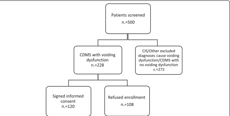
Five hundred patients attending Kasr Al-Ainy MS Clinic, Neurology Department, Cairo University Hospi-tals, Egypt, from December 2016 to March 2018, were evaluated for MS diagnosis and the presence of bladder dysfunction symptoms. Clinically definite multiple scler-osis (CDMS) according to the revised McDonald criteria (2010) [9] was verified in subjects; 228 of them had void-ing dysfunction durvoid-ing their disease course. Eventually, 120 eligible subjects accepted to sign the informed con-sent (Fig. 1). Patients with other diagnoses such as his-tory of diabetes mellitus, bladder neck surgery, prostate enlargement and fracture spine, and urinary tract infec-tion at time of assessment were excluded.
Full history and neurological examination were carried out via a specialized neurologist, with assessment of dis-ease severity using Expanded Disability Status Scale (EDSS) [10] at baseline and at the end of 1-year follow-up. A careful analysis of urological symptoms was done by a urologist from the “voiding dysfunction unit” Ur-ology Department, Cairo University Hospitals, with the severity of symptoms rated by the American Urological Association Symptom Score (AUASS) [11] at baseline, 3, 6, and 12 months. The AUASS range from 0 to 35, where 0–7 points are considered mild, 8–19 are moder-ate, and 20–35 are severe.
MRI brain and spinal cord (cervical and dorsal) with and without contrast were done for all patients using 1.5-T MRI machine (Achiva, Philips Medical system, the Netherlands) during the period of patient admission with the following sequences: Axial T1WI, T2WI, Sagit-tal T1WI, coronal T2WI, and Fluid Attenuated Inversion Recovery (FLAIR).
Post voiding abdominal and pelvic ultrasound, where a residual urine volume ≥100 ml is considered significant [12], ascending cystography, renal functions, and urine analysis were evaluated for all patients at baseline.
Urodynamic assessment (Laborie, Delphis KT, version 12 with ilist reporting system, Canada 2010) was done for all patients initially at baseline including filling cysto-flowmetry, pressure flow study, and EMG of external sphincter, and the assessment was recorded by a computer-based device that consists of similar input sensors and amplification with the cystoflowmetry in the standing position or sitting in a urodynamic chair using 6- or 7-Fr dual-lumen urethral catheter along with either a rectal or vaginal catheter to assess extravesical pressure fluctuations. Urodynamic changes including the bladder volume at the first desire to void (ml), maximum bladder volume (ml), PdetQmax (cmH2O), postvoiding residual (PVR) (ml), electromyographic, Qmax (ml/s), and blad-der compliance were measured. Detrusor overactivity, detrusor-sphincter dyssynergia, detrusor hypocontracti-lity, and areflexia were defined according to the stan-dardized definitions of lower urinary tract function by the international continence society (ICS) [13].
The patients were prescribed pharmacological treat-ment based on the nature of their symptoms and the therapeutic plan was modified according to patient’s re-sponses, and interventions as clean intermittent catheterization (CIC) and botulinum toxin-A (Botox) were used whenever needed.
Statistical methods
Data was analyzed using the computer program Statistical package for the Social Science (SPSS version 22, IBM Corp, Armonk, NY, USA) released 2013 for Microsoft Windows. Mean and standard deviation (± SD) was used for numerical data while frequency and percentage for non-numerical data. Studentttest was used to assess the statistical significance of the difference between means of the two studied groups. Mann-Whitney test (Utest) was used to assess the statistical significance of the difference of a non-parametric variable between the two studied groups. Correlation analysis (using Pearson’s and Spear-man’s rho method) assessed the strength of association between two quantitative variables. The correlation coeffi-cient (r) defines the magnitude and direction (+ or−) of the linear relationship between two variables. Multivariate logistic regression analysis was done to test for the signifi-cant independent predictors of abnormal urodynamic, and linear model was used to test for the significant independ-ent predictors of the urinary symptoms severity score. The probability/significance value (Pvalue)≥0.05 is not statis-tically significant and < 0.05 is statisstatis-tically significant.
Results
The demographics, neurological and urological character-istics of MS patients are illustrated in (Tables1and2).
Patients were prescribed medications to control void-ing dysfunction and the most frequently used anticholin-ergics (76.6%), selective alpha blockers (35%), and
parasympathomimetics (11.6%) in addition to
intervention, clean intermittent catheterization CIC (20%), and Botox injection for the bladder (15%).
Urinary symptoms severity showed a steady decrease by time, the highest at the 1st month with mean score 18 ± 6.6, then 15.8 ± 5.4 at the 3rd month, 12.5 ± 4.8 at the 6th month and the least after 1 year with mean score 10.3 ± 4.7 andPvalue < 0.001 (Fig.2).
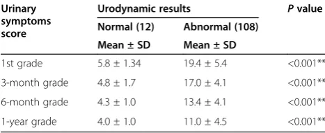
There is statistical significance in the relation of urin-ary symptoms severity and urodynamic results at all measurements through the year as P value was highly significant < 0.001 (Table3).
A statistically significant relation was found between the type of bladder symptoms and the pattern of urody-namic abnormality (P = 0.000) as 72% of patients with irritative symptoms had overactive bladder, and 66.7% of patients with obstructive symptoms had DSD while 64% of patients with mixed urinary symptoms had overactive bladder + DSD (Table4).
Table 1Demographics and clinical characteristics of multiple sclerosis
Age(years) 35.2 ± 10
Sex Males 38 (31.7)
Females 82(68.3)
Disease duration(years) 6.1 ± 4.9
EDSS 1st 4.8 ± 1.8
last 4.7 ± 1.8
Total relapse number 4.1 ± 2.3
Type of MS CIS 2(1.7)
RRMS 88(37.3)
PPMS 18(15)
SPMS 12(10)
CISclinical isolated syndrome,RRMSrelapsing remitting multiple sclerosis, PPMSprimary progressive multiple sclerosis,SPMSsecondary progressive multiple sclerosis
Data are expressed as the mean ± SD or number (percentage)
Table 2Urological characteristics of multiple sclerosis
Bladder symptoms Irritative 58(48.3)
Obstructive 12(10)
Mixed 50(41.7)
Residual urine via post voiding sonar ≥100 ml 66 (55)
Urodynamic studies Normal 12(10)
Abnormal Atonic bladder 14(11.7)
Overactive bladder 42(35)
Overactive bladder + DSD 32(26.7)
DI 12(10)
DSD 8(6.7)
Total 108 (90)
There was a statistically significant positive correlation between 1st total EDSS versus urinary symptoms score at 1st assessmentr= 0.39,P < 0.001, and also last EDSS versus urinary symptoms score at 1 year follow-up r = 0.27, P = 0.003, but no correlation was found between neither disease duration nor number of relapses and urinary symptoms score (Table5).
On regression analysis, baseline total EDSS was a predictor for higher urinary symptoms severity score (P = 0.001), however not a predictor for abnormal urodynamic. All other tested variables were not asso-ciated with higher urinary symptoms score or abnor-mal urodynamic (Table 6).
Discussion
All patients in the current study complained from urin-ary symptoms with the irritative symptoms which were the most common and this came in agreement with multiple studies which reported that storage symptoms as urgency and frequency are the recurrent urinary symptoms [14–16], while other study stated that ob-structive symptoms were more common than storage symptoms in MS patients and interrupted urinary flow was the most frequent symptom [17].
The severity of urinary symptoms in our study im-proved with compliance to symptomatic urological medication as anticholinergics decrease bladder pres-sure, improve muscle spasms, and reduce pain result-ing from urine storage. The alpha-1 receptor blockers decrease urethral resistance and relieve emptying symptoms, in addition to intermittent catheterization for MS patients with urinary retention and high re-sidual urine [14].
Urodynamic abnormalities were found in most of our studied MS patients with detrusor overactivity represent-ing the predominant findrepresent-ing. Similarly, it was reported that the incidence of urodynamic changes in MS patients reaches up to 99% with neurogenic detrusor overactivity is the most frequent abnormality followed by DSD and detrusor hyporeflexia [17–19].
Intracranial plaques present in up to 90% of MS pa-tients [20] but sacral plaques are much less common be-ing found only in about 18% of MS patients. Lesions in the pons, lateral corticospinal and reticulospinal tracts and sacral cord neurons can cause voiding problem and the site of lesion is responsible for the resulting urinary syndrome. Suprasacral lesions especially at cervical level are common interrupting descending inhibitory signals producing irritative symptoms. Plaques in the reticulosp-inal tract disturbs fibers originating in the pons thus impairing synergistic contraction of the detrusor muscle and relaxation of the external sphincter with (DSD), while lesions in the pons with loss of stimulatory signals result in detrusor hyporeflexia [21,22].
In our study we initially evaluated the MS patients with urodynamic assessment to provide better diagnosis and improve clinical management and quality of life. On the contrary, other study did not support the use of in-vasive urodynamic in the initial evaluation of patients with MS and prefer to be restricted to patients with se-vere urinary symptoms refractory to treatment [23]. Fig. 2Urinary symptoms severity. m, month
Table 3The relationship of urinary symptoms severity through the follow-up between abnormal and normal urodynamic results
Urinary symptoms score
Urodynamic results Pvalue
Normal (12) Abnormal (108)
Mean ± SD Mean ± SD
1st grade 5.8 ± 1.34 19.4 ± 5.4 <0.001**
3-month grade 4.8 ± 1.7 17.0 ± 4.1 <0.001**
6-month grade 4.3 ± 1.0 13.4 ± 4.1 <0.001**
1-year grade 4.0 ± 1.0 11.0 ± 4.5 <0.001**
Through the 1-year follow-up, our study confirmed the re-sult of a previous one with the severity of urinary symptoms in patients who had abnormal urodynamic differ significantly [15]. In contrast to our results, another urodynamic study re-vealed that there is no association between urinary symptoms severity and urodynamic changes [24].
Our studied MS patients suffered from irritative symp-toms resulting from detrusor overactivity and/or incompe-tent sphincter, while obstructive symptoms denote detrusor-sphincter dyssynergia or detrusor underactivity [6].
Babovic and colleagues agree with our results and mentioned that urgency, frequency, and urge incontin-ence were well correlated with detrusor hyperreflexia [3]. On the other side, Haggiag and colleagues did not report the value of urinary symptoms assessment in de-termining the urodynamic changes as they used different questionnaire and limited urological parameters and the effect of other MS disabling symptoms [25].
EDSS initial and 1 year assessment in our study was correlated with urinary symptoms score while no correl-ation was found with disease durcorrel-ation. This result agrees with a study that showed irritative urinary symptoms were significantly affected by EDSS while obstructed symptoms were not affected by disease duration [6].
High EDSS score was the only predictor for severe urinary symptoms in our studied MS patients while it was not a predictor for abnormal urodynamic. This was consistent with a prospective study conclusion that high EDSS was associated with frequent urinary incontinence [26]. However, another study demonstrated a significant relationship between EDSS and urodynamic abnormality with risk to upper urinary tract damage. We attributed absence of predictive value of EDSS and other disease parameters in our study to the low number of patients with normal urodynamic that did not reach a statistical significant value [27].
The observed limitation of our study was that all included MS patients had urinary complaints so no chance for value of urodynamic in patients without urinary symptoms.
Conclusion
Irritative bladder symptoms and overactive bladder are the most frequent lower urinary tract problem in MS pa-tients. EDSS is a predictor for urinary symptoms
sever-ity. The type of bladder symptoms can reflect
urodynamic abnormalities. Table 4Relationship of bladder symptoms type and urodynamic results
Urodynamic results Pvalue
Atonic bladder
Overactive bladder
Overactive + DSD DI DSD
Bladder symptoms type Irritative,n= 58 Number 0 42 0% 12 0 0.000**
% within bladder symptoms 0% 72.4% 0% 20.7% 0%
% within urodynamic 0% 100% 0% 100% 0%
Obstructive,n= 12 Number 2 0 0 0 8
% within bladder symptoms 16.7% 0% 0% 0% 66.7%
% within urodynamic 14.3% 0% 0% 0% 100%
Mixed,n= 50 Number 12 0 32 0 0
% within bladder symptoms 24% 0% 64% 0% 0%
% within urodynamic 85.7% 0% 100% 0% 0%
DSDdetrusor sphincter dyssynergia,DIdetrusor instability
**
Pvalue < 0.01 (highly significant)
Table 5Correlation of urinary symptoms severity with EDSS (1st month vs 1st EDSS, 1 year vs last EDSS), disease duration, and number of relapses
Urinary symptoms severity
1st month 1 year
rvalue Pvalue rvalue Pvalue
EDSS 1st 0.39 < 0.001**
---Last --- 0.27 0.003**
Disease duration in years 0.17 0.19 0.06 0.60
Number of relapses 0.15 0.23 0.1 0.36
EDSSexpanded disability status scale
Pvalue≥0.05 (non-significant), **Pvalue < 0.01 (highly significant)
Table 6Predictors of abnormal urodynamic and severity of urinary symptoms
Predictors Abnormal urodynamic Severity of urinary symptoms
OR 95% confidence interval
Pvalue Regression coefficient
95% confidence interval
Pvalue
EDSS 1.38 0.92–2.08 0.11 1.49 0.89–2.10 < 0.01**
Disease duration (years)
1.10 0.82–1.48 0.49 0.21 −0.08 to 0.52 0.15
Number of relapses
1.42 0.73–2.72 0.29 0.30 −0.32 to 0.94 0.34
MS type 0.36 0.00 1.00 −1.02 −2.64 to 0.59 0.21
Acknowledgements
Not applicable.
Authors’contribution
HT participated in study design, urodynamic study, collection, and analysis of data. NS participated in study design and helped to select the type of patients. SH helped to draft the manuscript and in the diagnosis of selected participants. HA participated in study design. SZ participated in the follow-up of urodynamic study. MS participated in study design sequence alignment. AY performed the laboratory work. SM performed abdominal and pelvic ultrasound for all participants. All authors read and approved the final manuscript.
Funding
Authors did not receive any funding for this work.
Availability of data and materials
The datasets used during the current study are available from the corresponding author on reasonable request with permission of Faculty of Medicine, Cairo University, Egypt.
Ethics approval and consent to participate
The study was approved by the ethical committee of the Urology Department, Faculty of Medicine, Cairo University (I-131016, on the 26th of December 2016). The procedures and follow-up were explained to every participant and an informed written consent was obtained from all participants.
Consent for publication
Not applicable.
Competing interests
The authors declare that they have no competing interests.
Author details
1
Department of Urology, Cairo University, Giza, Egypt.2Department of Neurology, Cairo university, Giza, Egypt.
Received: 21 April 2019 Accepted: 24 April 2020
References
1. Compston A, Coles A. Multiple sclerosis. Lancet. 2008;372:1502–17. 2. Wein AJ. Lower urinary tract dysfunction in neurologic injury and disease.
In: Wein AJ, Kavoussi LR, Novick AC, Partin AW, Peters CA, editors. Campbell-Walsh Urology. 9th ed. USA; 2007. p. 2011-2045.
3. BabovićR, MilićevićS, RadovanovićS, JančićJ. Testing of urodynamic dysfunctions in patients with multiple sclerosis. Vojnosanit pregled. 2014; 71(5):446–50.
4. Shah P. Symptomatic management in multiple sclerosis. Ann Indian Acad Neurol. 2015;18(Suppl 1):S35.
5. Kirchhof K, Fowler CJ. The value of the Kurtzke Functional Systems Scales in predicting incomplete bladder emptying. Spinal Cord. 2000;38(7):409. 6. Araki I, Matsui M, Ozawa K, Nishimura M, Kuno S, Saida T. Relationship
between urinary symptoms and disease-related parameters in multiple sclerosis. J Urol. 2002;249(8):1010–5.
7. DasGupta R, Fowler CJ. Bladder, bowel and sexual dysfunction in multiple sclerosis: management strategies. Drugs Rev. 2003;63(2):153–66. 8. World Medical Association. Declaration of Helsinki. Ethical principles for
medical research involving human subjects. Adopted by the 18th World Medical Association General Assembly. Helsinki, Finland. 1964. 9. Polman CH, Reingold SC, Banwell B, Clanet M, Cohen JA, Filippi M, et al.
Diagnostic criteria for multiple sclerosis: 2010 revisions to the McDonald criteria. Ann Neurol. 2011;69(2):292–302.
10. Kurtzke JF. Rating neurologic impairment in multiple sclerosis: an expanded disability status scale (EDSS). Neurology. 1983;33(11):1444.
11. Roehrborn CG. The American Urological Association Symptom Index– concerns and confirmation. J Urol. 1996;6(155):1975–6.
12. Kelly CE. Evaluation of voiding dysfunction and measurement of bladder volume. Rev Urol. 2004;6(Suppl 1):S32.
13. Abrams P, Cardozo L, Fall M, Griffiths D, Rosier P, Ulmsten U, et al. The standardisation of terminology of lower urinary tract function: report from the Standardisation Sub-committee of the International Continence Society. Neurourol Urodyn. 2002;21(2):167–78.
14. Wang T, Huang W, Zhang Y. Clinical characteristics and urodynamic analysis of urinary dysfunction in multiple sclerosis. Chin Med J. 2016;129(6):645. 15. Tadayyon F, Etemadifar M, Bzeih H, Zargham M, Nouri-Mahdavi K, Akbari M,
et al. Association of urodynamic findings in new onset multiple sclerosis with subsequent occurrence of urinary symptoms and acute episode of disease in females. J Res Med Sci. 2012;17(4):382.
16. Engeler DS, Meyer D, Abt D, Müller S, Schmid HP. Sacral neuromodulation for the treatment of neurogenic lower urinary tract dysfunction caused by multiple sclerosis: a single-centre prospective series. BMC Urol. 2015;15(1):105. 17. Nakipoglu GF, Kaya AZ, Orhan G, Tezen O, Tunc H, Ozgirgin N, et al. Urinary
dysfunction in multiple sclerosis. J Clin Neurosci. 2009;16(10):1321–4. 18. De Sèze M, Ruffion A, Denys P, Joseph PA, Perrouin-Verbe B. International
Francophone Neuro-Urological expert study group (GENULF). The neurogenic bladder in multiple sclerosis: review of the literature and proposal of management guidelines. Mult Scler. 2007;13(7):915–28. 19. Kobashi KC. Review of the 2008 annual meeting of the Society of
Urodynamics and Female Urology (SUFU) at the American Urological Association. Curr Bladder Dysfunct Rep. 2009;4(1):5–10.
20. Aharony SM, Lam O, Corcos J. Evaluation of lower urinary tract symptoms in multiple sclerosis patients: review of the literature and current guidelines. Can Urol Assoc J. 2017;11(1-2):61.
21. Araki I, Matsui M, Ozawa K, Takeda M, Kuno S. Relationship of bladder dysfunction to lesion site in multiple sclerosis. J Urol. 2003;169(4):1384–7. 22. Litwiller SE, Frohman EM, Zimmern PE. Multiple sclerosis and the urologist. J
Urol. 1999;161(3):743–57.
23. Çetinel B, Tarcan T, Demirkesen O, Özyurt C,Şenİ, Erdoğan S, et al. Management of lower urinary tract dysfunction in multiple sclerosis: a systematic review and Turkish consensus report. Neurourol Urodyn. 2013; 32(8):1047–57.
24. Onal B, Siva A, Buldu I, Demirkesen O, Cetinel B. Voiding dysfunction due to multiple sclerosis: a large scale retrospective analysis. Int Braz J Urol. 2009; 35(3):326–33.
25. Haggiag S, Bolla G, Picconi O, Galgani S, Gasperini C. Discrepancies between urinary symptoms assessment and objective bladder dysfunctions in multiple sclerosis. Mult Scler Demyelinating Disord. 2017;2(1):11. 26. Wiedemann A, Kaeder M, Greulich W, Lax H, Priebel J, Kirschner-Hermanns
R, et al. Which clinical risk factors determine a pathological urodynamic evaluation in patients with multiple sclerosis? An analysis of 100 prospective cases. World J Urol. 2013;31(1):229–33.
27. Ineichen BV, Schneider MP, Hlavica M, Hagenbuch N, Linnebank M, Kessler TM. High EDSS can predict risk for upper urinary tract damage in patients with multiple sclerosis. Mult Scler. 2018;24(4):529–34.
Publisher’s Note