A.Yuvaraj et al, International Journal of Computer Science and Mobile Applications,
Vol.2 Issue. 3, March- 2014, pg. 75-84 ISSN: 2321-8363
©2014, IJCSMA All Rights Reserved, www.ijcsma.com 75
Exudates Detection Strategies in Retinal
Images Exploitation Process Techniques
A.Yuvaraj
1, Ms. N.Radhika
2 1M.TECH Student, Department of Computer Science and Engineering, PRIST University, Trichy
2
Assistant Professor, Department of Computer Science and Engineering, PRIST University, Trichy
Abstract
Exudates are one in every of the foremost common occurring lesions in diabetic retinopathy. Exudates are often known as areas with laborious white or chromatic colours and ranging sizes, shapes and locations close to the leaky capillaries at intervals the tissue layer. The detection of exudates is that the major goal. For this the pre-requisite stage is that the detection of blind spot. Once the blind spot is found sure algorithms can be accustomed sight the presence of exudates. During this paper few ways are used for the detection and also the performance of all the ways are compared.
Keywords — Capillaries, diabetic retinopathy, exudates, optic disks
1. Introduction
India and China area unit, and can stay, the leading countries in terms of the amount of individuals with polygenic disease. Among the ten leading countries during this respect, 5 area unit in Asia. Though solely a moderate increase within the total population in China is anticipated within the next twenty five years, China is calculable to contribute virtually thirty eight million individuals to the world burden of polygenic disease. India, owing to its Brobdingnagian population size and high polygenic disease prevalence, can contribute fifty seven million. Calculable growth, population ageing, and urbanization, however they are doing not take into consideration changes in alternative diabetes-related risk factors.
So, Diabetic screening programmes area unit necessary in addressing all of those factors once operating to eradicate preventable vision loss in diabetic patients. Once playing retinal screening for Diabetic Retinopathy some of these clinical displays area unit expected to be imaged. Diabetic retinopathy is globally the first cause of visual defect not as a result of, it's the very best incidence and it typically remains undetected till severe vision loss happens. Advances in form analysis, the event methods for the detection and quantitative characterization of vas changes within the tissue layer area unit of nice importance. Machine-driven early detection of the presence of exudates will assist the ophthalmologists to stop the unfold of sickness a lot of expeditiously.
A.Yuvaraj et al, International Journal of Computer Science and Mobile Applications,
Vol.2 Issue. 3, March- 2014, pg. 75-84 ISSN: 2321-8363
©2014, IJCSMA All Rights Reserved, www.ijcsma.com 76
screening. The conventional options of bodily structure pictures embody optic disc, fovea centralis and blood vessels. Exudates and haemorrhages area unit the most abnormal options that is that the leading reason for visual defect within the operating age population.
Optic disk is that the brightest half within the traditional bodily structure pictures which might be seen as a pale, spherical or vertically slightly oval disk. Finding the most parts within the bodily structure pictures helps in characterizing detected lesions and in distinctive false positives. Abnormality detection in pictures is found to play a crucial role in several real life applications recommended neural network approach for the detection and classification of exudates. A decision support frame work for deducing the presence or absence of DR area unit developed and tested. The detection rule is based on binary-hypothesis testing downside that simplifies the matter to yes/no selections. The results recommend that by biasing the classifier towards DR detection, it's potential to create the classifier succeeds good sensitivity.
2. METHODS
2.1 Feature Extraction
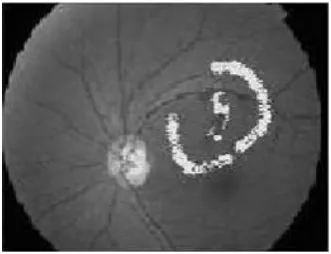
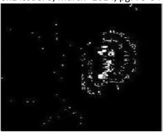
Here, during this methodology we have a tendency to use the construct that in traditional retinal pictures the optic disk is that the brightest half and next to it comes the exudates. therefore once when detection the optic disk, the centre purpose is set for extraction of varied features within the image. Then the optic disk is off from the image, therefore we have a tendency to square measure currently left with exudates because the next brightest region. Here once more we will apply Binary Image and correct threshold price is about and also the exudatescan be simply known from the check image. The results square measure shown in figures one and two.
Figure1. INPUT OPTIC DISK EXTRACTED IMAGE
A.Yuvaraj et al, International Journal of Computer Science and Mobile Applications,
Vol.2 Issue. 3, March- 2014, pg. 75-84 ISSN: 2321-8363
©2014, IJCSMA All Rights Reserved, www.ijcsma.com 77 2.2 Model Matching
For the construct behind this methodology is that, a standard and healthy retinal image is taken and it's unbroken because the reference to isolate the abnormalities within the check image. This reference image acts because the model. each the reference image and check pictures square measure born-again from RGB to grey levels then component by component each the photographs square measure compared.
During comparison, the extra objects gift within the check image get isolated and that they square measure clearly visible in the output. If the check image is traditional, then whereas comparison it gets off as there's no distinction of component value between the two, wherever as within the check image with exudates, the optic disk gets off and solely exudates are separated within the output. and is shown in figure three to five
Figure 3. REFERENCE IMAGE
A.Yuvaraj et al, International Journal of Computer Science and Mobile Applications,
Vol.2 Issue. 3, March- 2014, pg. 75-84 ISSN: 2321-8363
©2014, IJCSMA All Rights Reserved, www.ijcsma.com 78
Figure 5. OUTPUT IMAGE WITH EXUDATES DETECTED
The basic demand of this methodology is that, should always have a standard and healthy retinal image as reference and also the check pictures must be taken within the same orientation because the reference, it ought to be of same lighting, angle, etc… It ought to be taken within the same manner as that of the reference, then solely this algorithmic rule can work well instead it might manufacture wrong result. Thus this basic want should be glad to figure with this methodology.
2.3 Minimum Distance Discriminant Classifier
Color info has shown to be effective for lesions detection beneath bound conditions. On the premise of color info, the presence of lesions will be preliminarily detected by victimization MDD (Minimum Distance Discriminant) classifier supported applied mathematics pattern recognition techniques. If the background color of a decent quality retinal image is sufficiently uniform, then an easy and effective methodology to separate exhausting lesions from such background will be simply applied by choosing a correct threshold. However, the limitation of thosethresholding techniques is that they generally solely work well for the coaching pictures, however once associate degree unseen image comes on, they will not be ready to accurately find the exudates. this can be as a result of the process steps need completely different threshold parameters for various sorts of retinal pictures and want user‟s intervention on a case by case basis. As a result, these thresholding based mostly algorithms are not ascendible for analyzing sizable amount of retinal pictures.
A.Yuvaraj et al, International Journal of Computer Science and Mobile Applications,
Vol.2 Issue. 3, March- 2014, pg. 75-84 ISSN: 2321-8363
©2014, IJCSMA All Rights Reserved, www.ijcsma.com 79
feature vector Y, we have a tendency to category if y as happiness to class i if Di(Y) is that the most on all Dj(Y), where j=1, 2,….N and j not up to i.
The color options are taken because the feature area, R. The color structure retinal image consists of 3 planes-red, inexperienced and blue, every plane with 256 levels of intensity denoted as (R, G and B). Color will be additionally diagrammatical by ș, ij, and P within the spherical coordinates. The relation between the 2 color areas is expressed as:
P =(R2+G2+B2)1/2
β =Arctan(G/R)
α =Arccos(B/P)
P denotes the exposure or brightness of a picture, whereas β,α emphasize the variations or changes of colours. When P is command constant, β and α describe the color property is an fuel surface. Since our focus is to differentiate between chromatic lesions and different darker objects within the color retinal pictures, we don‟t like to incorporate both the brightness of the image moreover because the changes of color data. Hence, we have got elect P, β,α feature area, R (rP, rβ, rα). Then we don‟t like to derive acceptable discriminant perform. Our discriminant D(Y) is
derived from Thomas Bayes rule that is given as, Di(Y) = (Y-Ci) T(Y-Ci).This is additionally known as a minimum distance discriminant (MDD).
Applying Di(Y) as outlined on top of to the matter of police work presence of exudates in retinal pictures, we define only two classes-yellow patches (lesions) and dark cerise background. The feature centers of lesions and classes severally. for every picture element Y (yP, yβ, yα) from the retinal image, the discriminant Dlesion and Dbkgnd(Y) square measure calculated. If Dlesion(Y) is a smaller amount than Dbkgnd(Y), then picture element Y is assessed as lesion otherwise it\'s being classified as background. during this method, exudates or alternative chromatic lesions is quickly detected. this straightforward and quick algorithmic program is able to smarts good accuracy within the detection of exudates in color bodily structure pictures. The results square measure shown in figures six to eight.
A.Yuvaraj et al, International Journal of Computer Science and Mobile Applications,
Vol.2 Issue. 3, March- 2014, pg. 75-84 ISSN: 2321-8363
©2014, IJCSMA All Rights Reserved, www.ijcsma.com 80
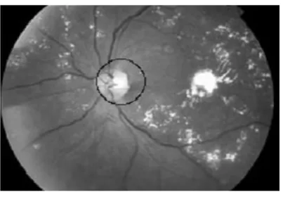
Figure 7. INPUT IMAGE WITH OPTIC DISC CIRCLED
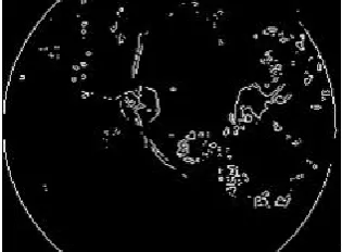
Figure 8. OUTPUT IMAGE WITH EXUDATES MARKED AS BLACK
3. Increased MDD classifier
This image works on the RGB co-ordinates instead of spherical co-ordinates. within the Minimum Distance Discriminant (MDD) Classifier technique, the centre of sophistication is found employing a coaching set and thence remains mounted. however this could cause drawback as a result of distinction in image illumination and their average intensity. So a way is used such the centre of sophistication (CZ and Cbgnd) varies dynamically relying on the image. From previous optic disk
detection technique.
we know the position of the point for the image. mistreatment this data we have a tendency to choose a gaggle of pixels that surrounds the point and also the mean of those pixels type the Cbgnd. point sometimes has an equivalent color and intensity as that of exudates. that the pixels that belong to the OD ar used for calculation for Cz.
m
Cz = 1/m∑ yi
i=1
A.Yuvaraj et al, International Journal of Computer Science and Mobile Applications,
Vol.2 Issue. 3, March- 2014, pg. 75-84 ISSN: 2321-8363
©2014, IJCSMA All Rights Reserved, www.ijcsma.com 81
Cbgnd = 1/n ∑ Bi
i=1
Where m & n are variety of pixels in chromatic and background region severally, that are wont to calculate these centers and Loloish and bismuth are the vectors of the three color options within the totally different region of point and background. The methodology tries to find exudates by mistreatment the two necessary options of exudates, its color and its sharp edges.
it's administrated within the following steps.
Detection of point.
Detection of chromatic objects within the image.
Detection of objects within the image with sharp edges.
Combination of the previous steps to find chromatic objects with sharp edges.
3.1 Detection of Blind Spot
Principal part Analysis between clusters and propagation through radii square measure wont to observe blind spot. The area envelopment the blind spot is derived out and off from the retinal image.
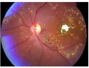
Figure 9 INPUT IMAGE WITH OPTIC DISK CIRCLED
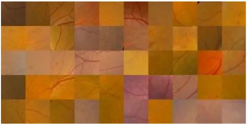
3.2 Detection of Yellow Objects
The detection of yellow objects is disbursed playacting color segmentation supported applied mathematics classification method. it's supported the actual fact that if a gaggle of options are often outlined, so the objects in a picture map to non-intersecting categories in feature area, then we are able to simply determine totally different objects classifying them into corresponding classes. we tend to outline two categories yellow objects and background that square measure characterised victimisation solely three color features(R, G, and B).Using Baye‟s theory the Minimum Distance Discriminant (MDD) is found as,
Di(Y) = -(Y-Ci)T(Y-Ci)
A.Yuvaraj et al, International Journal of Computer Science and Mobile Applications,
Vol.2 Issue. 3, March- 2014, pg. 75-84 ISSN: 2321-8363
©2014, IJCSMA All Rights Reserved, www.ijcsma.com 82
Next we tend to performed AN adjustment for unsimilarity of illumination, due to lighting variation, decreasing color saturation, skin pigmentation etc… the colour of lesions in some regions of a picture could seem rheostat than the background color that's set in another region and would be incorrectly classified. We used a new color image; this image is obtained playacting AN operation of channels (T1, T2, T3) of the NTSC color area.
T1‟=1.5T1-T2-T3
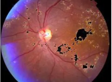
The changing the image obtained (T1, T2, and T3) into the RGB color area once more. we tend to improve each contrasting attributes of lesions and overall color saturation in image creating optic disk and exudates to look with same color freelance of their location. Minimum Distance Discriminant (MDD) is applied to all or any pixels and the exudates square measure known. whereas changing the ntsc image to rgb the colour map is scaled to worth „1‟.Hence in mathematical computation the distinction improved image‟s worth has got to be increased by 255 since each the centre of class were obtained from the initial RGB image wherever most intensity worth is pictured by 255. Along with exudates, alternative lesions like drusens, artifacts, optic disk square measure are known and therefore the exudates are shown in figure ten as black color.
Figure 10. DETECTION OF YELLOW OBJECTS FROM
THE IMAGE
There square measure numerous algorithms to search out the perimeters of a picture like sobel, cagy etc…In our case we tend to used sobel operator to find the sharp edges. We have a binary image with edges being shown white. This image contains the edges of optic disk, blood vessels, exudates and conjointly the image boundary. therefore this can't be severally wont to determine the exudates.
A.Yuvaraj et al, International Journal of Computer Science and Mobile Applications,
Vol.2 Issue. 3, March- 2014, pg. 75-84 ISSN: 2321-8363
©2014, IJCSMA All Rights Reserved, www.ijcsma.com 83
THE IMAGE
3.3. Combination of Two Images
To observe solely exudates and to get rid of all the false detections within the previous stages, we tend to combined the two images obtained victimisation Minimum Distance Discriminant (MDD) and edge police investigation technique through a Boolean operation, feature primarily based AND. In feature primarily based AND, ON pixels in one binary image square measure wont to choose object in another image. we tend to use the image with objects having sharp edges to pick out objects within the image with yellow elements, as a result of within the last one the lesions square measure detected utterly, not solely their contours. Therefore we tend to get lesions characterized by two desired features-yellowish color and sharp edge. The boundary region encloses the exudates and is shown in figure12
Figure 12. OUTPUT IMAGE GIVING BOUNDARY OF EXUDATES
4. Conclusion
The feature extraction once more desires the correct thresholding values. The essential demand in model matching is that we'd like each traditional and abnormal picture. The orientation, angle, lighting of each reference and therefore the abnormal image ought to be same otherwise it'd provide wrong identification of the presence of exudates. Minimum distance discriminant (MDD) classifier is predicated on applied mathematics recognition technique and this provides higher result. However this works on spherical coordinates and therefore the center is found employing a coaching set and thence stay fastened. This might cause drawback and utilized such the centre of sophistication varies dynamically, counting on the image. Increased minimum distance discriminant (MDD) classifier uses rgb values of the image and therefore the abnormality is characterised by the options yellow color and sharp edges.
References
[1] King H, Aubert RE, Herman WH. “Global burden of diabetes Care 1998; Vol.21: Page 1414- 31.
A.Yuvaraj et al, International Journal of Computer Science and Mobile Applications,
Vol.2 Issue. 3, March- 2014, pg. 75-84 ISSN: 2321-8363
©2014, IJCSMA All Rights Reserved, www.ijcsma.com 84
tal Fundus Retinal Images” Computing: Theory and Applica tions, ICCTA‟07. International Conference on Issue Date: 5-7 March 2007 PP: 705-710 ISBN: 0-7695- INSPEC Accession Num ber: 9420643 Digital Object Identifier: 10.1109/ICCTA.2007.16
[3] Fong DS, Aiello L, Gardner TW, King GL, Blankenship G, Caval lerano JD, Ferris FL, II, Klein R: Diabetic retinopathy. Diabetes Care 26:226-229, 2003
[4] Huiqili, and Opas Chutatape, (2004) “Automated Feature Ex traction in Color Retinal Images by a Model based Approach”, IEEE transactions on biomedical engineering, vol.51, no.2, February 2004 Digital Object Identifier : 10.1109/tbme.2003.820400
[5] Nguyenl, H.T., M. Butler, A. Roychoudhryl, A.G. Shannonl, J. Flack and P. Mitchell, 1996. “Classification of diabetic retinopathy using neural networks”. Proceedings of the 18th
Annual International Conference of the IEEE Engineering in Medicine and Biology Society, Oct. 31-Nov. 3, Amsterdam, pp: 1548-1549
[6] Kahai, P., K.R. Namuduri and H. Thompson, 2006. A decision support framework for automated screening of diabetic retinopathy. Int. J. Biomed. Imaging., 2006: 1-8.
[7] Milan Sonka,Hlavac and Roger Boyle(2008),Digital Image Processing and Computer Vision, Cengage Learning India Private Limited.