Available online at www.macvetrev.mk
Corresponding author: Dr. Avo Karus, PhD
E-mail address: [email protected]
Present address: Department of Food Sciences and Food Technology, Institute of Veterinary Medicine and Animal Science,
Estonian University of Life Sciences, Kreutzwaldi 62, Tartu, Estonia
Phone: ++372 7313740; Fax: ++372 7313741
Copyright: © 2017 Karus A. This is an open-access article published under the terms of the Creative Commons Attribution License which permits unrestricted use, distribution, and reproduction in any medium, provided the original author and source are credited.
Competing Interests: The authors have declared that no competing interests exist.
Available Online First: 3 January 2017 Published on: 15 March 2017
http://dx.doi.org/10.1515/macvetrev-2017-0010
Original Scientific Article
DEVELOPMENT OF SIMPLE MULTIPLEX REAL-TIME PCR ASSAYS
FOR FOODBORNE PATHOGENS DETECTION AND
IDENTIFICATION ON LIGHTCYCLER
Avo Karus
1, Fabrizio Ceciliani
2,
Armand Sanches Bonastre
3,
Virge Karus
11
Department of Food Sciences and Food Technology, Institute of Veterinary Medicine
and Animal Science, Estonian University of Life Sciences,
Kreutzwaldi 62, Tartu, Estonia
2
Department of Veterinary Science and Public Health, University of Milan,
via Celoria 10, 20133 Milan, Italy
3
Universitat Autonoma de Barcelona, Campus de la UAB, Bellaterra, Spain
Received 18 November 2016; Received in revised form 22 December 2016; Accepted 24 December 2016
ABSTRACT
Most acute intestinal diseases are caused by food-borne pathogens. A fast and simple real-time PCR-based procedure for simultaneous detection of food contamination by any of the five food-borne pathogens: Campylobacter jejuni, Mycobacterium bovis, Enterobacter sakazaki, Shigella boydii, Clostridium perfrigens using multiplex EvaGreen real-time PCR for LightCycler was developed and evaluated. Real-time qPCR showed excellent sensitivity. Tm calling and Melting Curve Genotyping (MCG) were used for analysis of PCR product melting curves. The Melting Curve Genotyping option showed good performance for discrimination of positive samples containing DNA of single pathogen or pathogen mixtures from negative samples.
Key words: food-borne pathogens, multiplex real-time PCR, melting curve genotyping
INTRODUCTION
The frequency of outbreaks of food-borne infection cases worldwide is still extremely high (1) in both the Western world and Third World countries (2). Food pathogens are commonly found in the intestines of healthy food-producing animals, and can be transmitted to humans through contamination in the food chain. Thus, a strict control is required of the whole food chain aimed at enforcing contamination detection measures.
Molecular methods for detection and identification of food pathogens have significant benefits as compared to traditional methods due
several singleplex analyses might reduce the total costs for testing (8).
The objective of the present study was to provide a specific protocol to detect simultaneously food contamination by any of the five food-borne pathogens, namely Campylobacter jejuni, Mycobacterium bovis, Enterobacter sakazaki, Shigella boydii, Clostridium perfrigens. The developed assay can be used to analyse any kind of food, including but not limited to milk, cheese and meat products. The performance of this assay was assessed by using DNA purified by ATCC strains.
MATERIAL AND METHODS
Samples
The lyofilized Shigella boydii and Enterobacter sakazaki strains were aerobically grown in a Brain Heart Infusion Broth (Oxoid, Italy) at 37°C for 24 hours. Campylobacter jejuni strain was grown in a Brain Heart Infusion Broth (Oxoid, Italy) under a microaerophilic atmosphere (CO2Gen, Oxoid, Italy) at 42°C for 24-48 hours. Clostridum perfringens was grown anaerobically, using BBL GasPak™ Systems (BD, USA), in a Brain Heart Infusion Broth (Oxoid, Italy) at 37 °C for 48 hours. After overnight incubation from fresh colonies, bacterial cells were collected by centrifugation and subjected to genomic DNA isolation, as previously described (9). The bacterial strains and the isolated genomic DNA are described in Table 1.
For Mycobacterium bovis only genomic DNA was available. The concentration of DNA used for reference target strains varied from 3.9 to 11.2 ng/ml. Pure and mixed samples were prepared using isolated bacterial DNA and NA-free water from Roche Diagnostics (Germany). No real food samples were used.
Primer design
Primers for real-time PCR amplification were designed using PRIMER EXPRESS (ver. 3.0). Primer pairs were ordered from Tib-MolBiol (Germany) and tested for EvaGreen assays on LightCycler 2.0® (Roche Diagnostics GmbH)
using 5x HOTFIREPol® EvaGreen® qPCR Mix
Plus (Capillary) with 7.5 mM MgCl2 ready-to use mastermix (Solis BioDyne). Primer sequences and the accession number of target genes are listed in Table 2.
EvaGreen PCR assay
Quantitative PCR reactions followed by melting curve analysis were performed in a final volume of 20 ml containing 2 µl of template genomic DNA, 4 µl of 5x HOT FIREPol® EvaGreen® qPCR Mix
Plus (Capillary) with 7,5 mM MgCl2, 5 µl of primer cocktail (0.5 µl of each forward and reverse primer) and 10 ml of molecular grade water. Tenfold dilutions of the target genomic DNA (1 ng/ml and 100 pg/ml) were tested to determine the fair amount of template DNA detected by the assay (PCR sensitivity). The reactions were carried out by using LC Capillaries 20 µl. Each sample was tested in triplicate with the following thermal profile: 95°C for 15 min for hot-start, followed by 45 cycles at 95°C for 15 s, 60°C for 20 s and 72°C for 30 s. The melting point analysis was preceded by a denaturation step 95°C for 5 s and an annealing step 65°C for 15 s, followed by continuous fluorescence recording from 65°C till 90°C 0.05°C/s and a cooling step 40°C for 15 s.
Comparison of two different analysis methods: Tm calling versus Melting Curve Genotyping
The melting curve results at λ 530 nm were
analyzed with LightCycler480 SW1.5.0 using two different options: automated Tm Calling
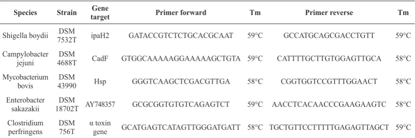
Table 1. Reference target bacterial strains used in this study and primers designed using PRIMER EXPRESS (ver. 3.0)
Species Strain targetGene Primer forward Tm Primer reverse Tm
Shigella boydii 7532TDSM ipaH2 GATACCGTCTCTGCACGCAAT 59°C GCCATGCAGCGACCTGTT 59°C Campylobacter
jejuni 4688TDSM CadF GTGGCAAAAAGGAAAAAGCTGTA 59°C CATTTTGCTTGTGGAGTTGCA 58°C Mycobacterium
bovis 43990DSM Hsp GGGTCAAGCTCGACGTTGA 58°C CGGTGGTCCGTTTGGAACT 58°C Enterobacter
sakazakii 18702T AY748357DSM GCGCGGTGTGTCAGAGTCT 59°C AACCTCACAACCCGAAGAAGTC 58°C Clostridium
and Melting Curve Genotyping to investigate the potential to identify the pathogens via qPCR product melting peak and melting profile. Both analyses were used only for samples with Ct less than 37 in quantitative analysis.
RESULTS
Analytical sensitivity and specificity
In multiplex testing, sensitivity was detected by analysis of tenfold dilution series of bacterial DNA. The detection of 100 pg of pathogen DNA was confirmed (Table 2).
The melting curves for PCR products were analyzed by two methods, while specificity of the multiplex assay was confirmed by testing against all five targets: Campylobacter jejuni, Mycobacterium bovis, Enterobacter sakazaki,
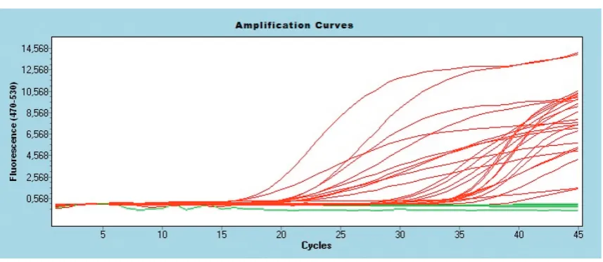
Shigella boydii, Clostridium perfrigens. In samples containing artificial mixtures of pathogens DNA, usually no more than two major pathogens were identified by melting curve analysis. However, in all capillaries no false-positive or false-negative results were generated, confirming the excellence of the developed assay (Fig. 1).
Intra- and inter-assay reproducibility
To obtain values for the intra- and inter-assay variation of the assay, 1 ng and 100 pg of purified genomic DNA was analyzed in triplicate. The coefficients of variation (CV) of the Ct values ranged from 0.5 to 2.1 % for intra-assay experiments
and from 2.3 to 4.5 % for inter-assay experiments. The coefficients of variation of the Tm values ranged from 0.05 to 0.21 % for intra-assay experiments and from 0.06 to 0.25 % for inter-assay experiments.
Table 2. Threshold cycle (CT) of quantification analysis obtained in 10-fold dilution of the pure culture genomic DNA (1 ng and 100 pg) and major melting peaks of amplicons
1 ng/μl 100 pg/μl Melting peaks (Tm)*
EvaGreen assay Avg CT sd Avg CT sd Tm1 sd Tm2 sd
Shigella boydii 31.72 0.82 35.12 0.83 81.85 0.19
Campylobacter jejuni 33.65 1.18 37.01 1.24 80.99 0.03
Mycobacterium bovis 31.54 0.84 34.92 0.81 81.16 0.15 84.32 0.05
Enterobacter sakazaki 22.14 0.88 25.49 0.96 80.71 0.16
Clostridium perfrigens 22.10 1.00 25.43 1.03 77.38 0.19
* Variability of Tm was calculated in DNA cocktail analyses and standard deviations (sd) are shown; Mycobacterium bovis
showed two melting peaks
Figure 1. Amplification curves of EvaGreen assays in multiplex test with DNA cocktails Positive and/or spiked samples
Tm calling versus Melting Curve Genotyping The robustness of the assay was evaluated by comparative analysis of PCR product melting curves using different software features – Tm calling and Melting Curve Genotyping (MCG).
Tm calling allowed the identification of PCR products by major product(s) melting temperature, however in MCG the pattern of the melting curve was identified. Tm calling showed clear differentiation of samples with single pathogen genomic DNA as shown in Table 2. In mixed samples (containing DNA from two or more pathogen) the major component was always identified, thus the food would be rejected in any pathogen contamination included in the assay. However, the procedure
did not allow identifying the minor components, especially if three pathogens were mixed for analysis.
Melting Curve Genotyping showed similar performances, but because the basis for identification is the melting curve pattern,
Mycobacterium bovis got the priority for decision. Mycobacterium bovis amplification resulted with products identified with two melting peaks and it fit best under the term ‘pattern’. Thus, Mycobacterium bovis was identified as a major pathogen even in cases where it was in a mixture even in a three times lower concentration than other pathogens. The Melting Curve Genotyping results are shown in Figure 2 and Table 3.
Table 3. Melting Curve Genotyping results for different pathogen DNA cocktails
MC Genotype DNA cocktails included by MCG SW
1 ES+MB+CJ+CP+SB
2 ES ES+MB+CJ ES+CJ ES+MB(1/10)
3 CP CP+SB CP+MB(1/10)
4 CJ+ES+MB CJ+SB CJ+CP+SB+MB
5 MB MB(1 - 1/3)+ES MB(1 - 1/3)+CJ
6 CJ CJ+ES(1/3)
Negative Negative control Other genotypes All other variants
CJ - Campylobacter jejuni; MB - Mycobacterium bovis; ES - Enterobacter sakazaki; SB - Shigella boydii; CP - Clostridium perfrigens
Figure 2. Species identification in melting curves using melting point genotyping feature of LightCycler 480 software for DNA cocktails
MCGenotype 1 MCGenotype 5
MCGenotype 2 MCGenotype 6
MCGenotype 3 Negative
DISCUSSION
It is generally accepted (10), that prevention of food-borne disease basically depends on surveillance and prompt identification of pathogens in food products. The major advantage of the current molecular method is the lower price and the short time necessary to obtain the results.
A crucial step for molecular assessment of microbial communities is the selection of a gene or a genetic marker that can be used to differentiate a wide variety of organisms (11). Usually, the specificity of assays is ensured by using hydrolysis or hybridization probes (3). Indeed, any of all additional chemistries, even the most widely used TaqMan® chemistry, will significantly increase the costs of assay.
Several real-time PCR assays for single reaction have been developed for the detection of the pathogens subjects of our interest and the trend has been moving towards strategies for rapid identification of more than one pathogen through the development of multiple analysis platforms (12, 13, 14, 15).
The real-time PCR assay described in this study has the potential to be a fast screening assay for several pathogens, enabling simultaneous processing of many food samples. Because the assay development does not include the sample preparation steps, the only prerequisite is to obtain good quality (good purity and sufficient concentration) purified DNA from various samples and is not limited to food, but can also be used for HACCP (analysing surfaces, etc.) risk analysis or other goals.
CONCLUSION
In conclusion this assay may be used for accurate and rapid diagnosis of food-borne outbreaks, it has the potential to be used in routine diagnostic laboratories providing a simple, fast, cheap and sensitive alternative method to culture-based or TaqMan qPCR methods.
CONFLICT OF INTEREST STATEMENT
The authors declared that they have no potential conflict of interest with respect to the authorship and/or publication of this article.
ACKNOWLEDGEMENT
This work was supported by a Grant from INTERREG IVC, financed by the European Regional Development Fund within the project “Innovation 4
Welfare” – subproject “FOBOS -Sharing molecular techniques for food-borne pathogen detection”
REFERENCES
1. Fleckenstein, JM., Bartels, SR., Drevets, PD., Bronze, MS., Drevets, DA. (2010). Infectious agents of food- and water-borne illnesses. Am J Med Sci, 340(3): 238-246.
https://doi.org/10.1097/MAJ.0b013e3181e99893 2. Postollec, F., Falentin, H., Pavan, S., Combrisson, J.,
Sohier, D. (2011). Recent advances in quantitative PCR (qPCR) applications in food microbiology. Food Microbiol, 28(5): 848-861.
https://doi.org/10.1016/j.fm.2011.02.008 PMid:21569926
3. Severgnini, M., Cremonesi, P., Consolandi, C., De Bellis, G., Castiglioni, B. (2011). Advances in DNA Microarray technology for the detection of foodborne pathogens. Food Bioproc Tech, 4, 936-953.
https://doi.org/10.1007/s11947-010-0430-5
4. Jošić D., Petković J., Bunčić O., Lepšanović Z., Pivić R., Rašić Z., Katić V. (2016). Typing of
indigenous Campylobacter spp. from Serbia by M-PCR and RAPD. Acta Veterinaria-Beograd, 66 (2): 203-213.
https://doi.org/10.1515/acve-2016-0017
5. Fukushima, H., Katsube, K., Hata, Y., Kishi, R., Fujiwara, S. (2007). Rapid separation and concentration of food-borne pathogens in food samples prior to quantification by viable-cell counting and real-time PCR. Appl Environ Microbiol, 73(1): 92-100.
https://doi.org/10.1128/AEM.01772-06 PMid:17056684 PMCid:PMC1797114
6. Fukushima, H., Kawase, J., Etoh, Y., Sugama, K., Yashiro, S., Iida, N., Yamaguchi, K. (2010). Simultaneous screening of 24 target genes of foodborne pathogens in 35 foodborne outbreaks using multiplex real-time SYBR green PCR analysis. International Journal of Microbiology 12010, Article ID 864817, 18 pages.
https://doi.org/10.1155/2010/864817
pathogens. J.Microbiol.Biotechnol 24(3): 297-312. https://doi.org/10.4014/jmb.1310.10013
PMid:24375418
8. Binnicker, MJ. (2015). Multiplex molecular panels for diagnosis of gastrointestinal infection: performance, result interpretation, and cost-effectiveness. J.Clin.Microbiol. 53,3723-3728. https://doi.org/10.1128/JCM.02103-15
9. Cremonesi, P., Pisani, L. F., Lecchi, C., Ceciliani, F., Martino, P., Bonastre, A. S., Karus, A., Balzaretti, C., Castiglioni, B. (2014). Development of 23 individual TaqMan® real-time PCR assays for identifying common foodborne pathogens using a single set of amplification conditions. Food Microbiol, 43, 35 - 40. https://doi.org/10.1016/j.fm.2014.04.007
PMid:24929880
10. Amagliani, G., Omiccioli, E., Campo, A., Bruce, IJ., Brandi, G., Magnani, M. (2006). Development of a magnetic capture hybridization-PCR assay for Listeria monocytogenes direct detection in milk samples. J Appl Microbiol. 100(2): 375-383.
https://doi.org/10.1111/j.1365-2672.2005.02761.x PMid:16430514
11. Justé, A., Thomma, BP., Lievens, B. (2008). Recent advances in molecular techniques to study microbial communities in food-associated matrices and processes. Food Microbiol. 25(6): 745-761. https://doi.org/10.1016/j.fm.2008.04.009 PMid:18620966
12. Elizaquível, P., Aznar, R. (2008). A multiplex RTi-PCR reaction for simultaneous detection of Escherichia coli O157:H7, Salmonella spp. and Staphylococcus aureus on fresh, minimally processed vegetables. Food Microbiol. 25(5): 705-713.
https://doi.org/10.1016/j.fm.2008.03.002 PMid:18541170
13. Kawasaki, S., Fratamico, PM., Horikoshi, N., Okada, Y., Takeshita, K., Sameshima, T., Kawamoto, S. (2010). Multiplex real-time polymerase chain reaction assay for simultaneous detection and quantification of Salmonella species, Listeria monocytogenes, and Escherichia coli O157:H7 in ground pork samples. Foodborne Pathog Dis. 7(5): 549-554.
https://doi.org/10.1089/fpd.2009.0465 PMid:20132032
14. Omiccioli, E., Amagliani, G., Brandi, G., Magnani, M. (2009). A new platform for Real-Time PCR detection of Salmonella spp., Listeria monocytogenes and Escherichia coli O157 in milk. Food Microbiol. 26(6): 615-622.
https://doi.org/10.1016/j.fm.2009.04.008 PMid:19527837
15. Suo, B., He, Y., Tu, SI., Shi, X. (2010). A multiplex real-time polymerase chain reaction for simultaneous detection of Salmonella spp., Escherichia coli O157, and Listeria monocytogenes in meat products. Foodborne Pathog Dis. 7(6): 619-628.
https://doi.org/10.1089/fpd.2009.0430 PMid:20113204
Please cite this article as: Karus A., Ceciliani F., Bonastre SA., Karus V. Development of simple multiplex real-time PCR assays for foodborne pathogens detection and identification on LightCycler. Mac Vet Rev 2017; 40 (1): 53-58.