Arindhom Kakati, Dhananjaya Bhat, Bhagavathula Indira Devi, Dhaval Shukla
Department of Neurosurgery, National Institute of Mental Health and Neurosciences, Bangalore, Karnataka, India
Injection nerve palsy
Introduction
Although considered a basic technique, intramuscular injections (IMI) are far from innocuous. Injury to the peripheral nerves from accidental injection of drugs is known as injection nerve palsy (INP). INP is known from the inception of IMI over 100 years ago.[1] In developed
countries, the incidence of INP following IMI has decreased over the last decade.[1‑7] However, INP appears
to be a much greater problem in developing countries because of indiscriminate use of IMI for treating common illnesses.[4,8‑11]
It is estimated that in developing countries each person receives two injections per year, and 50% of these injections are unsafe.[12] In 68% of visits to private clinics patients
were given an IMI.[13] Data collected in cross sectional
studies in India and Pakistan has found a high frequency of indiscriminate use of injections in these regions. More than 50% of these injections are administered in unregistered health care facilities, non‑formal health care systems, and at home by friends and relatives for indications that may include fever, pain, infections, and injuries. The purpose of this study was to evaluate the clinical profile and surgical outcome of patients with nerve injury following IMI managed at our institute.
Materials and Methods
This is a retrospective study of patients with INP who were treated at our institute during May 2000 to May 2009. The study was undertaken following clearance from our Institutional Scientific Ethics Committee. Surgically as well as conservatively management patients were included. The data of all patients with nerve injury are entered in a comprehensive structured “Nerve Injury Proforma” at the time of admission. The details of which are verified by the consultant. From this proforma the demographic data, indication for IMI, the person who administered IMI, preceding systemic symptoms, timing of onset of neurological symptoms, and clinical and electrophysiological findings were reviewed.
Address for correspondence:
Dr. Dhaval Shukla, Department of Neurosurgery, National Institute of Mental Health and Neurosciences, Hosur Road, Bangalore, Karnataka,
India. E-mail: [email protected]
ABSTRACT
Objective: To study the clinical profile and outcome of surgery for injection nerve palsies. Materials and Methods: This is a retrospective study of patients with INP who were treated at our institute during May 2000 to May 2009. Clinical, electroneuromyography (ENMG), and operative findings were noted. Intraoperative nerve action potential monitoring was not used in any case. Outcome of patients who were followed was reviewed. Results: INP comprised 92 (11%) of 837 nerve injury patients. Seventy one patients were children less than 16 years. The nerves involved were sciatic in 80 patients, radial in 8, and others in four. Fifty seven patients had power, grade 0/5. ENMG studies revealed absent compound muscle action potential in 64 and absent sensory nerve action potential in 67 patients. Thirty nine (42.3%) of 92 patients underwent surgery. The mean duration since injury in these patients was 5.2 months (3 months to 11 months). All underwent neurolysis. Only 18 patients who underwent surgery had a follow up of more than 3 months. Ten (55.5%) patients had good or fair outcome after surgery. Except for grade of motor deficit prior to surgery, none of the variables were found to significantly affect the outcome. Conclusion: The outcome of INP is generally good and many patients recover spontaneously. The outcome of surgery is dependent on preoperative motor power.
Key words: Injection, lesion in continuity, nerve injury, paralysis
Access this article online Quick Response Code:
Website:
www.ruralneuropractice.com
DOI:
10.4103/0976-3147.105603
Medical Research Council (MRC) grading was used to quantify motor and sensory deficits. For analysis purpose, quantification of neurological deficits was restricted to key muscle/muscle group supplied by the involved nerve distal to the level of injury and autonomous sensory zone of that nerve; e.g., wrist extensors and anatomical snuffbox respectively for radial nerve. Electroneuromyography (ENMG) findings; compound muscle action potential (CMAP) and sensory nerve action potential (SNAP) were also noted. Outcome of patients who were followed was reviewed. For statistical analysis, only those patients with a follow up of more than 3 months were included. The outcome was graded as good if there was grade 2 or more motor power improvement, fair if grade 1 motor power improvement, and poor if there was no motor improvement. Functional outcome was graded as used by Kline et al. as more than 3 MRC grade as good functional recovery. MRC grade 3 as fair functional outcome and MRC grade <3 as poor functional outcome.[14] All statistical analyses were performed using SPSS software version 17 (SPSS Inc., Chicago, IL, USA. Feature 1200‑SPSS Statistics Base 17.0: Local license for version 17.0‑Network Expiration: None). Chi‑square tests were used to assess the univariate relationship between the variables of interest and the outcome. There are many other variables which could influence the prognosis; however analysis was restricted to few because of less number of patients available for follow up. Multivariate analysis could not used because of less number of patients.
Results
INP comprised 92 (11%) of 837 nerve injury patients treated between May 2000 to May 2009. Their demographic profile is shown in Table 1. Seventy one (77.2%) patients were children less than 16 years age, most of them received injections by unqualified medical practitioners to treat fever. The symptoms are summarized in Table 2. Most patients developed severe pain along the distribution of nerve with neurological deficits immediately after receiving injection however they were referred late after injury. The commonest nerve involved was sciatic nerve due to gluteal injection. Within sciatic nerve, common peroneal division was more often involved.
The MRC grading of key muscles and sensory zones of the involved nerve is summarized in Table 3. All patients had motor deficits and most patients had complete paralysis of involved muscle. Most patients had partial sensory loss in the sensory zone of involved nerve. The ENMG findings are summarized in Table 4.
Treatment
All patients were advised physiotherapy and appropriate orthoses. Seven (7.6%) patients consulted us for purpose of certification for litigation, and did not want any treatment.
Nine patients (9.8%) made some spontaneous recovery and were happy with it. Five (5.4%) patients presented
Table 1: Demographic profile of the patients Age
2 years to 60 years (mean 8 years) Sex (%)
75 (81.1) Male, 17 (18.9) Female Injection administered by (%)
Unqualified medical practitioner 50 (60.4)
Medical Doctor 18 (19.8)
Alternative medicine practitioner 10 (10.5) Nurse 8 (9.3)
Indication for injection (%) Fever 58 (65.6) Cough 5 (5.6) Pain 18 (18.9) Infection 6 (6.7) Femoral puncture 2 (2.2) Abscess 2 (2.2) Snake bite 1 (1.1)
Table 2: Neurological symptoms and distribution of nerves involved
Onset of symptoms (%)
83 (90) immediate, 9 (10) delayed Duration of symptoms
3 months (1 month to 15 months) Nerves involved (%)
Sciatic 80 (86.9)
Common peroneal division 56.6
Common peroneal more than tibial division 24.5 Both common peroneal and tibial division 18.9 Radial 8 (8.7)
Femoral 2 (2.2) Median 1 (1.1)
Common peroneal 1 (1.1)
Table 3: Number of patients with deficits according to medical research council (MRC) grading of the nerve palsies
MRC Grade Sensory (%) Motor (%)
0 13 (14.1) 57 (61.4)
1 5 (5.6) 16 (17.0)
2 16 (16.9) 7 (8.0)
3 24 (25.8) 9 (8.0)
4 10 (11.3) 5 (5.7)
late (more than one year after injury) and had developed contractures hence were not offered surgery.
Surgery
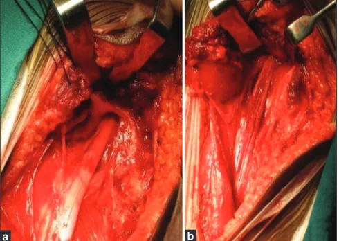
All patients who did not improve beyond 3 months of sustaining injury were offered surgery. As most of the injuries were of sciatic nerve at the buttock level, the surgical technique of exposure of sciatic nerve deep to the buttock is described [Figure 1]. The patient is positioned prone with knee slightly flexed at the thigh and sand bag placed under the anterior iliac crest on the side of the exposure. A curvilinear skin incision is made, starting cephalad and adjacent to the posterior iliac spine, curving around the lateral aspect of the buttock mass and then medially, to end below the buttock crease in the midline of the posterior upper thigh.
The gluteus maximus muscle is detached along its lateral aspect, 2 to 3 cm from its insertion into the greater trochanter, leaving a cuff of muscle and tendon to attach to bone for closure. The entire muscle mass including some gluteus medius is reflected medially. Blunt followed by sharp dissection is performed in a relatively avascular plane deep to the muscle and medially to expose the sciatic nerve. Sharp dissection is performed on and along the nerve itself. The general principle is to work alternatively from above and below the presumed level of injury, gaining circumferential exposure. The last area to be dissected is the lesion site itself.
External internal and neurolysis (making longitudinal incisions) is done over the lesion till healthy fascicles are seen below and above the lesion. The neurolysis is extended superiorly towards the sciatic notch, with care taken to preserve the hamstring, the posterior femoral cutaneous nerve branches, and gluteal nerves and vessels. Piriformis muscle was often sectioned to facilitate exposure in the region of the sciatic notch.
After ensuring decompression of the fascicles, incision is closed in layers; muscle and tendon with non absorbable sutures, and subcutaneous tissue and dermis with absorbable sutures.
Thirty nine (42.3%) of 92 patients underwent surgery. The mean duration to surgery since injury in these
patients was 5.2 months (3 months to 11 months). All underwent exploration of the involved nerve based on preoperative clinical and ENMG findings. All underwent external and internal neurolysis. On exposing the injured nerve all patients were found to have lesion in continuity; 30 (10.3%) had scar, 4 (10.3%) had neuroma, and 5 (12.8%) had thinned out nerve. Intraoperative nerve action potential (NAP) monitoring was not used in any case. None of the patients underwent resection of lesion and nerve grafting. The only complication which occurred was wound infection and it was treated with appropriate antibiotics.
Outcome
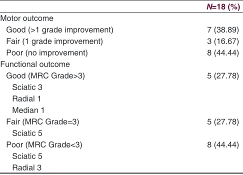
Only 18 (46.1%) patients who underwent surgery had a follow up of more than 3 months (mean 5.1 months; range 3‑18 months). The outcome of these patients is summarized in Table 5. Except for grade of motor deficits prior to surgery none of the variables significantly affected the outcome [Table 6].
Discussion
Intramuscular injection is an often abused form of medical intervention in developing countries. It is the commonest cause of iatrogenic nerve injuries. The exact incidence of INP is not known. During acute flaccid paralysis (AFP) surveillance in children, it was found that INP comprised 12% to 20% cases of AFP.[8,15] Injection injury of peripheral
nerves constitutes a sizeable burden of all nerve injuries. In our study 11% of the patients with nerve injury were due to injections. This is higher than the rates of around 2% reported in literature.[7,16] In spite of regulations, injections
in developing countries are still administered by untrained personnel. In our study 70% of the patients received IMI from persons other than doctors or nurses. In a study
Figure 1: Exposure of sciatic nerve in buttock (a) Sciatic nerve after
reflection of gluteus muscle (b) Sciatic nerve after neurolysis
Table 4: Electroneuromyography (ENMG) findings
ENMG parameter Number (%)
Absent CMAP 64 (70.5)
Reduced CMAP 28 (29.5)
Absent SNAP 67 (72.9)
Reduced SNAP 25 (27.1)
CMAP=Compound muscle action potential, SNAP=Sensory nerve action potential
done from North India,[16] It was found that uncertified
Medical Practitioners were responsible for 86% of INPs. Though most of our patients received injection for febrile illness, the nature of drug injected was not available. In an African study it was found that the common drugs, given intramuscularly in children, resulting in nerve injury were chloroquine, novalgin, penicillin, and sulfadoxine‑pyrimethamine.[11] Certain drugs are
much more damaging than others when injected into a peripheral nerve. The most toxic agents are penicillin, diazepam, chlorpromazine, meperidine, dimenhydrinate, tetanus toxoid, procaine and hydrocortisone.[17]
Clinical features
The mean age of our patients was 8 years, and most were children. In a similar study from North India, the mean age was 28 years.[16] However western studies
have reported higher incidences in the elderly.[14,18] The
reason of increased possibility of INP in children and elderly is thin built and less gluteal covering. The onset of symptoms was immediate in 90% of our cases. The immediate symptoms are due to injection within the nerve causing severe radicular pain and paresthesias along the distribution of nerve, with immediate onset of variable motor and sensory deficit. The cause of delayed symptoms is injection in epineurium or close to nerve, and in tissue plane from which the drug can seep to the nerve resulting in neurotoxicity later.[19] Other cause of delayed worsening is perineural fibrosis.
Most of our patients presented very late after injury. This delay is due to lack of awareness of IMI as a cause of nerve palsy, the hope of spontaneous recovery, and misattribution of nerve palsy to other disease. In our study the commonest nerves involved were the sciatic and radial, which is similar to other studies.[8,16] This
distribution is due to preference of gluteal and shoulder sites for IMI. However injury to every major nerve has
been observed following IMI.[19] In a review of 280 cases
of INP; sciatic (84.3%) was the commonest nerve involved followed by radial (5.4%), median (3.6%), sciatic with posterior femoral cutaneous nerve/pudendal/inferior gluteal (1.4%), axillary (1.1%), ulnar (1.1%), posterior femoral cutaneous nerve (1.1%), superior gluteal nerve (0.7%), femoral (0.7%), lateral femoral cutaneous nerve (0.4%) and medial cutaneous nerve of forearm (0.4%).[1]
Anatomic proximity of the injection is considered the single, most critical factor in determining the degree of damage, with injection directly into the nerve being the most destructive. The large size of sciatic nerve makes it more vulnerable to injury. In accordance with other studies we have also found that common peroneal part of the sciatic nerve is affected more often. The lateral and superficial location of peroneal division makes it more vulnerable to injury.[14] Sciatic nerve injury can result
when the injection is given in outer upper quadrant of the dorsogluteal region, even in patients with normal anatomy. The needles used can be too long for a particular patient or inserted at an angle after entry into the tissue. If the hub of the needle depresses soft tissue, even if the needle is inserted in an outer quadrant, injury can still occur. Positioning is also a predisposing factor, when the patient is placed into a lateral decubitus, upright, or bent forward position instead of prone, probability of nerve injury increases. This changes the relationship of the quadrants to the sciatic nerve.[18]
Most of our patients had severe motor deficits at presentation (about 60% had motor power grade 0), and had inexcitable nerves (about 70%). This observation is similar to other Indian studies[16] but significantly higher
than western reports.[14] This may be due to the poor
referral system in our country, as most of the patients with partial deficits are not referred.
Management
Evaluation of patients with INP includes proper history and clinical evaluation, with quantification of motor and sensory deficits, supplemented with ENMG studies. Table 5: Outcome of patients after surgery
N=18 (%) Motor outcome
Good (>1 grade improvement) 7 (38.89)
Fair (1 grade improvement) 3 (16.67)
Poor (no improvement) 8 (44.44)
Functional outcome
Good (MRC Grade>3) 5 (27.78)
Sciatic 3 Radial 1 Median 1
Fair (MRC Grade=3) 5 (27.78)
Sciatic 5
Poor (MRC Grade<3) 8 (44.44)
Sciatic 5 Radial 3
Table 6: Univariate analysis of the variables affecting outcome
Variable P value
Age 0.391
Duration of symptoms 0.328
ENMG 0.202
Motor deficit grade prior to surgery 0.004*
Operative findings 0.616
Scar in continuity Neuroma in continuity Thinned out
The management of INP is similar to the principles of any other nerve lesions in continuity. The patient should be managed conservatively initially with physiotherapy and appropriate orthoses, with serial clinical and electrophysiological monitoring.
Patients with partial deficits uncomplicated by severe pain, with significant spontaneous recovery or late referral can be managed without surgery. Surgical exploration is not indicated in as many as 50% of injuries.[14] Most of these patients achieve partial but
good spontaneous recovery. However conservative management should not be prolonged in patients who do not exhibit recovery at the expected time following injury.[14]
If intraoperative nerve action potential (NAP) facility is available then these injuries can be explored at 3 months. The timing of exploration is influenced by whether a sensory or motor deficit exists. If a motor deficit exists, some proximal muscle recovery is anticipated, either clinically or electrophysiologically within 3 months. If only a sensory deficit exists; a longer period of observation is appropriate, as successful reinnervation of sensory receptors is not as time dependent as are motor targets. Only exception to this is severe pain; not responding to neuropathic analgesics; which can be relieved with neurolysis. Controversy exists regarding the role of immediate surgical intervention in the treatment of nerve injection injuries. Experimental studies have shown good outcome of immediate exploration for neruolysis or irrigation of the involved nerve with normal saline.[20]
This approach is not practical for human beings. Most of these cases are seen by specialist much later for non improvement. The timing of surgery in our series was 5.2 months since surgery. The surgical treatment for INP is neurolysis. After neurolysis if NAP is not elicitable across the lesion in a particular division of nerve, only that portion needs to be excised and repaired using nerve graft. Most of these lesions are within 1 to 2 cms of sciatic notch hence it is difficult to get good length of proximal stump after resection for anastomosis. In a study of sciatic nerve injection palsy, less than 15% of patients required either suture or graft repair of the nerve. Rest could be managed with neurolysis only based on NAP monitoring.[18] We do not have facility for intraoperative
NAP monitoring and hence only neurolysis was done.
Outcome
Some patients recover function spontaneously, but others do not. Results are generally good because many injuries are focal. As with other injuries, the prognosis depends on the part or divisions involved, level of injury, timing of surgery, and the patient’s overall medical
status.[18] Outcome of tibial division is better than
peroneal division of sciatic nerve. In a study of 64 INPs of sciatic nerve, 84% of patients with tibial division had good functional outcome after neurolysis, as opposed to only 68% of patients with peroneal division injury.[18]
However improvement in inversion (peroneal nerve function) leads to better functional outcome as patient is able to put the foot flat on the ground for taking steps. Outcome is not so good after resection of lesion and graft repair. In the same study only 57% of patients with tibial division had good outcome after graft repair. Outcome of surgery for INP is better than that for other iatrogenic injuries like total hip arthroplasty. Only 75% patients with tibial division and 25% with peroneal division injuries after hip arthroplasty had good outcome. Patients with nerve injuries improve over several months following surgery and if they are followed up for longer duration many will show good outcome eventually. Of 164 INPs of sciatic nerve, 57% and 78% in the early and late stage, respectively had excellent to good nerve recovery.[21] The
functional outcome was good or fair in 55% of our cases. The reason of less number of good outcomes in our study was short duration of follow up. The only significant factor which affected outcome was motor power grade before surgery.
Prevention
The need to discourage indiscriminate use of intramuscular injections and choice of a proper site of injection should be stressed. Only trained staff should be allowed to administer intramuscular injections. The choice of site for injection must be based on good clinical judgment, using the best evidence available and individualized assessment of the patient. There is wide agreement in the literature that the ventrogluteal site is preferable site for intramuscular injections in the gluteal region. According to royal college of nursing recommendation, the anterolateral thigh should be used in infants and the deltoid in older children. The buttock is not recommended. The size of the needle should also be according to age and built of children.[22] Physicians and
nurses should be educated about INP. When they suspect INP they should refer patients early to the specialist. If motor deficits occur, physiotherapy should be started and appropriated orthoses should be prescribed for prevention of contractures. INP from erroneous injection causes discomfort, morbidity, lasting disability, and also provides the basis for negligence suits.[17]
Conclusion
injuries. The outcome of INP is generally good and many patients recover spontaneously. The outcome of surgery is good if performed within reasonable time after injury. Outcome is influenced by motor power after injury. However prevention of nerve injuries due to intramuscular injections cannot be under emphasized.
References
1. Iyer VG, Shields CB. Isolated injection injury to the posterior femoral cutaneous nerve. Neurosurgery 1989;25:835-8.
2. Gilles FH, French JH. Postinjection sciatic nerve palsies in infants and children. J Pediatr 1961;58:195-204.
3. Gilles FH, Matson DD. Sciatic nerve injury following misplaced gluteal injection. J Pediatr 1970;76:247-54.
4. Kane A, Lloyd J, Zaffran M, Simonsen L, Kane M. Transmission of hepatitis B, hepatitis C and human immunodeficiency viruses through unsafe injections in the developing world: Model-based regional estimates. Bull World Health Organ 1999;77:801-7.
5. Mayer M, Romain O. Sciatic paralysis after a buttock intramuscular injection in children: An ongoing risk factor. Arch Pediatr 2001;8:321-3. 6. Villarejo FJ, Pascual AM. Injection injury of the sciatic nerve (370 cases).
Childs Nerv Syst 1993;9:229-32.
7. Yuen EC, So YT, Olney RK. The electrophysiologic features of sciatic neuropathy in 100 patients. Muscle Nerve 1995;18:414-20.
8. Ahuja B. Post injection sciatic nerve injury. Indian Pediatr 2003;40:368-9. 9. Anand K, Pandav CS, Kapoor SK. Injection use in a village in north
India. Natl Med J India 2001;14:143-4.
10. Bhatia M, Jindal AK. Injection induced nerve injury: An iatrogenic tragedy. J Assoc Physicians India 1996;44:532-3.
11. Fatunde OJ, Familusi JB. Injection-induced sciatic nerve injury in Nigerian children. Cent Afr J Med 2001;47:35-8.
12. Kotwal A, Priya R, Thakur R, Gupta V, Kotwal J, Seth T. Injection practices in a metropolis of North India: Perceptions, determinants and issues of safety. Indian J Med Sci 2004;58:334-44.
13. Kumar S. Traditional Indian remedy for asthma challenged in court. BMJ 2004;328:1457.
14. Kline DG, Kim D, Midha R, Harsh C, Tiel R. Management and results of sciatic nerve injuries: A 24-year experience. J Neurosurg 1998;89:13-23. 15. Sharma S, Kale R. Post injection palsy in Chhatisgarh region. Indian
Pediatr 2003;40:580-1.
16. Pandian JD, Bose S, Daniel V, Singh Y, Abraham AP. Nerve injuries following intramuscular injections: A clinical and neurophysiological study from Northwest India. J Peripher Nerv Syst 2006;11:165-71. 17. Shukla DP, Bhat DI, Devi BI, Gopalakrishnan MS, Moiyadi A. Injection
injuries of peripheral nerves. Indian J Neurotrauma 2004;1:21-3. 18. Yeremeyeva E, Kline DG, Kim DH. Iatrogenic sciatic nerve injuries
at buttock and thigh levels: The Louisiana State University experience review. Neurosurgery 2009;65:A63-6.
19. Midha R. Mechanism and pathology of injury. In: Kline and Hudson’s nerve injuries: Operative results for major nerve injuries, entrapments, and tumors. 2nd ed. China: Saunders Elsevier; 2008. p. 23-42.
20. Zhu Q. Peripheral nerves injection injury: The clinical and experimental study. Zhonghua Wai Ke Za Zhi 1992;30:522-4.
21. Huang Y, Yan Q, Lei W. Gluteal sciatic nerve injury and its treatment. Zhongguo Xiu Fu Chong Jian Wai Ke Za Zhi 2000;14:83-6.
22. Position Statement on Injection Technique. March 2002. Available from: http://www.rcn.org.uk/__data/assets/pdf_file/0010/78535/001753. pdf. [Last cited on 2011 May 16].
How to cite this article: Kakati A, Bhat D, Devi BI, Shukla D. Injection nerve palsy. J Neurosci Rural Pract 2013;4:13-8.
Source of Support: Nil. Conflict of Interest: None declared.
Staying in touch with the journal
1) Table of Contents (TOC) email alert
Receive an email alert containing the TOC when a new complete issue of the journal is made available online. To register for TOC alerts go to www.ruralneuropractice.com/signup.asp.
2) RSS feeds