Available Online at www.ijpret.com 835
INTERNATIONAL JOURNAL OF PURE AND
APPLIED RESEARCH IN ENGINEERING AND
TECHNOLOGY
A PATH FOR HORIZING YOUR INNOVATIVE WORK
BLOOD VESSEL SEGMENTATION
PROF. SAGAR P. MORE1, PROF. S. M. AGRAWAL2, PROF. M. A. KHAN3
1. Assistant Professor: Electrical (Electronics & Power) Babasaheb Naik College of Engineering, Pusad, India(M.S).
2. Associate Professor & Head: Electrical (Electronics & Power) Engnineering Department. Babsaheb Naik College of Engineering, Pusad, India (M.S).
3. Associate Professor: Electronics & Telecommunication Babsaheb Naik College of Engineering, Pusad, India (M.S).
Accepted Date: 05/03/2015; Published Date: 01/05/2015
\
Abstract: The blood vessel segmentation is an important issue for assessing retinal abnormalities and diagnoses of many diseases. The segmentation of vessels is complicated by huge variations in local contrast, particularly in case of the minor vessels. In this paper, proposed a new method of texture based vessel segmentation to overcome this problem. It uses Gaussian and L*a*b*perceptually uniform color spaces with original RGB for texture feature extraction on retinal images. A bank of Gabor energy filters are used to analyze the texture features from which a feature vector is constructed for each pixel. The Fuzzy C-Means (FCM) clustering algorithm is used to classify the feature vectors into vessel or non-vessel based on the texture properties. After the FCM clustering output it attains the final output segmented image after a post processing step.
Keywords: Medical Images; Texture Classification; Gabor Energy Filter Bank; FCM Clustering; Image Segmentation.
Corresponding Author: PROF. SAGAR P. MORE
Access Online On:
www.ijpret.com
How to Cite This Article:
Sagar P. More, IJPRET, 2015; Volume 3 (9): 835-843
Available Online at www.ijpret.com 836
INTRODUCTION
The detection of blood vessels is very important as ophthalmologists can potentially screen larger populations for vessel abnormalities. In contrast, manual delineation of vessels becomes tedious or even impossible when the number of vessels in an image is large or when a large number of images are acquired. Blood vessel appearance can provide information on pathological changes caused by some diseases including diabetes, hypertension, and arteriosclerosis. Changes in retinal vasculature, such as hemorrhages, angiogenesis; increases in vessel tortuosity, blockages and arteriolar-venulardiameter ratios are important indicators of, for example, diabetic retinopathy, and retinopathy of prematurity and cardiovascular risk. Information about blood vessels in retinal images can be used in grading disease severity or as part of the process of diagnosis of diseases.
Retinal segmentation is complicated by the fact that the width of the retinal vessels can vary from large to very small, and the local contrast of vessels is unstable, especially in unhealthy retinal images. Although a large number of schemes have been proposed for the detection of blood vessels, a huge improvement in detection procedures remains a necessity for the detection of minor vessels.
In this report novel approach is proposed for vessel segmentation which is equally efficient to detect major and minor vessels. It considers Gaussian and L*a *b *perceptually uniform color spaces with the original RGB image for texture feature extraction. To extract features, a bank of Gabor energy filters with three wavelengths and twenty-four orientations is applied in each selected color channel. Then a texture image is constructed from the maximum response of all orientations for a particular wavelength in each color channel. From the texture images, a feature vector is constructed for each pixel. These feature vectors are classified using the FCM clustering algorithm. Finally, it segments the image based on the cluster centroid value.
II. LITRETURE SURVEY.
Available Online at www.ijpret.com 837 Title Author Description
1 Retinal vessel extraction by Bob Zhang, Lin
matched filter response with Zhang, Lei Zhang first order derivative of, Fakhri karry. Gaussian.
The propose MF-FDOG is compose of original MF which is zero mean Gaussian function and first order derivative of Gaussian. The Vessel is detected by Thresholding the retinal images response to the MF.
On the adaptive detection of Di Wu, Ming blood vessel in retinal Zhang , Jyh-charn image. Liu.
It as adaptive detection scheme for large and small blood vessel in color retinal images Consisting three functions adaptive contrast enhancement, feature extraction of blood vessel and tracing. The average of 75% of small vessels was capture by this method.
A. Retinal vessel extraction by matched filter response with first order derivative of gaussian.
Accurate extraction of retinal blood vessels is an important task in computer aided diagnosis of retinopathy. The matched filter (MF) is a simple yet effective method for vessel extraction. However, a MF will respond not only to vessels but also to non-vessel edges. This will lead to frequent false vessel detection. In this paper proposed a novel extension of the MF approach, namely the MF-FDOG, to detect retinal blood vessels. The proposed MF-FDOG is composed of the original MF, which is a zero- mean Gaussian function, and the first-order derivative of Gaussian (FDOG). The vessels are detected by thresholding the retinal image's response to the MF, while the threshold is adjusted by the image's response to the FDOG. The proposed MF-FDOG method is very simple; however, it reduces significantly the false detections produced by the original MF and detects many fine vessels that are missed by the MF. It achieves competitive vessel detection results as compared with those state-of-the-art schemes but with much lower complexity. In addition, it performs well at extracting vessels from pathological retinal images.
B. On the adaptive detection of blood vessel in retinal image.
Available Online at www.ijpret.com 838 for truepositive rate (TPR), and 3.9% for false positive rate (FPR). For normal images, the TPRs range from 80%to 91%, and their corresponding FPRs range from 2.8% to 5.5%. For abnormal images, the TPRs range from 73.8% to 86.5% and the FPRs range from 2.1% to 5.3%, respectively. Small vessels take up 42% of overall vessel pixels, where 75% of small vessels were captured by our method. Enhancement of blood vessels is achieved from the extension of the adaptive histogram equalization technique. Feature extraction of small blood vessels is by using the standard deviation of Gabor filter responses along different orientations. Tracing of the vascular network consists of three major functions: forward detection, backward verification, and bifurcation detection. Combining extrapolation and local greedy search reduces the prediction errors of vessel directions by 15-20%. Only two sample images and their hand-traced maps are needed for parameter training and calibration.
III. PROPOSED METHOD
Here a new method is proposed for blood vessel segmentation which is based on the texture property analysis of vessel and non-vessel parts in the color retinal images. The reasons are as follows. Firstly, due to large variation of local contrast in the retinal image, texture analysis is more appropriate to extract features from vessel and non-vessel parts in the retinal images. Secondly, a color texture is a patio-chromatic pattern and can be defined as the "distribution of colors over a surface"; therefore, incorporating color into texture analysis is enhancing the procedure. The original retinal images are in RGB color space which is not perceptually uniform and Euclidean distances in 3D RGB space do not correspond to differences as perceived by humans. In addition, perceptually uniform color spaces are very effective in rotation invariant color texture analysis. So, here perceptually uniform color spaces along with original RGB color channels are used to extract texture features.
Available Online at www.ijpret.com 839 At first apply the transformation of original RGB image into Gaussian and L*a*b* color space. Then choose first two components of Gaussian color space "E and E"X, Luminance L from L*a*b*color space and Green channel G from RGB color space due to the higher contrast of vessel and background which is convenient for texture analysis. After this apply Adaptive
Histogram Equalization (AHE) method to these four different color channel images for contrast enhancement. For each of these color channels, apply a bank of Gabor filters with twenty-four orientations and three wavelengths for texture feature extraction. Then construct the texture image in each color channel for every wavelength considering the maximum response of all twenty-four orientations. These texture images are used to analyze the number of clusters which latter will be used as the classifier input. Consequently, we construct twelve texture images for each original retinal image.
Here a feature vector for every pixel mapping each pixel position of all these texture image constructed (i.e. each feature vector is in the length of twelve elements). These feature vectors are classified as a vessel or background part using unsupervised FCM clustering algorithm. From the output of the FCM clustering algorithm construct a 2D matrix (as original image dimension) with cluster numbers which have the highest membership values (for each position). Finally, it produces the ultimate segmented image with converting the cluster numbers into binary values considering the cluster centroid values. Figure 3.1 portrays the overall technique of our proposed method.
A. Channel selection.
First apply the transformation of original RGB image into Gaussian and L*a*b* color space. Then choose first two components of Gaussian color space "E and ETX, Luminance L from
L*a*b* color space and Green channel G from RGB color space due to the higher contrast of
vessel and background which is convenient for texture analysis. Here for simplicity we have used only one color channel i.e. green. The green channel possess high contrast level which will be helpful in further processing.
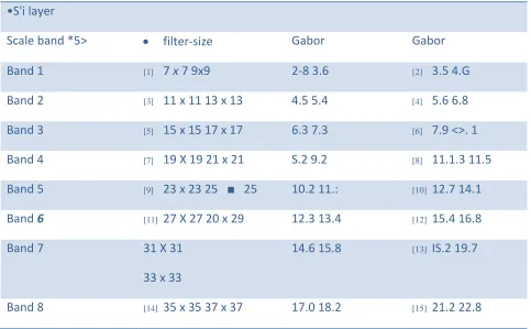
B. Texture features extraction
Available Online at www.ijpret.com 840 retinal images is very promising. In the following two subsections the Gabor filter based texture analysis method.
•S'i layer
Scale band *5> filter-size Gabor Gabor
Band 1 [1] 7 x 7 9x9 2-8 3.6 [2] 3.5 4.G
Band 2 [3] 11 x 11 13 x 13 4.5 5.4 [4] 5.6 6.8
Band 3 [5] 15 x 15 17 x 17 6.3 7.3 [6] 7.9 <>. 1
Band 4 [7] 19 X 19 21 x 21 S.2 9.2 [8] 11.1.3 11.5
Band 5 [9] 23 x 23 25 ■ 25 10.2 11.: [10] 12.7 14.1
Band 6 [11] 27 X 27 20 x 29 12.3 13.4 [12] 15.4 16.8
Band 7 31 X 31
33 x 33
14.6 15.8 [13] IS.2 19.7
Band 8 [14] 35 x 35 37 x 37 17.0 18.2 [15] 21.2 22.8
Fig 3.2 table of Gabor filter bank
C. TEXTURE CLASSIFICATION AND IMAGE SEGMENTATION The FCM is a data clustering
technique where in each data point belongs to a cluster to some degree that is specified by a membership grade. Let X = x1, x2,xn where x eRN present a given set of feature data. The objective of the
FCM clustering algorithm is to minimize the Fuzzy C-Means cost function formulated as
j{h\v)=Y^{PlJT\\^-vi\?
V = {v1, v2, ,vC} are the cluster centers. U = (p.tj)N^C is fuzzy partition matrix, in which each
Available Online at www.ijpret.com 841
Fig. 3.3 Original images (left) and segmented vessel center line images (right).
The exponent m £[1,°°] is the weighting exponent, which determines the fuzziness of the clusters. The most commonly used distance norm is the Euclidean distance diJ= \\xi-vj\\.
The Matlab Fuzzy Logic Toolbox is used for clustering 253440 vectors (the size of the retinal image is 512x495) in length twelve for each retinal image. In each retinal image clustering procedure, the number of clusters was assigned after analyzing the histogram of the texture image. The parameter values used for the FCM clustering were as follows. The exponent value of 2 for the partition matrix, maximum number of iterations was set to 1000 for the stopping criterion and the minimum amount of improvement being 0.00001. Here the membership values on each cluster for every vector are received, from which the cluster number that belonged to the highest membership value for each vector are taken out and converted it into a 2D matrix. From this matrix produced the binary image considering the cluster central intensity value which identifies the blood vessels only.
Available Online at www.ijpret.com 842
EXPERIMENTAL RESULTS
In this project segmented vessel are found from color retinal images with software approached. The output image of fundus camera is directly inputted to the process and after processing output image is segmented image of vessel. This method is very useful to find smallest blood vessel from the color retinal image. The segmented blood vessel appearance can provide information on pathological changes caused by some diseases example diabetes, hypertension, and arteriosclerosis.
Iv. CONCLUSION
In this project proposed a novel approach for a texture based vessel segmentation technique. The automated vessel detection reduces manual delineation. This method is very efficient in detecting both major and minor blood vessels.
REFERENCES
1. A. Hoover, V. Kouznetsova, and M. Goldbaum, "Locatingblood vessels in retinal images by piece-wise threshold probing of a matched filter response," IEEE Transactionson Medical Imaging, vol. 19(3), pp. 203-210,2000.
2. D. Wu, M. Zhang, and J. Liu, "On the adaptive detection of blood vessels in retinal images," IEEE Transactionson Biomedical Engineering, vol. 53(2), pp. 341-343, 2006.
3. G. W. Wyszecki and S. W. Stiles, "Color science: Concepts and methods, quantitative data and formulas," NewYork, Wiley, 1982. [4] J. Geusebroek, R. V. D. Boomgaard, A. W. M. Smeulders, and H.
4. Geerts, "Color invariance," IEEE Transactionson Pattern Analysis and Machine Intelligence, vol. 23(2), pp. 1338-1350, 2001.
5. C. Sinthanayothin, J.F. Boyce, H.L. Cook, T.H. Williamson Automated localisation of the optic disc, fovea, and retinal blood vessels from digital colour fundus images British Journal of Ophthalmology, 83 (1999), pp. 902-910.
Available Online at www.ijpret.com 843 7. J. Staal, M.D. Abramoff, M. Niemeijer, M.A. Viergever, B. van Ginneken Ridge-based vessel segmentation in color images of the retina, IEEE Transactions on Medical Imaging, 23 (2004), pp. 501-509.
8. J.V.B. Soares, J.J.G. Leandro, R.M. Cesar, H.F. Jelinek, M.J. Cree Retinal vessel segmentation using the 2-D Gabor wavelet and supervised classification, IEEE Transactions on Medical Imaging, 25 (2006), pp. 1214-1222.
9. E. Ricci, R. Perfetti Retinal blood vessel segmentation using line operators and support vector classification,IEEE Transactions on Medical Imaging, 26 (2007), pp. 1357-1365.
10.L. Xu, S. Luo, A novel method for blood vessel detection from retinal images, BioMedical Engineering Online, 9 (2010), p. 14
11.. Kaba , A. G. Salazar-Gonzalez , Y. Li , X. Liu and A. Serag Health Information Science, pp.105 -112 2013 :Springer-Verlag. [12] A. Efros and T. Leung "Texture synthesis by non-parametric sampling", Proc. IEEE Int. Conf. Comput. Vis, pp.1033 -1038 1999.
12. X. You, Q. Peng, Y. Yuan, Y.-m. Cheung, J. Lei, Segmentation of retinal blood vessels using the radial projection and semi-supervised approach Pattern Recognition, 44 (2011), pp. 2314-2324.
13.A. G. Salazar-Gonzalez , Y. Li and X. Liu "Retinal blood vessel segmentation via graph cut", Proc. IEEE 11th Int. Conf. Contr. Autom. Robot. Vis., pp.225 -230 2010.