toxicity. We also have shown that the well-knownSaccharomyces cerevisiaesulfite efflux permease Ssu1 is also able to excrete nitrite and nitrate. These results characterize for the first time essential components of the nitrate/nitrite efflux system and their impact on net nitrate uptake and its regulation.
T
he yeastHansenula polymorphais able to use nitrate as the sole nitrogen source. Nitrate is transported into the cell and then reduced to ammonium by the consecutive action of nitrate and nitrite reductase (NR) (1–3). Nitrate assimilation genes are in-duced by nitrate (4,5) and repressed by preferred nitrogen sources (6). High-affinity nitrate and nitrite transport is mainly mediated by Ynt1, which also is posttranslationally regulated in response to nitrogen source quality (7,8). In algae and yeast, nitrate acts as an inducer once it enters the cell, and therefore, intracellular nitrate levels play a key role in regulating nitrate assimilation genes (9). In this framework, nitrate and nitrite effluxes from the cell could play an important role in net nitrate/nitrite uptake and also in keeping nitrite below toxic levels. Nitrite efflux has been observed in most organisms, includingH. polymorpha, growing in nitrate (1,10– 13), indicating a clear imbalance between nitrate uptake and re-duction to nitrite and its further transformation to ammonium. In contrast, nitrate efflux has not been found in fungi. However, in plants, nitrate efflux can even exceed nitrate uptake in various stress situations. A nitrate excretion transporter, NAXT1, belong-ing to the NRT1/PTR family has been found in the root plasma membrane ofArabidopsis thaliana, although its role is scarcely understood (14). Moreover, it has been reported thatArabidopsis NRT1.1 (CHL1) is a bidirectional transporter involved in root-to-shoot nitrate translocation (15).InSaccharomyces cerevisiae, Ssu1 is involved in sulfite efflux (16). It belongs to the tellurite resistance/dicarboxylate trans-porter (TDT) family, which includes theEscherichia colitellurite transporter TehAp and the Schizosaccharomyces pombe malate transporter Mae1 (17). Upregulation ofSSU1andYHB1has been found inS. cerevisiaeandCandida albicansin response to nitric oxide (NO)-generating compounds (18,19).YHB1encodes a fla-vohemoglobin that presents NO dioxygenase activity, which cat-alyzes the transformation of NO to nontoxic nitrate and thereby protects against nitrosylation of cellular targets and inhibition of cell growth, under both aerobic and anaerobic conditions (20). However, the role of Ssu1 in NO detoxification is unknown, al-though it has been suggested that besides transporting sulfite, Ssu1
may also transport NO-derived metabolites, such as nitrite or ni-trate, out of the cell (19).
Aspergillus nidulansNitA (AnNitA), belonging to the formate-nitrite transporter family (FNT), mediates specific high-affinity transport of nitrite inA. nidulansand also has some role in nitrite efflux in that fungus (13). FNT members have been found in bac-teria, archaea, fungi, algae, and protozoan parasites. InE. coli, FocA and NirC have been characterized and implicated in the transport of formate and nitrite, respectively (21,22). Moreover, NirC is also involved in nitrite efflux (11). The structure of FocA strongly suggests that it is a channel rather than a transporter (23). In Chlamydomonas reinhardtii, some of the NAR1 genes are clearly regulated by carbon or nitrogen (24) and involved in nitrite transport in the chloroplast (25).
In this study, we aimed to explore at a molecular level the nitrate and nitrite extrusion systems in the nitrate-assimilatory yeastH. polymorpha. The rationale of our approach was to search theH. polymorphagenome database for genes encoding mem-brane proteins with similarity to nitrate/nitrite transporters. Ssu1/2, encoding a sulfite permease, were included because of the structural resemblance between sulfite and nitrite and also since SSU1is induced by NO precursor donors inS. cerevisiae(19). We have uncovered some of the molecular entities involved in nitrate/ nitrite efflux in fungi. Ssu2 and to a lesser extent Ssu1 extrude
Received2 October 2013Accepted16 December 2013 Published ahead of print20 December 2013
Address correspondence to José M. Siverio, [email protected].
* Present address: Elisa Cabrera, Research Unit, University Hospital of Canary Islands, La Laguna, Tenerife, Spain; Rafaela González-Montelongo, Research Unit, University Hospital N.S. Candelaria, Santa Cruz de Tenerife, Tenerife, Spain.
E.C. and R.G.-M. contributed equally to this work.
Supplemental material for this article may be found athttp://dx.doi.org/10.1128
/EC.00268-13.
Copyright © 2014, American Society for Microbiology. All Rights Reserved.
doi:10.1128/EC.00268-13
on September 8, 2020 by guest
nitrate, while Nar1 extrudes nitrate and nitrite. We also have shown thatS. cerevisiaeSsu1 extrudes nitrite and nitrate, in addi-tion to sulfite.
MATERIALS AND METHODS
Strains and growth conditions.TheH. polymorphastrains used in this work are listed in Table S1 in the supplemental material. All strains are derivatives of the NCYC495leu2 ura3strain. Yeast cells were grown with shaking at 37°C in YPD medium (1% [wt/vol] yeast extract, 2% [wt/vol] peptone, and 2% [wt/vol] glucose) or synthetic medium containing 0.17% (wt/vol) yeast nitrogen base without amino acids and ammonium sulfate (Difco), 2% (wt/vol) glucose, and the nitrogen source indicated in each case. Nitrogen deprivation medium (nitrogen-free medium) con-tains 0.17% (wt/vol) yeast nitrogen base without amino acids and ammo-nium sulfate (Difco) and 2% (wt/vol) glucose (YG). Whenever necessary, media were supplemented with 30g/mlL-leucine, 20g/ml uracil, or 100g/ml Zeocin (Invitrogen). Sulfite plates were made as described previously (26). To test yeast chlorate sensitivity, potassium chlorate was added to medium before sterilization at the concentration indicated in each case. One OD660(optical density at 660 nm) unit was about 3.5 mg
cells · ml⫺1(approximately 7⫻107cells · ml⫺1).
Plasmids.All of the primers for gene disruption, tagging, or quanti-tative real-time PCR (qRT-PCR) are described in Table S2 in the supple-mental material. All vectors used in this work are listed in Table S3. The
pHPI 359vector (27) was used to fuse the promoter of theSSU2orNAR1
gene to thelacZgene to obtainpPSSU2-lacZorpNAR1-lacZ, respectively.
The region from⫺1006 to⫹45 relative to ATG ofSSU2was amplified by PCR from genomic DNA, using the primers SSU2Prom-F and SSU2Prom-R. To obtain the promoter ofNAR1, the region from⫺900 to
⫹31 was amplified with the primers proNAR1-F and proNAR1-R. Both constructs were linearized at BstEII inLEU2before yeast transformation.
pPSSU2I-ScSSU1LEU2 was generated to express S. cerevisiae SSU1
(ScSSU1) (NC_001148.4, NCBI reference sequence) under theH. poly-morpha SSU2(HpSSU2) gene promoter and was obtained by inserting a 1,883-bp DNA fragment containing the ScSSU1open reading frame into the plasmidpGEMT-PSSU2ILEU2. This last vector was constructed by
in-serting a 1,200-bp DNA fragment containing the HpSSU2gene promoter into the plasmid pGEM-T Easy (Promega) and by inserting theLEU2gene marker. To transform yeast, DNA was linearized at BstEII inLEU2.
pSSU2-GFPandpNAR1-GFPcarry theSSU2orNAR1C-terminal region fused in frame to GFP (green fluorescence protein) by inserting each open reading frame without its stop codon into the BglII site ofpANL31(28).
pSSU2-GFPwas linearized atSSU2with BclI whilepNAR1-GFPwas lin-earized atNAR1with KpnI to transform strains bearingSSU2orNAR1.
pSSU2-YFPcontains theSSU2open reading frame without a stop codon fused to the 5=-end cDNA of the enhanced yellow fluorescent protein (eYFP) in EcoRI-SalI sites ofpEYFP-N1(BD Biosciences Clontech) and subcloning into EcoRI-SalI sites ofpGEMHE. A 1,200-bp DNA fragment containingSSU2-YFPwas subcloned intopGEMHE, which contains 5=
and 3=untranslated regions of theXenopus laevis-globin gene (29) to enhance protein expression inX. laevisoocytes.
Disruption ofH. polymorpha SSU1,SSU2, andNAR1genes.To disruptSSU1, the region from⫺905 to⫹1947 relative to the ATG start codon was amplified by PCR usingPfufrom genomic DNA by using the oligonucleotides SSU1-F and SSU1-R. This fragment was cloned into the plasmid pGEM-T Easy (Promega), obtaining the vectorpGEMT-SSU1. A 1,642 bp-internal region fromSSU1(from nucleotide⫺427 to⫹1215) was removed with XhoI and BglII and replaced by theURA3gene marker to generate the vectorpssu1⌬URA3.ssu1⌬strains were then generated by transforming the wild type (WT) with the 3,134-bp fragment amplified frompssu1⌬URA3with the SSU1-F and SSU1-R oligonucleotides. Trans-formants bearing the disrupted target gene were identified by PCR. The region from⫺999 to⫹1828 relative to ATG of theSSU2gene was ampli-fied as above using the oligonucleotides SSU2-F and SSU2-R. This frag-ment was cloned into the plasmid pGEM-T Easy (Promega), obtaining
the vectorpGEM-SSU2 int. A 1,599-bp internal region fromSSU2(from nucleotides⫺174 to⫹1425) was removed with NruI and replaced by the
zeocinresistance gene (blegene) as a selective marker frompREMIZ(30) to generate the vectorpssu2⌬ble.ssu2⌬strains were then generated by transforming the WT with the 3,217-bp fragment amplified from
pssu2⌬blewith the SSU2-F and SSU2-R oligonucleotides. Transformants bearing the disrupted target gene were identified by PCR and by sulfite sensitivity. To disruptNAR1, the region from⫺777 to⫹2096 relative to ATG was amplified by PCR usingPfufrom genomic DNA by using the oligonucleotides 334int-F and 334int-R. This fragment was cloned into the plasmid pGEM-T Easy (Promega), obtaining the vectorpNAR1. A 347-bp internal region fromNAR1(from nucleotides⫹430 to⫹777) was removed with BamHI and KpnI and replaced by theURA3gene marker to generate the vectorpnar1⌬. Thenar1⌬::URA3strain was generated by transforming the WT with the 4,499-bp fragment amplified frompnar1⌬
with the 334int-F and 334int-R oligonucleotides. Transformants bearing the disrupted target gene were identified by PCR.
nSSU2and nNAR1strains.Strains bearing several copies of theSSU2
gene (nSSU2) were obtained by transforming the WT strain with the plasmidpSSU2-URA3orpSSU2-LEU2linearized at theURA3gene with BglII or atLEU2with BstEII. Strains bearing multiple integrations of
pSSU2URA3orpSSU2LEU2were screened for increased sulfite resistance and low nitrate uptake. The nNAR1strain was obtained by transforming
nar1⌬with the plasmid pNAR1-LEU2linearized atLEU2 with NarI. nNAR1strains were screened for increased nitrite resistance and nitrite excretion.
Determination of intracellular nitrate and nitrite.Cells grown in ammonium were resuspended at 10 mg/ml (wet weight) in YG and incu-bated with shaking for 120 min, and then nitrate and nitrite were added to the cells at the concentration indicated in each case. Cells (250 mg [wet weight]) were collected over 25 ml cold water by centrifugation for 5 min at 4,863⫻gat 4°C, washed with cold water, and kept below⫺20°C until use. Cells were resuspended in 1 ml of a boiling solution made of 75% ethanol (vol/vol) buffered with 70 mM HEPES, pH 7.5, and incubated 5 min at 80°C, as described previously (31). After cooling down on ice for 5 min, samples were centrifuged for 15 min at 20,500⫻gat 4°C to remove the cells. Volume was reduced to 500l by evaporation at 40°C using a vacuum concentrator (Heto). Nitrate and nitrite uptake activity was mea-sured as described in a previous report (32) as extracellular nitrate or nitrite depletion. Purified H. polymorpha nitrate reductase enzyme (NECi) was used to determine the nitrate concentration. Nitrite was colo-rimetrically measured as described previously (33). Nitrate uptake is ex-pressed as nmol of NO3⫺/NO2⫺transported · min⫺1· mg of cell⫺1.
Re-sults are reported as mean values⫾standard deviations (SD) from at least three independent experiments. Sulfite up to 240M does not interfere with nitrate determination assays using purifiedH. polymorphanitrate reductase (NECi) (data not shown).
Functional expression of HpSSU2 in Xenopus oocytes.Capped mRNA was transcribed in vitrofrom linearizedpSSU2-YFP by using mMESSAGE mMACHINE kits (Ambion). All procedures involving Xe-nopus laeviswere approved by the University of La Laguna Research Ethics Committee in agreement with local and national legislation. Oocytes were harvested from adult females under benzocaine anesthesia by partial ovariectomy and collagenase IA dispersion. Stage V to VI oocytes were selected and microinjected with 20, 10, and 5 ng ofSSU2cRNA. Cells were then incubated for 7 days at 18°C in oocyte Ringer’s medium (containing [mmol/liter] NaCl [82.5], KCl [2], CaCl2[2], MgCl2[2], Na2HPO4[1],
and HEPES [10], at pH 7.5).SSU2-YFPprotein expression was detected by Western blot analysis ofXenopusoocyte extracts using an anti-GFP monoclonal antibody (Roche) as previously described (34). Cell surface expression of fluorescently labeledSSU2was detected from whole oocytes using a laser scanning confocal microscope (Olympus FluoView 1000). Background fluorescence was assessed by imaging noninjected or water-injected oocytes (35). Nitrate efflux was measured by microinjecting oocytes expressing or not expressing Ssu2 with 30 nl of 30 mM KNO3or
Cabrera et al.
on September 8, 2020 by guest
http://ec.asm.org/
30 mM NaNO2, followed by a 15-min incubation in Ringer’s medium.
Afterwards, nitrate in the medium was measured as described above. Ni-trate efflux is expressed as nmol of niNi-trate determined in the medium after 15 min of nitrate microinjection.
Cell viability.WT,ssu2⌬,nar1⌬,ynr1⌬, andynr1⌬ssu2⌬strains were grown in ammonium and resuspended at 10 mg/ml (wet weight) in 10 mM nitrate, 5 mM ammonium, and 1 mM nitrite. This point represented 100% of viability. To calculate the percentage of viable cells, approxi-mately 100 cells were plated over YPD in triplicate. These experiments were repeated at least three times.
Miscellaneous methods.Electrotransformation of yeast cells was per-formed as described previously (36).-Galactosidase activity was deter-mined as described in reference27. Yeast cell extract preparation, SDS-PAGE, and immunoblotting were done as described in reference 9. Fluorescence microscopy ofSSU2-GFPwas performed as described pre-viously (8). RNA extraction and qRT-PCR were done as described in reference6.
Nucleotide sequence accession numbers.The sequences ofSSU1,
SSU2, andNAR1have been deposited in GenBank under accession num-bersHF585084,HF585085, andHF585083, respectively.
RESULTS
H. polymorphaSsu1 and Ssu2 are involved in nitrate efflux.The S. cerevisiaesulfite efflux permeaseSSU1gene is induced by NO-generating compounds, and its involvement in NO-derived me-tabolite efflux has been suggested (19). This prompted us to study the role of ScSSU1orthologs in nitrate efflux in the yeastH. poly-morpha. Two open reading frames (ORFs), termed HpSSU1and HpSSU2, encoding proteins similar to ScSsu1, are present in the
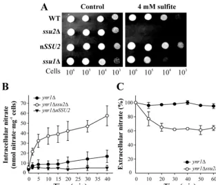
H. polymorphagenome database. Strains bearing disruptedSSU1 orSSU2showed sensitivity to sulfite, which was much greater in thessu2⌬strain. In contrast, the nSSU2strain, bearing several copies ofSSU2, was more resistant (Fig. 1A).
To analyze whether Ssu2 is involved in nitrate efflux, we mea-sured intracellular nitrate in thessu2⌬strain bearing disrupted YNR1(nitrate reductase) to avoid nitrate reduction to nitrite. Ni-trate accumulated in theynr1⌬ssu2⌬strain at a higher level than in theynr1⌬strain, while in the nSSU2strain, no intracellular nitrate accumulation was detected (Fig. 1B). Consistently, we also observed greater net nitrate uptake in theynr1⌬ssu2⌬strain than in theynr1⌬strain (Fig. 1C). We also measured intracellular ni-trite in thessu2⌬strain lacking nitrite reductase (yni1⌬ssu2⌬), showing that Ssu2 was not involved in nitrite efflux (data not shown). These results strongly suggest that Ssu2 plays a role in nitrate efflux.
The ability of Ssu2 to extrude nitrate was also studied using a heterologous system. As expected for a permease,Xenopusoocytes expressed Ssu2 at the plasma membrane (Fig. 2A), with a good correlation between the amount of cSSU2 injected and the Ssu2 levels (Fig. 2B). Oocytes preloaded with nitrate or nitrite showed nitrate efflux levels according to the Ssu2 levels (Fig. 2C), but this was not the case with nitrite (data not shown).
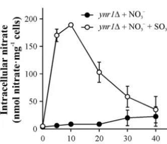
Involvement of Ssu2 in nitrate and sulfite extrusion was also found in cells previously incubated in nitrate plus sulfite. Under these conditions, a transitory nitrate accumulation in theynr1⌬ strain was observed only when sulfite was present (Fig. 3). This FIG 1Ssu1 and Ssu2 are involved in sulfite sensitivity and nitrate efflux. (A) Deletion of theSSU1orSSU2gene produces sulfite sensitivity. Thessu1⌬andssu2⌬
strains were grown in YPD. Serial 10-fold dilutions were spotted on pH 3.5 buffered synthetic medium containing 5 mM ammonium chloride plus sodium sulfite at the concentration indicated. Plates were incubated at 37°C for 2 days. (B) Thessu2⌬strain accumulates nitrate. Ammonium-grown cells were resuspended in synthetic medium at an OD660of 2 to 3 and then nitrogen starved for 120 min. Nitrate accumulation assays were triggered with 1 mM nitrate. Intracellular nitrate
was determined in ethanolic cell extracts. (C) Net nitrate uptake increases in thessu2⌬ynr⌬strain. Ammonium-grown cells were resuspended to an OD660of 10
in nitrogen-free medium buffered at pH 5.5 for 60 min. Nitrate uptake assays were triggered with 0.1 mM nitrate. Nitrate uptake was determined as extracellular nitrate depletion for 60 min. Data⫾SE from three independent experiments are shown.
on September 8, 2020 by guest
http://ec.asm.org/
increase in intracellular nitrate could be explained by a decrease in nitrate efflux or an increased uptake. Considering the fact that Ssu2 extrudes sulfite as well as nitrate, competition of sulfite with nitrate for Ssu2 and Ssu1 would be expected in theynr1⌬strain, leading to the observed nitrate accumulation. Once sulfite is me-tabolized, nitrate is extruded from the cell. Consistent with our results, on solid medium the presence of nitrate increased the sensitivity of the WT andssu1⌬strains to sulfite. This was hard to observe in thessu2⌬strain, probably due to higher sensitivity of
this strain to sulfite (see Fig. S1 in the supplemental material). The fact that nitrate accumulation in theynr1⌬strain in the presence of sulfite (Fig. 3) is higher than that in theynr1⌬ssu2⌬strain (Fig. 1B) suggested that sulfite could inhibit other nitrate efflux trans-porters apart from Ssu1 and Ssu2.
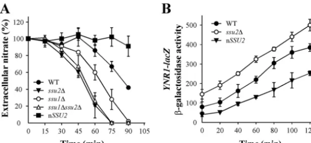
Involvement of Ssu2 in net nitrate uptake and nitrate-in-duced gene expression.Next, we evaluated the role of Ssu1 and particularly Ssu2 in nitrate uptake. We found that net nitrate up-take was almost negligible in the strain overexpressing SSU2 (nSSU2) (Fig. 4A). Both thessu1⌬andssu2⌬strains present a significant increase in nitrate uptake with respect to the WT, that of thessu2⌬strain being higher. The ssu1⌬ ssu2⌬and ssu2⌬ strains presented equal nitrate uptake rates, indicating the lower participation of Ssu1 in nitrate extrusion than of Ssu2. These find-ings clearly show that Ssu2 is affecting net nitrate uptake levels, acting directly on nitrate efflux and indirectly on nitrate induc-tion, which is mediated by intracellular nitrate. Indeed the lag phase preceding nitrate uptake is shorter in thessu1⌬strain than in the WT and even shorter in thessu2⌬strain. This suggests that due to the lesser efflux of nitrate in thessu2⌬strain and conse-quently the higher nitrate accumulation, nitrate induction be-comes quicker and nitrate uptake is triggered sooner. To test the role of Ssu2 in nitrate induction, we measured nitrate reductase gene expression (YNR1-lacZ) as a readout of nitrate-induced gene expression in the nSSU2andssu2⌬strains. Indeed,YNR1-lacZ expression was about 50% lower in the nSSU2strain than in the WT. In the ssu2⌬ strain, however, YNR1-lacZexpression was higher and took place earlier than in in the WT (Fig. 4B). The nitrateYNR1-lacZinduction time course is essentially the same in FIG 2Ssu2 is involved nitrate efflux in oocytes fromXenopus laevis. Oocytes were injected with different amounts ofSSU2cRNA and incubated for 7 days at 17°C. (A) Ssu2-YFP is localized in injected oocytes at the cell surface. (B) The amount of Ssu2-YFP is directly proportional to the amount of cRNA injected. (C) Oocytes injected or not withSSU2cRNA were preloaded with 0.9 nmol of nitrate and incubated for 15 min. Afterward, nitrate was determined in the extracellular medium. Nitrate (nmol) excreted in 15 min⫾SE from 5 independent experiments is shown.
FIG 3Sulfite raises intracellular nitrate levels. Theynr1⌬strain, lacking ni-trate reductase, grown in ammonium, was transferred to nitrogen-free syn-thetic medium buffered at pH 3.5 for 120 min. Nitrate accumulation assays were triggered with 0.75 mM sodium nitrate or 0.75 mM sodium nitrate plus 1.5 mM sodium sulfite. Intracellular nitrate was determined in ethanolic cell extracts. Data⫾SE from three independent experiments are shown.
Cabrera et al.
on September 8, 2020 by guest
http://ec.asm.org/
both the WT and thessu2⌬strain. However, in thessu2⌬ YNR1-lacZstrain, levels are higher at time zero, after the cells have been depleted of nitrogen for 90 min. This can be explained if nitrate traces present in a nitrogen-free medium are still able to slightly induceYNR1-lacZexpression (7). These traces are not excreted to the medium because ofSSU2deletion. We also measured YNR1-lacZexpression in theynr1⌬ssu2⌬andynr1⌬strains in very low nitrate (micromolar level of nitrate). The induction ofYNR1-lacZ expression was faster and its levels higher in theynr1⌬ssu2⌬strain than in theynr1⌬strain. This indicates that nitrate traces are not excreted once enteringynr1⌬ssu2⌬cells, unlike the case with ynr1⌬and WT cells, increasing the levels of intracellular nitrate and as a result the rate of nitrate assimilation gene induction (see Fig. S2 in the supplemental material). Likewise, in the presence of nitrate, nitrite efflux peaked earlier in thessu2⌬strain than in the WT, while in the nSSU2strain it remained very low (see Fig. S3). We conclude that Ssu2 plays key roles in net nitrate uptake and also in modulating the response of nitrate-induced genes to nitrate.
Nar1 is involved in nitrite and nitrate efflux.The levels of intracellular nitrate in theynr1⌬strain incubated in nitrate plus sulfite are higher than in those in theynr1⌬ssu2⌬strain This pointed to the presence inH. polymorphaof nitrate efflux system components other than Ssu2 and Ssu1. To check this, we searched for genes encoding proteins with similarity to nitrate and nitrite transporters in theH. polymorphagenome database. We found two genes encoding putative nitrate/nitrite transporters with similarity toA. thalianaCHL1 andChlamydomonas reinhardtii NAR1. Chl1 belongs to the nitrate transporter family (NRT1/ PTR), as doesA. thaliananitrate transporter CHL1 (37). We dis-ruptedCHL1but could not find any involvement of Chl1 in ni-trate efflux or influx inH. polymorpha(data not shown). Nar1 belongs to the formate nitrite transporter family (FNT).H. poly-morphaNar1 (HpNar1) showed about 20% identity with different members of the FNT family, such as NirC fromE. coliand NAR1.1 fromC. reinhardtii, all involved in nitrite transport (11,22,38).
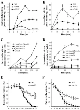
To study the role of Nar1, we determined nitrite excretion in the WT, nar1⌬ and nNAR1 strain (bearing several copies of NAR1). We observed that nitrite excretion was higher in the WT than in thenar1⌬strain when they were incubated in 5 mM
ni-trate, while in the nNAR1strain it increased strongly (Fig. 5A). This suggests that Nar1 could be involved in nitrite efflux. There-fore, we analyzed nitrite accumulation in the WT,nar1⌬, and nNAR1strains. There was a clear accumulation of nitrite in the nar1⌬strain, unlike the case with the nNAR1strain, where intra-cellular nitrite was almost nil (Fig. 5B). This allows us to conclude that Nar1 is involved in nitrite efflux.
We then asked if Nar1 excretes nitrate. To address this, intra-cellular nitrate was determined for different strains lacking nitrate reductase (ynr1⌬) incubated in nitrate. The highest accumulation was in theynr1⌬nar1⌬ssu2⌬strain, followed by theynr1⌬nar1⌬, ynr1⌬ssu2⌬, andynr1⌬strains Therefore, Nar1 has a high capac-ity to extrude nitrate, even higher than that of Ssu2 (Fig. 5C). Short-term nitrate accumulation was analyzed in the same strains for 20 min (Fig. 5D) and found to be higher in theynr1⌬ssu2⌬ strain than in theynr1⌬nar1⌬strain. In contrast, nitrate accumu-lation after 20 min was higher in theynr1⌬nar1⌬strain than in theynr1⌬ssu2⌬strain (Fig. 5C). This suggests that Ssu2 is the high-affinity transport system that copes with nitrate excretion at low intracellular nitrate levels, while Nar1 seems to be important at high intracellular nitrate levels.
We also explore the role of Nar1 in nitrate and nitrite uptake in the WT andnar1⌬strains, finding that nitrate (Fig. 5E) and nitrite (Fig. 5F) uptake was less in thenar1⌬strain. This could be ex-plained by the accumulation of nitrite in thenar1⌬strain, which leads to a downregulation of nitrate assimilation genes. Indeed, YNR1-lacZlevels decrease in thenar1⌬strain incubated in nitrate (see Fig. S4 in the supplemental material). However, we cannot rule out that Nar1 could be also involved in nitrate and nitrite influx.
SSU2 is upregulated by nitrite, unlikeNAR1. To monitor
SSU2andNAR1 gene expression, SSU2-lacZ, NAR1-lacZ, and
qRT-PCR were used. We measuredSSU2expression bearing in
mind that ScSSU1is induced by NO-generating compounds (19) and that NO could be also produced by NR from nitrite (39). The WT andynr1⌬strains were incubated in either ammonium or nitrate so as to focus on the role of NR. As depicted inFig. 6A, we observed that nitrate induced Ssu2 levels about 4-fold, although this induction was abolished in strains lacking nitrate reductase FIG 4SSU2deletion raises nitrate uptake and nitrate induction. (A) Nitrate uptake. Cells grown in synthetic medium plus 5 mM ammonium were resuspended in the same medium at an OD660of 2 to 3 but without a nitrogen source and incubated for 90 min. Nitrate uptake was determined as extracellular nitrate depletion
for 90 min. Assays were triggered with 0.5 mM nitrate. Data⫾SE from three independent experiments are shown. (B) Nitrate induction. Strains bearing
YNR1-lacZgrown in synthetic medium plus 5 mM ammonium were resuspended in nitrogen-free medium for 90 min.YNR1-lacZinduction in response to 1 mM nitrate was determined as-galactosidase activity and expressed as nmolo-nitrophenol · min⫺1(mU) · mg⫺1of protein. Data⫾SE from three independent
experiments are shown.
on September 8, 2020 by guest
http://ec.asm.org/
(ynr1⌬). This suggests thatSSU2upregulation is due to the nitrite from nitrate reduction or NO generated from nitrite by NR. Fur-ther experiments showed that nitrite inducedSSU2expression whether NR was present or not (Fig. 6B). Therefore, nitrite is clearly involved inSSU2upregulation, even though further trans-formations of nitrite to NO independently of NR (39) cannot be excluded. Other enzymes, such as xanthine oxidase, mitochon-drial cytochromes, or even nonenzymatic reduction, could ac-count for this (40–42). We also measured Ssu2 levels in a strain bearing Ssu2-GFP, in different nitrogen sources, finding them well correlated withSSU2expression (Fig. 6C). This was con-firmed by epifluorescence microscopy, which also showed that
Ssu2 is localized mainly at the plasma membrane (Fig. 6D), con-sistent with data obtained forXenopusoocytes (Fig. 2A). Interest-ingly, we observed a small proportion of Ssu2-GFP intracellular retention in some yeast cells, which could be transporting some nitrate into an internal compartment. However, this does not af-fect our total cell nitrate content measurements after complete disruption of cell membranes (31).
Unlike the case withSSU2, no significant differences were ob-served in the response ofNAR1or Nar1 to different nitrogen sources (data not shown). Epifluorescence microscopy showed that Nar1-GFP was localized mainly at the cell surface in nitrate and ammonium (data not shown).
FIG 5Nar1 is involved in nitrite and nitrate efflux. (A) Nitrite excretion. Ammonium-grown cells were resuspended at an OD660of 2 to 3 in synthetic medium
plus 5 mM nitrate. Appearance of nitrite in the medium was determined for 7 h. (B) Intracellular nitrite. Ammonium-grown cells were nitrogen starved in synthetic medium at an OD660of 2 to 3 for 120 min. Intracellular nitrite was determined in ethanolic cell extracts in assays triggered with 1 mM nitrite. (C and
D) Intracellular nitrate is highest in theynr1⌬nar1⌬ssu2⌬strain. Ammonium-grown cells were nitrogen starved in synthetic medium for 60 min at an OD660
of 2 to 3. Nitrate accumulation assays were triggered with 1 mM nitrate. Intracellular nitrate was determined in ethanolic cell extracts from long (C) or short (D) assays. (E and F) Thenar1⌬strain shows lower net nitrate and nitrite uptake. Ammonium-grown cells were nitrogen starved on pH 5.5 buffered synthetic medium at an OD660of 2 to 3 for 90 min. Net nitrate (E) or nitrite uptake (F) assays were triggered with 1 mM nitrate or 0.5 mM nitrite. Net nitrate or nitrite
uptake was determined as extracellular nitrate or nitrite depletion. Data⫾SE from three independent experiments are shown.
Cabrera et al.
on September 8, 2020 by guest
http://ec.asm.org/
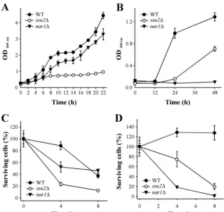
Involvement of Ssu2 and Nar1 in growth and cell viability. We further studied the role of Ssu2 and Nar1 in cell growth and viability. A liquid medium assay in 5 mM nitrate showed that the ssu2⌬strain grew more slowly than the WT (Fig. 7A). In contrast, under the same conditions,nar1⌬strain growth was slightly less than that of the WT. However, in 1 mM nitrite, thenar1⌬strain was unable to grow, while growth of thessu2⌬strain was about 50% less than that of the WT (Fig. 7B).
We also studied the effects of Ssu2 and Nar1 on cell viability in
nitrate and nitrite. Cells grown in ammonium up to an OD660of 3 were resuspended at the same cell density in 5 mM ammonium, 10 mM nitrate, or 1 mM nitrite. Cell suspensions were incubated with shaking for 8 h. Thessu2⌬strain showed lower viability in nitrate (Fig. 7C). These results raise the question of whether ni-trate, or nitrite from nitrate reduction, was responsible for cell viability reduction of thessu2⌬strain in nitrate. However, to avoid nitrite production, strains lacking NR, theynr1⌬ssu2⌬andynr1⌬ strains, were incubated in nitrate and did not show any difference FIG 6SSU2is upregulated by nitrite. (A)SSU2expression levels.SSU2expression was followed by assaying-galactosidase activity in the WT and the
ynr1⌬strain bearingSSU2-lacZ. Strains grown in YPD were transferred to synthetic medium plus 5 mM nitrate and 0.5 mM proline or 5 mM ammonium at an OD660of 3. Data⫾SE from three independent experiments are shown. (B) Effect of nitrite onSSU2expression levels in WT andynr1⌬strains.
Ammonium-grown cells were incubated at an OD660of 3 in synthetic medium plus 5 mM ammonium or 2 mM nitrite for 120 min. Relative expression
was determined by qRT-PCR. Data⫾SE from three independent experiments are shown. The expression is normalized to cells incubated in ammonium. HpACT1was used as a reference gene. (C) Nitrate and nitrite raise Ssu2 levels. Ssu2 was determined by immunoblot analysis of Ssu2-GFP. Strains grown in YPD were resuspended at an OD660of 0.8 in synthetic medium plus 5 mM ammonium, 10 mM nitrate, or 10 mM nitrate plus 2 mM nitrite. Pma1 was
used as a loading control. (D) Ssu2 is located mainly at the cell surface, and its levels increased in nitrate or nitrate plus nitrite. Cells bearing Ssu2-GFP fusion grown in YPD were transferred to synthetic medium plus 5 mM ammonium, 10 mM nitrate, or 10 mM nitrate plus 2 mM nitrite at an OD660of 2
to 3. Ssu2-GFP was monitored by fluorescence microscopy.
on September 8, 2020 by guest
http://ec.asm.org/
in cell viability (data not shown). This suggests that nitrite, and not nitrate, was involved in the lower cell viability of thessu2⌬ strain in nitrate. In the case of Nar1 (Fig. 7C), the differences between thenar1⌬and WT strains are noticeable only after 4 h of incubation in nitrate. The fact that after 8 h no differences were observed could be due to a weaker nitrate assimilation gene induc-tion in thenar1⌬strain due to nitrite accumulation. Nitrite tox-icity was further confirmed in the WT,ssu2⌬, andnar1⌬strains. Thenar1⌬strain was almost killed after 4 h in nitrite, while unex-pectedly, thessu2⌬strain presented a moderate sensitivity to ni-trite (Fig. 7D). We conclude that Ssu2 is as essential for cell growth and viability in nitrate as is Nar1 in nitrite.
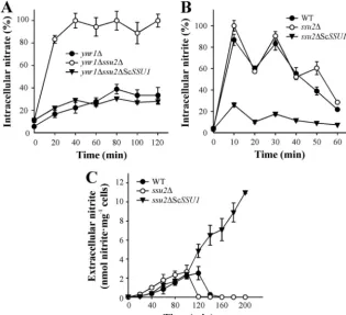
Besides sulfite,S. cerevisiaeSsu1 is able to mediate the efflux of nitrite and nitrate.We studied whether ScSSU1, in addition to sulfite, is involved in nitrate and nitrite efflux. This was tackled by expressing ScSSU1in theH. polymorpha ssu2⌬strain under the HpSSU2promoter to avoid misinterpretations due to expression level alterations. Thessu2⌬strain expressing ScSSU1was able to recover sulfite tolerance, confirming the suitability of this con-struct to functionally render ScSSU1inH. polymorpha(see Fig. S5A in the supplemental material). Nitrate uptake experiments revealed that thessu2⌬strain expressing ScSSU1presented a lower net nitrate uptake, almost nil net nitrite uptake (see Fig. S5B and S5C), and incapacity to grow in nitrate and nitrite (data not shown). However, determination of intracellular nitrate in the
ynr1⌬ssu2⌬ScSSU1,ynr1⌬ssu2⌬, andynr1⌬strains showed that theynr1⌬ssu2⌬ScSSU1strain does not accumulate nitrate, unlike theynr1⌬ssu2⌬strain (Fig. 8A). This indicates that ScSSU1is involved in nitrate efflux, since nitrate is not accumulated in the presence of ScSsu1 in thessu2⌬strain. Likewise, we measured intracellular nitrite in the WT,ssu2⌬, andssu2⌬ScSSU1strains. As shown inFig. 8B, the latter strain does not accumulate nitrite, indicating that ScSsu1 is also involved in nitrite efflux. To obtain further confirmation of this, we measured nitrite efflux in the ssu2⌬ScSSU1, ssu2⌬, and WT strains incubated in 1 mM nitrate. The ability of thessu2⌬ScSSU1strain to efflux nitrite was about 10-fold higher than that of the WT andssu2⌬strains (Fig. 8C). This did not decrease after 4 h, unlike the case with thessu2⌬and WT strains, where nitrite efflux disappears after about 2 h. Further evidence on this was obtained by transforming the WT with ScSSU1. The resultant strains were better able to extrude nitrite and lower the intracellular nitrite (see Fig. S6). This indicates that ScSsu1 presents high nitrite efflux activity. We also proposed that the incapacity of thessu2⌬ScSSU1strain to grow in nitrate is due to the very high nitrite efflux activity of this strain. The high activ-ity ofssu2⌬ScSSU1strain suggests that ScSSU1preferentially cretes nitrite instead of nitrate, since in the latter case nitrite ex-cretion would be lower, but it is not. In addition to sulfite efflux, we concluded that ScSSU1also extrudes nitrite and nitrate. FIG 7Thessu2⌬strain presents reduced cell viability in nitrate while thenar1⌬strain does so in nitrite. (A) Growth in nitrate. Strains grown in YPD were transferred to synthetic medium plus 5 mM nitrate at an OD660of 0.2. Cell growth was determined by measuring the OD660. (B) Growth in nitrite. The procedure
was the same as that for panel A except that the medium contained only 1 mM nitrite. (C) Cell viability in nitrate. Ammonium-grown cells were resuspended at an OD660of 3 in synthetic medium plus 10 mM nitrate and incubated with shaking at 37°C for 8 h. (D) Cell viability in nitrite. Ammonium-grown cells were
resuspended at an OD660of 3 in synthetic medium plus 1 mM nitrite and incubated with shaking at 37°C for 8 h. Cell viability was determined after different
incubation times in nitrate or nitrite by measuring growth on YPD plates at 37°C for 2 days. Data⫾SE from three independent experiments are shown.
Cabrera et al.
on September 8, 2020 by guest
http://ec.asm.org/
DISCUSSION
Using the yeastH. polymorphaas a model system, we have char-acterized the molecular components of nitrate and nitrite efflux and its impact on nitrate assimilation. We wish to suggest here an essential role for nitrate and nitrite efflux in nitrate assimilation in terms of net nitrate transport, growth, and viability. We found that the sulfite efflux permease Ssu1 and especially Ssu2 were also able to extrude nitrate, while Nar1 extrudes nitrite and nitrate.
Intracellular nitrate accumulation and net nitrate uptake show that deletion ofSSU2led to higher levels of intracellular nitrate and net nitrate uptake than were seen with the control strains (the WT and theynr1⌬strain). In contrast, in the nSSU2strain, bear-ing several copies ofSSU2, intracellular nitrate accumulation and net nitrate uptake were almost nil. We reasoned that the high net nitrate uptake in thessu1⌬,ssu2⌬, andssu1⌬ssu2⌬strains was due to these strains presenting lower nitrate efflux than the WT and therefore higher net nitrate uptake (Fig. 4A). Furthermore, in these mutants, nitrate accumulation is greater, and as a result, nitrate assimilation gene induction rises, also contributing to the increased net nitrate uptake. Indeed, we found that deletion of SSU2increased nitrate assimilation gene expression whileSSU2 overexpression downregulated these genes (Fig. 4B). These results suggest that Ssu2 negatively regulates nitrate assimilation gene expression by acting on intracellular nitrate levels, since nitrate acts as an inducer once inside the cell (7). In this framework of regulation, we also found that Ssu2 levels are positively regulated by nitrate (Fig. 6C).
Experiments withXenopusoocytes preloaded with nitrate or nitrite also confirmed that Ssu2 was able to efflux nitrate but not nitrite. This inability is also seen from the scarce accumulation of nitrite in theyni1⌬ssu2⌬strain (data not shown).
Ssu2 appears to be more important in sulfite efflux than Ssu1, since thessu2⌬strain was more sensitive to sulfite than thessu1⌬ strain (Fig. 1A). Accordingly, under the conditions used, our re-sults indicate that the contribution of Ssu1 to nitrate efflux was much lower than that of Ssu2 (Fig. 4A).
In our search for genes involved in nitrate and nitrite transport, we also found that deletion of one ORF, encoding a protein termed Nar1, belonging to the FNT family, led to lower nitrite excretion. In contrast, a strain bearing multiple copies ofNAR1 (nNAR1) increased it dramatically. Consistent with this, the nar1⌬strain presents high intracellular nitrite accumulation (Fig. 5B), and this is even more the case with theyni1⌬nar1⌬strain (data not shown). However, contrary to what was expected for a member of the FNT family, Nar1 was also involved in nitrate extrusion. Thus, theynr1⌬nar1⌬strain accumulated more nitrate than theynr1⌬strain and even more than theynr1⌬ssu2⌬strain. Moreover, theynr1⌬nar1⌬ssu2⌬triple mutant yielded the high-est intracellular levels of nitrate under the thigh-ested conditions. The time courses of nitrate accumulation in theynr1⌬ssu2⌬,ynr1⌬ nar1⌬, andynr1⌬nar1⌬ssu2⌬strains suggest that Ssu2 was re-sponsible for short-term nitrate extrusion, while intracellular ni-trate levels remained lower (60 nmol mg⫺1of cells). In accordance with this, in theynr1⌬nar1⌬strain, unlike the case with theynr1⌬ FIG 8ScSSU1is involved in nitrate and nitrite efflux. Ammonium-grown cells were resuspended at an OD660of 2to 3 in synthetic medium and then nitrogen
starved for 120 min. Intracellular nitrate (A) or nitrite (B) assays were triggered with 1 mM nitrate or 1 mM nitrite. Nitrate and nitrite were determined in ethanolic cell extracts. (C) Nitrite excretion increased in thessu2⌬ScSSU1strain. Ammonium-grown cells were resuspended at an OD660of 2 to 3 in synthetic
medium buffered at pH 5.5 and then nitrogen starved for 120 min; afterward, 1 mM nitrate was added. Nitrite excretion was determined in the medium. Data⫾ SE from three independent experiments are shown.
on September 8, 2020 by guest
http://ec.asm.org/
ssu2⌬strain, no nitrate accumulation is observed in the short term. In contrast, once intracellular nitrate levels increase, Nar1 seems to be mainly responsible for nitrate excretion. This conclu-sion is also supported by the fact that theynr1⌬nar1⌬strain accumulates nitrate only after 80 min in nitrate, when intracellular levels of nitrate rise (Fig. 5C). These results also suggest that Ssu2 presents greater affinity for nitrate than Nar1. Indeed, Nar1 be-longs to the FNT family, whose members are involved in nitrite transport and efflux (11,21–23,25). Consistent with this, in the yni1⌬ nar1⌬ strain, nitrite is quickly accumulated (data not shown), while nitrate is slowly accumulated in theynr1⌬nar1⌬ strain, suggesting again that Nar1 presents a higher affinity for nitrite than for nitrate (Fig. 5BandD).
Uptake assays showed a lower net uptake of nitrate and nitrite by thenar1⌬strain. This is explained because nitrite, coming from nitrate reduction or from the medium, is intracellularly accumu-lated in thenar1⌬strain and represses the nitrate assimilation gene (1). Indeed, decreased net uptake of nitrate and nitrite is observed after a 40-min incubation. Nevertheless, the involve-ment of Nar1 in their influx cannot be absolutely ruled out.
Our results challenge the idea that NR plays an important role in net nitrate uptake in yeast and filamentous fungi. This had been concluded from uptake assays using the tracer13NO
3⫺, which showed that net nitrate uptake is negligible in an NR deletion mutant yeast (43). However, nitrate efflux could also account for the absence of intracellular nitrate accumulation in mutants lack-ing NR. Thus, we observed that theynr1⌬ssu2⌬strain and par-ticularly theynr1⌬nar1⌬ssu2⌬strain were able to accumulate nitrate, unlike theynr1⌬strain. Therefore, nitrate influx was op-erative in mutants lacking NR. The accumulation of nitrate in the ynr1⌬ssu2⌬andynr1⌬nar1⌬strains is therefore consistent with the absence of a nitrate efflux system (Fig. 5CandD).
The role of nitrite excretion seems to be a response of the cell to cope with the toxicity of nitrite. However, this could be regulated at the nitrate uptake step to avoid the imbalance between nitrate transported into the cell and that reduced to ammonium by NR. Since nitrite is toxic for most organisms (44–47), this could con-tribute to the success of nitrate-assimilating microorganisms in colonizing nitrate-containing media in competition with non-ni-trate assimilators. In contrast, the precise role(s) of ninon-ni-trate efflux is difficult to explain, since nitrate appears not to be toxic for cells, because strains lacking NR, theynr1⌬ssu2⌬andynr1⌬strains, were viable in nitrate (data not shown). Nitrate uptake takes place against an electrochemical gradient, and therefore its efflux appar-ently seems to be a waste of energy for the cell. Nevertheless, we observed that thessu2⌬strain grew poorly in nitrate (Fig. 7A) and also presented lower cell viability after incubation in it (Fig. 7C). This suggests that intracellular nitrate levels must also be tightly regulated. In this regard, we have observed in thessu2⌬strain that nitrate induction of nitrate assimilation genes is quicker and higher. This could produce an imbalance between the capacity of the cells to take up nitrate and its reduction to nitrite and then ammonium, as a result increasing intracellular nitrite. In any case, the question of whether nitrate itself is toxic for the cells or the toxicity is due to nitrite was also addressed. We observed that when the ssu2⌬ strain is incubated in nitrate, nitrite efflux is higher than that in the WT, suggesting that nitrite could actually be the main cause of the low growth and viability of this mutant in nitrate. In fact, when we compared growth and viability in nitrate of thessu2⌬strain with those of theynr1⌬ssu2⌬strain, we
ob-served that the latter becomes more viable since it is unable to produce nitrite (data not shown). We conclude that Ssu2 contrib-utes to nitrate homeostasis, avoiding nitrite accumulation. Unlike thessu2⌬strain, thenar1⌬strain is unviable in nitrite due to its capacity to accumulate it. These results show that Nar1 is crucial for growth and cell viability in nitrite. In contrast, Ssu2 is essential in nitrate. However, the lower growth of thessu2⌬strain in nitrite could be due to the conversion of nitrite to NO via NR. Nitric oxide could be oxidized to nitrate by flavohemoglobin, as shown with FhbA fromA. nidulans(48). So, in thessu2⌬strain, the in-tracellular nitrate from flavohemoglobin activity is not excreted into the medium at the same levels as in the WT. This would increase nitrite accumulation, which would be toxic for the cell and limit growth. However, the ability of Ssu2 to extrude some nitrite cannot be excluded. The capacity of Ssu2 to excrete nitrate, the inducer of the nitrate assimilation genes, also acts to balance nitrate levels by downregulating these genes. Thus,SSU2was in-duced about 5-fold by the presence of nitrate, although this induc-tion almost disappears in mutants lacking nitrate reductase. Fur-thermore, several experiments showed a correlation between nitrite andSSU2upregulation. This again suggests that nitrite, or nitric oxide from nitrite, could be involved in the induction of SSU2, as seen in the ScSSU1strain. Unlike that ofSSU2,NAR1 expression was not significantly modified in response to nitrate or ammonium (data not shown). The apparent lack ofNAR1 regu-lation in nitrate and ammonium does not rule out other mecha-nisms of regulating Nar1. However, Nar1 levels determined by Western blotting confirm that the regulation of protein levels seems unimportant (data not shown).
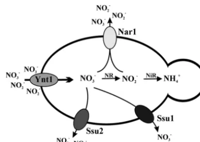
We also addressed the question of whether an ScSSU1strain was able to efflux nitrate/nitrite. ScSSU1expressed in the Hpssu2⌬ strain under theSSU2gene promoter restores sulfite tolerance. Our experiments clearly show that ScSsu1 extrudes nitrate and nitrite (Fig. 8). It is difficult to reach a conclusion regarding the affinity of ScSsu1 for nitrate and nitrite, but it seems that ScSsu1 possesses a high capacity to extrude nitrite. This capacity was clearly shown in a WT strain expressing ScSSU1. We suggested FIG 9Nitrate and nitrite efflux systems inH. polymorpha. The high-affinity nitrate transporter (Ynt1) is involved in nitrate and nitrite influx. The nitrate is reduced by nitrate reductase (NR) to nitrite, which is catalyzed to ammonium by nitrite reductase (NiR). Ssu2 and to a lesser extent Ssu1 are involved nitrate efflux. Nar1 is involved in nitrite and nitrate efflux. Nar1-dependent ni-trate efflux is observed when intracellular nini-trate reaches higher levels. The nitrate and nitrite influx systems allow cells to cope with intracellular ni-trite in cells growing in nitrate or nini-trite as the sole nitrogen source.
Cabrera et al.
on September 8, 2020 by guest
http://ec.asm.org/
de Ciencia e Innovación (MICINN) (Spain) and PI 2008/338 from Gobi-erno de Canarias to J.M. Siverio and Consolider CSD2008-00005 to D. Alvarez de la Rosa and T. Giraldez from the Ministerio de Ciencia e Inno-vación (MICINN) (Spain). The work was also supported by an IMBRAIN grant (FP7-REGPOT-2012-CT2012-316137-IMBRAIN) from the Euro-pean Union. E. Cabrera was a recipient of predoctoral fellowships from Agencia Canaria de Investigación e Innovación y Sociedad de la Infor-mación (ACIISI) from Gobierno de Canarias and from SEGAI (Univer-sidad de La Laguna, 2011-2012).
We thank Rhein Biotech (Germany) for providingSSU1,SSU2, and
NAR1DNA sequences. We are grateful to R. Serrano (Valencia) for Pma1 antiserum and to Guido Jones for proofreading the manuscript.
REFERENCES
1.Brito N, Ávila J, Pérez MD, González C, Siverio JM.1996. The genes
YNI1andYNR1, encoding nitrite reductase and nitrate reductase respec-tively in the yeastHansenula polymorpha, are clustered and coordinately regulated. Biochem. J.317:89 –95.
2.Pérez MD, González C, Ávila J, Brito N, Siverio JM.1997. TheYNT1
gene encoding the nitrate transporter in the yeastHansenula polymorphais clustered with genesYNI1andYNR1encoding nitrite reductase and ni-trate reductase, and its disruption causes inability to grow in nini-trate. Biochem. J.321:397– 403.
3.Siverio JM.2002. Assimilation of nitrate by yeasts. FEMS Microbiol. Rev.
26:277–284.http://dx.doi.org/10.1111/j.1574-6976.2002.tb00615.x. 4.Ávila J, González C, Brito N, Siverio JM.1998. Clustering of theYNA1
gene encoding a Zn(II)2Cys6transcriptional factor in the yeastHansenula
polymorphawith the nitrate assimilation genesYNT1,YNI1and YNR1, and its involvement in their transcriptional activation. Biochem. J.335:
647– 652.
5.Ávila J, González C, Brito N, Machín F, Pérez MD, Siverio JM.2002. A second Zn(II)2Cys6transcriptional factor encoded by theYNA2gene is
indispensable for the transcriptional activation of the genes involved in nitrate assimilation in the yeastHansenula polymorpha. Yeast19:537–544.
http://dx.doi.org/10.1002/yea.847.
6.Rodríguez C, Tejera P, Medina B, Guillén RM, Domínguez A, Ramos J, Siverio JM.2010. Ure2 is involved in nitrogen catabolite repression and salt tolerance via Ca2⫹homeostasis and calcineurin activation in the yeast Hansenula polymorpha. J. Biol. Chem.285:37551–37560. http://dx.doi .org/10.1074/jbc.M110.146902.
7.Navarro FJ, Perdomo G, Tejera P, Medina B, Machín F, Guillén RM, Lancha A, Siverio JM.2003. The role of nitrate reductase in the regulation of the nitrate assimilation pathway in the yeastHansenula polymorpha. FEMS Yeast Res.4:149 –155.http://dx.doi.org/10.1016/S1567-1356(03)00163-6. 8.Navarro FJ, Machín F, Martín Y, Siverio JM.2006. Down-regulation of
eukaryotic nitrate transporter by nitrogen-dependent ubiquitinylation. J. Biol. Chem.281:13268 –13274.http://dx.doi.org/10.1074/jbc.M601253200. 9.Navarro FJ, Martín Y, Siverio JM.2008. Phosphorylation of the yeast
nitrate transporter Ynt1 is essential for delivery to the plasma membrane during nitrogen limitation. J. Biol. Chem.283:31208 –31217.http://dx.doi .org/10.1074/jbc.M802170200.
10. Azuara M, Aparicio P.1983.In vivoblue-light activation of Chlamydomo-nas reinhardiinitrate reductase. Plant Physiol.71:286 –290.http://dx.doi .org/10.1104/pp.71.2.286.
11. Jia W, Tovell N, Clegg S, Trimmer M, Cole J.2009. A single channel for nitrate uptake, nitrite export and nitrite uptake byEscherichia coliNarU
15. Leran S, Muños S, Brachet C, Tillard P, Gojon A, Lacombe B.2013.
ArabidopsisNRT1.1 is a bidirectional transporter involved in root-to-shoot nitrate translocation. Mol. Plant6:1984 –1987.http://dx.doi.org/10 .1093/mp/sst068.
16. Park H, Bakalinsky AT.2000.SSU1mediates sulphite efflux in Saccha-romyces cerevisiae. Yeast 16:881– 888. http://dx.doi.org/10.1002/1097 -0061(200007)16:10⬍881::AID-YEA576⬎3.0.CO;2-3.
17. Léchenne B, Reichard U, Zaugg C, Fratti M, Kunert J, Boulat O, Monod M.2007. Sulphite efflux pumps inAspergillus fumigatusand dermato-phytes. Microbiology153:905–913.http://dx.doi.org/10.1099/mic.0.2006 /003335-0.
18. Chiranand W, McLeod I, Zhou H, Lynn JJ, Vega LA, Myers H, Yates JR, Lorenz MC, Gustin MC.2008.CTA4transcription factor mediates in-duction of nitrosative stress response inCandida albicans. Eukaryot. Cell
7:268 –278.http://dx.doi.org/10.1128/EC.00240-07.
19. Sarver S, DeRisi J.2005. Fzf1p regulates an inducible response to nitro-sative stress inSaccharomyces cerevisiae. Mol. Biol. Cell16:4781– 4791.
http://dx.doi.org/10.1091/mbc.E05-05-0436.
20. Ullmann BD, Myers H, Chiranand W, Lazzell AL, Zhao Q, Vega LA, Lopez-Ribot JL, Gardner PR, Gustin MC.2004. Inducible defense mech-anism against nitric oxide inCandida albicans. Eukaryot. Cell3:715–723.
http://dx.doi.org/10.1128/EC.3.3.715-723.2004.
21. Clegg S, Yu F, Griffiths L, Cole JA.2002. The roles of the polytopic membrane proteins NarK, NarU and NirC inEscherichia coliK-12: two nitrate and three nitrite transporters. Mol. Microbiol.44:143–155.http: //dx.doi.org/10.1046/j.1365-2958.2002.02858.x.
22.Jia W, Cole JA. 2005. Nitrate and nitrite transport inEscherichia coli. Biochem. Soc. Trans.33:159 –161.http://dx.doi.org/10.1042/BST0330159. 23. Wang Y, Huang Y, Wang J, Cheng C, Huang W, Lu P, Xu Y-N, Wang
P, Yan N, Shi Y.2009. Structure of the formate transporter FocA reveals a pentameric aquaporin-like channel. Nature462:467– 472.http://dx.doi .org/10.1038/nature08610.
24. Mariscal V, Moulin P, Orsel M, Miller AJ, Fernández E, Galván A.2006. Differential regulation of theChlamydomonas Nar1gene family by carbon and nitrogen. Protist 157:421– 433. http://dx.doi.org/10.1016/j.protis .2006.06.003.
25. Rexach J, Fernández E, Galván A.2000. TheChlamydomonas reinhardtii Nar1gene encodes a chloroplast membrane protein involved in nitrite transport. Plant Cell 12:1441–1453.http://dx.doi.org/10.1105/tpc.12.8 .1441.
26. Xu X, Wightman JD, Geller BL, Avram D, Bakalinsky AT. 1994. Isolation and characterization of sulfite mutants ofSaccharomyces cerevi-siae. Curr. Genet.25:488 – 496.http://dx.doi.org/10.1007/BF00351667. 27. Brito N, Pérez MD, Perdomo G, González C, García-Lugo P, Siverio
JM.1999. A set ofHansenula polymorphaintegrative vectors to construct
lacZ fusions. Appl. Microbiol. Biotechnol.53:23–29.http://dx.doi.org/10 .1007/s002530051609.
28. Leão-Helder AN, Krikken AM, van der Klei IJ, Kiel JKAW, Veenhuis M.
2003. Transcriptional down-regulation of peroxisome numbers affects se-lective peroxisome degradation inHansenula polymorpha. J. Biol. Chem.
278:40749 – 40756.http://dx.doi.org/10.1074/jbc.M304029200. 29. Liman ER, Tytgat J, Hess P.1992. Subunit stoichiometry of a
mamma-lian K⫹channel determined by construction of multimeric cDNAs. Neu-ron9:861– 871.http://dx.doi.org/10.1016/0896-6273(92)90239-A. 30. van Dijk R, Faber KN, Hammond AT, Glick BS, Veenhuis M, Kiel
JKAW.2001. TaggingHansenula polymorphagenes by random integra-tion of linear DNA fragments (RALF). Mol. Genet. Genomics266:646 – 656.http://dx.doi.org/10.1007/s004380100584.
on September 8, 2020 by guest
http://ec.asm.org/
31. Gonzalez B, François J, Renaud M.1997. A rapid and reliable method for metabolite extraction in yeast using boiling buffered ethanol. Yeast13:1347– 1355.http://dx.doi.org/10.1002/(SICI)1097-0061(199711)13:14⬍1347::AID -YEA176⬎3.0.CO;2-O.
32. Machín F, Medina B, Navarro FJ, Pérez MD, Veenhuis M, Tejera P, Lorenzo H, Lancha A, Siverio JM.2004. The role of Ynt1 in nitrate and nitrite transport in the yeastHansenula polymorpha. Yeast21:265–276.
http://dx.doi.org/10.1002/yea.1075.
33. Snell FD, Snell CT.1949. Colorimetric methods of analysis, 3rd ed, p 804 – 805. Van Nostrand, New York, NY.
34. Giraldez T, Hughes TE, Sigworth FJ.2005. Generation of functional fluorescent BK channels by random insertion of GFP variants. J. Gen. Physiol.126:429 – 438.http://dx.doi.org/10.1085/jgp.200509368. 35. Giraldez T, Afonso-Oramas D, Cruz-Muros I, Garcia-Marin V, Pagel P,
González-Hernández T, De La Rosa DA.2007. Cloning and functional expression of a new epithelial sodium channel␦subunit isoform differen-tially expressed in neurons of the human and monkey telencephalon. J. Neurochem.102:1304 –1315.http://dx.doi.org/10.1111/j.1471-4159.2007 .04622.x.
36. Faber KN, Haima P, Harder W, Veenhuis M, Ab G.1994. Highly-efficient electrotransformation of the yeastHansenula polymorpha. Curr. Genet.25:305–310.http://dx.doi.org/10.1007/BF00351482.
37. Steiner H-Y, Naider F, Becker JM.1995. The PTR family: a new group of peptide transporters. Mol. Microbiol.16:825– 834.http://dx.doi.org/10 .1111/j.1365-2958.1995.tb02310.x.
38. Galván A, Rexach J, Mariscal V, Fernández E.2002. Nitrite transport to the chloroplast inChlamydomonas reinhardtii: molecular evidence for a regulated process. J. Exp. Bot. 53:845– 853. http://dx.doi.org/10.1093 /jexbot/53.370.845.
39. Rockel P, Strube F, Rockel A, Wildt J, Kaiser WM.2002. Regulation of nitric oxide (NO) production by plant nitrate reductasein vivoandin vitro. J. Exp. Bot. 53:103–110. http://dx.doi.org/10.1093/jexbot/53.366 .103.
40. Godber BLJ, Doel JJ, Sapkota GP, Blake DR, Stevens CR, Eisenthal R,
Harrison R.2000. Reduction of nitrite to nitric oxide catalyzed by xan-thine oxidoreductase. J. Biol. Chem.275:7757–7763.http://dx.doi.org/10 .1074/jbc.275.11.7757.
41. Lundberg JO, Weitzberg E.2005. NO generation from nitrite and its role in vascular control. Arterioscler. Thromb. Vasc. Biol.25:915–922.http: //dx.doi.org/10.1161/01.ATV.0000161048.72004.c2.
42. Tischner R, Planchet E, Kaiser WM.2004. Mitochondrial electron trans-port as a source for nitric oxide in the unicellular green algaChlorella sorokiniana. FEBS Lett.576:151–155.http://dx.doi.org/10.1016/j.febslet .2004.09.004.
43. Unkles SE, Rouch DA, Wang Y, Siddiqi MY, Glass ADM, Kinghorn JR.
2004. Two perfectly conserved arginine residues are required for substrate binding in a high-affinity nitrate transporter. Proc. Natl. Acad. Sci. U. S. A.
101:17549 –17554.http://dx.doi.org/10.1073/pnas.0405054101. 44. Hinze H, Holzer H.1985. Accumulation of nitrite and sulfite in yeast cells
and synergistic depletion of the intracellular ATP content. Z. Lebensm. Unters. Forsch.180:117–120.http://dx.doi.org/10.1007/BF01042634. 45. Kohn MC, Melnick RL, Ye F, Portier CJ.2002. Pharmacokinetics of
sodium nitrite-induced methemoglobinemia in the rat. Drug Metab. Dis-pos.30:676 – 683.http://dx.doi.org/10.1124/dmd.30.6.676.
46. Mortensen HD, Jacobsen T, Koch AG, Ameborg N.2008. Intracellular pH homeostasis plays a role in the tolerance ofDebaryomyces hanseniiand
Candida zeylanoidesto acidified nitrite. Appl. Environ. Microbiol.74:
4835– 4840.http://dx.doi.org/10.1128/AEM.00571-08.
47. Spencer JPE, Whiteman M, Jenner A, Halliwell B.2000. Nitrite-induced deamination and hypochlorite-induced oxidation of DNA in intact hu-man respiratory tract epithelial cells. Free Radic. Biol. Med.28:1039 – 1050.http://dx.doi.org/10.1016/S0891-5849(00)00190-8.
48. Schinko T, Berger H, Lee W, Gallmetzer A, Pirker K, Pachlinger R, Buchner I, Reichenauer T, Güldener U, Strauss J.2010. Transcriptome analysis of nitrate assimilation inAspergillus nidulansreveals connections to nitric oxide metabolism. Mol. Microbiol.78:720 –738.http://dx.doi.org /10.1111/j.1365-2958.2010.07363.x.
Cabrera et al.