Open Access
Methodology
Protocol: A high-throughput DNA extraction system suitable for
conifers
Stanislav Bashalkhanov and Om P Rajora*
Address: Faculty of Forestry and Environmental Management, University of New Brunswick, 28 Dineen Drive, Fredericton, New Brunswick, E3B 6C2, Canada
Email: Stanislav Bashalkhanov - [email protected]; Om P Rajora* - [email protected] * Corresponding author
Abstract
Background: High throughput DNA isolation from plants is a major bottleneck for most studies requiring large sample sizes. A variety of protocols have been developed for DNA isolation from plants. However, many species, including conifers, have high contents of secondary metabolites that interfere with the extraction process or the subsequent analysis steps. Here, we describe a procedure for high-throughput DNA isolation from conifers.
Results: We have developed a high-throughput DNA extraction protocol for conifers using an automated liquid handler and modifying the Qiagen MagAttract Plant Kit protocol. The modifications involve change to the buffer system and improving the protocol so that it almost doubles the number of samples processed per kit, which significantly reduces the overall costs. We describe two versions of the protocol: one for medium-throughput (MTP) and another for high-throughput (HTP) DNA isolation. The HTP version works from start to end in the industry-standard 96-well format, while the MTP version provides higher DNA yields per sample processed. We have successfully used the protocol for DNA extraction and genotyping of thousands of individuals of several spruce and a pine species.
Conclusion: A high-throughput system for DNA extraction from conifer needles and seeds has been developed and validated. The quality of the isolated DNA was comparable with that obtained from two commonly used methods: the silica-spin column and the classic CTAB protocol. Our protocol provides a fully automatable and cost effective solution for processing large numbers of conifer samples.
Introduction
Most genetic linkage mapping, marker-assisted selection, population and conservation genetic studies require processing of a large number of samples. Cost-effective high-throughput DNA extraction is a major bottleneck for many of these applications because handling and quanti-fication of non-liquid samples, such as plant tissues, are labour-intensive and difficult to automate.
Traditional methods of DNA extraction involve multiple time-consuming steps, including organic solvent extrac-tion and alcohol precipitaextrac-tion. Although commercially available spin column-based DNA preparation kits pro-vide higher throughput, they are relatively expensive and difficult to automate. Recently-introduced magnetic fish-ing protocols allow for fully-automated, flexible through-put DNA isolation from certain samples, such as blood, Published: 1 August 2008
Plant Methods 2008, 4:20 doi:10.1186/1746-4811-4-20
Received: 10 July 2008 Accepted: 1 August 2008
This article is available from: http://www.plantmethods.com/content/4/1/20
© 2008 Bashalkhanov and Rajora; licensee BioMed Central Ltd.
but these methods are less tolerant to the secondary metabolites present in conifers and other plants [1]. Polysaccharides and phenolic compounds either impede DNA extraction or inhibit enzymatic reactions in the downstream applications. As a cheaper alternative to commercial DNA extraction kits, many labs use in-house developed simplified high throughput protocols, or use crude lysate as template for PCR [2,3]. However, majority of these protocols provide little quantitative information on the DNA yields, and were designed for crop species where the concentrations of PCR inhibitors are relatively low. Conifers require a more efficient DNA purification procedure. Although some protocols have been published for higher throughput DNA extraction from conifers [4,5], they still suffer from labour-intensive tissue grinding, high cost of silica columns or inconsistent DNA quality and yield.
Here we describe a cost-effective protocol based on the Qiagen MagAttract Plant DNA kit (QIAGEN Inc., Missis-sauga, Ontario) for high throughput DNA extraction from conifer needles and seeds. Most steps, including DNA quantification and normalization, can be done using an automated liquid handler.
Materials
Sample collection
DNA was extracted from silica-dried needles of red spruce (Picea rubens). Needles were collected in the field into plastic bags containing 5–10 g silica gel pouches as desic-cant, and then stored in a freezer at -20°C upon arrival to the laboratory. With silica gel, the plant material could be kept at the ambient temperature for up to 14 days without degradation of DNA quality. The protocol was also tested and operationally used on seeds and fresh needles of red spruce and black spruce (Picea mariana), as well as needles of white spruce (Picea glauca) and eastern white pine (Pinus strobus).
Reagents and consumables
The DNA isolation kit – Qiagen MagAttract Plant DNA Kit (Qiagen, Cat # 67161)
Lysis buffer – Qiagen AP1 buffer (Qiagen, Material # 1014630)
Ethanol
Disposable pipette tips
TE buffer, pH 7.5
2.0 ml microcentrifuge tubes, e.g. Sarstedt SafeLock tubes (Sarstedt, Cat # 72695)
Collection tubes in 96-well format racks (Qiagen, Cat # 19560)
Cap strips (Qiagen, Cat # 19566)
5 mm stainless steel balls (Glen Mills, Cat # 7400-004763-6)
Flat bottom 96-well microtiter plates (Greiner, Cat # 655101)
Round bottom 96-well microtiter plates (Greiner, Cat # 650101)
PCR plates (Greiner, Cat # 652270)
PicoGreen dsDNA quantification reagent (Invitrogen, Cat # P7581)
Equipment
Laboratory balances with 0.001 g resolution
Multichannel pipette or an automated liquid handler
Mixer Mill MM300 (Retsch GmbH) or Qiagen TissueLyser mill
Water bath
Microcentrifuge
Centrifuge equipped with plate rotor, e.g. Eppendorf 5417 with the A-2-DWP-plate rotor or equivalent
96-well magnet, e.g. Qiagen Type B
Fluorescent plate reader or other instruments for measur-ing the DNA concentration
Protocol
There are two versions of the protocol. Protocol A is designed for medium throughput and provides higher DNA yields per sample processed, whereas Protocol B works in the high-throughput 96-well format from the initial tissue homogenization to the purified DNA at the end.
Protocol A – MTP version
1. Weigh approximately 150 mg of plant material in 2 ml microcentrifuge tubes
3. Incubate the samples at 65°C for 15 min
4. Clear the lysates by centrifugation at 18,000 g for 15 min
5. Transfer 170 μl of cleared lysate into a flat bottom 96-well plate
6. Add premixed 200 μl 98% ethanol and 15 μl MagAt-tract A suspension to lysate. Mix by pipetting and then incubate the mixture at the ambient temperature for 2 min. Mix by pipetting again and incubate for another 2 min
7. Place the plate onto the magnet and remove the super-natant from the sample
8. Resuspend the beads by pipetting in 200 μl of RPW buffer containing RNAse A as per the manufacturer's pro-tocol
9. Place the plate onto the magnet and remove the super-natant from the sample
10. Resuspend the beads in 200 μl of 98% ethanol
11. Place the plate on the magnet and remove the super-natant, repeat 2 more times
12. Air dry the beads for 10 min
13. Resuspend the beads in 100 μl of 10 mM Tris-HCl pH 7.5, incubate 5 min at ambient temperature
14. Place the plate on the magnet and transfer the DNA into a 96-well PCR plate for storage. Normally 95–100 μl (DNA concentrations varied with species and are pro-vided below) can be recovered.
NOTE: The original MagAttract buffer system didn't work well with conifers. The yield and quality of DNA were poor when original MagAttract buffers and protocol were used. The resulting DNA concentrations were less than 1 ng/μl and no PCR products could be obtained using these DNA extracts. We were able to overcome these problems by using AP1 buffer instead of RLT for lysis and ethanol instead of RB buffer for the DNA binding step.
We recommend the fluorimetric assay (e.g. PicoGreen by Invitrogen) as the best way to determine the DNA concen-tration in MagAttract-processed samples. Spectrophoto-metric (OD260) measurements in 96-well plates tend to give unstable results due to the possible sample-to-sample variation in the liquid meniscus shape which in turn leads to biased pathlength correction. The resulting DNA
con-centrations were 70 ± 15 ng/μl (~7 μg DNA/150 mg nee-dle tissue) in Picea rubens, and 49 ± 13 ng/μl (~5 μg DNA/ 150 mg needle tissue) in Picea glauca. For subsequent gen-otyping, working plates containing 10 ng/μl DNA dilu-tions were prepared using an automated liquid handler.
Protocol B – HTP version
1. Place 1–2 needles (3–5 mg dry weight) or one seed per sample in 96-racked collection tubes. Add 50 μl of buffer AP1 and one 5 mm stainless steel bead. Seal the tubes with cap strips
2. Grind the samples in a Mixer Mill for two rounds, 3 min each at 30 Hz
3. Briefly spin the racked tubes in a plate rotor at 2000 g and add another 150 μl of buffer AP1. Re-cap the tubes and mix the contents by shaking
4. Incubate the samples at 65°C for 15 min
5. Clear the lysates by centrifugation at 2200 g for 10 min in a plate rotor
6. Transfer 85 μl of cleared lysate into a round bottom 96-well plate
7. Add premixed 130 μl 98% ethanol and 12 μl MagAt-tract A suspension. Mix by pipetting. Incubate at the ambi-ent temperature for 2 min. Mix by pipetting again and incubate for another 2 min
8. Place the plate onto the magnet and remove the super-natant from the sample
9. Resuspend the beads by pipetting in 150 μl of RPW buffer containing RNAse A as per the manufacturer's pro-tocol
10. Place the plate onto the magnet and remove the super-natant
11. Resuspend the beads in 200 μl of 98% ethanol
12. Place the plate on the magnet and remove the super-natant, repeat 2 more times
13. Air dry the beads for 10 min
14. Resuspend the beads in 130 μl of 10 mM Tris-HCl pH 7.5, incubate 5 min at ambient temperature
130 μl (DNA concentrations varied with species and tis-sues and are provided below) can be recovered.
NOTE: We have eliminated the weighing step as one of the most time-consuming operations by using a few nee-dles instead. Depending on the foliage size and shape, the amount of the plant material should be adjusted at the beginning to keep the dry weight of sample around 3–5 mg. The resulting DNA concentrations varied from 5.6 ± 2.0 ng/μl (~1 μg DNA/3–5 mg needle tissues) for Picea rubens to 9.7 ± 6.4 ng/μl for Pinus strobus, according to the fluorimetric PicoGreen assay. For Picea rubens and Picea mariana seeds, the DNA concentrations were 28.3 ± 13.8 ng/μl (~3.5 μg DNA/seed) and 6.2 ± 1.8 ng/μl (~0.8 μg DNA/seed), respectively. Seeds were processed as they the came from the storage without additional manipulations except for the visual inspection. Seed coats were not removed. 1.0–1.2 μl of eluates were used as templates for PCR.
Comments
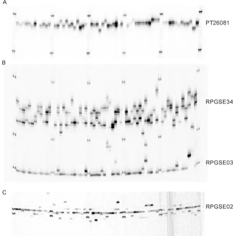
We validated the performance of the resulting DNA prep-arations by multiplex PCR assays. Amplicons of expected size were obtained. Later on more than 3000 needle and seed samples of Picea rubens, Picea glauca, and Picea mari-ana were processed and genotyped in our lab using more than a dozen nuclear and chloroplast microsatellites (Fig-ure 1). Reproducible assays were possible with 4-plex reactions. DNA isolation for one 96-well plate takes approximately 3.5 hours start to finish, and the use of a robotic liquid handler further reduces the hands-on time to about 40 min.
Comparison with other DNA extraction methods
To determine the reproducibility and efficiency of DNA isolation, we have compared our Protocol A-MTP with a slightly modified protocol using Qiagen DNeasy spin col-umns (Qiagen), and a version of widely used CTAB method [6]. Since some variability in the efficiency of mechanical tissue disruption is unavoidable due to the natural heterogeneity of plant material, we performed parallel DNA extractions using all three protocols in 8 rep-licates from the same bulked homogenized sample. Twenty grams of spruce needles were ground into fine powder under liquid nitrogen using mortar and pestle. 150 ± 2 mg of ground tissue were transferred into 2 ml Eppendorf tubes along with two 5 mm stainless steel balls normally used for tissue grinding. Then the DNA isolation was performed as follows:
Modified MagAttract protocol
As outlined above in the section Protocol A.
CTAB
The plant material was resuspended in 900 μl of 2× CTAB buffer (2% cetyltrimethylammonium bromide (CTAB); 1.4 M NaCl; 100 mM Tris-HCl, pH 8.0; 20 mM EDTA; 1% PVP; 0.2% β-mercaptoethanol) and 4 μl RNAse A (4 mg/ ml, Qiagen) for 1 min, then incubated for 40 min at 65°C. The lysates were extracted twice with equal volume of chlorophorm:isoamyl alcohol (24:1). DNA was precipitated with 0.7 volume of 100% isopropanol (30 min at -20°C, followed by centrifugation at 18000 g for 10 min). The DNA pellet was washed with cold 70% ethanol twice, vacuum dried, and resuspended in 100 μl of TE buffer.
Qiagen DNeasy
DNA was isolated according to the manufacturer's instruc-tions, except for increased volumes of AP1 and AP2 buff-ers (700 μl and 250 μl per prep, respectively). Purified DNA was eluted into 150 μl of 10 mM Tris-HCl pH 7.5.
Microsatellite genotyping of 60 spruce trees with SSRs con-taining mono-, di-, and trinucleotide repeats
Figure 1
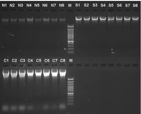
All three methods resulted in high quality DNA (Figure 2) with A260/280 values in the range 1.69–1.85. The DNA yield varied greatly among the methods (Table 1). Signif-icant variance of DNA concentration in our protocol can be attributed to relatively high pipetting errors when han-dling magnetic particles.
Conclusion
The CTAB method demonstrated the best yield and good reproducibility and probably remains the method of choice where large amounts of high quality DNA are required. For research projects requiring processing of larger sample sizes, our MagAttract-based protocol can be a flexible and cost-efficient solution (around $1 per sam-ple, compared to $4 for spin columns).
Medium-through-put version of our protocol allows for processing of large sample sets while maintaining higher DNA yield. The high-throughput variation is the best solution for geno-typing projects where speed is the ultimate priority. It allows for processing up to 200 samples per person per day, in comparison to other protocols where expected output would be around 40–60 samples. The protocol works from start to end in the standard SBS 96 well micro-plate format and is fully automatable.
Competing interests
The authors declare that they have no competing interests.
Authors' contributions
SB carried out the laboratory work, developed the proto-col and designed the robotic liquid handler software methods. OPR is the principal investigator of the project. He supervised the research work and provided funding and direction. Both authors participated in writing and revising the article, and approved the final manuscript.
Acknowledgements
The research was funded by the Canada Research Chair Program (CRC950-201869) funds and the Natural Sciences and Engineering Research Council of Canada Discovery Grant RGPIN 170651 to O.P. Rajora. S. Bashalkhanov was supported by the University of New Brunswick start up funds provided to O.P. Rajora and a Canadian Forest Service grad-uate student's supplemental stipend. The gel image and DNA concentration data for Picea glauca were kindly provided by Dr. M. Fageria and for Pinus strobus by Mr. J.W.R. Zinck.
References
1. Manen JF, Sinitsyna O, Aeschbach L, Markov A, Sinitsyn A: A fully automatable enzymatic method for DNA extraction from plant tissues. BMC Plant Biology 2005, 5(1):23.
2. Dilworth E, Frey JE: A rapid method for high throughput DNA extraction from plant material for PCR amplification. Plant Molecular Biology Reporter 2000, 18:61-64.
3. Paris M, Carter M: Cereal DNA: a rapid high-throughput extraction method for marker assisted selection. Plant Molec-ular Biology Reporter 2000, 18:357-360.
4. Vuosku J, Jaakola L, Jokipii S, Karppinen K, Kämäräinen T, Pelkonen VP, Jokela A, Sarjala T, Hohtola A, Häggman H: Does Extraction of DNA and RNA by Magnetic Fishing Work for Diverse Plant Species? Molecular Biotechnology 2004, 27:209-216.
5. Shepherd M, Cross M, Stokoe RL, Scott LJ, Jones ME: High-through-put DNA extraction from forest trees. Plant Molecular Biology Reporter 2002, 20:425a-425j.
Table 1: Comparison of MagAttract-based protocol with Qiagen DNeasy spin columns and CTAB protocol.
Method DNA
concentration, ng/μl
DNA yield,
μg/preparation
CV, %
MagAttract-based protocol OD260 75 7.5 30.66
fluorimetric 70 7.0 37.98
DNeasy OD260 121 18.1 9.59
fluorimetric 190 28.5 10.73
CTAB OD260 875 87.5 8.10
fluorimetric 1079 107.9 13.12
Comparison of DNA yield and quality across different isola-tion methods
Figure 2
Publish with BioMed Central and every scientist can read your work free of charge
"BioMed Central will be the most significant development for disseminating the results of biomedical researc h in our lifetime."
Sir Paul Nurse, Cancer Research UK
Your research papers will be:
available free of charge to the entire biomedical community
peer reviewed and published immediately upon acceptance
cited in PubMed and archived on PubMed Central
yours — you keep the copyright
Submit your manuscript here:
http://www.biomedcentral.com/info/publishing_adv.asp
BioMedcentral 6. Doyle JJ, Doyle JL: A rapid DNA isolation procedure for small
quantities of fresh leaf tissue. Phytochemical Bulletin 1987, 19:11-15.