Tomasz Fuchs
A, B, D, Michał Pomorski
B, Krzysztof Grobelak
C, D,
Marek Tomiałowicz
B, Mariusz Zimmer
E, FSignal Loss During Fetal Heart Rate Monitoring
Using Maternal Abdominal Surface Electrodes
Between 28 and 42 Weeks of Pregnancy
2nd Department and Clinic of Gynaecology, Obstetrics and Neonatology, Wroclaw Medical University, Poland
A – research concept and design; B – collection and/or assembly of data; C – data analysis and interpretation;
D – writing the article; E – critical revision of the article; F – final approval of article; G – other
Abstract
Background. Fetal electrocardiography is one of the methods for monitoring the well-being of the fetus. Signal loss limits the proper interpretation of electrocardiogram traces.
Objectives. The aim of this study was to assess the average signal loss in non-invasive abdominal fetal electro-cardiogram (fECG) monitoring using the KOMPOREL fetal monitoring system (ITAM, Zabrze, Poland) in women between 28 and 42 week of pregnancy. The results were compared to FIGO (International Federation of Gynaecology and Obstetric) and DGGG (Deutsche Gesellschaft für Gynäkologie und Geburtshilfe e.V.) recom-mendations concerning fetal heart monitoring. The correlation between fetal ECG signal quality, week of preg-nancy and patient BMI was evaluated.
Material and Methods. 773 pregnant women, hospitalized and diagnosed in the Department of Gynecology and Obstetrics, Wroclaw Medical University, underwent 30 min of abdominal fECG recordings using the KOMPOREL fetal monitoring system.
Results. The average signal loss in abdominal fECG monitoring in the study group was 32%. FIGO recommenda-tions describe an acceptable fetal signal loss of 20%. In our study, 46% (357/773) of the recordings were up to FIGO standards, with fetal heart rate success rates above 80%. According to DGGG guidelines, with acceptable fetal signal loss of 15%, only 39% (303/773) of the recordings could be assessed as accurate. No correlation between fECG signal quality, week of pregnancy and patient BMI was proved.
Conclusions. The average signal loss in abdominal fECG monitoring in our study group was 32%. Low fECG signal quality may constitute a potentially limiting factor of the described fetal heart monitoring system.No relationship between fECG signal quality, week of pregnancy and patient BMI was proved (Adv Clin Exp Med 2014, 23, 5, 813–819).
Key words: non-invasive fetal electrocardiography, fetal heart rate monitoring, abdominal fetal electrocardiogra-phy, signal loss in abdominal fetal electrocardiography.
Adv Clin Exp Med 2014, 23, 5, 813–819 ISSN 1899–5276
ORIGINAL PAPERS
© Copyright by Wroclaw Medical University
Fetal electrocardiography (fECG) is one the methods for registering the electrical activity of the heart of the fetus, which makes it possible to assess fetal well-being during pregnancy and labor. This method was presented for the first time by Cre-mer at the beginning of the 20th century. He
mea-sured the electric signal from the heart of the fetus using a string galvanometer invented by Willem Einthoven, the device used for recording the heart’s electrical activity in adults [1]. Cremer’s studies on obtaining the fECG signal were carried out with
electrodes located on the abdomen, in the vagina, the esophagus and the rectum of pregnant woman. In this way, he demonstrated the capabilities of di-agnostic techniques in those times. The quality of his recordings was poor, mainly due to unwanted back-ground interference such as the maternal electro-cardiography waveform, noise from adjacent tissues and the device. In practice, the low quality did not al-low accurate assessment of fetal intrauterine status.
signal, but only the use of modern computers, am-plifiers and dedicated software have made it pos-sible to obtain a clear signal and a complete im-age of the atrio-ventricular complexes of the fetal heart and typical fECG traces of the fetal cardiac cycle [2]. In 1953, Smyth et al. used an electrode attached to the amniotic membranes for the first time, while Hon et al. in 1962 designed and de-scribed an electrode which could be attached di-rectly to the fetal scalp or other presenting part of the fetus [1]. In this way, it was possible to pre-cisely assess the characteristics of P and T waves. Currently, a similar type of electrode is used in the STAN monitoring system (Neoventa Medical, Mölndal, Sweden), whereas in British systems such as Monica AN24 Monitor (Monica Healthcare, Nottingham, UK), American – MindChild Medi-cal (North Andover, MA, USA) and Polish KOM-POREL (ITAM, Zabrze, Poland), transabdominal electrodes are used [3]. Depending on the applica-tion mode on the abdominal wall, the number of electrodes varies from 6 to 16.
Since the 60s of the last century, classical car-diotocography (CTG) with fetal heart rate analysis is a standard, non-invasive procedure for monitor-ing fetal well-bemonitor-ing before and durmonitor-ing labor. The fetal monitor is an ultrasound transducer which uses the Doppler effect to detect the heart beat of the fetus. The fECG is an alternative to the classi-cal CTG. The advantage of this method is the ad-ditional information which can not be obtained by CTG [4].
Aim of the Study
Assessment of the average signal loss obtained during recording of fetal heart rate and uter-ine electrical activity using the KOMPOREL fe-tal monitoring system (ITAM, Zabrze, Poland) in women between 28 and 42 weeks’ gestation. The results obtained were compared to the FIGO cri-teria (International Federation of Gynecology and Obstetrics) and DGGG (Deutsche Gesellschaft für Gynäkologie und Geburtshilfe e.V.) recommenda-tions [5, 6].
Correlation between fECG signal quality, week of pregnancy and patient’s BMI was evaluated.
Material and Methods
The study group consisted of 773 pregnant women whose pregnancies ranged from 28 to 42 weeks gestational age, either hospitalized or di-agnosed in outpatient settings in the Second De-partment of Gynecology and Obstetrics, Wroclaw
Medical University. The study group was divided into 3 subgroups:
1. Physiological pregnancies, in which ultra-sound examination confirmed normal, singleton pregnancy with eutrophic fetus – 601 cases.
2. Pregnancies complicated by intrauterine growth retardation between 28 and 38 weeks of gestation and by pregnancy induced hypertension between 28 and 41 weeks of gestation – 91 cases.
3. Woman threatening preterm labor between 28 and 37 weeks of gestation – 81 cases.
The study protocol was approved by the Com-mission of Bioethics at Wroclaw Medical Universi-ty. Each patient was instructed in study procedures and methodology prior to entering the study. All women gave written informed consent.
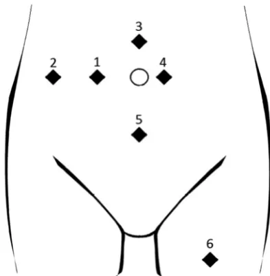
With each patient, the recording of the elec-trical signal of fetal heart and uterine contraction activity lasted 30 min. The KOMPOREL (ITAM, Zabrze, Poland) fetal monitoring system was used for the signal registration. The device re-cords and analyses bioelectric signals. During the examination, the woman laid in the supine or left lateral recumbent position, depending on which of them provided the best signal quality. Prior to electrode placement, the skin was prepared with a mild skin abrasion to the electrode site using sand paper material for electrocardiography from 3M in order to remove part of the stratum corne-um. Disposable electrodes of 3M type 2222 were used. In order to obtain the best electrode adhe-sion, an additional gel layer on the sensing ele-ment was applied. Six electrodes were placed as follows:
1) on the level of the umbilicus, 5 cm on the right side,
2) on the level of the umbilicus, 10 cm on the right side,
3) in the midline, 5 cm above the umbilicus, 4) on the left side, 1 cm from the umbilicus, 5) in the midline, 10 cm below the umbilicus, the so-called ground reference electrode,
6) 10 cm below the inguinal region on the front side of thigh, the so-called return electrode.
The KOMPOREL software is used for:
– filtering interference, including those from the maternal abdominal muscle,
– extracting and canceling the maternal electrocardiogram,
– detecting the fetal QRS complex and calcu-lating fetal heart rate,
– identifying the P-QRS-T complex,
– determining T-wave amplitude in relation to QRS – T/QRS complex,
– calculating baseline fetal heart rate,
– determining the short-term and long-term variability,
– filtering and analysis of uterine electrical activity.
The statistical analysis was performed with STATISTICA 10 PL. The results are expressed as mean, standard deviation (SD), confidence inter-val (CI). In order to determine correlations among the studied parameters, a Pearson correlation anal-ysis was carried out. A value of p < 0.05 was con-sidered significant.
Results
The average signal loss in abdominal fECG monitoring in the study group was 32% (SD 25.26; CI 29.82–33.38). In the group of 601 pregnant wom-en with normal pregnancies, mean signal loss was 30.38% (SD 25.02; CI 28.38–32.38). In the group of women (n = 91) with pregnancies complicated by intrauterine growth restriction and pregnancy in-duced hypertension, it was 31.44% (SD 25.12; CI 26.21–36.67), while the highest percentage of sig-nal loss occurred in the group of woman threaten-ing preterm labor (n = 81) – 40.86% (SD 25.4; CI 35.25–46.48). According to the FIGO criteria, the
maximum acceptable level of signal loss determin-ing the correct interpretation of the record is 20%. In the present study, over 46% (357/773) of total records met this criterion, yielding efficiency of at least 80%. According to DGGG guidelines, which are stricter, the maximum level of acceptable fe-tal signal loss is 15%. The percentage of correct re-cordings drops then to 39% (303/773).
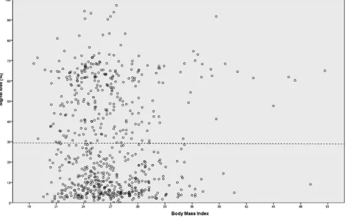
Body mass index (BMI) was calculated in 687 patients. The values obtained ranged from 18.5 to 50.7 (mean 26.9; SD 4.31).
Assessing the relationship between signal loss, gestational age and patient’s BMI, the following re-sults were obtained:
– no correlation between the percentage of sig-nal loss and gestatiosig-nal age was found (correlation coefficient r – 0.059; CI 0.129–0.0107; p > 0.096); scatter plots of signal loss in gestational week are shown in Fig. 2,
– no correlation between the percentage of signal loss and patient’s BMI was found (cor-relation coefficient r – 0.005; CI – 0.079–0.069; p > 0.892); scatter plots of signal loss and BMI val-ues are shown in Fig. 3.
Discussion
CTG, since the 60s of the last century, has be-come a standard non-invasive method in monitor-ing fetal well-bemonitor-ing before and durmonitor-ing labor. It was introduced in order to identify events that might result in complications such as hypoxic isch-emic encephalopathy, cerebral palsy and perinatal death. The use of the method in clinical practice is still controversial, especially in terms of efficacy. A study conducted by Zimmer et al. on a group of 10,983 labors showed that continuous electronic fetal monitoring was not associated with a reduc-tion in cesarean secreduc-tions and an increase in neo-natal Apgar score given to newborns [7]. Studies conducted by Alfirevic et al. proved that continu-ous monitoring of fetal heart rate during labor re-sulted in an increase of cesarean sections and in-strumental vaginal deliveries but did not reduce neonatal mortality and the frequency of cerebral palsy [8]. The only noticeable advantage of this method was a reduction in neonatal seizures [9]. The major factor limiting the effectiveness and re-liability of continuous electronic fetal monitoring might be the difficulty and variability in the inter-pretation of intrapartum cardiotocogram traces by obstetricians and midwives [9–11]. Therefore, methods for more accurate assessment of heart rate and intrauterine fetal well-being have been sought. An alternative to CTG may be the rapidly developing fECG.
Fig. 2. Pearson correlation – signal loss vs. week of pregnancy: r – 0.059, CI – 0.129–0.0107, p > 0.096
The STAN monitoring system has been under development for many years. It provides a comput-erized analysis of ST interval of the fECG. The de-vice is attached to the fetus by fetal scalp electrode. Unlike transabdominal fECG, it is an invasive method as it requires dilated cervix (sometimes in-strumentally) and ruptured amniotic membranes, which increases the risk of intrauterine infec-tion [12]. Salmelin et al., in a meta-analysis carried out in 2013, concluded that there is not enough sci-entific evidence that computerized ST analysis re-duces the incidence of metabolic acidosis in new-borns [13]. At the same time, it was shown that the incidence of cesarean sections and instrumen-tal vaginal deliveries due to feinstrumen-tal distress did not change, regardless of the fetal heart rate monitor-ing method. However, there was a significant re-duction in the number of fetal scalp pH testing.
In 1986 FIGO introduced the first recommen-dations for the nomenclature of changes in fetal heart rate recorded on cardiotocographic traces and guidelines for their interpretation. This helped to standardize maternity care [5]. Organizations such as the American College of Obstetricians and Gynecologists (ACOG), the National Institute of Child Health and Human Development (NICH-HD), the Royal College of Obstetricians and Gyn-aecologists (RCOG), and the National Institute of Clinical Excellence (NICE) also undertook to in-troduce their own guidelines for the interpretation of CTG, though regarding mainly records during labor. However, their use is limited to the coun-tries in which they were developed [14].
According to the FIGO criteria, fetal heart rate recording can be considered possible to interpret and reliable if signal loss during the examination does not exceed 20%. Among the available meth-ods of fetal heart rate monitoring, magnetocardiog-raphy seems to be the most precise. In their stud-ies, Crowe et al. as well as Stinstra et al. showed nearly 100% efficacy of fetal magnetocardiography in registering the electrical activity of the heart of the fetus [15, 16]. In comparison to transabdom-inal fECG, the electrical activity of the maternal heart and the presence of vernix did not affect the quality of the obtained recording [17]. However, due to the high cost of the equipment and condi-tions under which it operates, this method is not widely used in the assessment of fetal intrauterine well-being [18]. High efficacy can be also obtained in recordings of fetal electrical activity of the heart using electrodes placed on the presenting part of the fetus. The average signal loss of fECG in this method usually does not exceed 10%, although Bakker et al. achieved an average efficacy in the second stage of labor at less than 80% [19, 20]. In the studies in which researchers evaluated the
quality of the signal recorded during intrapartum cardiotocography, the signal loss ranged between 15 and 40% [19, 21].
In the published literature, the authors did not find any reference to the quality description of the signal obtained during the recording of transab-dominal electrocardiograms of the fetus in the an-tepartum period. In the studies conducted to date, researchers have focused mainly on the descrip-tion of the efficacy of this method during labor.
Cohen et al. compared the accuracy and reliabil-ity of 3 methods used for heart rate detection: trans-abdominal fECG, cardiotocography, and monitoring with fetal scalp electrode. The study group consisted of 75 laboring women. All the pregnancies included in the study were of > 37 weeks’ gestation. The mean value of signal loss in transabdominal fECG did not exceed 17% (success rate 83.4% ± 20.1%). During the first stage of labor, mean signal loss was 13.6% (success rate 86.4% ± 21.1%), and 24.8% in the sec-ond stage of labor (success rate 75.2 % ± 19.2%) [22]. Reinhard et al. conducted a study on 144 laboring women in order to assess the fetal heart rate signal quality of abdominal fECG. The signal loss in the first and second stages of labor was 4.3% and 19.8% with a median success rate 95.7% and 80.2%, respec-tively [23]. Considering the FIGO criteria, in the Re-inhard’s et al. study group, the percentage of patients having fECG signal loss below 20% during the first and second stages of labor was 78.5% and 46.9%, respectively. Signal loss below 15% (DGGG guide-lines) occurred in 73.3% and 36.7% of women in the first and second stages of labor, respectively. These results show the poor quality of the recordings, par-ticularly in the second stage of labor. In a Cohen et al. paper, corresponding calculations were not pro-vided. Both Cohen et al. and Reinhard et al. used the Monica AN24 (Monica Healthcare, Notting-ham, UK) fECG monitoring system. Clifford et al. evaluated the accuracy and fidelity of the E-TROLZ physiologic monitoring platform for measuring fe-tal heart rate and fefe-tal electrocardiogram morpholo-gy, especially ST segment changes [24]. Data was re-corded from 32 laboring women with the use of 29 electrodes placed over the maternal abdomen. This made it possible to obtain records with a mean val-ue of signal loss below 11% (success rate 89.9%). The results of the above-mentioned studies are summa-rized in Table 1.
between 27 and 32 gestational weeks was report-ed [25, 26]. It concernreport-ed both an increasreport-ed per-centage of signal loss of electrical heart activity of the fetus and a reduced quality of various registered cardiac waveforms. This is presumably caused by the layer of vernix caseosa, which begins to form intensively on the entire body of the fetus at this stage of pregnancy. As the pregnancy advances, the mass of the cardiac muscle increases, the fe-tus grows and matures, and gaps appear in the ver-nix covering the body of the fetus. The above-men-tioned factors contribute to increased quality of the fECG. In the present study, the average signal loss in pregnancies between 28 and 32 weeks’ gestation was 29.9%. In contrast to the results of the above-mentioned authors, that value is lower than that obtained in the present study on pregnancies be-tween 33 and 42 weeks’ gestation (37.1%).
Table 1. Signal loss in abdominal fECG monitoring in different studies
Author Fuchs et al. Cohen et al. Reinhard et al. Gari et al.
n 773 75 144 32
BMI (range) 26.9 (18.5–50.7) 32.6 (25–40.2) 29.8 (20.6–49.5) 30.4 (21.7–45.1) Monitoring system KOMPOREL Monica AN24
Monitor Monica AN24 Monitor E-TROLZ success rate m (%)
Antepartum recording 68.4%; n-773 n/a n/a n/a Intrapartum recording n/a 83.4% 20.1%; n-75 n/a 89.9%; n-32 First stage of labor n/a 86.4% 21.1%; n-72 95.7%; n-135 n/a Second stage of labor n/a 75.2%; n-41 80.2%; n-98 n/a
m – arithmetic mean; n – number of pregnant/laboring women, n/a – not applicable.
Reinhard et al. did not show correlation be-tween a patient’s BMI and the signal loss of ab-dominal fECG registered with the use of electrodes placed over maternal abdomen. Also in the pres-ent study, which was conducted on a 5-fold larg-er research group (144 vs. 773) this correlation was not demonstrated (r – 0.005; CI 0.079–0.069; p > 0.892). In the case of intrapartum electrocardi-ography, Solum et al. showed an inverse relation-ship between patient’s BMI and the quality of the fECG signal [27].
The authors concluded that the average signal loss in abdominal fECG monitoring in our study group was 32%. Low fECG signal quality may con-stitute a potentially limiting factor of the described fetal heart monitoring system. No relationship be-tween fECG signal quality, week of pregnancy and patient’s BMI was proved.
References
[1] Jenkins HM: Technical progress in fetal electrocardiography – a review. J Perinat Med 1986, 14, 365–370.
[2] Martens SM, Rabotti C, Mischi M, Sluijter RJ: A robust fetal ECG detection method for abdominal recordings. Physiol Meas 2007, 28, 373–388.
[3] Adam J: The future of fetal monitoring. Rev Obstet Gynecol 2012, 5, 132–136.
[4] Rosén KG, Amer-Wåhlin I, Luzietti R, Norén H: Fetal ECG waveform analysis. Best Pract Res Clin Obstet Gynaecol 2004, 18, 485–514.
[5] Rooth G, Huch A, Huch R: Guidelines for the use of fetal monitoring. Int J Gynaecol Obstet 1987, 25, 159–167.
[6] Deutsche Gesellschaft fur perinatale Medizin, AG fur maternofetale Medizin, deutsche Gesellschaft fur Gynakologie und Geburtshilfe Anwendung des CTG wahrend chwangerschaft und Geburt. Frauenarzt 2004, 45, 979–989.
[7] Zimmer M, Hirnle L, Fuchs T, Florjański J, Tomiałowicz M, Kłósek A, Milnerowicz-Nabzdyk E: The influence of computer supervision of deliveries on the medical procedures during labor and neonatal post-delivery status. Ginekol Pol 2000, 71, 187–191.
[8] Alfirevic Z, Devane D, Gyte GM: Continuous cardiotocography (CTG) as a form of electronic fetal monitoring (EFM) for fetal assessment during labour. Cochrane Database Syst Rev 2006.
[9] Schiermeier S, Westhof G, Leven A, Hatzmann H, Reinhard J: Intra- and interobserver variability of intrapar-tum cardiotocography: a multicenter study comparing the FIGO classification with computer analysis software. Gynecol Obstet Invest 2011, 72, 169–173.
[10] Sweha A, Hacker TW, Nuovo J: Interpretation of the electronic fetal heart rate during labor. Am Fam Physician 1999, 59, 2487–2500.
[12] Leatherman J, Parchman ML, Lawler FH: Infection of fetal scalp electrode monitoring sites. Am Fam Physician 1992, 45, 579–582.
[13] Salmelin A, Wiklund I, Bottinga R, Brorsson B, Ekman-Ordeberg G, Grimfors EE, Hanson U, Blom M, Persson E: Fetal monitoring with computerized ST analysis during labor: a systematic review and meta-analysis. Acta Obstet Gynecol Scand 2013, 92, 28–39.
[14] Ayres-de-Campos D, Bernardes J: Twenty-five years after the FIGO guidelines for the use of fetal monitoring: Time for a simplified approach? Int J Gynaecol Obstet 2010, 110, 1–6.
[15] Crowe JA, Herbert JM, Huang XB, Reed N, Wolfson MS, Rassi D, Zhuravlev YE, Emery SJ: Sequential record-ing of the abdominal fetal electrocardiogram and magnetocardiogram. Physiol Meas 1995, 16, 43–47.
[16] Stinstra J, Golbach E, van Leeuwen P, Lange S, Menendez T, Moshage W, Schleussner E, Kaehler C, Horigome H, Shigemitsu S, Peters MJ: Multicentre study of fetal cardiac time intervals using magnetocardiography. BJOG 2002, 109, 1235–1243.
[17] Quinn A, Weir A, Shahani U, Bain R, Maas P, Donaldson G: Antenatal fetal magnetocardiography – a new method for fetal surveillance? Br J Obstet Gynaecol 1994, 101, 866–870.
[18] Quartero HW, Stinstra JG, Golbach EG, Meijboom EJ, Peters MJ: Clinical implications of fetal magnetocardi-ography. Ultrasound Obstet Gynecol 2002, 20, 142–153.
[19] Spencer JAD, Belcher R, Dawes GS: The influence of signal loss on the comparison between computer analyses of the fetal heart-rate in labor using pulsed Doppler ultrasound (with autocorrelation) and simultaneous scalp electrocardiogram. Eur J Obstet Gynecol Reprod Biol 1987, 25, 29–34.
[20] Bakker PC, Colenbrander GJ, Verstraeten AA, Van Geijn HP: The quality of intrapartum fetal heart rate moni-toring. Eur J Obstet Gynecol Reprod Biol 2004, 116, 22–27.
[21] Dawes GS, Visser GH, Goodman JD, Redman CW: Numerical analysis of the human fetal heart rate – the quality of ultrasound records. Am J Obstet Gynecol 1981, 141, 43–52.
[22] Cohen WR, Ommani S, Hassan S, Mirza FG, Solomon M, Brown R, Schifrin BS, Himsworth JM, Hayes-Gill BR:
Accuracy and reliability of fetal heart rate monitoring using maternal abdominal surface electrodes. Acta Obstet Gynecol Scand 2012, 91, 1306–1313.
[23] Reinhard J, Hayes-Gill BR, Schiermeier S, Hatzmann W, Herrmann E, Heinrich TM, Louwen F: Intrapartum signal quality with external fetal heart rate monitoring: a two way trial of external Doppler CTG ultrasound and the abdominal fetal electrocardiogram. Arch Gynecol Obstet 2012, 286, 1103–1107.
[24] Clifford G, Sameni R, Ward J, Robinson J, Wolfberg AJ: Clinically accurate fetal ECG parameters acquired from maternal abdominal sensors. Am J Obstet Gynecol 2011, 205, 47, 1–5.
[25] Taylor MJ, Smith MJ, Thomas M, Green AR, Cheng F, Oseku-Afful S, Wee LY, Fisk NM, Gardiner HM: Non-invasive fetal electrocardiography in singleton and multiple pregnancies.Br J Obstet Gynaecol 2003, 110, 668–678.
[26] Chia EL, Ho TF, Rauff M, Yip WC: Cardiac time intervals of normal fetuses using noninvasive fetal electrocardi-ography. Prenat Diagn 2005, 25, 546–552.
[27] Solum T: A comparison of the three methods for external fetal cardiography. Acta Obstet Gynecol Scand 1980, 59, 123–126.
Address for correspondence:
Tomasz Fuchs2nd Department and Clinic of Gynaecology, Obstetrics and Neonatology
Wroclaw Medical University Borowska 213
50-556 Wrocław Poland
E-mail: [email protected]
Conflict of interest: None declared