R E S E A R C H A R T I C L E
Open Access
Ino80 is essential for proximal-distal axis
asymmetry in part by regulating Bmp4
expression
Zhijun Qiu
†, Zeinab Elsayed
†, Veronica Peterkin, Suehyb Alkatib, Dorothy Bennett and Joseph W. Landry
*Abstract
Background:Understanding how embryos specify asymmetric axes is a major focus of biology. While much has
been done to discover signaling pathways and transcription factors important for axis specification, comparatively little is known about how epigenetic regulators are involved. Epigenetic regulators operate downstream of signaling pathways and transcription factors to promote nuclear processes, most prominently transcription. To discover novel functions for these complexes in axis establishment during early embryonic development, we characterized phenotypes of a mouse knockout (KO) allele of the chromatin remodeling Ino80 ATPase.
Results:Ino80KO embryos implant, but fail to develop beyond the egg cylinder stage.Ino80KO embryonic stem
cells (ESCs) are viable and maintain alkaline phosphatase activity, which is suggestive of pluripotency, but they fail to fully differentiate as either embryoid bodies or teratomas. Gene expression analysis ofIno80KO early embryos by in situ hybridization and embryoid bodies by RT-PCR shows elevatedBmp4expression and reduced expression of distal visceral endoderm (DVE) markersCer1,Hex,andLefty1. In culture,Bmp4maintains stem cell pluripotency and when overexpressed is a known negative regulator of DVE differentiation in the early embryo. Consistent with the early embryo, we observed upregulatedBmp4expression and down-regulatedCer1,Hex,andLefty1expression whenIno80KO ESCs are differentiated in a monolayer. Molecular studies in these same cells demonstrate that Ino80 bound to theBmp4promoter regulates its chromatin structure, which correlates with enhanced SP1 binding. These results in combination suggest that Ino80 directly regulates the chromatin structure of theBmp4promoter with consequences to gene expression.
Conclusions:In contrast toIno80KO differentiated cells, our experiments show that undifferentiatedIno80KO ESCs are viable, but fail to differentiate in culture and in the early embryo.Ino80KO ESCs and the early embryo up-regulateBmp4expression and down-regulate the expression of DVE markersCer1,HexandLefty1. Based on this data, we propose a model where the Ino80 chromatin remodeling complex repressesBmp4expression in the early embryo, thus promoting DVE differentiation and successful proximal-distal axis establishment. These results are significant because they show that epigenetic regulators have specific roles in establishing embryonic axes. By further characterizing these complexes, we will deepen our understanding of how the mammalian embryo is patterned by epigenetic regulators.
Keywords:Ino80, Chromatin remodeling, Bmp4, DVE, PD Axis, Gastrulation, Embryonic ectoderm
* Correspondence:[email protected] †Equal contributors
Department of Human and Molecular Genetics, Virginia Institute of Molecular Medicine, Massey Cancer Center, Virginia Commonwealth University School of Medicine, Richmond, VA 23298, USA
Background
Mammalian embryonic development is best understood using the mouse model [1]. Fertilized eggs develop into a blastocyst by embryonic day 3.5 (E3.5). At this stage, the blastocyst is composed of both an inner cell mass (ICM), which is fated to become the embryo, and an extraembry-onic trophectoderm (TE). The blastocyst implants into the uterus at E4.5, and during this time the embryo forms the epiblast (EPI) and primitive endoderm (PrE). During implantation the EPI rapidly proliferates and expands to form an egg cylinder, which is composed of both embry-onic ectoderm (EmE) and extraembryembry-onic ectoderm (ExE) covered by visceral endoderm (VE). During egg cylinder expansion, the distal tip of the embryo differentiates into the distal visceral endoderm (DVE), establishing a proximal-distal (P-D) axis. During the transition from E5.5 to E6.0, the DVE migrates up the anterior portion of the embryo to create the anterior visceral endoderm (AVE) where it has organizer activity through secreting several inhibitors of the Tgfb and Wnt family ligands. The secretion of these inhibitors constrains Tgfb and Wnt sig-naling activity to the posterior portion of the embryo, which promotes the differentiation of the primitive streak and the anterior-posterior (A-P) axis.
Early mammalian development, and the establishment of asymmetric axes (P-D and A-P axes), require coordi-nated gene expression [2]. Gene expression is regulated by both transcription factors and epigenetic regulators. Both operate within chromatin, which is composed of nucleosomes at its fundamental level [3, 4]. Essential epigenetic regulators in eukaryotes are chromatin re-modeling complexes. Chromatin rere-modeling complexes are usually multi-subunit enzymes that slide or evict nu-cleosomes, or exchange its histone subunits. These activ-ities change chromatin structure by altering the position, occupancy or composition of nucleosomes [5]. In turn, changes in chromatin structure regulate access to the underlying DNA, that in turn influences nuclear pro-cesses like transcription.
Chromatin remodeling complexes are classified into the SWI/SNF, ISWI, CHD or INO80 families based upon the sequence homology of their ATPase subunit [5]. In mammals, the INO80 family is composed of the Srcap, p400, and Ino80 remodeling complexes. These com-plexes are large, 12-15 subunit, comcom-plexes that are unique among chromatin remodeling complexes because they catalyze histone exchange reactions [6]. In addition to histone exchange, Ino80 has significant nucleosome sliding activity, suggesting that it can either alter nucleo-some position or occupancy, or change nucleonucleo-some composition in vivo. Presumably through these activities, Ino80 regulates a variety of nuclear processes which in-clude transcription, DNA repair, DNA replication, and telomere structure [6].
While much has been done to characterize the nuclear functions of Ino80, little has been done to determine its functions in metazoan development. In plants, INO80 is essential for flowering and reproductive organ develop-ment, possibly through functions in homologous recom-bination and regulated transcription [7, 8]. In flies, INO80 mutants are late embryonic lethal, deregulate Hox gene expression, and manifest homeotic transfor-mations [9]. In addition to Hox genes, INO80 regulates ecdysone response genes, which are essential for pupal development and molting [10]. Localization and nucleo-some mapping studies in insect cells show that INO80 is widely distributed throughout the genome and remodels nucleosomes onto energetically unfavorable DNA se-quences [11]. Similar to studies in plants and flies, mam-malian Ino80 is also essential for development. Ex vivo studies with Ino80 knockout (KO) or shRNA knock-down (KD) pre-implantation embryos show that Ino80 maintains stem self-renewal by promoting the expres-sion of pluripotency factors like Oct4 and stabilizing DNA replication forks [12, 13]. In utero,Ino80 KO em-bryos implant into the uterus but fail to develop to mid-gestation, possibly due to roles for Ino80 in regulating telomere structure or DNA damage repair [14]. Because Ino80 is a well-documented regulator of gene expres-sion, developmental defects could also result from ab-normal gene expression [13].
Building on these earlier studies, we show that Ino80
KO embryos fail to specify a DVE and a P-D axis in utero. Coincident with defects in the DVE, Ino80 KO embryos aberrantly express Bmp4in the EmE, a known repressor of DVE specification [15]. Molecular studies using differentiating ESC models show that Ino80 is spe-cifically localized to the Bmp4 promoter, remodels its chromatin structure, and regulates the binding of tran-scription activators to its DNA sequence. These results in combination suggest that Ino80 directly represses
Bmp4 expression in the EmE through its chromatin re-modeling activity to promote DVE specification and P-D axis establishment.
Results
Ino80KO embryos fail to develop beyond the egg cylinder stage
mice constitutively expressing the Flp or Cre recombi-nases. Successful recombination and germ line transmis-sion of the Floxed and KO alleles were identified by Southern blotting and PCR-based genotyping strategies (Additional file 1b-d).
To determine if our targeted allele resulted in a loss of Ino80 protein, we crossed a Tet inducible Cre expression system (M2-rtTA and TetO-Cre alleles) into our Ino80
Floxed mice [17, 18]. Homozygous Ino80 Floxed (F/F) mouse embryonic fibroblasts (MEFs), with or without TetO-Cre, were isolated from mid-gestation E12.5 em-bryos. As expected, the addition of doxycycline to TetO-Cre,Ino80F/F fibroblast cultures resulted in the conver-sion of the Ino80 Floxed alleles to Ino80 KO alleles in two days (Additional file 2a). Over this same time course, Western blotting demonstrated a complete de-pletion of the Ino80 protein by day 6 (Additional file 2b). Coincident with the depletion of the Ino80 protein, we observed a loss of the exons 1-7Ino80transcript and the appearance of two smaller Ino80 transcripts (KO-1 and KO-2) by RT-PCR (Additional file 2c, d). Cloning and sequencing these smaller PCR products identified aberrant exon 1-5 and exon 1-6 splice events for the
Ino80 transcript, with the deletion of exons 2-4 by Cre-mediated recombination (Additional file 2d). In silico translation from the first ATG of each aberrant splice product results in an out of frame transcript and a ran-dom protein product (Additional file 2e). These results demonstrate that ourIno80Floxed allele can be success-fully excised by Cre recombinase, resulting in loss of the Ino80 protein.
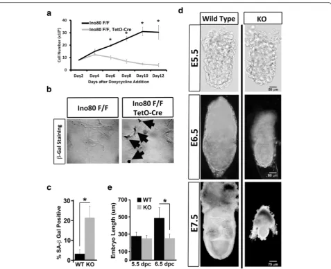
A previous publication showed that MEFs depleted of Ino80 arrest the cell cycle and senesce [14]. To deter-mine if depletion of Ino80 using ourIno80 Floxed allele resulted in similar phenotypes, we counted the number of viable fibroblasts over 12 days post-Cre expression by doxycycline exposure. As described previously [14], we observed that after doxycycline exposure Ino80 KO MEFs stopped proliferating and senesced as identified by an increased number of cells positive for endogenous b-Gal activity, an indicator of senescence [19] (Fig. 1a-c). Reproducing previous phenotypes of Ino80 KO MEFs supports the conclusion that our Ino80 KO allele is a null allele.
Phenotypes forIno80 KO embryos are largely unchar-acterized. To identify the functions of Ino80 in mamma-lian development, we intercrossed mice heterozygous for theIno80KO allele. Genotyping litters of mice from this intercross identified no homozygous Ino80 KO mice, demonstrating that Ino80 is essential for mouse viability (Additional file 3). To stage the earliest point where Ino80 is required for development, we genotyped litters of embryos at mid-gestation (E13.5), gastrulation (E6.5), and peri-implantation (E4.5). Results from these
experiments showed that Ino80 KO homozygous em-bryos were absent at E13.5, but present at the expected Mendelian ratios at gastrulation (E6.5) and prior to im-plantation (E4.5) (Additional file 3). These results dem-onstrate that Ino80 is essential for mammalian development post-gastrulation. A gross analysis of Ino80
KO embryos prior to (E5.5), during (E6.5), and post-gastrulation (E7.5) reveled that Ino80 is important for expansion of the egg cylinder during gastrulation (Fig. 1d). Quantitation of embryo length showed that the size of Ino80 KO embryos at E6.5 is similar to that ob-served for Ino80 KO embryos at E5.5 (Fig. 1e). Embryo reabsorption likely occurs after E7.5 because Ino80 KO embryos after E7.5 begin to show a loss in structural in-tegrity (data not shown).
Ino80KO embryonic stem cells are viable but exhibit an unstable pluripotent state
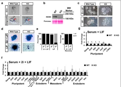
To characterize roles for Ino80 in the early embryo, we isolatedIno80KO embryonic stem cells (ESCs) from the ICM of pre-implantation E3.5 Ino80 KO embryos. Col-lection and genotyping pre-implantation E3.5-day blas-tocysts from Ino80 KO heterozygous intercrosses showed that Ino80 KO homozygous blastocysts look morphologically similar to wild-type littermates by gross inspection (Fig. 2a). These same isolated blastocysts suc-cessfully attached and outgrew onto gelatinized plates in media containing serum and leukemia inhibitory factor (LIF). After seven days of outgrowth, we observed a similar expansion of trophoblasts and the ICM between wild-type andIno80KO blastocysts (Fig. 2a). From these outgrowths, we successfully cloned Ino80 KO ESCs under culture conditions that maintain ground state pluripotency (media containing serum + 2i + LIF) [20, 21]. Western blotting showed that the Ino80 KO ESCs do not have the Ino80 protein, further confirming that our Ino80 KO allele is a null allele (Fig. 2b). Unlike
was recovered if cells maintained in the metastable state are returned to ground state conditions (Fig. 2e). These results demonstrate that Ino80 KO ESCs are viable, AP positive, and morphologically show evidence of slight cell scattering when compared to wild-type controls when maintained at ground state pluripotency. However,
Ino80KO ESCs lose AP staining and exhibit cell scatter-ing when grown under conditions that maintain a meta-stable pluripotent state.
To further characterize phenotypes ofIno80 KO ESCs grown under differing culture conditions, we measured
transcript levels of several markers of pluripotency, stem cell differentiation, and lineage commitment. When maintained at ground state pluripotency,Ino80KO ESCs have equivalent expression of pluripotency markers
Nanog, Oct4, Klf4, Sox2, and Essrb. Markers of ecto-derm, mesoecto-derm, and endoderm lineages were also ap-proximately equivalent between wild-type andIno80 KO ESCs (Fig. 2f ). In contrast, we observed reduced pluripo-tency marker expression (Nanog, Oct4, Klf4, and Essrb) whenIno80KO ESCs were maintained in the metastable state for five days (Fig. 2g) [13]. These results are
Fig. 1Ino80 is essential for embryonic fibroblast and early embryonic development.aGrowth curve of conditionalIno80KO MEF induced by doxycycline treatment. 1.0 × 10⁴cells of control (Ino80F/F) orIno80KO (Ino80F/F, TetO-Cre) were seeded into media containing 10μg/ml doxycycline. Numbers of trypan blue negative cells were counted every two days over a 12-day period (N= 3 independent measurements per group, representative of three biological replicates; * =ttestp≤0.05).bDay-12 control andIno80KO cells from panel a were stained for endogenousβ-galactosidase activity at pH 6.0 to measure cellular senescence [19].Black arrowsdesignateβ-galactosidase positive cells.c
consistent with the metastable pluripotent state, but not ground state pluripotency requiring Ino80.
Ino80KO ESCs fail to differentiate using models of mammalian development
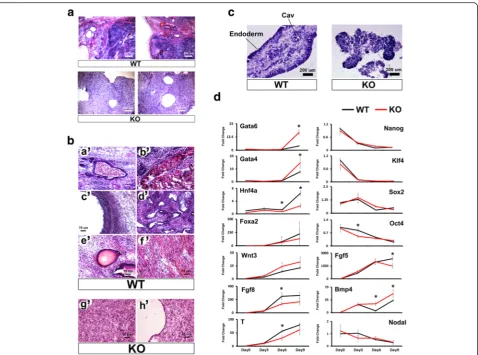
To further characterize roles for Ino80 during mammalian development, we differentiatedIno80KO ESCs using the teratoma model [22]. To create teratomas, both wild-type and Ino80KO ESCs maintained at ground state were in-troduced into opposing flanks of NOD/SCID mice. Tera-tomas were allowed to grow until control tumors were approximately 1 cm in diameter. From five inoculations, we harvested four wild-type tumors and twoIno80KO tu-mors. A histological analysis showed that control tumors
formed a wide range of differentiated tissues from each of the three germ lines. Differentiated tissues include blood islands, keratin pearls, and neural rosettes from the ecto-derm lineage, both striated and smooth muscle from mesoderm, and ciliated endoderm from the endoderm lineage (Fig. 3a, b). In contrast to the well-differentiated tissues observed in wild-type teratomas, we observed that
Ino80 KO teratomas are composed of undifferentiated mesenchyme with a thin layer of epithelial cells on the outer periphery (Fig. 3a, b).
In addition to forming teratomas, we utilized the em-bryoid body ESC differentiation model [23]. To create embryoid bodies, ESCs maintained at ground state were dispersed in clumps and aggregated in serum containing
Fig. 2Ino80KO ESCs are viable, but differentiate when maintained in a metastable pluripotent state.aMicroscopic analysis of wild-type andIno80 KO pre-implantation E3.5 blastocysts. Blastocysts were allowed to outgrow for seven days onto gelatinized plates in media containing serum + LIF. Wild-type andIno80KO are at identical magnification.Red arrowsdesignate ESC colony outgrowth.bWestern analysis of Ino80 protein expression in wild-type andIno80KO ESCs using a custom Ino80 antibody. Ponceau S was used as a loading control.cMicroscope analysis of wild-type andIno80KO ESCs grown in media containing serum + 2i + LIF or serum + LIF. Wild-type andIno80KO are at identical magnification.
media without 2i + LIF. ESC aggregates were then har-vested at three-day intervals over the course of nine days. Histological analysis of day-9 wild-type embryoid bodies provided evidence of differentiated cell types in-cluding a well-defined endoderm, and an organized mes-enchyme with evidence of cavitation as previously described [24] (Fig. 3c). In contrast, embryoid bodies de-rived from Ino80KO ESCs were not well organized and lacked a continuous endoderm (Fig. 3c). We subse-quently used RT-PCR to measure markers of pluripo-tency and differentiated tissues in the embryoid bodies. From these experiments, we observed that pluripotency marker repression during embryoid body differentiation
is largely Ino80-independent (Fig. 3d). In contrast to pluripotency markers, we observed Ino80-dependent ex-pression for several differentiation markers during em-bryoid body differentiation. Ino80 KO embryoid bodies showed enhanced expression of endoderm markers
Gata6 and Gata4 [25], and the stem cell maintenance factor Bmp4 (Fig. 3d) [26]. The expression of these markers could be linked sinceBmp4 positively regulates
Gata4 and Gata6 expression [27]. In contrast to these changes,Ino80KO embryoid bodies showed reduced ex-pression of the ectoderm marker Fgf5 [28], the endo-derm marker Hnf4a [29], and mesoderm markers Fgf8
[30] andT[31] (Fig. 3d). Because the expression of these
markers is essential for embryonic development, Ino80-dependece of these same genes could contribute to a post-implantation lethal phenotype.
Ino80is expressed in the embryonic and extraembryonic ectoderm of post-implantation embryos
In a first step towards understanding whyIno80KO em-bryos fail to develop beyond E6.5, we determined where
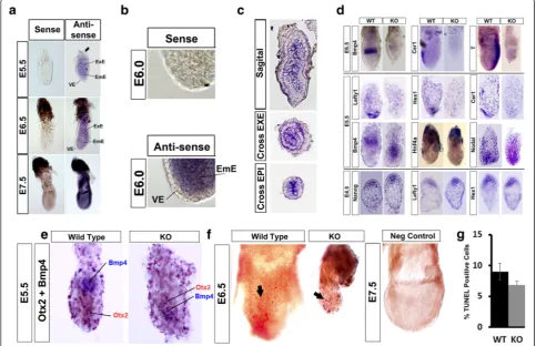
Ino80is expressed during embryonic development and in the adult. Towards this end, we performed in situ RNA hybridization (ISH) using both sense and antisense probes to the Ino80 transcript in E5.5- to E7.5-day embryos. Whole mount ISH of E5.5 to E6.5 with sense and anti-sense Ino80 probes showed widespread staining of
embryos with the antisense probe, but not the sense probe (Fig. 4a). The most intense staining with the antisense probe was observed in both the EmE and ExE, and com-paratively less staining was observed in the VE (Fig. 4a, b). The EmE contributes to all cell types of the embryo proper, whereas the ExE contributes to the placenta and umbilical cord [1]. To better visualizeIno80ISH, we sec-tioned E6.5-day embryos either sagittal or in cross section. Inspection of these sections showed that staining with the antisenseIno80probe is highest in the ExE and EmE, with less staining in the VE (Fig. 4c). Consistent with wide-spread expression in the early embryo, Northern blotting for Ino80 transcripts showed that it is expressed in all adult tissues analyzed (Additional file 5). The combination
Fig. 4Ino80is widely expressed in embryonic tissues and is essential for establishing the proximal-distal axis of the post-implantation embryo.
aWhole mount RNA in situ hybridization (ISH) of E5.5-, E6.5-, and E7.5-day embryos using either sense or antisenseIno80riboprobes. All embryos were photographed at identical magnification. Extraembryonic ectoderm (ExE), embryonic ectoderm (EmE) and visceral endoderm (VE) are designated.bIncreased magnification showing reduced antisenseIno80riboprobe staining in the visceral endoderm (VE) in E6.0-day embryos. Embryos were photographed at identical magnification.cFrozen sections of E6.5-day embryos stained whole mount with antisenseIno80 riboprobe. Sagittal and cross sections through the ExE and EPI were obtained for representative embryos.dWhole mount RNA in situ hybridization of E6.5-, E5.5-, and E4.5-day embryos was performed with antisense riboprobes for markers of tissue differentiation. Markers used include the ExE marker (Bmp4), EmE marker (Nodal), mesoderm marker (T), EPI marker (Nanog), VE marker (Hnf4a), and PrE/DVE/AVE markers (Cer1, Lefty1,andHex1). Embryos were photographed at identical magnification.eWhole mount ISH of E5.5-day embryos was performed with antisense probes to the ExE markerBmp4(purple color) and the EmE markerOtx2(magenta color). Embryos were photographed at identical magnification.f
of these results supports the conclusion that Ino80 is widely expressed in many tissues of post-implantation em-bryo and the adult.
Ino80KO embryos have a defective distal visceral endoderm and fail to gastrulate
To better understand whyIno80 KO embryos fail to de-velop, we used whole mount ISH to monitor marker ex-pression for several essential cell types of the gastrulating embryo. We focused our ISH on markers previously analyzed from our studies of Ino80 KO em-bryoid bodies (Fig. 3d). In the early embryo, gastrulation occurs at E6.5 when the posterior of the embryo differ-entiates into mesoderm and definitive endoderm [1]. Ini-tial ISH experiments documented a lack of mesoderm marker T expression in Ino80 KO embryos, suggesting that they do not differentiate mesoderm and likely do not gastrulate (Fig. 4d). Gastrulation requires the specifi-cation of the AVE and is the first organizer of the mam-malian embryo. The AVE secretes inhibitors to Tgfb and Wnt ligands including Cer, and its expression at E6.5 is a marker of the AVE [32]. At E6.5Cer1is not expressed in the Ino80 KO embryo, suggesting that Ino80 is re-quired for AVE establishment (Fig. 4d). In addition to the AVE, signaling molecules that originate from the ExE are essential for AVE establishment and gastrulation [33]. One such molecule is the Tgfb superfamily member Bmp4 [34]. ISH for Bmp4 transcripts showed that it is weakly expressed in the ExE in Ino80KO E6.5 embryos (Fig. 4d). The results from these analyses demonstrate that Ino80 KO embryos do not gastrulate and have de-fects in specifying both the AVE and ExE.
To determine if defects in gastrulation result from de-fects at earlier stages of development, we measured tis-sue differentiation in Ino80 KO E5.5 embryos. From these experiments, we observed that Bmp4, which is normally expressed in the ExE [34], is abnormally expressed in the EmE in Ino80 KO E5.5 embryos (Fig. 4d). To further confirm EmE Bmp4expression we performed a double ISH using Bmp4 and the EmE marker Otx2 [35]. From this experiment we show co-localization of Bmp4 and Otx2 expression in Ino80 KO E5.5-day embryos (Fig. 4e). To further monitor for de-fects in the EmE, we next measured Nodal expression. At E5.5 Nodal is expressed in the EmE where it pro-motes proliferation of the epiblast and differentiates the DVE [36]. In Ino80 KO E5.5 embryos, Nodal was nor-mally expressed suggesting that the EmE is partially spe-cified (Fig. 4d). Previous reports have shown that over expression of Bmp4 in the early embryo inhibits DVE differentiation [15]. To determine if Ino80 KO embryos have a defective DVE, we measured the expression of DVE markersCer1,Hex1,andLefty1[37]. None of these markers is expressed in E5.5 Ino80 KO embryos,
suggesting thatIno80KO embryos do not specify a DVE (Fig. 4d). Defects in DVE specification could be due to a general defect in the VE because we observed reduced expression of the extraembryonic VE marker Hnf4a in
Ino80 KO embryos (Fig. 4d) [29]. These results demon-strate that the DVE does not form inIno80KO E5.5 em-bryos, which could result from expression of the inhibitory molecule Bmp4 in the EmE and general de-fects in the VE.
Both the VE and the DVE originate from the PrE, which is specified during implantation at E4.5 [38]. To determine if defects in the DVE result from defects in the PrE, we stained Ino80 KO E4.5 embryos with the PrE markers,Lefty1 andHex1. From these experiments, we observed equivalent expression of Lefty1 and Hex1,
suggesting that the PrE ofIno80 KO embryos are speci-fied at E4.5 (Fig. 4c). In addition to the PrE, the E4.5 em-bryo has an EPI that is the precursor to the E5.5 EmE [38]. We next determined if the EPI is specified by stain-ing forNanog,a marker of the ICM and EPI. From these experiments we observed equivalent expression of
Nanog in the EPI, suggesting that it is specified inIno80
KO embryos (Fig. 4d).
We next stained E6.5-day embryos using TUNEL to determine if defects in development are due to increased apoptosis with Ino80 KO. From these experiments we observed no increase of TUNEL positive cells between wild-type andIno80KO embryos, suggesting that the ob-served defects are not due to increased cell death (Fig. 4f, g) and likely are due to reduced cell proliferation.
In combination, these expression studies demonstrate that Ino80 KO embryos do not gastrulate. Defects in mesoderm differentiation coincide with a lack of the AVE and its organizer activity, and are not due to in-creased cell death. The lack of an AVE at E6.5 is due to the inability of Ino80 KO embryos to specify a DVE at E5.5. The lack of a DVE coincides with the abnormal ex-pression ofBmp4 in the EmE, a known negative regula-tor of DVE differentiation [15]. Defects in the DVE are not likely due to defects prior to E5.5 because both the PrE and EPI appear to be normal in E4.5 Ino80 KO embryos.
Ino80 repressesBmp4expression during embryonic stem cell differentiation
embryonic development. When ESCs are grown under these conditions, they differentiate into EPI lineages in-cluding EmE and mesoderm precursors [39]. Under these conditions, we observe elevated expression of
Bmp4 in Ino80 KO ESCs (Fig. 5a). Elevated Bmp4 ex-pression coincides with reex-pression of DVE markers
Cer1, Hex, and Lefty1. Defects in DVE marker expres-sion are not due to general defects in endoderm differ-entiation because we observed equivalent expression of endoderm markers Gata4 and Gata6 (Fig. 5a). In em-bryonic tissue lineages Bmp4 is regulated by two up-stream enhancers (Fig. 5b) [40]. Chromatin immunoprecipitation (ChIP) experiments showed that Ino80 is localized to theBmp4promoter, but not the en-hancers, when ESCs are differentiated for six days in serum containing media lacking 2i + LIF (Fig. 5c). Under these same conditions, we next used formaldehyde assisted isolation of regulatory elements (FAIRE) to de-tect differences in open chromatin at the Bmp4 pro-moter in Ino80 KO ESCs. From these studies, we detected increased open chromatin ~1.5 Kb upstream of the Bmp4 transcription start site in Ino80 KO ESCs (Fig. 5d, e). These changes in chromatin structure at ~1.5 Kb upstream of Bmp4 correlated with increased H3K4me3 and SP1 occupancy (Fig. 5f, g). H3K4me3 is a well characterized mark of active promoters [41], and SP1 is a transcription factor known to activateBmp4 ex-pression [42]. In combination, these experiments suggest thatBmp4 is a direct target of Ino80 chromatin remod-eling activity with consequences to SP1 binding and
Bmp4 expression. From these studies, we present a model where Ino80 normally repressesBmp4expression in the EmE. When Ino80 is deleted,Bmp4expression in-creases, DVE differentiation is repressed, and embryonic development is stopped (Fig. 5h).
Discussion
In the last few decades it has been discovered that chro-matin remodeling complexes are essential for many as-pects of mammalian development [43, 44]. Several chromatin remodeling complexes are essential for pre-implantation development including SWI/SNF (Brg1
KO) [45], NURD [46], and TIP60/p400 [47] complexes. In contrast, several other complexes including NURF (Bptf KO) [48] and CHD7 [49] have post-implantation phenotypes, suggesting that they regulate specific devel-opmental pathways. Similarly, we showed thatIno80KO embryos implant, but fail to develop beyond the egg cy-linder stage. These findings contrast with the cell essen-tial functions for Ino80 in differentiated MEFs, where it regulates telomere structures and DNA replication [12, 14]. The viability of Ino80 KO ESCs, but not MEFs, could be due to differences in telomere structure and
DNA replication, which are known to occur between the cell types [50, 51].
To understand better how Ino80 regulates embryonic development, we derived Ino80 KO ESCs from pre-implantation embryos. We maintained Ino80 KO ESC under growth conditions that promote either ground state (serum + 2i + LIF) or metastable pluripotency (serum + LIF). Previous studies have shown that these culture conditions simulate growth conditions of the E3.5 blastocyst ICM (ground state) or E4.5 EPI and PrE (metastable state) [21].Ino80 KO ESCs remain undiffer-entiated at ground state pluripotency, but begin to show signs of differentiation in the metastable state, including loss of robust AP staining, and down-regulation of pluri-potency markersNanog, Oct4, Klf4,andEsrrb. These re-sults support a model where Ino80 is not required to maintain the pluripotent E3.5 blastocyst ICM in utero, but rather has essential functions in regulating cellular differentiation of the E4.5 peri-implantation embryo. Our conclusions deviate slightly from previous studies which document that Ino80 is required to maintain ESC pluripotency and pre-implantation development [13]. Differences in methods used to achieve loss of function could explain these contrasting reports. The use of a siRNA KD approach could deplete maternal Ino80 tran-scripts, thus revealing pre-implantation phenotypes [13]. Conversely, a genetic KO approach would leave maternal
Ino80transcripts intact, mask any pre-implantation phe-notypes, and allow the embryo to progress to post-implantation development (this study and [12, 14]).
In addition, the discrepancy could arise because Ino80 is required for Oct4, Nanog,and Sox2 expression in the metastable state (serum + LIF) [13], but not the ground state (2i + LIF) [this study]. The ability of 2i + LIF to mask the self-renewal defects of Ino80 KO ESC is not unprecedented as it has also been reported for Mbd3
KO ESC [46, 52]. In the metastable state, FGF4/MAPK signaling upregulates pro differentiation transcription factors promoting differentiation [53, 54]. It is plausible that depletion of Ino80, and the accompanying reduction in Oct4expression [13], could synergize with aberrantly expressed pro differentiation factors to promote differ-entiation in the metastable state. Alternatively, increased H3K4me3 modified nucleosomes at the promoters of pluripotency regulators in the ground state [21] could recruit a redundant set of activating histone readers, masking the Ino80 dependency ofOct4expression.
To characterize functions for Ino80 during early em-bryonic development we performed RNA ISH of several differentiation markers in peri- and post-implantation embryos. Our analysis documents that the DVE markers
Nodal and Bmp4 ligands [15]. Nodal was the first dis-covered ligand to promote the DVE [55], then later it was discovered that Bmp4 signaling repressed the DVE [15]. Bmp4 is normally expressed in the ExE, which is proximally located from the distal tip of the embryo, and future site of the DVE [34]. During egg cylinder expan-sion (E4.5-E5.5), the ExE is rapidly moved away from the distal tip of the embryo. During this period, the repres-sive effects of Bmp4 on the distal tip of the embryo sub-side due to the increased distance between the ExE and distal tip during egg cylinder expansion [34], and activat-ing Nodal signals become dominant due to increased
Nodalexpression in the EmE [55, 56]. In this model, the combined loss of Bmp4 signaling and increased Nodal signaling to the distal VE, establishes the DVE [15]. From our studies, we observed that Nodal is Ino80-independent during embryoid body differentiation and embryonic development. In contrast to Nodal, we ob-serve that Bmp4 is upregulated during embryoid body differentiation and in the E5.5 EmE. The abnormal ex-pression ofBmp4in the EmE could result in down regu-lation of DVE markers [15]. Alternatively, defects in the DVE could result from general defects in the VE as sup-ported by reduced Hnf4aexpression with Ino80 KO, or due to reduction in epiblast proliferation. Ino80 repres-sion ofBmp4expression in the EmE could be direct be-cause it localizes to, regulates chromatin structure and transcription factor occupancy at, the Bmp4 promoter during ESC differentiation. In addition to these factors, we observed reduced Bmp4expression in the ExE, sug-gesting that Ino80 could also be an activator ofBmp4in extraembryonic tissues. These activities could also be direct, and the study ofBmp4expression in differentiat-ing Ino80 KO trophoblast stem cells could test this hy-pothesis [57].
Conclusions
From the data presented in this manuscript, we show that Ino80 is required to pattern the early embryo. Using several models of development, we showed that Ino80
KO embryos upregulate Bmp4 expression, and when measurable, we observed a coincident repression of the DVE markers Cer1, Hex,and Lefty1. Because Bmp4 re-presses the DVE, we propose that the lack of a DVE in
Ino80 KO embryos is in part due to elevated Bmp4 ex-pression in the EmE. Ino80 functions in regulating
Bmp4 expression are likely direct because we observed specific Ino80 localization to its promoter in differentiat-ing ESCs. We also observed evidence that Ino80 re-models chromatin and regulates SP1 binding at the
Bmp4promoter under these same conditions. These re-sults support a model where Ino80 directly represses
Bmp4 expression in the EmE, and its repression is
essential for DVE establishment and ultimately a func-tional P-D axis (Fig. 5h).
Methods
Gene targeting and animal husbandry
The RPC121 mouse PAC library (MRC, UK, London) was screened using probe P2 internal to theIno80gene. Probe hybridization was performed using 0.25 M sodium phosphate pH 7.2, 1 mM EDTA, 7 % SDS at 65 °C over-night. Blots were washed three times for 30 minutes with 0.25XSSC, 0.1 % SDS at 65 °C. Five positive P1 clones were identified, and recombineering was used to move the chr2: 119,464,212-119,447,795 (mm10 assem-bly) DNA sequence from clone 386-C4 into DNDF-7. Recombineering was subsequently used to insert an up-stream loxp site and a downup-stream frt-Neo-frt-loxp se-lection cassette.
Linearized Ino80 targeting vector was electroporated into R1 ESC (129 Sv X 129Sv-CP F1) on feeder layers. Neomycin resistant clones were isolated and screened for successful Ino80 targeting using a BglI digest and probe P2, BglI digest and probe P3, and an EcoRV digest using probe P4. Primers used to amplify probes from genomic DNA are available in Additional file 6. South-ern blotting was performed as described for probe hybridization. Ino80 was successfully targeted in 1 of 456 neomycin resistant clones. Germ line transmission of Ino80 Floxed-Neo ESCs from two founder chimera mice was confirmed by Southern blotting.
The Ino80 Floxed-Neo mice were crossed to Cre del-eter (Jackson labs Cat#006054) and Flp deldel-eter strains (Jackson labs Cat#011065) to create Ino80 Floxed and
Ino80 KO alleles, respectively. Ino80 Floxed and Ino80
KO mice were then backcrossed to C57BL/6 J for five generations and confirmed Cre and Flp negative by PCR genotyping. Once backcrossed founder lines were estab-lished, the Ino80 Floxed mice were cryopreserved at Jackson Laboratory (Jackson labs Cat#027920). To create
The mice used in this study were housed in a specific pathogen-free facility at Virginia Commonwealth Uni-versity, Richmond VA, USA. Mice were maintained on a 12-hour light/dark cycle, and provided a low fat diet and hypochlorinated water ad labium throughout the dur-ation of the study. All experiments and animal proce-dures were approved by the Animal Care and Use Committee of Virginia Commonwealth University under protocol AD10000372 and its modifications.
Cell line isolation and maintenance
To createIno80KO ESCs, we harvested and maintained E2.5-day embryos in KSOM media until the blastocyst stage. Expanded blastocysts were hatched using Tyrodes solution (Sigma T1788) and allowed to attach and out-grow on gelatinized plates for seven days in defined 2i + LIF media without serum (Millipore Cat# SF016-100). After outgrowth, the ICM was removed with a fine glass pipette, dispersed with 0.25 % trypsin + EDTA, and ex-panded in 2i + LIF + 15 % ESC grade serum (Life Tech-nologies) media on gelatinized plates. Depending on the experiment, ESC lines were also grown in 2i + LIF growth media lacking serum (Millipore SF016-100), or a serum + LIF formulation (15 % ESC grade FCS (Life Technologies), DMEM, nonessential amino acids, 2 mM glutamine, 10 μM β-mercaptoethanol, penicillin, and streptomycin, 1000 U/ml LIF (Life Technologies)). ESC lines were genotyped by PCR methods (see above) and routinely maintained in serum + 2i + LIF containing media on gelatinized plates. Ino80 KO at the protein level was confirmed by Western blotting using a custom rabbit polyclonal antibody and standard techniques. For gene expression studies, cells were harvested from cul-ture using Tri-Reagent (Sigma) and total RNA purified according to the manufacture’s protocol. Quantitative RT-PCR was performed as described below.
Primary MEFs were produced from E12.5-day embryos using standard methods. MEFs were maintained in DMEM containing 10 % FCS, 2 mM glutamine, 1 mM nonessential amino acids, penicillin, and streptomycin. All experiments were performed using early passage (P0-1) fibroblasts.Ino80deletion was achieved by adding 10 ng/ml doxycycline to the growth media for two days. Following doxycycline exposure, the medium was re-placed and MEFs were maintained at subconfluence using standard tissue culture techniques. Efficiency of
Ino80 deletion was confirmed by PCR as described above. Analysis of Ino80 transcripts was accomplished by RT-PCR using Superscript II (Life Technologies) followed by amplification using Phusion polymerase (NEB). PCR fragments were cloned into pTOPO Blunt Zero vector (Life Technologies) and sequenced.
To stain for β-galactosidase activity, MEFs were washed in phosphate buffered saline (PBS) and then
fixed in 4 % buffered formaldehyde for five minutes. Fixed cells were washed and then stained in 40 mM cit-ric acid/sodium phosphate buffer, pH 6.0, containing 5 mM potassium ferrocyanide, 5 mM potassium ferri-cyanide, 150 mM sodium chloride, and 2 mM magne-sium chloride overnight with gentle rocking. Cells were washed in PBS and blue cells counted with a microscope.
Annexin V + 7AAD staining of ESC lines was performed using AnnexinV-PE (BD Biosciences Cat#556421) using the protocol provided by BD Biosciences.
To stain for AP activity, ESC colonies were first briefly washed in PBS. Colonies were then fixed for five minutes at room temperature in PBS containing 0.2 % glutaralde-hyde, 0.02 % NP-40, and 0.01 % sodium DOC. Colonies were then washed for ten minutes at room temperature in 100 mM Tris-HCl buffer, pH 9.5, containing 100 mM NaCl and 10 mM MgCl2. Colonies were then stained in
BM purple AP substrate (Roche Cat# 11442074001) until the desired level of staining was achieved. Cells were then washed in PBS containing 0.1 % tween-20 and 2 mM MgCl2. The plates were then air dried.
To determine doubling time cells were plated at 5 × 10⁵ cells per well in a 24-well plate and incubated for the indicated time points, trypsinized, and counted using a hemocytometer. Doubling time was calculated using standard equations.
Teratoma and embryoid body production
To produce teratomas, 1 × 10⁶ ESC were injected into the flank of NOD/SCID mice (Jackson Labs Cat# 001303), after which the tumors were allowed to grow for three weeks. The mice were then sacrificed, and the tumors were removed and frozen in liquid nitrogen. Tu-mors were sectioned in OCT medium using a cryostat, fixed in 10 % neutral buffered formalin, stained with hematoxylin and eosin, and visualized with light micros-copy using standard techniques.
RNA in situ hybridization and TUNEL assays
Embryos were dissected from timed pregnancies and photographed or prepared for in situ hybridization as de-scribed previously [48]. Alkaline phosphatase (AP) coupled anti-DIG Fab fragments and BM Purple substrate (Roche) were used to detect DIG labeled antisense probes as previously described [48]. For co-localization staining, DIG-Bmp4 and FITC-Otx2 antisense labeled probes were detected by AP conjugated anti-DIG Fab and anti-FITC Fab fragment, subsequently. The two probes were visual-ized by two different AP substrates; the Bmp4 detection used BM Purple (Roche) and Otx2 detection used INT/ BCIP (Roche) after inactivating the first AP activity (Otx2) with 4 % formaldehyde for 30 min.
To measure apoptosis E6.5 embryos were fixed for two hours at 4C, washed with PBST and kept in 100 % a methanol until use. Embryos were treated with 5:1 methanol:30 % hydrogen peroxide for three hours. Then, they were washed in a methanol and treated with 20 ug/ ul proteinase K for three minutes and fixed with 0.2 % glutaraldehyde in 4 % paraformaldehyde for 20 minutes at room temperature. Embryos were washed three times with PBST and treated with sodium borohydride for 20 minutes. Embryos were then washed with TdT buffer and apoptotic cells were detected by TUNEL staining using a fluorescein in situ cell death detection kit (Sigma). Apoptosis was calculated for each embryo by averaging the percentage TUNEL+ cells (brown nuclei) to all nuclei (DAPI staining) in three representative 50 um fields using microscopy.
Formaldehyde Assisted Isolation of Regulatory Elements (FAIRE)
FAIRE-qPCR was performed as described [58]. The qRT-PCR cycle threshold (Ct)value from the Input and FAIRE samples (control andIno80 KO) were normalized to signal from the reference region in each sample. Next, the relative enrichment for site in theBmp4promoter in comparison to the reference primer set was calculated using the comparative ΔΔCt method. Primers used for FAIRE are available as Additional file 6.
Chromatin immunoprecipitation (ChIP)
ChIP was performed as described previously [48]. Anti-bodies used include Ino80 (VCU23, custom rabbit poly-clonal), SP1 (Santa Cruz Biotech # sc-59), pan histone H3 (Abcam # ab1791), and H3K4me3 (Abcam # ab1012). DNA quantification was accomplished using SsoAdvanced universal SYBR Green supermix (Biorad) according to the manufacturer’s established protocol. Data collection was performed using a 7900HT Fast Real Time PCR System (Applied Biosystems) and quantified as % Input DNA for Ino80 or SP1, or as a ratio of %
Input H3K4me3/pan H3. The primers used for ChIP are available in Additional file 6.
Mouse Ino80 antibody VCU23 was raised in rabbits as a GST fusion to aa792-892 of Genbank # AAH59235.1. It was subsequently affinity purified by column chroma-tography using 6 × His-aa792-892. Specificity to Ino80 was confirmed by Western blotting using Ino80 control and KO ESC extracts. As a reference we observed identi-cal results using the commercially available Ino80 anti-body from ProteinTech (Cat# 18810-1-AP) (See Additional file 7).
Gene expression assays
Quantitative RT-PCR was performed on cDNA libraries converted using Superscript II (Life Technologies). cDNA libraries were amplified using SsoAdvanced uni-versal SYBR green supermix (Biorad) according to the manufacturer’s established protocols. Primers used for gene specific quantitative PCR were previously described [48]. Data collection was performed using a 7900HT Fast Real Time PCR System (Applied Biosystems) and theΔΔCt method was used to estimate transcript abun-dance relative to the normalization controlGapdh.
Northern blotting was performed as previously de-scribed [48]. A total of 1 ug of polyA+ RNA was re-solved by 1 % formaldehyde agarose electrophoresis and transferred to Hybond N+ (GE Health Sciences) using the manufacturer’s suggested protocol. The Ino80 probe was amplified from ESC cDNA library using Taq poly-merase, sequenced to confirm identity and labeled with P32 using random hexamer labeling. Detection was per-formed by phosphoimager.
Statistics
Standard deviation was used to calculate error bars throughout the study. P values were calculated using two tailed T tests. Number of replicates are designated in the figure legend.
Availability of supporting data
Data supporting the results of this article are available in Additional file 8.
Additional files
Floxed (Floxed) andIno80knockout (KO) alleles are shown. DNA fragment sizes obtained from restriction enzyme digestion and Southern blotting with P2, P3, and P4 probes are as expected from diagrams in panel a. c Diagram describing PCR-based genotyping strategy.Ino80alleles as defined in panel a are shown in a cartoon format with exons and introns not to scale. Position of the Loxp sites (red dots) and the Neo selectable marker are shown relative toIno80exons 2-4, which are designated by numbers. Positions of primers P1, P2, and P3 are shown as arrows designating 5′-3′orientation. PCR amplicons and their size are shown as black bars above amplifying primers. d Results from the PCR genotyping strategy using DNA from wild type and mice heterozygous for theIno80 Floxed-Neo,Ino80Floxed, andIno80KO alleles. (TIF 10353 kb)
Additional file 2: Figure S2.Molecular characterization of theIno80KO allele. a Efficiency of Cre-mediated excision ofIno80exons 2-4 from controlIno80Floxed/Floxed (Cre-Neg) andIno80Floxed/Floxed, TetO-Cre (TetO-Cre) primary MEFs were monitored using the PCR genotyping strategy. Cells were incubated with 10μg/ml doxycycline for two days, and then shifted to culture medium without doxycycline. DNA was taken for analysis prior to the addition of doxycycline (Day 0), and at two, four and ten days post-doxycycline addition. b Western blot analysis of Ino80 protein from total cell extracts prior to doxycycline addition (Day 0) and six days after initial doxycycline treatment using a custom Ino80 antibody. Ponceau S was used as a loading control. c Cartoon showing the position of RT-PCR primers used for characterizing theIno80KO allele transcript. Location of the forward primer in exon 1 and reverse primer in exon 7 are shown. The position of the loxp sites flanking exons 2-4 are shown as black boxes. The position of the initiating ATG is shown in exon 2. d RT-PCR results using primers shown in panel c on cDNAs converted from total RNA harvested from controlIno80Floxed/Floxed (Cre-Neg) and Ino80Floxed/Floxed, TetO-Cre (TetO-Cre) MEFs six days post-doxycycline treatment. PCR products were gel purified, cloned and sequenced. Exon composition of PCR fragments is shown. e Predicted reading frames for the KO-1 and KO-2 transcripts initiating from the first available ATG. (TIF 10038 kb)
Additional file 3:Genotyping results from intercross of heterozygous Ino80KO mice. Litters of mice or embryos at specified stages were collected and genotyped using the PCR-based genotyping strategy. Significant deviations from the expected genotype ratios were identified using a Chi-Square test (**p< 0.01). (XLSX 10 kb)
Additional file 4: Figure S4.Ino80 is not essential for the growth and viability of ESCs maintained in ground state pluripotency. a Growth curve of wild-type andIno80KO ESCs. 1.0 × 10⁴cells were seeded onto gelati-nized plates containing serum + 2i + LIF media. Numbers of trypan blue negative cells were counted every day over a four-day period. (Doubling Time, WT = 36 ± 4.5 hours, KO = 44 ± 4.2 hours; t test p≤0.05, N = 3 bio-logical replicates) b AnnexinV + 7AAD staining of wild-type andIno80KO ESCs maintained in serum + 2i + LIF (N= 3 biological replicates). (TIF 8494 kb)
Additional file 5: Figure S5.Ino80is widely expressed in adult tissues. Abundance ofIno80transcripts in a variety of adult tissues was determined by Northern blotting. Ethidium bromide stained rRNA serves as a loading control. (TIF 1828 kb)
Additional file 6:Primers used in this Study. (XLSX 12 kb)
Additional file 7: Figure S7.Western blotting confirming custom Ino80 antibody. Western analysis of Ino80 protein expression in wild-type and Ino80 KO ESCs using custom antibody VCU23 (a) and a commercially available Ino80 antibody (ProteinTech Cat# 18810-1-AP) (b). In each case Ponceau S was used as a loading control. (TIF 6874 kb)
Additional file 8:Supporting data. (XLSX 36 kb)
Competing interests
The authors declare that they have no competing interests.
Authors’contributions
ZQ, ZE, and JWL designed experiments. ZQ, ZE, VP, SA, DB, and JWL performed experiments, JWL wrote the manuscript. All authors read and approved the final manuscript.
Acknowledgements
The authors would like to thank members of the Landry, Valery, and Kuehn laboratories for critical reading of the manuscript and Kevin Hogan for editing the manuscript. Gene targeting and cryopreservation services for this project were provided by the VCU Massey Cancer Center Transgenic/Knock-Out Mouse Facility, supported in part, with funding from the NIH-NCI Cancer Center Support Grant P30 CA016059. This research was supported by start-up funds from the Department of Human and Molecular Genetics and Massey Cancer Center at Virginia Commonwealth University.
Received: 10 January 2016 Accepted: 16 February 2016
References
1. Tam PP, Loebel DA. Gene function in mouse embryogenesis: get set for gastrulation. Nat Rev Genet. 2007;8(5):368–81.
2. Kojima Y, Tam OH, Tam PP. Timing of developmental events in the early mouse embryo. Semin Cell Dev Biol. 2014;34:65–75.
3. Cutter AR, Hayes JJ. A brief review of nucleosome structure. FEBS Lett. 2015; 589(20 PtA):2914–22. doi:10.1016/j.febslet.2015.05.016.
4. Li G, Zhu P. Structure and organization of chromatin fiber in the nucleus. FEBS Lett. 2015;589(20 PtA):2893–904. doi:10.1016/j.febslet.2015.04.023. 5. Becker PB, Workman JL. Nucleosome remodeling and epigenetics. Cold
Spring Harb Perspect Biol. 2013;5(9):a017905.
6. Gerhold CB, Gasser SM. INO80 and SWR complexes: relating structure to function in chromatin remodeling. Trends Cell Biol. 2014;24(11):619–31. 7. Fritsch O, Benvenuto G, Bowler C, Molinier J, Hohn B. The INO80 protein
controls homologous recombination in Arabidopsis thaliana. Mol Cell. 2004;16(3):479–85.
8. Zhang C, Cao L, Rong L, An Z, Zhou W, Ma J, et al. The chromatin-remodeling factor AtINO80 plays crucial roles in genome stability maintenance and in plant development. Plant J. 2015;82(4):655–68. 9. Bhatia S, Pawar H, Dasari V, Mishra RK, Chandrashekaran S, Brahmachari V.
Chromatin remodeling protein INO80 has a role in regulation of homeotic gene expression in Drosophila. Genes Cells. 2010;15(7):725–35.
10. Neuman SD, Ihry RJ, Gruetzmacher KM, Bashirullah A. INO80-dependent regression of ecdysone-induced transcriptional responses regulates developmental timing in Drosophila. Dev Biol. 2014;387(2):229–39. 11. Moshkin YM, Chalkley GE, Kan TW, Reddy BA, Ozgur Z, van Ijcken WF, et al.
Remodelers organize cellular chromatin by counteracting intrinsic histone-DNA sequence preferences in a class-specific manner. Mol Cell Biol. 2012;32(3):675–88.
12. Lee HS, Lee SA, Hur SK, Seo JW, Kwon J. Stabilization and targeting of INO80 to replication forks by BAP1 during normal DNA synthesis. Nat Commun. 2014;5:5128.
13. Wang L, Du Y, Ward JM, Shimbo T, Lackford B, Zheng X, et al. INO80 facilitates pluripotency gene activation in embryonic stem cell self-renewal, reprogramming, and blastocyst development. Cell Stem Cell.
2014;14(5):575–91.
14. Min JN, Tian Y, Xiao Y, Wu L, Li L, Chang S. The mINO80 chromatin remodeling complex is required for efficient telomere replication and maintenance of genome stability. Cell Res. 2013;23(12):1396–413. 15. Yamamoto M, Beppu H, Takaoka K, Meno C, Li E, Miyazono K, et al.
Antagonism between Smad1 and Smad2 signaling determines the site of distal visceral endoderm formation in the mouse embryo. J Cell Biol. 2009;184(2):323–34.
16. Bouabe H, Okkenhaug K. Gene targeting in mice: a review. Methods Mol Biol. 2013;1064:315–36.
17. Hochedlinger K, Yamada Y, Beard C, Jaenisch R. Ectopic expression of Oct-4 blocks progenitor-cell differentiation and causes dysplasia in epithelial tissues. Cell. 2005;121(3):465–77.
18. Perl AK, Wert SE, Nagy A, Lobe CG, Whitsett JA. Early restriction of peripheral and proximal cell lineages during formation of the lung. Proc Natl Acad Sci U S A. 2002;99(16):10482–7.
19. Dimri GP, Lee X, Basile G, Acosta M, Scott G, Roskelley C, et al. A biomarker that identifies senescent human cells in culture and in aging skin in vivo. Proc Natl Acad Sci U S A. 1995;92(20):9363–7.
20. Ying QL, Wray J, Nichols J, Batlle-Morera L, Doble B, Woodgett J, et al. The ground state of embryonic stem cell self-renewal. Nature.
21. Marks H, Kalkan T, Menafra R, Denissov S, Jones K, Hofemeister H, et al. The transcriptional and epigenomic foundations of ground state pluripotency. Cell. 2012;149(3):590–604.
22. Hultman I, Bjork L, Blomberg E, Sandstedt B, Ahrlund-Richter L. Experimental teratoma: at the crossroad of fetal- and onco-development. Semin Cancer Biol. 2014;29:75–9.
23. Weitzer G. Embryonic stem cell-derived embryoid bodies: an in vitro model of eutherian pregastrulation development and early gastrulation. Handb Exp Pharmacol. 2006;174:21–51.
24. Coucouvanis E, Martin GR. BMP signaling plays a role in visceral endoderm differentiation and cavitation in the early mouse embryo. Development. 1999;126(3):535–46.
25. Cai KQ, Capo-Chichi CD, Rula ME, Yang DH, Xu XX. Dynamic GATA6 expression in primitive endoderm formation and maturation in early mouse embryogenesis. Dev Dyn. 2008;237(10):2820–9.
26. Ying QL, Nichols J, Chambers I, Smith A. BMP induction of Id proteins suppresses differentiation and sustains embryonic stem cell self-renewal in collaboration with STAT3. Cell. 2003;115(3):281–92.
27. Nemer G, Nemer M. Transcriptional activation of BMP-4 and regulation of mammalian organogenesis by GATA-4 and -6. Dev Biol. 2003;254(1):131–48. 28. Hebert JM, Basilico C, Goldfarb M, Haub O, Martin GR. Isolation of cDNAs
encoding four mouse FGF family members and characterization of their expression patterns during embryogenesis. Dev Biol. 1990;138(2):454–63. 29. Duncan SA, Manova K, Chen WS, Hoodless P, Weinstein DC, Bachvarova RF,
et al. Expression of transcription factor HNF-4 in the extraembryonic endoderm, gut, and nephrogenic tissue of the developing mouse embryo: HNF-4 is a marker for primary endoderm in the implanting blastocyst. Proc Natl Acad Sci U S A. 1994;91(16):7598–602.
30. Crossley PH, Martin GR. The mouse Fgf8 gene encodes a family of polypeptides and is expressed in regions that direct outgrowth and patterning in the developing embryo. Development. 1995;121(2):439–51. 31. Wilkinson DG, Bhatt S, Herrmann BG. Expression pattern of the mouse T gene and its role in mesoderm formation. Nature. 1990;343(6259):657–9. 32. Stower MJ, Srinivas S. Heading forwards: anterior visceral endoderm
migration in patterning the mouse embryo. Philos Trans R Soc Lond B Biol Sci. 2014;369(1657):20130546. doi:10.1098/rstb.2013.0546.
33. Richardson L, Torres-Padilla ME, Zernicka-Goetz M. Regionalised signalling within the extraembryonic ectoderm regulates anterior visceral endoderm positioning in the mouse embryo. Mech Dev. 2006;123(4):288–96. 34. Winnier G, Blessing M, Labosky PA, Hogan BL. Bone morphogenetic
protein-4 is required for mesoderm formation and patterning in the mouse. Genes Dev. 1995;9(17):2105–16.
35. Ang SL, Conlon RA, Jin O, Rossant J. Positive and negative signals from mesoderm regulate the expression of mouse Otx2 in ectoderm explants. Development. 1994;120(10):2979–89.
36. Robertson EJ. Dose-dependent Nodal/Smad signals pattern the early mouse embryo. Semin Cell Dev Biol. 2014;32:73–9.
37. Mesnard D, Guzman-Ayala M, Constam DB. Nodal specifies embryonic visceral endoderm and sustains pluripotent cells in the epiblast before overt axial patterning. Development. 2006;133(13):2497–505.
38. Stephenson RO, Rossant J, Tam PP. Intercellular interactions, position, and polarity in establishing blastocyst cell lineages and embryonic axes. Cold Spring Harb Perspect Biol 2012, 4(11): a008235. doi:10.1101/cshperspect.a008235. 39. Sharova LV, Sharov AA, Piao Y, Shaik N, Sullivan T, Stewart CL, et al. Global gene
expression profiling reveals similarities and differences among mouse pluripotent stem cells of different origins and strains. Dev Biol. 2007;307(2):446–59. 40. Zhu W, Yao X, Liang Y, Liang D, Song L, Jing N, et al. Mediator Med23
deficiency enhances neural differentiation of murine embryonic stem cells through modulating BMP signaling. Development. 2015;142(3):465–76. 41. Harikumar A, Meshorer E. Chromatin remodeling and bivalent histone modifications in embryonic stem cells. EMBO Rep. 2015;16(12):1609–19. 42. Ebara S, Kawasaki S, Nakamura I, Tsutsumimoto T, Nakayama K, Nikaido T,
et al. Transcriptional regulation of the mBMP-4 gene through an E-box in the 5′-flanking promoter region involving USF. Biochem Biophys Res Commun. 1997;240(1):136–41.
43. Ho L, Crabtree GR. Chromatin remodelling during development. Nature. 2010;463(7280):474–84.
44. Chen T, Dent SY. Chromatin modifiers and remodellers: regulators of cellular differentiation. Nat Rev Genet. 2014;15(2):93–106.
45. Bultman S, Gebuhr T, Yee D, La Mantia C, Nicholson J, Gilliam A, et al. A Brg1 null mutation in the mouse reveals functional differences among mammalian SWI/SNF complexes. Mol Cell. 2000;6(6):1287–95.
46. Kaji K, Nichols J, Hendrich B. Mbd3, a component of the NuRD co-repressor complex, is required for development of pluripotent cells. Development. 2007;134(6):1123–32.
47. Thomas T, Dixon MP, Kueh AJ, Voss AK. Mof (MYST1 or KAT8) is essential for progression of embryonic development past the blastocyst stage and required for normal chromatin architecture. Mol Cell Biol. 2008;28(16):5093–105. 48. Landry J, Sharov AA, Piao Y, Sharova LV, Xiao H, Southon E, et al. Essential
role of chromatin remodeling protein Bptf in early mouse embryos and embryonic stem cells. PLoS Genet. 2008;4(10):e1000241.
49. Hurd EA, Capers PL, Blauwkamp MN, Adams ME, Raphael Y, Poucher HK, et al. Loss of Chd7 function in gene-trapped reporter mice is embryonic lethal and associated with severe defects in multiple developing tissues. Mamm Genome. 2007;18(2):94–104.
50. Batista LF. Telomere biology in stem cells and reprogramming. Prog Mol Biol Transl Sci. 2014;125:67–88.
51. Momcilovic O, Navara C, Schatten G. Cell cycle adaptations and maintenance of genomic integrity in embryonic stem cells and induced pluripotent stem cells. Results Probl Cell Differ. 2011;53:415–58. 52. Rais Y, Zviran A, Geula S, Gafni O, Chomsky E, Viukov S, et al. Deterministic
direct reprogramming of somatic cells to pluripotency. Nature. 2013; 502(7469):65–70.
53. Lanner F, Lee KL, Sohl M, Holmborn K, Yang H, Wilbertz J, et al. Heparan sulfation-dependent fibroblast growth factor signaling maintains embryonic stem cells primed for differentiation in a heterogeneous state. Stem Cells. 2010;28(2):191–200.
54. Silva J, Nichols J, Theunissen TW, Guo G, van Oosten AL, Barrandon O, et al. Nanog is the gateway to the pluripotent ground state. Cell. 2009;138(4):722–37. 55. Brennan J, Lu CC, Norris DP, Rodriguez TA, Beddington RS, Robertson EJ.
Nodal signalling in the epiblast patterns the early mouse embryo. Nature. 2001;411(6840):965–9.
56. Kumar A, Lualdi M, Lyozin GT, Sharma P, Loncarek J, Fu XY, et al. Nodal signaling from the visceral endoderm is required to maintain Nodal gene expression in the epiblast and drive DVE/AVE migration. Dev Biol. 2015;400(1):1–9.
57. Roberts RM, Fisher SJ. Trophoblast stem cells. Biol Reprod. 2011;84(3):412–21. 58. Giresi PG, Lieb JD. Isolation of active regulatory elements from eukaryotic
chromatin using FAIRE (Formaldehyde Assisted Isolation of Regulatory Elements). Methods. 2009;48(3):233–9.
• We accept pre-submission inquiries
• Our selector tool helps you to find the most relevant journal • We provide round the clock customer support
• Convenient online submission • Thorough peer review
• Inclusion in PubMed and all major indexing services • Maximum visibility for your research
Submit your manuscript at www.biomedcentral.com/submit