DEEP BITE AND ITS TREATMENT MODALITIES WITH FIXED ORTHODONTICS
*1
Dr. Amit Bhardawaj,
1
Dean, Professor and Head, Department of Orthodontics, Modern Dental College
2
PG Student, Department of Orthodontics, Modern Dental College and Research Centre, Indore, India
3
Assistant Professor, Department of Orthodontics, Mo
ARTICLE INFO ABSTRACT
Deep bite is one of the most common malocclusions seen in children as well as adults that can occur along with other associated malocclusions. It is said to be one of t
when considered from the viewpoint of the future health of the masticatory apparatus and the dental units. The correction of the deep bite is one of the primary objectives of orthodontic treatment. Deep bite has been consid
successfully. Thus an optimal treatment of deep bite requires a proper diagnosis, a careful treatment plan, and an efficient appliance design.
Copyright©2019, Amit Bhardawaj et al. This is an open use, distribution, and reproduction in any medium, provided
INTRODUCTION
Malocclusion can occur in three planes of space i.e. sagittal, transverse and vertical plane. Dental vertical relationships can be divided into four major categories: anterior open bite, anterior deep bite, posterior open bite and posterior collapsed bite with overclosure. Since the same occlusal relations can result from skeletal jaw proportions or from infraeruption or supraeruption of teeth, the descriptive terminology should be as precise as possible in indicating whether jaw or tooth positions are basically at fault in producing a vertical problem. The maxillary dental arch being larger than the mandibular dental arch allows the maxillary anteriors to overlap the mandibular anteriors. This overlapping of the mandibular teeth occurs in both the horizontal as well as vertical direction. The horizontal overlap is called as overjet while the vertical overlap is termed overbite. Thus some degree of vertical overlapping or overbite is a normal feature of human
However, some patients present with excessive overbite. Thus a condition where there is an excessive vertical overlapping of the mandibular anteriors by maxillary anteriors is termed as deep bite. The excessive overbite is a complex orthodontic problem that may involve group of teeth or whole dentition, alveolar bone, mandible and maxilla and / or soft tissues of the face.
ISSN: 0975-833X
Article History:
Received 15th March, 2019
Received in revised form 17th April, 2019
Accepted 10th May, 2019 Published online 30th June, 2019
Citation: Dr. Amit Bhardawaj, Dr. Shalini Patel and
International Journal of Current Research, 11, (06), 4901
Key Words:
Deepbite, Deep-overbite.
*Corresponding author: Dr. Amit Bhardawaj
REVIEW ARTICLE
DEEP BITE AND ITS TREATMENT MODALITIES WITH FIXED ORTHODONTICS
Dr. Amit Bhardawaj,
2Dr. Shalini Patel and
3Dr. Kratika Mishra
Professor and Head, Department of Orthodontics, Modern Dental College
and Research Centre, Indore, India
PG Student, Department of Orthodontics, Modern Dental College and Research Centre, Indore, India
Assistant Professor, Department of Orthodontics, Modern Dental College and Research Centre, Indore, India
ABSTRACT
Deep bite is one of the most common malocclusions seen in children as well as adults that can occur along with other associated malocclusions. It is said to be one of t
when considered from the viewpoint of the future health of the masticatory apparatus and the dental units. The correction of the deep bite is one of the primary objectives of orthodontic treatment. Deep bite has been considered one of the most common malocclusions and the most difficult to treat successfully. Thus an optimal treatment of deep bite requires a proper diagnosis, a careful treatment plan, and an efficient appliance design.
open access article distributed under the Creative Commons Attribution provided the original work is properly cited.
Malocclusion can occur in three planes of space i.e. sagittal, transverse and vertical plane. Dental vertical relationships can be divided into four major categories: anterior open bite, , posterior open bite and posterior collapsed bite with overclosure. Since the same occlusal relations can result from skeletal jaw proportions or from infraeruption or supraeruption of teeth, the descriptive terminology should be indicating whether jaw or tooth positions are basically at fault in producing a vertical problem. The maxillary dental arch being larger than the mandibular dental arch allows the maxillary anteriors to overlap the f the mandibular teeth occurs in both the horizontal as well as vertical direction. The horizontal overlap is called as overjet while the vertical overlap is termed overbite. Thus some degree of vertical overlapping or overbite is a normal feature of human dentition. However, some patients present with excessive overbite. Thus a condition where there is an excessive vertical overlapping of the mandibular anteriors by maxillary anteriors is termed as deep bite. The excessive overbite is a complex orthodontic problem that may involve group of teeth or whole dentition, alveolar bone, mandible and maxilla and / or soft tissues of the
Overbite can be described in millimeter measurement or as percentage of overlap of the mandibular incisors by the maxillary incisors. The later method of description is preferred because of the variation in the size of the mandibular incisors. Depth of the bite must be related to the health of the soft tissue and supporting structures, temporomandibular joint function and effect of future skeletal growth on these factors.
Definition
Graber (Graber, 1975
condition of excessive overbite, where the vertical measurement between the maxillary and mandibular incisor margins is excessive when the mandible is brought into habitual or centric occlusion.
Bishara (Bishara, 1989
aberration in the overlap between the maxillary and mandibular incisors.
Bishara (Bishara, 1989
which the mandibular incisor crowns are excessively overlapped vertically by the maxillary incisors when the teeth are in centric occlusion.
International Journal of Current Research Vol. 11, Issue, 06, pp.4901-4907, June, 2019
DOI: https://doi.org/10.24941/ijcr.35723.06.2019
Dr. Amit Bhardawaj, Dr. Shalini Patel and Dr. Kratika Mishra, 2019. “Deep bite and its treatment modalities with fixed orthodontics 4901-4907.
Available online at http://www.journalcra.com
z
DEEP BITE AND ITS TREATMENT MODALITIES WITH FIXED ORTHODONTICS- A REVIEW
Dr. Kratika Mishra
Professor and Head, Department of Orthodontics, Modern Dental College
PG Student, Department of Orthodontics, Modern Dental College and Research Centre, Indore, India
dern Dental College and Research Centre, Indore, India
Deep bite is one of the most common malocclusions seen in children as well as adults that can occur along with other associated malocclusions. It is said to be one of the most deleterious malocclusion when considered from the viewpoint of the future health of the masticatory apparatus and the dental units. The correction of the deep bite is one of the primary objectives of orthodontic treatment. Deep ered one of the most common malocclusions and the most difficult to treat successfully. Thus an optimal treatment of deep bite requires a proper diagnosis, a careful treatment
Attribution License, which permits unrestricted
Overbite can be described in millimeter measurement or as percentage of overlap of the mandibular incisors by the ary incisors. The later method of description is preferred because of the variation in the size of the mandibular incisors. Depth of the bite must be related to the health of the soft tissue and supporting structures, temporomandibular joint function
ffect of future skeletal growth on these factors.
, 1975) has defined deep bite as a
condition of excessive overbite, where the vertical measurement between the maxillary and mandibular incisor margins is excessive when the mandible is brought into habitual or centric occlusion.
, 1989) defined deep bite as
aberration in the overlap between the maxillary and
, 1989), (Glossary) Malocclusion in
which the mandibular incisor crowns are excessively overlapped vertically by the maxillary incisors when
teeth are in centric occlusion.
INTERNATIONAL JOURNAL OF CURRENT RESEARCH
Classification of Deep Bite
A) Dentoalveolar deep bite (Graber, 1997) B) Skeletal deep bite (Graber, 1997)
Dentoalveolar deep bite (Graber, 1997)
The dentoalveolar deep bite is characterized by infraocclusion of molars and / or supraocclusion of incisors. The growth pattern usually is average or toward the vertical.
The deep overbite that is due to the infraocclusion of molars has the following features:
The molars are partially erupted.
The interocclusal space is large
A lateral tongue thrust or posture is present.
The distances between the maxillary and mandibular basal planes and occlusal plane are short.
Skeletal deep bite (Graber, 1997)
Skeletal deep bite is usually of genetic origin.
It is characterized by horizontal growth pattern.
The anterior face height is short particularly the lower facial third (ANS to Me), while the posterior facial height is long (S to Go).
The normal ratio of upper to lower anterior face height is 2 : 3, it is reduced in skeletal deep bite to a ratio of 2:2.5 or 2:2.8
Cephalometric examination reveals that most of the horizontal cephalometric planes i.e. sella-nasion, palatal, occlusal and mandibular are approximately parallel to each other.
The interocclusal clearance is usually small.
Skeletal deep bite is due to (upward and forward) anticlockwise rotation of mandible or clockwise rotation of maxilla or due to combination of both. (Fig. 4.1)
It is also due to vertical maxillary deficiency.
Akerly (1993) (1977) has classified deep bite into four basic
types
Type I - Mandibular incisors extrudes and impinges into the palate.
Type II - The mandibular incisors impinge into the gingival sulci of the maxillary incisors.
Type III - Both maxillary and mandibular incisors incline lingually with impingement of the gingival tissues of each other.
Type IV - The mandibular incisors move or extrude into the abraded lingual surfaces of the maxillary anterior teeth.
Treatment Objectives
In deciduous and mixed dentition (Geiger, 1974):
Occasionally, the developing dentition may be deficient in vertical growth, and interalveolar space sufficient for the eruption of the permanent molars is not available. It is desirable to wait for further growth.
In general, the deep overbite is a reflection of a more complex malocclusion or of a disturbance of the growth pattern of the dentofacial complex
In Permanent Dentition (Geiger, 1974): Treatment of deep
overbite may be necessary for the preservation and rehabilitation of the mature adult dentition. The extents of the tooth movement required as well as the rationale of treatment are dependent on the etiologic factors and the objectives of the treatment plan. While some cases require a more complex approach, some of these procedures fall within the scope of minor tooth movement.
Assistance in extensive restorative procedures (Geiger,
1974): Many cases requiring restoration have a deep overbite that may have been created or accentuated by the loss of posterior teeth and concomitant tipping into abnormal axial inclinations. Because these cases frequently have some degree of alveolar bone loss, merely increasing the clinical crowns of the teeth prosthetically would be contraindicated. Tipping the abnormally inclined teeth into upright positions can reduce the overbite without increasing the stress on the periodontium and will provide more physiologic distribution of the occlusal forces.
In management of temporomandibular joint disorders
(Geiger, 1974): An individual with a deep overbite may
present pain and dysfunction of the temporomandibular joint. Reduction of the overbite by extensive crown or bridge procedures is not indicated unless the joint syndrome is chronic.
Improvement of facial esthetics (Geiger, 1974)
The deep overbite may be reflected in the facial contours of the patient. Overclosure of the mandible shortens the distance from alae of the nose to the chin. The musculature and soft tissues of the face present a characteristic posture of tense contraction on closure. Deep lines appear at the corners of the lips that may cause a chronic cheiliosis. These esthetic problems may be complicated by the protrusion, spacing or crowding of anterior teeth that are frequently associated with deep overbite.
Treatment modalities of deep bite
Deep overbite can be corrected by following methods.
1. Intrusion of anterior teeth 2. Extrusion of posterior teeth 3. Combination of both. 4. Proclination of incisors. 5. Surgical.
Correction of deep bite with fixed appliance therapy
1) Bite opening with fixed bite plane 2) Bite opening with Begg’s technique 3) Bite opening with Edge-wise technique
4) Bite opening with pre-adjusted edge-wise technique 5) Bite opening with segmented arch technique 6) Bite opening with utility arches
7) Bite opening with bite opening and space closing arch wire
9) Bite opening with lingual arch
10) Bite opening with mini-screw anchorage system 11) Bite opening with magnets
Correction of deep bite with fixed bite planes
Modified nance appliance (Northcutt, 1995
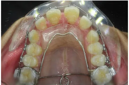
[image:3.595.310.558.51.161.2]version of the Nance appliance or palatal acrylic button is that which incorporates a bite plate. This appliance is invaluable as a treatment accelerator, because it allows immediate placement of brackets on the lower anterior teeth. It can also maintain vertical dimension in patients with early loss of primary teeth. In many TMJ cases, it is better than a removable splint because it frees the buccal occlusion from prematurities while allowing a natural path of lateral excursion.
Fig. 1. Modified Nance appliance with bite plane
Fixed bite planes with glass ionomercement
1996)
Bite Turbo: Glass ionomer cement is the material of choice
for fixed bite planes. Mix the cement powder with distilled water to a consistency thick enough for transportation on a spatula. Manual mixing should be kept to less than 30 seconds to allow sufficient working time, although this time can be extended by mixing on a frozen glass slab.
anterior bite plane, clean and dry the palatal te
enough cement to ensure that disclusion of the posterior teeth occurs when the lower incisal tips contact the bite plane. This can be checked by gently closing the mandible in centric relation until an indentation can be seen on the bite plane j before the molar make contact. The bite opening will then allow molar eruption without detracting from esthetics or speech. Once the desired opening has been achieved and the patient is in heavy rectangular archwire, the bite plane can be discarded.
Bonded Acrylic Lingual bite planes (direct technique
(Madsen, 1998): Acrylic extensions are bonded to the lingual
surfaces of the maxillary incisors, producing an intrusive effect or growth restrain on the incisors while allowing the extrusion of the posterior teeth. It is also called as lingual bite steps or bite turbos. (Fig. 2)
Correction of deep bite with begg’s technique ( 1998 and Jayade, 2001)
Unless otherwise indicated, the preference during bite opening is for incisor intrusion and for avoiding molar extrusion, because of the following reasons:
4903 International Journal
screw anchorage system
Correction of deep bite with fixed bite planes
Northcutt, 1995): A modified version of the Nance appliance or palatal acrylic button is that which incorporates a bite plate. This appliance is invaluable as a treatment accelerator, because it allows immediate placement of brackets on the lower anterior teeth. It can also maintain the vertical dimension in patients with early loss of primary teeth. In many TMJ cases, it is better than a removable splint because it frees the buccal occlusion from prematurities while allowing
Modified Nance appliance with bite plane
Fixed bite planes with glass ionomercement (Jackson,
Glass ionomer cement is the material of choice for fixed bite planes. Mix the cement powder with distilled h for transportation on a spatula. Manual mixing should be kept to less than 30 seconds to allow sufficient working time, although this time can be extended by mixing on a frozen glass slab. To construct an anterior bite plane, clean and dry the palatal teeth. Apply enough cement to ensure that disclusion of the posterior teeth occurs when the lower incisal tips contact the bite plane. This can be checked by gently closing the mandible in centric relation until an indentation can be seen on the bite plane just before the molar make contact. The bite opening will then allow molar eruption without detracting from esthetics or Once the desired opening has been achieved and the patient is in heavy rectangular archwire, the bite plane can be
onded Acrylic Lingual bite planes (direct technique)
Acrylic extensions are bonded to the lingual surfaces of the maxillary incisors, producing an intrusive effect or growth restrain on the incisors while allowing the extrusion erior teeth. It is also called as lingual bite steps or
Correction of deep bite with begg’s technique (Madsen,
[image:3.595.39.287.237.400.2]Unless otherwise indicated, the preference during bite opening avoiding molar extrusion,
Fig. 2. Bonded acrylic lingual bite planes
1. Incisor intrusion reduces or prevents gummy smile. 2. The intruded apices are placed in the thicker zone of
palatal and symphysealcancellous bone. This facilitates root movement required later, specially the root torquing movements.
3. Treatment of adult patients should be aimed at minimal molar extrusion: because bite opening by molar extrusion is not only unstable in these cases, but it also causes an incr
angle thereby worsening the Class II profile.
Whether to intrude both upper and lower incisors or only the upper ones, will be determined by esthetic requirement of case or in other words, exposure of incisors in repose and du speech and smile. In the first stage of Begg’s technique deep overbites are eliminated and overcorrected.
Mechanics of Intrusion
Arch wires: Teeth do not move on their own but move in
response to the forces imposed upon them by the active component of fixed appliances i.e. arch wires.
Sim: measured the forces generated by varying the wire size, degree of anchor bends and their location. It is apparent that 0.016” wire size is not suitable for producing higher force magnitudes, because the anchor bends will have to be made very steep (800 or more). This will result in a loss of molar control. According to Kesling0
opening the bite. Heavy arch wires tips the crowns of the upper and lower first permanent molars distally and the mesial marginal ridges of these molars elevates above the occlusal planes of dental arches. Also there is hardly any depression of six upper and six lower anterior teeth in their sockets. The bite is opened by elevating the mesial marginal ridges of anchor molars, and only to a small extent
teeth into their sockets. These elevated mesial marginal ridges of the molars sink back to the general plane of occlusion after active treatment, and the original deep overbite of upper and lower anterior teeth recurs to larg
Bite opening bends
The purpose of anchorage bend is
To give anchor molars the power to resist the forward pull of Class II elastics and also the space closing elastics.
To activate arch wires so that they depress the upper and lower anterior teeth in their sockets in order to open up anterior deep bites.
Many authors have proposed different sites for bite opening bends in the arch wires.
Journal of Current Research, Vol. 11, Issue, 06, pp.4901-4907, June, 201
Bonded acrylic lingual bite planes
Incisor intrusion reduces or prevents gummy smile. The intruded apices are placed in the thicker zone of palatal and symphysealcancellous bone. This facilitates root movement required later, specially the root torquing movements.
Treatment of adult patients should be aimed at minimal molar extrusion: because bite opening by molar extrusion is not only unstable in these cases, but it also causes an increase in the mandibular plane angle thereby worsening the Class II profile.
Whether to intrude both upper and lower incisors or only the upper ones, will be determined by esthetic requirement of case or in other words, exposure of incisors in repose and during In the first stage of Begg’s technique deep overbites are eliminated and overcorrected.
Teeth do not move on their own but move in response to the forces imposed upon them by the active
nt of fixed appliances i.e. arch wires.
measured the forces generated by varying the wire size, degree of anchor bends and their location. It is apparent that 0.016” wire size is not suitable for producing higher force magnitudes, because the anchor bends will have to be made more). This will result in a loss of molar 0.016” arch wires are better for opening the bite. Heavy arch wires tips the crowns of the upper and lower first permanent molars distally and the mesial ars elevates above the occlusal planes of dental arches. Also there is hardly any depression of six upper and six lower anterior teeth in their sockets. The bite is opened by elevating the mesial marginal ridges of anchor molars, and only to a small extent by depression of the anterior teeth into their sockets. These elevated mesial marginal ridges of the molars sink back to the general plane of occlusion after active treatment, and the original deep overbite of upper and lower anterior teeth recurs to large extent.
The purpose of anchorage bend is
To give anchor molars the power to resist the forward pull of Class II elastics and also the space closing elastics. To activate arch wires so that they depress the upper and
teeth in their sockets in order to open up
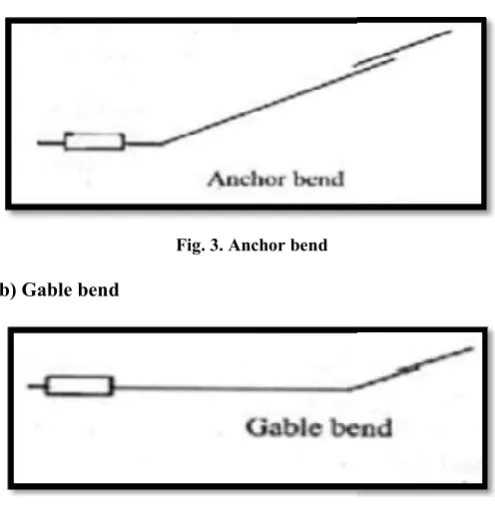
a) Anchor bend or conventional bite opening bends
Fig. 3. Anchor bend
b) Gable bend
Fig. 4. Gable bend
Hocevar’s modification: In the Hocevar’s modification a
[image:4.595.313.556.149.317.2]bend on either side of the canines are given. With this modification, the central incisors are subjected to intrusion while the canine and lateral incisors are extruded (canine more than lateral incisor) with respect to the central incisors (Fig. 12.19).
Fig. 5 Hocevar’s modification
Kameda’s modification: Using a simultaneous anchor and
gable bends, the canines and the premolars if engaged are extruded, while the lateral and central
[image:4.595.35.291.434.515.2] [image:4.595.34.289.635.726.2]progressively more intrusive effect. Thus the bite opening takes place by extrusion of posteriors and intrusion of anteriors (Fig. 6).
Fig. 6. Kameda’s modification
Dr. Jayade’s modification: A mild gingival curve is
incorporated in the anterior section, starting from mesial one cuspid circle to the corresponding point on the other side. This
a) Anchor bend or conventional bite opening bends
In the Hocevar’s modification a bend on either side of the canines are given. With this modification, the central incisors are subjected to intrusion while the canine and lateral incisors are extruded (canine more lateral incisor) with respect to the central incisors (Fig.
Fig. 5 Hocevar’s modification
Using a simultaneous anchor and gable bends, the canines and the premolars if engaged are extruded, while the lateral and centrals experience progressively more intrusive effect. Thus the bite opening takes place by extrusion of posteriors and intrusion of anteriors
Kameda’s modification
A mild gingival curve is incorporated in the anterior section, starting from mesial one cuspid circle to the corresponding point on the other side. This
should lift the archwire at the midpoint by about 3mm over the brackets.
Correction of deep bite with technique
Three-Piece Intrusion Arch (Shroff
Fig. 7. Three-piece base arch
A three-piece base arch is used to intrude the anterior segment. A heavy stainless steel segment (0.018” x 0.025”) with distal extensions below the center of resistance of anterior teeth is placed passively in the anterior brackets (Fig. 7). The distal extensions end 2 to 3 mm distal to the center of resistance of the anterior segment.
Correction of deep bite with utility arches
Ricketts Utility Arch: Late in the
and others attempted to counteract the tipping that occurred in the buccal segments in extraction cases by utilizing the supposedly immutable lower incisors as an anchor unit to hold the lower second bicuspids and molars upright in the retraction process. Round arch segments were laced from the lower molars and bicuspids to the lower incisors as the cuspids were retracted. It was noted that not only were the buccal segments maintained in an upright position, but that the lower incisors intruded with this light, continuous pressure. Later, there was a development of what is now classically described as the step down base arch or Rickett’s lower utility arch. Although the utility arch itself has not changed drastically in design from its early conception, understanding of its actions and reactions in the face of mechanics employed and growth response has been greatly enhanced.
Retraction Utility Arch: Most common type of utility arch
used in our practice is the retraction utility arch.( Figure 8) This type of utility arch can be used in both the mixed and the permanent dentitions to achieve retraction and intrusion of incisors. Loops are incorporated in the archwire anterior to the anterior vestibular segment. The loops add additional wire to the utility arch, facilitating retraction and intrusion of anterior teeth. This movement can be produced by activating arch wire in a manner like intrusion utility arch. The most common use of retraction utility arch is during the final steps of comprehensive edgewise treatment. A retraction utility arch can be used to close the space by retracting maxillary incisors. should lift the archwire at the midpoint by about 3mm over the
Correction of deep bite with preadjusted edgewise
Shroff, 1997 and Shroff, 1995)
piece base arch
piece base arch is used to intrude the anterior segment. A heavy stainless steel segment (0.018” x 0.025”) with distal extensions below the center of resistance of anterior teeth is placed passively in the anterior brackets (Fig. 7). The distal ensions end 2 to 3 mm distal to the center of resistance of
Correction of deep bite with utility arches
Late in the 1950’s Robert Ricketts13 and others attempted to counteract the tipping that occurred in the buccal segments in extraction cases by utilizing the supposedly immutable lower incisors as an anchor unit to hold the lower second bicuspids and molars upright in the retraction Round arch segments were laced from the lower molars and bicuspids to the lower incisors as the cuspids were retracted. It was noted that not only were the buccal segments maintained in an upright position, but that the lower incisors ight, continuous pressure. Later, there was a development of what is now classically described as the step-down base arch or Rickett’s lower utility arch. Although the utility arch itself has not changed drastically in design from its erstanding of its actions and reactions in the face of mechanics employed and growth response has been
This arch also provides the necessary intrusion that often must precede the retraction of anterior teeth.
Fig. 8 Retraction utility arch
Protraction Utility Arch (McNamara
protraction utility arch can be used to procline and intrude maxillary and mandibular incisors. (Figure 9) In the permanent dentition, this type of archwire commonly is used for proclining and intruding maxillary incisors in class II division 2 patients, especially in those with an impinging overbite. This arch wire is used to provide clearance between the maxillary and mandibular incisors to allow for placement of brackets on the mandibular dental arch. The protraction ut
used during the presurgical orthodontic phase of treatment to decompensate the position of the maxillary incisors in patients undergoing a mandibular advancement.
[image:5.595.311.558.203.363.2]
Fig. 8. Protraction utility arch
Simultaneous Intrusion and Retraction: The K
simultaneous Intrusion and Retraction) archwire given by Varun Kalra (Kalra, 1998), is a modification of the segmented loop mechanics of Burstone. It is a continuous 0.019” x 0.025” TMA archwire with closed 7mm x 2mm U
extraction sites (Image 9).
Fig. 9. K-SIR ARCH
4905 International Journal
[image:5.595.37.289.382.493.2]This arch also provides the necessary intrusion that often must
Fig. 8 Retraction utility arch
McNamara, 2001): The
protraction utility arch can be used to procline and intrude maxillary and mandibular incisors. (Figure 9) In the permanent dentition, this type of archwire commonly is used for in class II division 2 patients, especially in those with an impinging overbite. This arch wire is used to provide clearance between the maxillary and mandibular incisors to allow for placement of brackets on the mandibular dental arch. The protraction utility arch also is used during the presurgical orthodontic phase of treatment to decompensate the position of the maxillary incisors in patients
Protraction utility arch
The K-SIR (Kalra simultaneous Intrusion and Retraction) archwire given by is a modification of the segmented It is a continuous 0.019” x 0.025” TMA archwire with closed 7mm x 2mm U – loops at the
The connecticut intrusion arch
used for intrusion of incisors with CTA.
1. Insert a section of wire into the incisor bracket. 2. Choose the appropriate CTA.
3. Try in the CTA to determine the proper length. 4. Cut of the excess wire protruding from the molar
tubes, leaving 3 mm per side for cinch
5. Insert the posterior legs directly into the molar auxiliary tubes.
[image:5.595.314.561.603.765.2]6. Tie the CTA to the anterior segment between the central incisors.
Fig. 10. Connecticut intrusion arch
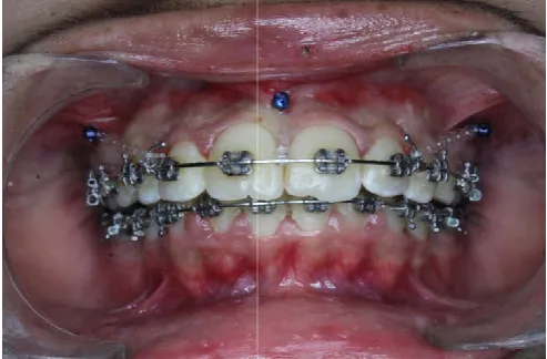
Correction of deep bite with mini screw anchorage system (Carano, 2004): Many malocclusions present a moderate to severe deep bite that requires pu
teeth as part of the treatment planning to level the occlusal plane. Using mini-screws simultaneous in the maxilla and in the mandible in young patients may be expensive unless the deep bite is severe enough to require absolute
those cases, the mini-screws can be used to reinforce the traditional orthodontic mechanics. To intrude the upper incisors, the best placement of mini
lateral incisors and the canines. Single implant in the midline can also be placed to inrude maxillary anterior segment. The placement of the mini-screws should be done after leveling and alignment, in order to maximize the interradicular space at the placement site (Image 11). In order to avoid tipping the upper incisors buccally during the intrusion, the end of the arch wire is cinched back (Ohnishi, 2005)
Fig. 11. Simultaneous intrusion and retraction with mini
Journal of Current Research, Vol. 11, Issue, 06, pp.4901-4907, June, 201
The connecticut intrusion arch: The following procedure is
used for intrusion of incisors with CTA.
Insert a section of wire into the incisor bracket. Choose the appropriate CTA.
Try in the CTA to determine the proper length. Cut of the excess wire protruding from the molar tubes, leaving 3 mm per side for cinch – back bends. Insert the posterior legs directly into the molar
Tie the CTA to the anterior segment between the
Connecticut intrusion arch
Correction of deep bite with mini screw anchorage system
Many malocclusions present a moderate to severe deep bite that requires pure intrusion of the anterior teeth as part of the treatment planning to level the occlusal screws simultaneous in the maxilla and in the mandible in young patients may be expensive unless the deep bite is severe enough to require absolute anchorage. In screws can be used to reinforce the traditional orthodontic mechanics. To intrude the upper incisors, the best placement of mini-screw is between the upper lateral incisors and the canines. Single implant in the midline can also be placed to inrude maxillary anterior segment. The screws should be done after leveling and alignment, in order to maximize the interradicular space at the placement site (Image 11). In order to avoid tipping the upper sors buccally during the intrusion, the end of the arch wire
).
Simultaneous intrusion and retraction with mini-screws
[image:5.595.35.291.606.774.2]Correction of deep bite with magnets (Blechman, 1985):
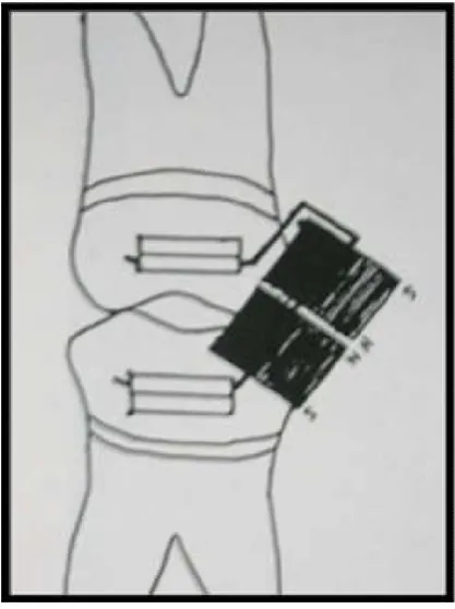
[image:6.595.59.268.155.435.2]Magnetic force is a valuable alternative to traditional force systems used in orthodontics. Magnets used in orthodontic are samarium cobalt (SmCo) magnets or aluminium – nickel – cobalt (AlNiCo) magnets. Samarium cobalt magnets are used more over aluminium – nickel – cobalt for their superior properties. They are used on buccal as well as lingual or palatal side, but commonly used on buccal side (Figure 12).
Fig. 12. Attracting magnets for bite opening
Certain physical properties of magnets, which have direct relation with generated force, are energy product, air gap, geometric configuration and size and mass of magnets.
Correction of deep bite with orthodontics and surgery: In
adults, it is important to decide in the beginning whether the treatment approach will be orthodontic or orthodontic – surgical. The reason is simple from the beginning; the treatment will be quite different with the two approaches. Will extractions be necessary? Probably yes with orthodontics alone, particularly in the upper arch but perhaps not in the lower, often not with surgical – orthodontic treatment perhaps in the lower arch but almost never in the upper. Cephalometric prediction of the results of alternative treatment is rational way to decide between orthodontic or orthodontic – surgical treatment. Fortunately, it is much easier to predict out comes when growth is not a variable.
The surgical treatment options in deep bite patients are
Orthodontics and interpositionalgenioplasty
Orthodontics and Inferior onlaymandibuloplasty
Orthodontics and mandibular advancement
Orthodontics and total subapical mandibular advancement
Orthodontics and inferior repositioning of maxilla and mandibular advancement
Orthodontics and combined maxillary and mandibular surgery
Retention and Relapse: Majority of patients require control of
the vertical overlap of incisors during retention of deepbite. This is accomplished most readily by using a removable upper retainer made so that the lower incisors will encounter the base plate of the retainer if they begin to slip vertically behind the upper incisors. The procedure is to build a potential bite plane into the retainer, which the lower incisors will contact if the bite begins to deepen. The retainer does not separate the posterior teeth. The long-term stability of deep overbite correction after orthodontic treatment is not well understood. In many cases the deep overbite returns as the maxillary and / or mandibular incisors overerupt following appliance removal. According to Lewis, Ricketts and Schudy,20 the maintenance of the overite is related to the torque or axial inclination of the incisors. If the maxillary and mandibular incisors are positioned too upright relative to one another after orthodontic treatment they will have an increased tendency to overerupt after appliance removal. The pattern of mandibular growth direction, as represented by the mandibular plane angulations could have an influence on overbite correction and stability. Patients with the more vertical growth pattern demonstrated smaller percentage of post retention overbite relapse as compared with patients with strong horizontal growth pattern.
Conclusion
The correction of deep bite is one of the primary objectives of orthodontic treatment. Deep bite can be corrected by many ways like intrusion of anteriors, extrusion of posteriors, combination of anterior intrusion and posterior extrusion, proclinatinganteriors or surgically. However, it should be decided which method will be more beneficial or which will improve the patients facial appearance and functional efficacy. Thus an optimal treatment of deep bite requires a proper diagnosis, a careful treatment plan and an efficient appliance design.
REFERENCES
Begg P.R., Kesling P.C. Begg Orthodontic therapy and technique 3rd Ed. W.B. Saunders Co. Philadelphia
Bench R.W., Gugino C.F., Hilgers J.J. Biopragressive therapy.
Bishara SE, Ziaja RR. Functional appliances: a review.
American Journal of Orthodontics and Dentofacial
Orthopedics. 1989 Mar 1;95(3):250-8.
Blechman AM. Magnetic force systems in orthodontics: clinical results of a pilot study. American Journal of Orthodontics. 1985 Mar 1;87(3):201-10.
Carano A, Velo S, Incorvati C, Poggio P. Mini-screw-anchorage-system (MAS) in the maxillary alveolar bone. J IndOrthod Soc. 2004 Apr 1;37:74-88.
Geiger A. Minor tooth movement in general practice. Mosby; 1974.
Graber TM, Rakosi T, Petrovic AG. Dentofacial orthopedics with functional appliances. Mosby Incorporated; 1997.
Graber TM, Swain BF. Current orthodontic concepts and techniques. WB Saunders Company; 1975.
Jackson S, Sandler PJ. Fixed biteplanes for treatment of deep bite. Journal of clinical orthodontics: JCO. 1996 May;30(5):283.
Kalra V. Simultaneous intrusion and retraction of the anterior teeth. Journal of clinical orthodontics: JCO. 1998 Sep;32(9):535-40.
Madsen R.Bonded acrylic lingual bite planes J Clin. Orthod 1998; 35 (5): 311- 317
McNamara JA, Brudon WL, Kokich VG. Orthodontics and dentofacial orthopedics. Needham Press; 2001.
Northcutt ME. The bite-plate Nance appliance. Journal of clinical orthodontics: JCO. 1995 Dec;29(12):760.
Ohnishi H, Yagi T, Yasuda Y, Takada K. A mini-implant for orthodontic anchorage in a deep overbite case. The Angle Orthodontist. 2005 May;75(3):444-52.
Parker CD, Nanda RS, Currier GF. Skeletal and dental changes associated with the treatment of deep bite malocclusion. American Journal of Orthodontics and Dentofacial Orthopedics. 1995 Apr 30;107(4):382-93.
Part 10 – Mechanics sequence for class II dinvision 1 cases J.
Clin. Orthod 1978; 12 (6): 427 – 439.
Part 11 – Mechanic Sequence for class II dinvision 2 cases J.
Clin. Orthod 1978; 12 (7): 505 – 521.
Part 2 – Principles of Bioprogressive therapy. J. Clin. Orthod.
1977; 11 (10): 661-682.
Part 7 – The utility arches and sectional arches in Bioprogressive therapy mechanics. J. Clin. Orthod 1978; 12 (3): 192 – 207.
Shroff B, Lindauer SJ, Burstone CJ, Leiss JB. Segmented approach to simultaneous intrusion and space closure: biomechanics of the three-piece base arch appliance.
American Journal of Orthodontics and Dentofacial
Orthopedics. 1995 Feb 28;107(2):136-43.
Shroff B, Yoon WM, Lindauer SJ, Burstone CJ. Simultaneous intrusion and retraction using a three-piece base arch. The Angle Orthodontist. 1997 Dec;67(6):455-61.
Stewart KL, Rudd KD, Kuebker WA. Clinical Removable Partial Prosthodontics. Implant Dentistry. 1993 Apr 1;2(1):59.
Zachrisson BU. Important aspects of long-term stability.
Journal of clinical orthodontics: JCO. 1997
Sep;31(9):562-83.