EXPRESSION OF B CELL POPULATION IN HASHIMOTO’S THYROIDITIS BY USING
*,1
Dr. Shaffy
1
Adesh Institute of Medical Sciences
2Meenakshi Medical College Hospital &
ARTICLE INFO ABSTRACT
Background:
thyroid and because of florid lymphocytic infiltrate it becomes difficult to distinguish it from lymphoma thereby posing a diagnostic challenge. The aim of the study was to demonstrate the demog
of B cell and T cell population using CD20 and CD 3 monoclonal antibodies for differentiating Hashimoto’s Thyroiditis with extensive lymphoplasmacytoid infiltrat
Materials and Methods:
Institute Hospital from September 2011 to July 2013. A total of 40 cases were studied. The specimen consisted of partial as well as total thyroidectomy specimens. Specimens were received in formalin and sections were processed
Eosin staining were done as routine in all the cases. Immunoreactivity with CD20 and CD3 was graded according to the scoring system and statistical analysis was carried out using SPSS vers 19.0 software with regression modules installed.
Results:
to be the most prevalent lesion followed by adenomatoid goitre with lymphoma to be least prevalent (1%). Immunohistochemical profile showed all cases (30) of Hashimoto’s thyroiditis to be positive for CD20 and CD3 positive in 29 of the 30 cases, all cases of Adenomatoid goitre (9) were negative for CD20 with single case of Lymphoma showing intensely positi
Conclusion:
Hashimoto’s thyroiditis from lymphoma. Cases with florid lymphocytic proliferation and any focus of atypical lymphocytes that
CD3 as well as Kappa and Lambda immunostaining.
Copyright©2017, Dr. Shaffyand Dr. Geetha Prakash.
unrestricted use, distribution, and reproduction in any medium, provided the original work is properly cited.
INTRODUCTION
Hashimoto’s Thyroiditis is organ specific autoimmune disease characterised by diffuse goitre, hypothyroidism and production of anti-thyroid microsomal and anti-thyroglobulin antibodies. It is an established risk factor for development of lymphoma in thyroid gland. Because of the florid lymphocytic infiltrate it becomes difficult to distinguish from a lymphoma thereby posing a diagnostic challenge to the pathologists. It has a polyclonal cell population in the lymphoid follicles composed of B cells positive for CD45RO and CD3. It is a well known fact that almost all thyroid lymphomas arise in
Hashimoto’s thyroiditis which can cause development of mucosa associated lymphoid tissue (MALT) lymphoma which
*Corresponding author: Dr. Shaffy
Adesh Institute of Medical Sciences and Research, Bathinda
ISSN: 0975-833X
Article History:
Received 17th October, 2016 Received in revised form 25th November, 2016 Accepted 12th December, 2016 Published online 31st January,2017
Citation: Dr. Shaffyand Dr. Geetha Prakash, 2017.
International Journal of Current Research, 9, (01), 45730
Key words:
Haematoxylin, Immunoreactivity, Adenomatoid goiter Scoring system and Statistical analysis.
RESEARCH ARTICLE
EXPRESSION OF B CELL POPULATION IN HASHIMOTO’S THYROIDITIS BY USING
IMMUNOHISTOCHEMISTRY
Dr. Shaffy
and
2Dr. Geetha Prakash
Adesh Institute of Medical Sciences and Research, Bathinda
Meenakshi Medical College Hospital & Research Institute, Kanchipuram
ABSTRACT
Background: Hashimoto’s Thyroiditis is an established risk factor for development of lymphoma in thyroid and because of florid lymphocytic infiltrate it becomes difficult to distinguish it from lymphoma thereby posing a diagnostic challenge. The aim of the study was to demonstrate the demographical and morphological profiles of Hashimoto’s thyroiditis and to ascertain the importance of B cell and T cell population using CD20 and CD 3 monoclonal antibodies for differentiating Hashimoto’s Thyroiditis with extensive lymphoplasmacytoid infiltrat
Materials and Methods: The study was conducted at the Meenakshi
Institute Hospital from September 2011 to July 2013. A total of 40 cases were studied. The specimen consisted of partial as well as total thyroidectomy specimens. Specimens were received in formalin and sections were processed and embedded in paraffin after gross examination. Haematoxylin and Eosin staining were done as routine in all the cases. Immunoreactivity with CD20 and CD3 was graded according to the scoring system and statistical analysis was carried out using SPSS vers 19.0 software with regression modules installed.
Results: Correlation between cytological and histological diagnosis revealed autoimmune thyroiditis to be the most prevalent lesion followed by adenomatoid goitre with lymphoma to be least prevalent ). Immunohistochemical profile showed all cases (30) of Hashimoto’s thyroiditis to be positive for CD20 and CD3 positive in 29 of the 30 cases, all cases of Adenomatoid goitre (9) were negative for CD20 with single case of Lymphoma showing intensely positive for CD20 and negative for CD3. Conclusion: Strict morphological and immunohistochemical criteria are required to differentiate Hashimoto’s thyroiditis from lymphoma. Cases with florid lymphocytic proliferation and any focus of atypical lymphocytes that masks earlylymphomatous transformation should be confirmed by CD20 & CD3 as well as Kappa and Lambda immunostaining.
This is an open access article distributed under the Creative Commons Att use, distribution, and reproduction in any medium, provided the original work is properly cited.
Hashimoto’s Thyroiditis is organ specific autoimmune disease goitre, hypothyroidism and production thyroglobulin antibodies. It is an established risk factor for development of lymphoma in thyroid gland. Because of the florid lymphocytic infiltrate it ish from a lymphoma thereby posing a diagnostic challenge to the pathologists. It has a polyclonal cell population in the lymphoid follicles composed It is a well known fact that almost all thyroid lymphomas arise in the setting of Hashimoto’s thyroiditis which can cause development of mucosa associated lymphoid tissue (MALT) lymphoma which
Adesh Institute of Medical Sciences and Research, Bathinda
can ultimately lead to an aggressive lymphoma. There is a need to learn how to identify individuals who are at increased risk of developing lymphoma and how to intervene against this risk. This has led to the immunohistochemistry and molecular techniques to confirm or exclude the diagnosis of lymphoma. Thus the aim of the study is to identify B cell and T cell population in Hashimoto’s Thyroiditis by means of immunohistochemistry using CD20 and CD3 monoclonal antibodies and to ascertain their usefulness in dif
Hashimoto’s Thyroiditis with extensive lymphoplasmacytoid infiltrate from MALT lymphoma.
Aims of the study
To demonstrate the demographical and morphological profiles of Hashimoto’s thyroiditis and to ascertain the importance of B cell and T cell population using CD20 and CD 3 monoclonal antibodies.
International Journal of Current Research
Vol. 9, Issue, 01, pp.45730-45735, January, 2017
INTERNATIONAL
OF CURRENT RESEARCH
2017. “Expression of b cell population in Hashimoto’s Thyroiditis
45730-45735.
EXPRESSION OF B CELL POPULATION IN HASHIMOTO’S THYROIDITIS BY USING
Research, Bathinda
Research Institute, Kanchipuram
is an established risk factor for development of lymphoma in thyroid and because of florid lymphocytic infiltrate it becomes difficult to distinguish it from lymphoma thereby posing a diagnostic challenge. The aim of the study was to demonstrate the raphical and morphological profiles of Hashimoto’s thyroiditis and to ascertain the importance of B cell and T cell population using CD20 and CD 3 monoclonal antibodies for differentiating Hashimoto’s Thyroiditis with extensive lymphoplasmacytoid infiltrate from MALT Lymphoma.
The study was conducted at the Meenakshi Medical College and Research Institute Hospital from September 2011 to July 2013. A total of 40 cases were studied. The specimen consisted of partial as well as total thyroidectomy specimens. Specimens were received in formalin and embedded in paraffin after gross examination. Haematoxylin and Eosin staining were done as routine in all the cases. Immunoreactivity with CD20 and CD3 was graded according to the scoring system and statistical analysis was carried out using SPSS version
Correlation between cytological and histological diagnosis revealed autoimmune thyroiditis to be the most prevalent lesion followed by adenomatoid goitre with lymphoma to be least prevalent ). Immunohistochemical profile showed all cases (30) of Hashimoto’s thyroiditis to be positive for CD20 and CD3 positive in 29 of the 30 cases, all cases of Adenomatoid goitre (9) were negative for
ve for CD20 and negative for CD3. Strict morphological and immunohistochemical criteria are required to differentiate Hashimoto’s thyroiditis from lymphoma. Cases with florid lymphocytic proliferation and any focus of masks earlylymphomatous transformation should be confirmed by CD20 &
is an open access article distributed under the Creative Commons Attribution License, which permits
ultimately lead to an aggressive lymphoma. There is a need to learn how to identify individuals who are at increased risk of developing lymphoma and how to intervene against this risk. This has led to the immunohistochemistry and molecular onfirm or exclude the diagnosis of lymphoma. Thus the aim of the study is to identify B cell and T cell population in Hashimoto’s Thyroiditis by means of immunohistochemistry using CD20 and CD3 monoclonal antibodies and to ascertain their usefulness in differentiating Hashimoto’s Thyroiditis with extensive lymphoplasmacytoid infiltrate from MALT lymphoma.
To demonstrate the demographical and morphological profiles and to ascertain the importance of B cell and T cell population using CD20 and CD 3 monoclonal
INTERNATIONAL JOURNAL OF CURRENT RESEARCH
MATERIALS AND METHODS
The study was conducted at the Meenakshi Medical College and Research Institute Hospital from September 2011 to July 2013. A total of 40 cases were studied. The specimen consisted of partial as well as total thyroidectomy specimens. Specimens were received in formalin and sections were processed and embedded in paraffin after gross examination. Haematoxylin and Eosin staining were done as routine in all the cases. Immunoreactivity with CD20 and CD3 was graded according to the scoring system and statistical analysis was carried out using SPSS version 19.0 software with regression modules installed.
Inclusion criteria
Cases from september 2011 to July 2013 were included in the study.
Exclusion criteria
Cases showing toxic changes were excluded.
RESULTS
[image:2.595.299.558.71.268.2]The study was conducted at the Meenakshi Medical College and Research Institute Hospital from September 2011 to July 2013 on a total of 40 cases. Of the 40 cases, Hashimoto’s Thyroiditis followed by Adenomatoid Goitre and Lymphoma with the maximum occurring in the age group of 40-49 years. Female preponderance was found. Correlation between cytological and histological diagnosis revealed autoimmune thyroiditis to be the most prevalent lesion followed by adenomatoid goitre with lymphoma to be the least prevalent (1 %). Immunohistochemical profile showed all cases (30) of Hashimoto’s thyroiditis to be positive for CD20 and CD3 positive in 29 of the 30 cases, all cases of Adenomatoid goitre (9) were negative for CD20 with single case of Lymphoma showing intensely positive for CD20 and negative for CD3.
Table 1. Distribution of Total No. of cases
S. No Diagnosis Number Percentage (%)
1. Hashimoto’s Thyroiditis 30 75
2. Adenomatoid Goitre 09 22
[image:2.595.305.555.310.568.2]3. Lymphoma 01 03
Figure 1. Distribution of total cases
Table 2. Sex wise distribution of lesions
S. No Diagnosis Male Female
1. Hashimoto’s Thyroiditis 2(6.6) 28(93.4)
2. Adenomatoid Goitre 3(33.3) 6(66.7)
3. Lymphoma 1(100) 0(0)
Figure 2. Sex distribution in total cases
Table 3. Correlation of Anatomical Diagnosis with Histopathological Diagnosis
S.No. Anatomical Diagnosis
Histopathological Diagnosis
No. of Cases (%)
Total (%) 1 Diffuse Hashimoto’s Thyroiditis 22 (73.4) 26 (65)
Adenomatoid Goiter 3 (33.3)
Lymphoma 1(100)
2 Solitary Hashimoto Thyroidits 7 (23.3) 11(27.5) Adenomatoid Goiter 4 (44.4)
3 Multinodular Hashimot Thyroidits 1 (3.3)
03 (7.5) Adenomatoid Goiter 2 (22.3)
4 Total 40 40
Figure 3. Anatomical and Histopathological correlation
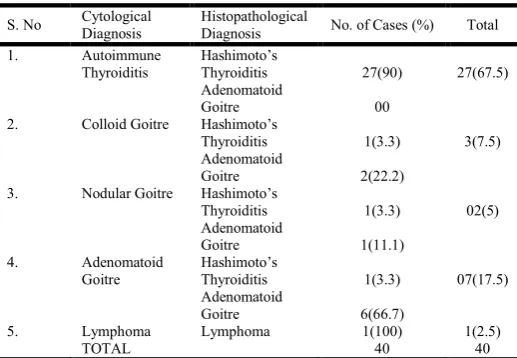
Table 4. Correlation of Cytological Diagnosis with Histopathological Diagnosis
S. No Cytological Diagnosis
Histopathological
Diagnosis No. of Cases (%) Total
1. Autoimmune
Thyroiditis
Hashimoto’s
Thyroiditis 27(90) 27(67.5)
Adenomatoid
Goitre 00
2. Colloid Goitre Hashimoto’s
Thyroiditis 1(3.3) 3(7.5)
Adenomatoid
Goitre 2(22.2)
3. Nodular Goitre Hashimoto’s
Thyroiditis 1(3.3) 02(5)
Adenomatoid
Goitre 1(11.1)
4. Adenomatoid
Goitre
Hashimoto’s
Thyroiditis 1(3.3) 07(17.5)
Adenomatoid
Goitre 6(66.7)
5. Lymphoma Lymphoma 1(100) 1(2.5)
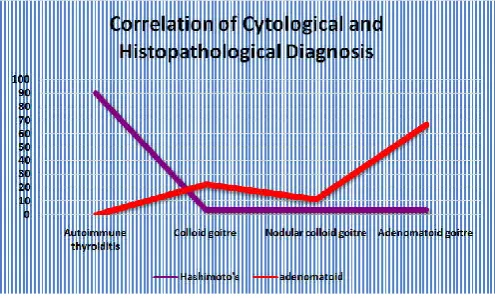
[image:2.595.39.286.540.766.2] [image:2.595.304.563.620.799.2]Figure 4. Cytological and Histopathological correlation
Table 5. Expression of CD20 and CD3 Positivity in total No. of cases
S.No. Antibody Histopathological
Diagnosis Positive Negative Total
1. CD20 Hashimoto’s
Thyroiditis
30(100) 00 30
Adenomatoid Goitre
00 09(100) 09
Lymphoma 1(100) 00 1
TOTAL 31 9 40
2. CD3 Hashimoto’s
Thyroiditis
29(96.6) 1(3.4) 30
Adenomatoid Goitre
00 09(100) 09
Lymphoma 00 1(100) 1
[image:3.595.310.553.267.439.2]TOTAL 40 40
Figure 5. CD20 Profile in total cases
Figure 6. CD3 Profile in total cases
[image:3.595.36.291.269.587.2]Figure 7. Intensity of CD20 Positivity
Figure 8. Intensity of CD3 Positivity
Gross pictures
[image:3.595.318.546.505.680.2]Microscopy
Figure 14. H & E (40X) Hashimoto’s Thyroiditis
DISCUSSION
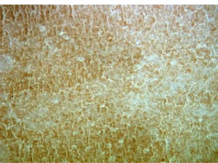
[image:3.595.42.286.619.783.2]Figure 15. IHC(40x) CD20 Positivity in Hashimoto’s Thyroiditis
[image:4.595.56.271.238.402.2]Figure 16. IHC(100X) CD20 Positivityin Hashimoto’s Thyroiditis
Figure 17. IHC(100x) CD3 Positivity in Hashimoto’s Thyroiditis
[image:4.595.322.546.242.400.2]Figure 18. H & E(100x): Adenomatoid goitre
[image:4.595.54.270.428.585.2]Figure 19. IHC(100x) CD20 Negativity in Adenomatoid goiter
Figure 20. IHC(100x) CD3 Negativity in Adenomatoid goitre
Figure 21. H & E(100x) Lymphoma showing effacement by monotonous population of lymphocytes
[image:4.595.322.546.430.592.2] [image:4.595.58.271.616.777.2] [image:4.595.325.542.621.783.2]The various types of thyroiditis encompasses a heterogenous group of disorders ranging from acute bacterial to chronic autoimmune diseases. In clinical practice, inflammatory disease of the thyroid may be commonest of the thyroid abnormalities encountered.
Classification of thyroiditis (Singer, 1991)
1. Acute thyroiditis
2. Subacute thyroiditis
a. Subacute granulomatous thyroiditis
b. Subacute lymphocytic thyroiditis
3. Chronic thyroiditis
a. Hashimoto’s thyroiditis
b. Riedel’s thyroiditis
Hashimoto’s thyroiditis can be graded pathologically as (Lakshman Rao and Reddy, 1991)
Grade I – Showing askanization of follicular cells and
lymphocytic infiltration.
Grade II – Askanization of the follicular cells and
lymphocytic infiltration with or without
lymphoid follicle formation, destruction of follicles and varying degrees of necrosis.
Grade III– Extensive fibrosis in the gland with almost total
disappearance of the follicle.
This is the stage of “burn out” disease.
In our study 40 patients were included, 30 of which were diagnosed as Hashimoto thyroiditis, 9 were diagnosed as adenomatoid goitre and 1 case diagnosed as lymphoma. Strong female preponderance was seen in concordance with the
studies by Sharma et al, Fenn et al and Kazem et al. (Amani,
2011; Sharma et al., 1990; Fenn, 1980). In our study the age
incidence ranged from 18 years to 58 years, youngest being a 18 year old girl and oldest being 58 year old woman. The mean age in study was 38.15 years with highest incidence being in between 40 -50 years. The mean age in Hashimoto’s thyroiditis
was 41.2 years. Kazem et al and Fenn et al. showed similar
findings and was in concordance with our study (Sharma, 1990; Fenn, 1980). In our study majority of cases presented as diffuse goitre (65%) followed by solitary nodule (27.5%) and multinodular goitre (7.5%) which was similar to the findings in
studies by Kazem et al and KusumKaplia et al. (Amani, 2011;
Kapila et al., 1995). Histopathology of Hashimoto’s thyroiditis
revealed lymphoid infiltrate arranged in lymphoid follicles with interfollicular small round lymphocytes, plasma cells, scattered lymphoplasmacytoid cells and a few large transformed cells. Hurthle cell metaplasia were seen in almost all the cases. These findings were in concordance to the study
conducted by Kazem et al. (Amani, 2011). Several studies
have linked certain autoimmune and chronic inflammatory conditions to an increased occurrence of lymphoma. The magnitude of the average lymphoma risk in each disorder differs considerably among various studies. It was found that almost all thyroid lymphomas arised in the setting of Hashimoto’s thyroiditis which induced reactive lymphoid proliferation that lead to the development of MALT lymphoma ultimately leading to aggressive lymphoma (Amani, 2011). The coexistence of reactive and neoplastic processes in the thyroid causes difficulty in diagnosing mucosa associated
lymphoid tissue lymphoma (MALTOMA) using cytology and
histology. This has led to the advancement of
immunohistochemistry amd molecular techniques to confirm and exclude the diagnosis (Amani, 2011). We used immunohistochemical markers (CD20 and CD3) to evaluate B cell and T cell population in Hashimoto’s thyroiditis. Our study showed 29 cases to be composed of small lymphocytes most of them are arranged as lymphoid follicles with CD20 positivity and CD3 negativity. The interfollicular lymphoid infiltrate were composed of T cells predominantly showing CD3 positivity and CD20 negativity. Our findings was in
concordance with the studies conducted by Saxena et al.
(2004) and Kazem et al. (Amani, 2011). All the cases of HT
showed an admixture of B and T lymphocytes with CD20 highlighting the germinal centres while CD3 demonstrated the well developed mantle zone and the interfollicular population
(His et al., 1998). Our study revealed one case of HT showing
focal effacement of architecture with CD20 positivity and CD3 negativity thereby raising the possibility of harbouring a
lymphomatous clone. D’Antonio et al also reported the
existence of a minute focus of extranodal marginal zone lymphoma in case of HT thereby necessitating careful examination to disclose small foci of lymphomatous
transformation (D’Antonio et al., 2009). Our study showed one
case of thyroid lymphoma which was used as positive control. Microscopy showed total effacement of thyroid architecture by lymphoid infiltrates.
Conclusion
Hashimoto’s thyroiditis is a common cause of goitrous enlargement of thyroid gland with hypothyroidism. It has a varied clinical presentation and can present as diffuse goitre, multinodular goitre or a solitary nodule. It is an established risk factor for the development of lymphoma but it differs both histopathologically and immunohistochemically from thyroid lymphoma and diagnosis is generally made by FNAC, antibody titre and histopathology. One case of thyroid lymphoma showed CD20 positive and CD3 negative thereby confirming B cell nature of the lymphoma. Clonal B cell proliferation in Hashimoto’s thyroiditis has been detected by means of immunohistochemistry. One case showed CD20 positivity and CD3 negativity thereby raised the possibility of a clone being harboured. Kappa and Lambda immunostaining is required to demonstrate clonal expansion. Hence to differentiate Hashimoto’s Thyroiditis from lymphoma strict morphological and immunohistochemical criteria has to be made. Cases of florid lymphocytic hyperplasia with focus showing atypical lymphocytes may mask early lymphomatous transformation thereby necessitating the use CD20 and CD3.
Kappa and Lambda immunostaining contributes in
demonstrating clonality and in ruling out the possibility of lymphoma arinsing in a background of Hashimoto’s Thyroiditis. Strict morphological and immunohistochemical criteria are required to differentiate Hashimoto’s thyroiditis from lymphoma. Cases with florid lymphocytic proliferation and any focus of atypical lymphocytes that masks earlylymphomatous transformationshould be confirmed by CD20 & CD3 as well as Kappa and Lambda immunostaining.
REFERENCES
Amani HK. 2011. Histopathologic and immunohistochemical
features of Hashimoto thyroiditis. Indian J Pathol
D’Antonio A, Caleo A, Licci S, Addesso M, De Palma M, Boscaino A, Nappi O. 2009. A minute focus of extranodal marginal zone B-cell lymphoma arising in Hashimoto thyroiditis diagnosed with PCR after laser capture
microdissection: a case report. Thyroid Res., 2(1):9.
Fenn AS. 1980. Job CK and Elizabeth George. Hashimoto’s
Thyroiditis. Indian J Surg., 4:123-125.
Hsi ED. Singleton TP, Svoboda SM, Schnitzer B, Ross CW. 1998. Characterization of the lymphoid infiltrate in Hashimoto’s thyroiditis by immunohistochemistry and Polymerase chain reaction for immunnoglobulin heavy
chain gene rearrangement. Am J ClinPathol., 110(3):
327-33.
Kapila K, Sathar SA, Al-Rabah NA, Prahash A, Seshadri MS. 1995. Chronic lymphocytic (Hashimoto's) thyroiditis in
Kuwait diagnosed by fine needle aspirates. Ann Saudi
Med., 15(4): 363-6.
Lakshman Rao KM. and Reddy SS. 1991. Hashimoto’s
disease- A clinicopathological study. Indian J Surg.,
53(8-9): 338-342.
Saxena A, Alport EC, Moshynska O, Kanthan R, Boctor MA. 2004. Clonal B cell populations in a minority of patients
with Hashimoto’s thyroiditis. J ClinPathol., 57(12):
1258-63.
Sharma AK, Paliwal RK and Pendse AK. 1990. Hashimoto
Thyroiditis clinical Review. J Post Grad Med.,
36(2):87-90.
Singer PA. 1991. The Medical Clinics of North America, 75(1):61-71.