1
IMMUNOMODULATION WITH
SELF-CROSSLINKED POLYELECTROLYTE
MULTILAYER BASED COATINGS
Helena Knopf-Marques,¥, Sonali Singhǂ, Su Su Htweǂ, Lucie WolfovaΘ, Radovan Buffa Θ, Jalal Bacharoucheᶲ, Grégory Franciusᶲ, Jean-Claude Voegel,¥, Pierre Schaaf,¥,, Amir M.
Ghaemmaghamiǂ, Nihal Engin Vrana ,£, Philippe Lavalle ,¥*
INSERM UMR 1121, 11 rue Humann, 67085 Strasbourg, France
¥Faculté de Chirurgie Dentaire, Fédération de Médecine Translationnelle de Strabourg,
Université de Strasbourg, 3 rue Sainte Elisabeth, 67000 Strasbourg, France
ǂDivision of Immunology, School of Life Sciences, Faculty of Medicine and Life Sciences,
University of Nottingham, Queen’s Medics, al Centre, Nottingham, NG7 2UH, UK
Θ Contipro a.s., Dolni Dobrouc 401 561 02 Dolni Dobrouc, Czech Republic
ᶲLaboratoire de Chimie Physique et Microbiologie pour l'Environnement CNRS UMR7564, 405
rue de Vandoeuvre, 54600 Villers-les-Nancy, France
Institut Charles Sadron, CNRS UPR 22, 23 rue du Lœss, 67034 Strasbourg, France
2 *Corresponding Author: [email protected]
KEYWORDS. Polyelectrolytes multilayers, poly(lysine), hyaluronic acid derivatives, cytokine, controlled release, monocytes, immunomodulation, macrophages
ABSTRACT. This study aims to design an optimal polyelectrolyte multilayer film of poly-L-lysine (PLL) and hyaluronic acid (HA) as an anti-inflammatory cytokine release system in order to decrease the implant failure due to any immune reactions. The chemical modification of the HA with aldehyde moieties allows self-crosslinking of the film and an improvement in the mechanical properties of the film. The crosslinking of the film and the release of immunomodulatory cytokine (IL-4) stimulate the differentiation of primary human monocytes seeded on the films into pro-healing macrophages phenotype. This induces the production of anti-inflammatory cytokines (IL1-RA and CCL18) and the decrease of pro-inflammatory cytokines secreted (IL-12, TNF-α and IL-1β). Moreover, we demonstrate that crosslinking PLL/HA film using HA-aldehyde is already effective by itself to limit inflammatory processes. Finally, this functionalized self-crosslinked PLL/HA-aldehyde films constitutes an innovative and efficient candidate for immunomodulation of any kind of implants of various architecture and properties.
Introduction
3 electrostatic attraction between polyanions and polycations, the advantage of this method is the possibility of preparing ultra-thin and well-defined films.2, 3 The spontaneous sequential adsorption of dissolved cationic and anionic polyelectrolytes on the substrate leads to the formation of ordered multilayer assemblies on the solid substrate surface.4 These multilayers are widely used in biotechnology as controlled release systems, cell growth surfaces, anti-adhesive coatings and stimuli responsive surfaces. As the system is composed of charged molecules, it is ideal to control the release of charged bioactive agents such as cytokines and the strength of electrostatic interactions between the film and the cytokine should be a key parameter to determine the rate of release.5, 6
4 reaction a long incubation time is required (at least 12 hours) and also some extensive rising steps after it, in order to eliminate excess products that can be toxic.19
5 any addition of elements or stimuli, will constitute an advantage compared to PEMs crosslinked with the chemicals like EDC/NHS for example. HA-aldehyde and PLL can interact i) via formation of ionic pairs where the cations are the protonated amino groups of PLL and the anions are the carboxylic groups of HA, and ii) via formation of a covalent imine bond –CH=N- between aldehyde moities of HA and amine groups of polylysine. HA-aldehyde and PLL interactions (ionic and covalent) are summarized in Scheme 1.
6 Frequently, adverse immune reactions against the implanted materials take place in the host body, which could compromise an orderly tissue repair. Indirect inflammatory responses can be induced by implants in the long term, leading to detrimental effects and damage to the surrounding tissues.30 One of the key components of inflammation and subsequent events are macrophages. They are cells with considerable phenotypic plasticity which are generally described within two extreme conditions. The pro-inflammatory M1 macrophage is associated with classic signs of inflammation whereas, the pro-healing M2 macrophage stimulates tissue repair, immunoregulation and tissue remodeling.31 In wound healing process, at early stages a pro-inflammatory M1 phenotype is dominant, whereas in later stages this is shifted towards a pro-healing M2 phenotype. It is known that M1 phenotype macrophages initiate angiogenesis, whereas M2 macrophages stabilizes the growing vasculature. The success or failure of a material device is strongly dependent on the inflammatory response.31-33 Thus, surface coatings which can provide long term release of M2 inducing cytokines would help in the resolution of inflammation around the implanted materials and can contribute to the faster healing and implant integration (more details can be found in the Supporting Information).
7 properties could enable the release of M2-promoting cytokines such as interleukin-4 (IL-4), to promote differentiation of incomming macrophages in the M2 phenotype (anti-inflammatory and remodeling promotion) (Scheme 2).
Scheme 2. Macrophage differentiation into M2 type is promoted by the implant surface and presence of IL-4 release system.
1. Experimental Section
Synthesis of HA-aldehyde.
8 stirred. The following step was the addition of 0.5 equivalent of sodium hypochlorite at 5oC (Scheme 3). The reaction solution was then stirred for 2h at this temperature. The final product was isolated by dialysis (cut off 12 kDa) against demineralized water and by freeze-drying procedure.
O
O O O
HO COOH O OH HO NH
CH3CO O
H
O
O O O
HO COOH O OH HO NH
CH3CO O
TEMPO
NaClO H2O
O
O O O
HO COOH O OH HO NH
CH3CO OH O H H2O HA-aldehyde
HA-aldehyde (hydrated form)
Scheme 3. Synthesis of HA-Aldehyde derivative verified by 1H NMR (D2O) (Supporting Information Figure S1).
Production of PLL/HA multilayers
9 described in the literature.27-29 HA-aldehyde derivative is crosslinked to PLL through hydrolytically labile imine bonds between amino groups of PLL and aldehydic derivative of HA.
Polyelectrolyte solutions were prepared in 0.15 M NaCl/10mM Tris buffer at a concentration of 0.5 mg.mL-1. Multilayer film build up was prepared with a dipping robot (Riegler & Kirstein GmbH, Berlin, Germany) on glass slides (12 mm in diameter). The first layer was Poly(L-lysine) and after each dipping step of 8 minutes the film was washed in rinsing buffer for 5 min (0.15 M NaCl/10mM Tris buffer). The polyanions hyaluronic acids (HA or HA-Ald) were deposited in the same way. The build-up process was continued until 24 bilayers of PLL and HA (or HA-Ald) were formed.
Monitoring of the build-up process by quartz crystal microbalance (QCM)
The build-up process of the multilayer were examined in situ by a QCM (Q-Sense 401 flow module QCM-D E1, Q-Sense, Västra Frölunda, Sweden). The molecule adsorption induces the quartz crystal, changing the resonance frequency f of it. The quartz crystal was excited at its fundamental frequency of 5 MHz and its third harmonic frequency (denoted by ν = 3) at 15 MHz was monitored. During each step of molecule adsorption the changes in the resonance frequency, (f) were obtained. The crystal used for these experiments is coated with a SiO2 film (100 nm thick - QSX 303 crystal from Q-Sense). In first approximation, the diminution of the normalized frequency shift, -f/ν, is be related with a growth of the adsorbed mass on the crystal.40
Determination of film thickness
10 films were labeled by deposition of a FITC (fluorescein-5-isothiocynanate) labelled PLL (PLL-FITC, Mw = 3.89×10² Da, Sigma Aldrich, France) on top of the film. After excitation, FITC fluorescence was detected by argon laser with a cut-off dichroic mirror of 488 nm and emission band-pass filter 493-556 nm (green emission). Virtual vertical sections (x,z) can be obtained, therefore allowing approximate thickness of the film.41
Determination of molecular mobility by FRAP (Fluorescence Recovery After
Photobleaching) experiments
The mobility of PLL-FITC and polyelectrolyte chains within the films was qualitatively determined by the FRAP experiments. Briefly, a circular area of 50 µm2 of the image (256x256 pixels) was bleached with the laser set at its maximum power. Then, the disappearance of the bleached area and the eventual recovery of the fluorescence were observed at various time periods. The rate of recovery of fluorescence in the bleached area is an indirect measurement of the mobility of the fluorescence molecule, i.e. PLL-FITC chains.10
Release kinetics of Bovine Serum Albumin (BSA) from the films
Release experiments of fluorescently labelled BSA (BSA-FITC) were carried out with a SAFAS Genius XC spectrofluorimeter (Monaco). PLL/HA films were incubated with BSA-FITC solution (1 mg.mL-1) for 1 hour and then rinsed 3 times with PBS during 5 minutes. The release experiments were performed in PBS solution and the data were monitored every 5 minutes during 24 hours. The measurement parameters were ex/em = 495nm/520nm.
11 fluorescence intensities of BSA-FITC solutions (image sections) as a function of their known concentrations. Then, fluorescence intensities of the image sections of the loaded film were measured and thanks to the calibration curve the concentration of BSA-FITC was determined.42
Release kinetics of IL-4 from the films
The levels of cytokines were measured with ELISA Development Kits. Standard curves were constructed with the included standard protein in phosphate buffer. Samples were analysed according to the manufacturer’s protocol. Absorbance was measured at 450 nm with SpectraMax Paradigm. Cytokine concentrations were calculated from the best fit line constructed from the standard sample dilutions’ OD values plotted against their concentrations. Cytokine concentrations were calculated from two parallels of one sample. OD cutoff represents the background OD value which corresponds to a standard concentration of 0 ng/mL on the best fit line. Minimal detection limit corresponds to the minimal cytokine concentration that can be detected with the used ELISA Development Kit according to the manufacturer.
AFM nanoindentation for elastic modulus analysis
12 mechanical properties. 15 nN were the maximal loading force. Elasticity maps were calculated based on Dimitriadis model: 43
1/2 3/2
2 3 4
2 1 1,133 1,283 0,769 0,0975
1 3
4
E R
F
where δ is the indentation depth, ν the Poisson coefficient, R is the colloids radius and h the sample thickness. The Dimitriadis correction for finite thickness is defined by χ parameter:
h R
All the curves were analyzed by mean of an automatic Matlab algorithm described elsewhere.44
Contrary to the classical Hertz model, Dimitradis model was used in order to take into account
the mechanical contribution of both the finite thickness of the soft polyelectrolyte film and the
influence of the supporting hard substrate (glass). Furthermore, Dimitriadis model described the
mechanical behavior of a finite elastic sample under sphere/plane contact. Such geometrical
configuration is more relevant than the Hertzian description of paraboloid/plane contact.43
Primary monocyte culture on PLL coating films
Buffy coats were obtained from the National Blood Service (U.K.) following Ethics committee approval. Peripheral blood mononuclear cells (PBMCs) were obtained from heparinised blood by Histopaque-1077 (Sigma-Aldrich) density gradient centrifugation. Monocytes were isolated from PBMCs using the MACS magnetic cell separation system (positive selection with CD14 MicroBeads and LS columns, Miltenyi Biotec) as we have previously described.45 This method
[1]
13 routinely yielded 95% pure monocytes as determined by flow cytometric analysis of CD14 expression.
Purified monocytes were cultured at 1 x 10⁶ cells / mL / well in RPMI-1640 supplemented with 10% fetal bovine serum (FBS), 2 mM L-glutamine, 100 U.mL-1 penicillin, and 100 µg.mL-1 streptomycin (all from Sigma-Aldrich) in 12-well tissue culture-treated plates containing the coated films. Samples and controls were incubated at 37°C, 5% CO2 for 1, 3 and 6 days. Scheme 4 shows the time line of the experiments and the readouts made.
Monocytes seeded on films and TC controls
Day 0 Day 1 Day 3 Day 6
Phase contrast images alamarBlue assay Live/Dead staining
Phase contrast images alamarBlue assay M1/M2 marker staining Cytokine analysis
Phase contrast images alamarBlue assay Cytokine analysis
Scheme 4. Time line of human primary monocyte culture on PLL/HA-Ald films and controls.
Phase contrast microscopy
Phase contrast images were captured using an Olympus CDX41 microscope (Olympus Corporation, Japan) with an Infinity 2 camera and Infinity Analyze software.
14 The AlamarBlue assay kit (Thermo Fisher Scientific, USA) was used to determine the cell viability as per the manufacturer’s instructions. Controls were monocytes plated in 12-well tissue culture-treated plates in the same medium in the absence of any cytokines (TC control).
Cytokine analysis
Supernatants were collected and assayed for the cytokines TNF-α, IL-12, IL-1β, CCL18 and IL-1RA by ELISA as per the manufacturer’s instructions (Table 1).
Table 1. ELISA kits and standard/sample dilutions
Cytokine ELISA Kit supplier
IL-4 PeproTech 900-K14 IL-1β
Antibody Solutions AS56-P, AS57-B (Capture and detection antibodies)
Peprotech 200-01B (Standard protein) IL-1RA PeproTech 900-K474
IL-12 PeproTech 900-K96 CCL18 R&D Systems DY394 TNFα Peprotech 900-K25
2. Results and Discussion
2.1. PLL/HA and PLL/HA-Aldehyde polyelectrolyte multilayer film characterizations
15 evolution of -f/ν with the number of deposited layers are exponential for both PLL/HA and PLL/HA-Ald, (Figure 1). However in the case of covalent multilayers, the mass adsorbed for a given number of layers appears slightly higher than for non-covalent multilayers. Many parameters could be at the origin of the difference in these growth like molecular weight of HA (300 kDa for HA and 436 kDa for HA-Ald) or the covalent binding of HA-Ald to PLL.
Crys tal
PLL1 HA1PLL2 HA2 PLL3 HA3 PLL4 HA4PLL5 HA5PLL6 HA6
[image:15.612.86.407.237.481.2] f ( H z ) 0 200 400 600 800 1000 PLL/HA PLL/HA-Ald
Figure 1. Buildup at pH 7.4 /150 mM NaCl of (PLL/HA)6 and (PLL/HA-Ald)6 multilayer films on a SiO2-coated crystal followed by QCM-D, evolution of normalized frequency -f/ν
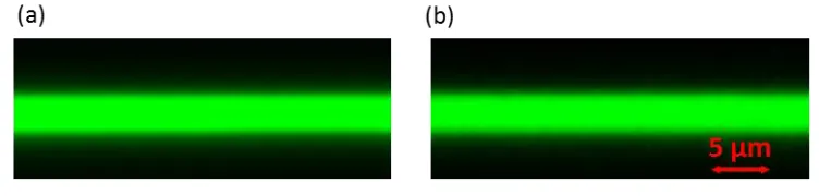
16 interlayer mixing of HA and PLL-FITC. This also suggests that despite formation of covalent bonding due to the presence of HA-Ald in (PLL/HA-Ald)24 multilayers, some PLL-FITC chains are able to diffuse at least during the build up process.
Figure 2. Section images, obtained by confocal laser scanning microscope, of multilayer films deposited on glass slides: (a) (PLL/HA)24/PLL-FITC/HA and (b) (PLL/HA-Ald)24/PLL-FITC/HA-Ald.
Nanoindentation measurements were then performed with AFM to determine the mechanical properties of both films. It is observed that PLL/HA-Ald film is more rigid than PLL/HA, with statistically significant difference in Young Moduli of 142 ± 63 kPa for PLL/HA-Ald and 10 ± 4 kPa for PLL/HA (P<0.001). This strongly indicates the crosslink of HA-Aldehyde chains through the reaction of hydrolytically labile imine bond between aldehydic derivative of HA and amino groups of PLL chains (Scheme 1). Increase in the Young modulus could be advantageous for biological applications where hard substrates are generally better for cell proliferation.
17 PLL/HA film.47 In comparison, when PLL/HA-Ald was used, the recovery of fluorescence was slower. This indicates that mobility of PLL-FITC in PLL/HA-Ald is lower than that on PLL/HA, and it could suggest that some PLL-FITC chains are crosslinked and could not diffuse any more. From Figure 3, we can conclude that almost 40% of the fluorescence is recovered in PLL/HA-Ald and thus about 60% of PLL chains are immobile. Finally FRAP experiments confirm that HA-Ald is able to crosslink the film. In comparison with the more conventional method using EDC, HA-Ald is a self-crosslinking method that do not need any additional steps with chemicals after the film buildup.
18 Release kinetics of BSA out of multilayer films in presence of PBS was investigated by fluorimetry. The release profiles seem to be dependent on the film crosslinking, the release of BSA-FITC from PLL/HA-Ald is slower for the first 24 hours than for non-crosslinked PLL/HA films (Figure 4a). This constitutes an interesting property as effectiveness of the PLL/HA film to release a protein can probably be tuned by playing with the crosslinking degree (HA/HA-Ald ratio for example) and moreover long-term release can probably be reached with PLL/HA-Ald films. The BSA-FITC release kinetics was also monitored in presence of medium containing serum, to mimic the concentration of proteins present in blood/interstitial fluid in vivo
(Supplementary Information Figure S3). For non-crosslinked PLL/HA films, the release of BSA-FITC is almost similar when PBS or media containing serum have been used as supernantant : 0.08 mg.mL-1 is released after 24h in both cases. However for crosslinked PLL/HA-Ald films the amount of BSA-FITC released after 24h is about 3 times higher in complete medium compared to PBS. Finally the slower release and thus the higher affinity of BSA to PLL/HA-Ald films can be attributed to higher hydrophobicity in these films because covalent PLL/HA interaction could be more tight and could release more water.
PLL/HA-19 Ald films have better mechanical property than PLL/HA films due to the crosslinking however they maintain the same ability to release IL-4.
(t)1/2
0 10 20 30 40 50 60
Nor
m
aliz
ed
Int
ensit
y
(a.
u.
)
0.0 0.2 0.4 0.6 0.8 1.0
PLL/HA PLL/HA-Ald
[image:19.612.85.401.142.391.2]20
Figure 4. Kinetic of biomolecules release from PLL/HA and PLL/HA-Ald in presence of PBS: (a) BSA-FTIC and (b) IL-4.
3.2 Monocyte phenotype and behavior on PLL/HA and PLL/HA-Ald multilayers
22
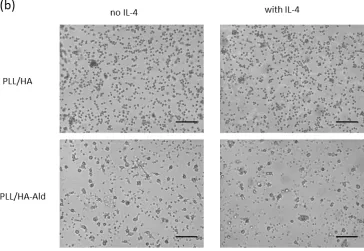
Figure 5. Phase contrast images of monocytes seeded on the films after (a) 3 and (b) 6 days of culture on PLL/HA and PLL/HA-Aldehyde in presence or not of IL-4. Scale bar = 100 µm.
23
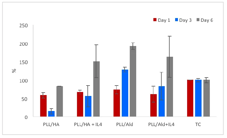
Figure 6. Metabolic activity of monocytes seeded on PLL/HA and PLL/HA-Ald with or without IL-4 after 1, 3 and 6 days (AlamarBlue assay). Controls were monocytes plated in well tissue culture-treated plates in the same medium in the absence of any cytokines (TC control, 100%).
25
Figure 7. Pro-inflammatory cytokines produced from monocytes seeded on PLL/HA, PLL/HA-Ald with or without 4: TNF-α after 3 (a) and 6 days (b); 12 after 3 (c) and 6 days (d), IL-1β after 3 (e) and 6 days (f).
26
Figure 8. Anti-inflammatory cytokines produced from monocytes seeded on PLL/HA, PLL/HA-Ald with or without IL-4: IL-1RA after 3 (a) and 6 days (b), CCL18 after 3 (c) and 6 days (d).
Finally, PLL/HA-Ald films could have applications where anti-inflammatory / pro-wound healing macrophage populations would be beneficial in improving the rate and quality of implant integration, protection of biomedical devices such as electrodes from adverse immune reactions.
Conclusions
27 modulated by tuning the physical property of the films, such as Young Modulus. The main objective of this study is to use the controlled release capacities of the PLL/HA films for the delivery of immunomodulatory cytokines such as IL-4. PLL/HA-Aldehyde films stimulate the anti-inflammatory response on monocytes even in the absence of IL-4. However IL-4 improves in an efficient way the anti-inflammatory properties of the coating. The designed film could be applied in the future to medical devices like implants to trigger differentiation of monocytes in a pro-healing phenotype. Such coatings can be used in the control of initial inflammatory reactions to permanent titanium implants such as dental, orthopaedic replacement systems or in neuroprosthetic devices in the brain. We plan to use these coatings also for improvement of integration of recently developed artificial larynx system that is clinical trial stage.
Supporting Information
Additional information regarding the Interleukin 4, inflammatory responses, characterization of HA-Aldehyde by 1H NMR and calibration curve for BSA-FITC are found in Supporting Information.
Author Contributions
The manuscript was written through contributions of all authors. All authors have given approval to the final version of the manuscript.
Funding Sources
28 ACKNOWLEDGMENT
This project has received funding from the European Union’s Seventh Framework Programme for research, technological development and demonstration under grant agreement no. 602694 (IMMODGEL). We thank B. Senger, K. Benmlih and C. Bouthier for their support.
REFERENCES
(1) Lavalle, P.; Voegel, J. C.; Vautier, D.; Senger, B.; Schaaf, P.; Ball, V., Dynamic aspects of films prepared by a sequential deposition of species: perspectives for smart and responsive materials. Adv. Mater. 2011,23, 1191-1221.
(2) Lvov, Y.; Decher, G.; Moehwald, H., Assembly, structural characterization, and thermal behavior of layer-by-layer deposited ultrathin films of poly(vinyl sulfate) and poly(allylamine).
Langmuir 1993,9, 481-486.
(3) Tieke, B.; Van Ackern, F.; Krasemann, L.; Toutianoush, A., Ultrathin self-assembled polyelectrolyte multilayer membranes. Europ. Phys. J. E. 2001,5, 29-39.
(4) Pargaonkar, N.; Lvov, Y. M.; Li, N.; Steenekamp, J. H.; de Villiers, M. M., Controlled release of dexamethasone from microcapsules produced by polyelectrolyte layer-by-layer nanoassembly. Pharmaceut. Res. 2005,22, 826-835.
(5) Prokopović, V. Z.; Duschl, C.; Volodkin, D., Hyaluronic Acid/Poly-l-Lysine Multilayers as Reservoirs for Storage and Release of Small Charged Molecules. Macromol. Biosci. 2015, 15, 1357-1363.
29 (7) Brunot, C.; Grosgogeat, B.; Picart, C.; Lagneau, C.; Jaffrezic-Renault, N.; Ponsonnet, L., Response of fibroblast activity and polyelectrolyte multilayer films coating titanium. Dent. Mater. 2008,24, 1025-1035.
(8) Almodóvar, J.; Guillot, R.; Monge, C.; Vollaire, J.; Selimović, Š.; Coll, J.-L.; Khademhosseini, A.; Picart, C., Spatial patterning of BMP-2 and BMP-7 on biopolymeric films and the guidance of muscle cell fate. Biomaterials 2014,35, 3975-3985.
(9) Müller, S.; Koenig, G.; Charpiot, A.; Debry, C.; Voegel, J.-C.; Lavalle, P.; Vautier, D., VEGF-Functionalized Polyelectrolyte Multilayers as Proangiogenic Prosthetic Coatings.
Advanced Functional Materials 2008,18, 1767-1775.
(10) Vrana, N.; Erdemli, O.; Francius, G.; Fahs, A.; Rabineau, M.; Debry, C.; Tezcaner, A.; Keskin, D.; Lavalle, P., Double entrapment of growth factors by nanoparticles loaded into polyelectrolyte multilayer films. J. Mater. Chem. B 2014,2, 999-1008.
(11) Gilde, F.; Guillot, R.; Fourel, L.; Almodovar, J.; Crouzier, T.; Boudou, T.; Picart, C., Matrix-Bound Presentation of Bone Morphogenetic Protein 2 by Multilayer Films: Fundamental Studies and Applications to Orthopedics. Eds C. Picart, F. Caruso and J.-C. Voegel, Wiley-VCH Verlag GmbH & Co. KGaA, Weinheim, Germany 2015.
(12) Yamanlar, S.; Sant, S.; Boudou, T.; Picart, C.; Khademhosseini, A., Surface functionalization of hyaluronic acid hydrogels by polyelectrolyte multilayer films. Biomaterials 2011,32, 5590-5599.
30 (14) Picart, C.; Senger, B.; Sengupta, K.; Dubreuil, F.; Fery, A., Measuring mechanical properties of polyelectrolyte multilayer thin films: Novel methods based on AFM and optical techniques. Colloids Surf. A 2007,303, 30-36.
(15) Picart, C., Polyelectrolyte multilayer films: from physico-chemical properties to the control of cellular processes. Curr. Med. Chem. 2008,15, 685-697.
(16) Ren, K.; Crouzier, T.; Roy, C.; Picart, C., Polyelectrolyte multilayer films of controlled stiffness modulate myoblast cells differentiation. Adv. Funct. Mater. 2008,18, 1378.
(17) Schneider, A.; Richert, L.; Francius, G.; Voegel, J.-C.; Picart, C., Elasticity, biodegradability and cell adhesive properties of chitosan/hyaluronan multilayer films. Biomed. Mater. 2007,2, S45.
(18) Picart, C.; Elkaim, R.; Richert, L.; Audoin, F.; Arntz, Y.; Da Silva Cardoso, M.; Schaaf, P.; Voegel, J.-C.; Frisch, B., Primary cell adhesion on RGD-functionalized and covalently crosslinked thin polyelectrolyte multilayer films. Adv. Funct. Mater. 2005,15, 83-94.
(19) Francius, G.; Hemmerlé, J.; Ohayon, J.; Schaaf, P.; Voegel, J.-C.; Picart, C.; Senger, B., Effect of crosslinking on the elasticity of polyelectrolyte multilayer films measured by colloidal probe AFM. Microsc. Res. Tech. 2006,69, 84-92.
(20) Collins, M. N.; Birkinshaw, C., Hyaluronic acid based scaffolds for tissue engineering—A review. Carbohyd. Polym. 2013,92, 1262-1279.
(21) Morra, M., Engineering of biomaterials surfaces by hyaluronan. Biomacromolecules 2005, 6, 1205-1223.
31 Bioconjugate Targeting Ovarian Cancer Affords a Potent In vivo Therapeutic Activity. Clin. Cancer Res. 2008,14, 3598-3606.
(23) Homma, A.; Sato, H.; Okamachi, A.; Emura, T.; Ishizawa, T.; Kato, T.; Matsuura, T.; Sato, S.; Tamura, T.; Higuchi, Y., Novel hyaluronic acid–methotrexate conjugates for osteoarthritis treatment. Bioorg. Med. Chem. 2009,17, 4647-4656.
(24) Pitarresi, G.; Palumbo, F. S.; Albanese, A.; Fiorica, C.; Picone, P.; Giammona, G., Self-assembled amphiphilic hyaluronic acid graft copolymers for targeted release of antitumoral drug.
J. Drug Target. 2010,18, 264-276.
(25) Saravanakumar, G.; Choi, K. Y.; Yoon, H. Y.; Kim, K.; Park, J. H.; Kwon, I. C.; Park, K., Hydrotropic hyaluronic acid conjugates: Synthesis, characterization, and implications as a carrier of paclitaxel. Int. J. Pharm. 2010,394, 154-161.
(26) Mero, A.; Pasqualin, M.; Campisi, M.; Renier, D.; Pasut, G., Conjugation of hyaluronan to proteins. Carbohyd. Polym. 2013,92, 2163-2170.
(27) Buffa, R.; Kettou, S.; Pospisilova, L.; Berkova, M.; Velebny, V. Oxidized derivative of hyaluronic acid, a method of preparation thereof and a method of modification thereof. 2010. (28) Buffa, R.; Kettou, S.; Pospisilova, L.; Huerta-Angeles, G.; Chladkova, D.; Velebny, V., method of preparation of an oxidized derivative of hyaluronic acid and a method of modification thereof. In Google Patents: 2010.
32 (30) Rostam, H.; Singh, S.; Vrana, N.; Alexander, M.; Ghaemmaghami, A., Impact of surface chemistry and topography on the function of antigen presenting cells. Biomater. Sci. 2015, 3, 424-441.
(31) Kzhyshkowska, J.; Gudima, A.; Riabov, V.; Dollinger, C.; Lavalle, P.; Vrana, N. E., Macrophage responses to implants: prospects for personalized medicine. Journal of leukocyte biology 2015, jlb. 5VMR0415-166R.
(32) Badylak, S. F.; Gilbert, T. W., Immune response to biologic scaffold materials. Semin. Immunol. 2008,20, 109-116.
(33) Ratner, B. D.; Hoffman, A. S.; Schoen, F. J.; Lemons, J. E., Biomaterials science: an introduction to materials in medicine. Academic Press: 2004.
(34) Decher, G.; Schlenoff, J. B., Multilayer thin films: sequential assembly of nanocomposite materials. John Wiley & Sons: 2006.
(35) Gribova, V.; Auzely-Velty, R.; Picart, C., Polyelectrolyte multilayer assemblies on materials surfaces: from cell adhesion to tissue engineering. Chem. Mater. 2011,24, 854-869.
(36) Tang, Z.; Wang, Y.; Podsiadlo, P.; Kotov, N. A., Biomedical applications of layer-by-layer assembly: from biomimetics to tissue engineering. Adv. Mater. 2006,18, 3203.
(37) Morton, S. W.; Poon, Z.; Hammond, P. T., The architecture and biological performance of drug-loaded LbL nanoparticles. Biomaterials 2013,34, 5328-5335.
33 (40) Chaubaroux, C.; Vrana, E.; Debry, C.; Schaaf, P.; Senger, B.; Voegel, J.-C.; Haikel, Y.; Ringwald, C.; Hemmerlé, J.; Lavalle, P., Collagen-based fibrillar multilayer films cross-linked by a natural agent. Biomacromolecules 2012,13, 2128-2135.
(41) Barthes, J.; Mertz, D.; Bach, C.; Metz-Boutigue, M.-H. l. n.; Senger, B.; Voegel, J.-C.; Schaaf, P.; Lavalle, P., Stretch-induced biodegradation of polyelectrolyte multilayer films for drug release. Langmuir 2012,28, 13550-13554.
(42) Vogt, C.; Ball, V.; Mutterer, J.; Schaaf, P.; Voegel, J.-C.; Senger, B.; Lavalle, P., Mobility of proteins in highly hydrated polyelectrolyte multilayer films. J. Phys. Chem. B 2012, 116, 5269-5278.
(43) Dimitriadis, E. K.; Horkay, F.; Maresca, J.; Kachar, B.; Chadwick, R. S., Determination of elastic moduli of thin layers of soft material using the atomic force microscope. Biophys. J. 2002, 82, 2798-2810.
(44) Polyakov, P.; Soussen, C.; Duan, J.; Duval, J. F.; Brie, D.; Francius, G., Automated force volume image processing for biological samples. PLoS One 2011,6, e18887.
(45) García-Nieto, S.; Johal, R. K.; Shakesheff, K. M.; Emara, M.; Royer, P.-J.; Chau, D. Y.; Shakib, F.; Ghaemmaghami, A. M., Laminin and fibronectin treatment leads to generation of dendritic cells with superior endocytic capacity. PLoS one 2010,5, e10123.
(46) Picart, C.; Mutterer, J.; Richert, L.; Luo, Y.; Prestwich, G.; Schaaf, P.; Voegel, J.-C.; Lavalle, P., Molecular basis for the explanation of the exponential growth of polyelectrolyte multilayers. Proc. Natl. Acad. Sci. USA 2002,99, 12531-12535.
34 multilayer films studied by fluorescence recovery after pattern photobleaching. Langmuir 2008, 24, 7842-7847.
(48) Vodouhê, C.; Schmittbuhl, M.; Boulmedais, F.; Bagnard, D.; Vautier, D.; Schaaf, P.; Egles, C.; Voegel, J.-C.; Ogier, J., Effect of functionalization of multilayered polyelectrolyte films on motoneuron growth. Biomaterials 2005,26, 545-554.
35