Original Article
Alhagi-honey and its compound abnormal savda munziq
(ASMq) inhibit proliferation and enhance apoptosis
of human hepatoma BEL-7402 cells
Wei Wang1, Halmurat Upur1, Junmin Chang2
1Uyghur Medical College, 2College of Pharmacy, Xinjiang Medical University, No. 393, New Medical Road, Urumqi
830011, Xinjiang, China
Received February 14, 2016; Accepted May 5, 2016; Epub August 15, 2016; Published August 30, 2016
Abstract: Objective: To investigate the effects of alhagi-honey and abnormal savda munziq (ASMq) on the prolifera-tion and apoptosis of human hepatoma BEL-7402 cells and the related molecular mechanisms. Method: Human hepatoma BEL-7402 cells were treated with alhagi-honey, ASMq, or 5-FU (positive control). Cell proliferation was assessed with the CCK-8 assay, and the cell cycle and cellular apoptotic process were detected with flow cytometry. The mRNA and protein expression levels of apoptosis-related proteins were determined with quantitative real-time PCR and Western blot analysis, respectively. Results: Both the treatments of alhagi-honey and ASMq significantly increased the inhibitory rates of cell proliferation in human hepatoma BEL-7402 cells, in a dose-dependent manner. Moreover, the treatments of alhagi-honey and ASMq induce the G0/G1 phase arrest of human hepatoma BEL-7402 cells. Furthermore, both the alhagi-honey and ASMq treatments significantly elevated the apoptosis rates of human hepatoma BEL-7402 cells. In addition, the treatments of alhagi-honey and ASMq significantly decreased the expres-sion levels of anti-apoptotic proteins Bcl-2 and EGFR, while increased the expresexpres-sion level of pro-apoptotic protein Bax in human hepatoma BEL-7402 cells. Conclusion: Alhagi-honey and its compound ASMq could significantly in-hibit the proliferation and enhance the apoptosis of human hepatoma BEL-7402 cells, which would be associated with the down-regulated Bcl-2 and EGFR expression levels and the up-regulated Bax expression level in these cells. These findings might provide evidence for the application of alhagi-honey and its compound ASMq in the treatment of hepatocellular carcinoma (HCC) in clinic.
Keywords: Alhagi-honey, abnormal savda munziq (ASMq), human hepatoma BEL-7402 cells, proliferation and apoptosis
Introduction
Hepatocellular carcinoma (HCC) is one of most common cancers, andthe third leading cause of cancer-induced death,throughout the world [1]. There are approximately one million newly-reported HCC cases each year, and 600 thou-sand subjects die of HCC in the world, making it a serious threat to human health [2]. Therefore, it is of great importance to discover and
devel-op efficient therapeutic strategies for HCC in
clinic.
Abnormal savda munziq (ASMq) is a commonly used clinical compound in Uyghur medicine. Recent studies have shown that, ASMq could effectively inhibit the tumor cells in liver cancer, lymphoma, and breast cancer [3]. As one of the
single medicines in ASMq, saccharum alhagi accounts for a large proportion (about 42.3%) in the prescription. Alhagi-honey is the main com-ponent in saccharum alhagi. In recent years, alhagi-honey has also been shown to be able to exert potent anti-tumor effects, and has been widely used in the clinical treatment of malig-nant tumors. Alhagi-honey could stimulate the maturation, differentiation, and proliferation of immunocompetent cells, regulating the body immune system, which could phagocyte and clear the tumor cells [4].
RPMI-1640 complete medium (Life Technolo- gies, Carlsbad, CA, USA) containing 10% fetal bovine serum and 1 mL penicillin and strepto-mycin, in a 5% CO2, 37°C incubator.
Cell counting kit-8 (CCK-8) assay
Cell proliferation was assessed with the CCK-8 assay (Beyotime, Haimen, Jiangsu, China). Human hepatoma BEL-7402 cells in the loga-rithmic growth phase were collected and plant-ed onto a 96-well plate at the density of 5 × 10⁴ cells/mL. After 48 h, the medium was discard-ed, and the cells were treated with alhagi-hon-ey (Xinjiang Maidisen Uyghur Pharmaceutical Co., Ltd., Hetian, Xinjiang, China; which had
been qualified by Prof. Palida Abuliziat Uyghur
Medical College, Xinjiang Medical University) or ASMq (Xinjiang Ciconhabo Uyghur Pharma- ceutical Co., Ltd., Urumqi, Xinjiang, China) at 0, 6, 8, 10, 12, 14, 16, and 20 mg/mL, respec-tively, for 48 h. The 5-FU was used as positive control, and the treatment concentrations were
set at 0, 0.1, 0.5, 1, 5, 20, 40, and 80 μg/mL,
respectively. After discarding the medium, the cells were treated with 10% CCK-8 at 37°C for 1.5 h. Then OD450 nm was read with a microplate reader (Bio-Rad, Hercules, CA, USA). The IC50 values were obtained, and the inhibitory rates were calculated according to the following for-mulation: inhibitory rate = (OD450 nm control - OD450 nm treatment)/OD450 nm control × 100%.
Flow cytometry
Cell cycle and cellular apoptotic process were
detected with flow cytometry. Human hepato -ma BEL-7402 cells were collected and planted onto the 25-cm2 culture bottle at the density of 1 × 10⁶ cells/bottle. After 24 h, the cells were treated with 8 mg/mL alhagi-honey, 8 mg/mL
ASMq, and 0.6 μg/mL 5-FU, respectively, for 48
sion was treated with 400 μL AnnexinV binding solution and 5 μL AnnexinV-FITC in dark for 15
min, and then 10 µL PI at 4°C in dark for 30
min, followed by flow cytometry.
Quantitative real-time PCR
The mRNA expression levels of Bcl-2, Bax, and EGFR in human hepatoma BEL-7402 cells were detected with quantitative real-time PCR. After drug administration for 48 h, the cells were col-lected and total RNA was extracted with Trizol (Invitrogen, Carlsbad, CA, USA). The cDNA tem-plate was obtained with the reverse transcrip-tion kit (Taingen, Beijing, China). Quantitative real-time PCR was performed with the SYBR Select Master Mix (ABI, Grand Island, NY, USA) on the MyCycler thermal cycler (Bio-Rad, Hercules, CA, USA). The primer sequences were as follows: Bcl-2, forward 5’-GATAACGGAGG- CTGGGATGC-3’ and reverse 5’-TCACTTGTGGCC- CAGATAGG-3’; Bax, forward 5’-CCCCCGAGAGG- TCTTTTTCC-3’ and reverse 5’-CTGATCAGTTCC- GGCACCTT-3’; EGFR, forward 5’-TTGCCGCAAA- GTGTGTAACG-3’ and reverse 5’-AGTCACCCCTA-
AATGCCACC-3’; and β-actin, forward 5’-ATGA- TGATATCGCCGCGCTC-3’ and reverse 5’-TCGA-
TGGGGTACTTCAGGG-3’. The 20-μL PCR system consisted of 0.4 μL primer each, 10 μL SYBRR Select Master Mix (2 ×), 1 μL template, and 8.2 μL ddH2O. The reaction condition was set as 50°C for 2 min, 95°C for 2 min, 95°C for 15 s, 60°C for 1 min, for totally 40 cycles.
Western blot analysis
to SDS-PAGE, and then electronically
trans-ferred onto a PVDF membrane. After blocking
with 5% non-fat milk for 1 h, the membrane was incubated with anti-Bcl-2 primary antibody (1:200 dilution; Abcam, Cambridge, MA, USA), anti-Bax primary antibody (1:500 dilution; Abcam), anti-EGFR primary antibody (1:500
dilution; Abcam), or anti-β-actin primary anti -body (1:2000 dilution; Abcam), at 4°C over-night. Then the membrane was treated with goat anti-mouse IgG (Pierce, Rockford, IL, USA)
Results
Effects of alhagi-honey and ASMq on prolifera-tion of human hepatoma BEL-7402 cells
To investigate the effects of alhagi-honey and its compound ASMq on the proliferation of human hepatoma BEL-7402 cells, these cells
were first treated with alhagi-honey (0, 6, 8, 10,
[image:3.612.91.482.67.520.2]12, 14, 16, and 20 mg/mL), ASMq (0, 6, 8, 10, 12, 14, 16, and 20 mg/mL), and 5-FU (0, 0.1,
Figure 1. Effects of alhagi-honey and ASMq on proliferation of human hepatoma BEL-7402 cells. Human hepatoma BEL-7402 cells were treated with alhagi-honey (A), ASMq (B), and 5-FU (C), respectively, at indicated concentra-tions, for 48 h. Cell proliferation was assessed with by the CCK-8 assay, in which the OD450 nm was read, and the proliferation rates were cal-culated accordingly. Compared with the control group (0 mg/mL), *P<0.05, **P<0.01; compared
with theformer group, #P<0.05, ##P<0.01.
Figure 2. Effects of alhagi-honey and ASMq on cell cycle of human hepatoma BEL-7402 cells. Human hepatoma BEL-7402 cells were treated with 8 mg/mL alhagi-honey, 8 mg/mL ASMq, and 6 mg/mL 5-FU (positive control), respec-tively, for 48 h. Then cell cycle was detected with flow cytometry.
at room temperature for 1 h. After incubated with coloriza-tion solucoloriza-tion A and B, the che-miluminescence was detect-ed with the ChemiScope mini-imaging system.
Statistical analysis
Data were expressed as me- an ± SD. SPSS 19.0 software was used for statistical
analy-sis. ANOVA was performed for
multiple comparisons, with LSD and Dunnett’s T tests. P<0.05 was considered as
0.5, 1, 5, 20, 40, and 80 μg/mL), respectively,
for 48 h, and the cell proliferation was assessed with the CCK-8 assay. Our results showed that, the IC50 values for alhagi-honey, ASMq, and 5-Fu treatments were 10.75 mg/mL, 11.30
mg/mL and 4.55 μg/mL respectively. For the
treatments of alhagi-honey and ASMq, the inhibitory rates of cell proliferation of human
Effects of alhagi-honey and ASMq on apopto-sis of human hepatoma BEL-7402 cells
To investigate the effects of alhagi-honey and ASMq on the apoptotic process of human
[image:4.612.90.392.72.539.2]hepa-toma BEL-7402 cells, the AnnexinV-FITC/PI staining was performed, and the fluorescence was detected with flow cytometry. As shown in
Figure 3. Effects of alhagi-honey and ASMq on apoptosis of human hepatoma BEL-7402 cells. Human hepatoma BEL-7402 cells were treated with 8 mg/mL alhagi-honey, 8 mg/mL ASMq, and 6 mg/mL 5-FU (positive control), respective-ly, for 48 h. A. Cellular apoptotic process was detected using AnnexinV-FITC/PI staining by flow cytometry. B. Statistical analysis of apoptosis rates of human hepatoma BEL-7402 cells. Compared with the control group, *P<0.05, **P<0.01.
Effects of alhagi-honey and ASMq on cell cycle of human hepatoma BEL-7402 cells
To investigate the effects of alhagi-honey and ASMq on the cell cycle of human hepatoma BEL-7402 cells,
flow cytometry was per -formed. As shown in Figure 2, after treated with 8 mg/ mL alhagi-honey for 48 h, the G0/G1 cell proportion
was significantly elevated
(P<0.05), while the propor-tions of S and G2/M cells
were significantly declined
(both P<0.05). Similar re- sults were obtained for the ASMq treatment, which si-
gnificantly increased the
Figure 3, the apoptosis rate for the control
group was 1.60±1.00%, which was significantly
elevated by the treatments of alhagi-honey and ASMq, to 14.97±0.15% and 15.07±0.29%, respectively (both P<0.05). These results sug-gest that, the treatments of alhagi-honey and
ASMq could significantly enhance the apopto -sis of human hepatoma BEL-7402 cells. Effects of alhagi-honey and ASMq on expres-sion of apoptosis-related proteins
The effects of alhagi-honey and ASMq on expression of apoptosis-related proteins in human hepatoma BEL-7402 cells were next
investigated. These cells were first treated with
8 mg/mL alhagi-honey or ASMq for 48 h, and the mRNA and protein expression levels of Bcl-2, Bax, and EGFR were determined with quanti-tative real-time PCR and Western blot analysis, respectively. Our results from the quantitative real-time PCR showed that, compared with the control group, the mRNA expression levels of
Bcl-2 and EGFR were significantly declined,
while the mRNA expression level of Bax was
significantly elevated, by the treatments of
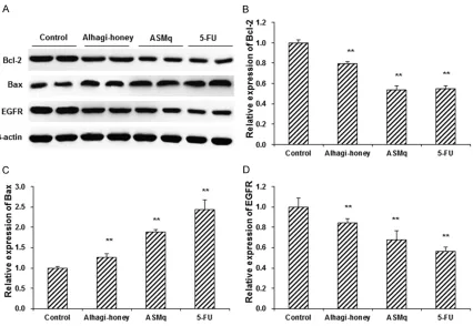
alhagi-honey and ASMq, in human hepatoma BEL-7402 cells (P<0.05) (Figure 4). Similar results were observed for the Western blot analysis. Compared with the control group, the
treatments of alhagi-honey and ASMq signifi -cantly decreased the expression levels of anti-apoptotic proteins Bcl-2 and EGFR, while increased the expression level of pro-apoptotic protein Bax in human hepatoma BEL-7402 cells (P<0.05) (Figure 5). These results suggest that, the treatments of alhagi-honey and ASMq could
significantly change the expression levels of
apoptosis-related proteins in human hepatoma BEL-7402 cells, which was in line with its apop-tosis-inducing effects.
Discussion
Modern researches have confirmed that ASMq could exert diuretic, anti-inflammatory, and
detoxication functions in clinic, with negligible-side effects. At present, the investigation and application of ASMq are no longer limited to the treatment of diabetes and cardiovascular dis-eases. The anti-tumor activity of ASMq has been attracting more and more attention, espe-cially concerning its apoptosis-inducing effects in tumor cells. Wang et al. [5] have found that, ASMq could down-regulate the expression of p53 in cervical cancer cells, thus promoting the cellular apoptotic process. Moreover, Yusup et al. [6] have shown that, the apoptosis-promot-ing effects of ASMq in human hepatoma cells might be associated with the down-regulated
Bcl-2 mRNA expression level and the up-regu-lated mRNA expression levels of p53, p21, and Bax. As one of the single medicines in ASMq, saccharum alhagi accounts for about 42.3% in the prescription, and alhagi-honey is the main component in saccharum alhagi [7]. Numerous studies have demonstrated that polysaccha-rides could exert potent anti-tumor effects. Polysaccharides do not directly kill the tumor
cells, but they work as highly efficientimmuno -modulators in the body. They stimulate the mat-uration, differentiation, and proliferation of immunocompetent cells, and recover the bal-ance of immune system, which could phago-cyte and clear the tumor cells [8-15]. Therefore, polysaccharides have been widely used in the clinical treatment of malignant tumors nowa-days [16-19].
[image:6.612.95.521.70.364.2]Due to the complex structure of polysaccha-rides, the investigation of their molecular mechanism and structure-activity relationship has become a research hotspot in recent years. In this study, the anti-tumor effects of both
Figure 5. Effects of alhagi-honey and ASMq on apoptosis-related protein expression. Human hepatoma BEL-7402 cells were treated with 8 mg/mL alhagi-honey, 8 mg/mL ASMq, or 6 mg/mL 5-FU (positive control), for 48 h. (A) The protein expression levels of Bcl-2, Bax, and EGFR were detected with Western blot analysis. (B-D) Statistical analysis of protein expression levels of Bcl-2 (B), Bax (C), and EGFR (D). Compared with the control group, *P<0.05, **P<0.01.
alhagi-honey and its compound ASMq have been analyzed and compared. The effects of alhagi-honey and ASMq on the proliferation and apoptosis of human hepatoma BEL-7402 cells
were investigated with the CCK-8 assay, flow
cytometry, quantitative real-time PCR, and Western blot analysis, respectively.
In this study, our results from the CCK-8 assay showed that, the treatments of alhagi-honey
and ASMq could significantly inhibit the prolif -eration of human hepatoma BEL-7402 cells, in a dose-dependent manner. Moreover, when the
cell cycle was detected with flow cytometry, our
results showed that, the alhagi-honey and
ASMq treatments could significantly elevate
the cell proportion in the G0/G1 phase, and decline the cell proportion in the S and G2/M phases. These results suggest that the treat-ments of alhagi-honey and ASMq could induce G0/G1 phase arrest in human hepatoma
BEL-7402 cells, indicating significantly inhibited cell
Induction of apoptosis in human hepatoma BEL-7402 cells is an important therapeutic method for the disease in clinic. In this study, the cellular apoptotic process was detected
using the AnnexinV-FITC/PI staining by flow
cytometry. Our results showed that, when these cells were treated with alhagi-honey and ASMq
for 48 h, the apoptosis rate would be signifi -cantly elevated. The expression levels of Bcl-2
and Bax could reflect the cellular apoptotic pro -cess [20, 21]. Bcl-2 family is well known for the anti-apoptotic properties, which could prevent and block cell shrinking, DNA cleavage, and chromatin shrinkage, thereby inhibiting apopto-sis. However, Bax in the cytoplasm would bind to the mitochondrial membrane and increas-ethe membrane permeability, thus inducinga-poptosis. Studies have shown that, the interac-tion between Bcl-2 and Bax could promote the apoptotic process. In this study, our results from the quantitative real-time PCR and Western blot analysis showed that, the
treat-ments of alhagi-honey and ASMq could signifi -cantly decline the Bcl-2 expression and elevate the Bax expression in human hepatoma BEL-7402 cells. On the other hand, EGFR is a com-mon receptor involved in the cellular prolifera-tion and signal transducprolifera-tion, whose mutaprolifera-tion and/or over-expression might lead to tumori-genesis. In this study, our results showed that, the treatments of alhagi-honey and ASMq could
significantly decrease the expression level of
EGFR in human hepatoma BEL-7402 cells, which might contribute to the apoptosis-enhancing effects of alhagi-honey and ASMq. In conclusion, our results showed that, the treat-
ments of alhagi-honey and ASMq could signifi -cantly inhibit the proliferation and enhance the apoptosis of human hepatoma BEL-7402 cells. Moreover, the treatments of alhagi-honey and ASMq could induce G0/G1 phase arrest in these cells. Furthermore, the treatments of
alhagi-honey and ASMq significantly down-reg -ulated Bcl-2 and EGFR expression levels and up-regulated Bax expression level in human
hepatoma BEL-7402 cells. These findings might
provide evidence for the application of alhagi-honey and its compound ASMq in the treat-ment of HCC in clinic.
Acknowledgements
This work was supported by the National Na- tural Science Foundation of China (81460633)
and the Science and Technology Supporting Project of Xinjiang Uygur Autonomous Re- gion (201491172).
Disclosure of conflict of interest
None.
Address correspondence to: Junmin Chang, College of Pharmacy, Xinjiang Medical University, No. 393, New Medical Road, Urumqi 830011, Xinjiang, China. Tel: 86-15699222131; E-mail: cjmcjn2471@163. com
References
[1] El-Serag HB, Rudolph KL. Hepatocellular carci-noma: epidemiology and molecular Carcino-genesis. Gastroenterology 2007; 132: 2557-2576.
[2] Jemal A, Bray F, Center MM, Ferlay J, Ward E, Forman D. Global cancer statistics. CA Cancer J Clin 2011; 61: 69-90.
[3] Hu H, Sheng L, Upur H. Anti-cancer effects of Uighur abnormal Savda Munzipand its influ-ence on cell migration. Sci Technol Rev 2011; 29: 62-65.
[4] Qin T, Chen J, Wang D, Hu Y, Zhang J, Wang M, Qiu S, Gao Z, liu R, Yu Y, Huang Y, Wang Q, Wang Q. Selenylation modification can en-hance immune-enhancing activity of Chinese angelica polysaccharide. Carbohydr Polym 2013; 95: 183-187.
[5] Wang G, Li H, Chen K. Effects of abnormal sav-da munziqtotal phenolcombined with cisplatin and docetaxel on proliferation and apoptosis of HeLa cells. Shandong Medical Journal 2011; 51: 22-24.
[6] Berke B, Baudrimont I, Moore N. Apoptosis-in-ducing effects of abnormalsavda munziq in HepG2 cells. China Journal of Chinese Materia Medica 2007; 32: 1068-1071.
[7] Jian LJ, Chang JM, Li GR, Cheng JN. Isolation, purification, and basic structural elucidation of poly saccharide from Alhagi-honey. Journal of Xinjiang Medical University 2013; 36: 460-463.
[8] Chen S, Yin DK, Yao WB, Wang YD, Zhang YR, Gao XD. Macrophage receptors of polysaccha-ride isolated from a marine filamentous fungus Phoma herbarum YS4108. Acta Pharmacol Sin 2009; 30: 1008-1014.
[9] Piotrowski J, Jędrzejewski T, Kozak W. Immuno-modulatory and antitumor properties of poly-saccharide peptide (PSP). Postepy Hig Med Dosw 2015; 69: 91-97.
gas-Biol Macromol 2012; 50: 768-772.
[14] Holderness J, Schepetkin IA, Freedman B, Kir-potina LN, Quinn MT, Hedges JF, Jutila MA. Polysaccharides isolated from Acai fruit induce innate immune responses. PLoS One 2011; 6: e17301.
[15] Aikemu A, Sulaiman X, Shan L, Yang T, Upur H. Study of abnormal savda munziq inhibitory ef-fect on mice EAC sarcoma. Journal of Xinjiang Medical University 2015; 38: 154-156. [16] Pan H, Han Y, Huang J, Yu X, Jiao C, Yang X,
Dhaliwal P, Xie Y, Yang BB. Purification and identification of a polysaccharide from medici-nal mushroom Amauroderma rude with immu-nomodulatory activity and inhibitory effect on tumor growth. Oncotarget 2015; 6: 17777-17791.
[20] Spampanato C, De Maria S, Sarnataro M, Gior-dano E, Zanfardino M, Baiano S, Cartenì M, Morelli F. Simvastatin inhibits cancer cell growth by inducing apoptosis correlated to ac-tivation of Bax and down-regulation of BCL-2 gene expression. Int J Oncol 2012; 40: 935-941.