1,4-Bis(4-pyridylsulfanylmethyl)benzene
Suk-Hee Moonaand Ki-Min Parkb*
aSubdivision of Food Science, Kyungnam College of Information and Technology,
Busan 616-701, Republic of Korea, andbResearch Institute of Natural Science,
Gyeongsang National University, Jinju 660-701, Republic of Korea Correspondence e-mail: [email protected]
Received 1 June 2008; accepted 11 June 2008
Key indicators: single-crystal X-ray study;T= 298 K; mean(C–C) = 0.004 A˚; Rfactor = 0.053;wRfactor = 0.122; data-to-parameter ratio = 17.2.
In the title compound, C18H16N2S2, a crystallographic
inver-sion centre lies at the centre of the benzene ring, and the two terminal 4-mercaptopyridyl groups adopt an anti geometry. Each benzene ring makes a dihedral angle of 55.4 (1)with the
plane of the benzene fragment. The crystal structure is stabilized by C—H interactions between a benzene H atom and a pyridyl ring of a neighbouring molecule. In addition, the crystal structure exhibits intermolecular C— H N interactions.
Related literature
For details of the preparation and related structures of 1,4-bis(2-pyridyl-sulfanylmethyl)benezene derivatives, see: Atherton et al. (1999); McMorran & Steel (2003); For the structures of Co(II) and Ag (I) complexes of 1,4-bis(2-pyridylsulfanylmethyl)benezene, see: Hartshorn & Steel (1998). For bond-length data, see: Allenet al.(1987).
Experimental
Crystal data
C18H16N2S2 Mr= 324.45
Monoclinic,P21=c
a= 7.145 (1) A˚
b= 6.1667 (8) A˚
c= 17.954 (2) A˚
= 90.391 (3)
V= 791.03 (18) A˚3
Z= 2
MoKradiation
= 0.33 mm1
T= 298 (2) K 0.350.200.15 mm
Data collection
Bruker SMART CCD area-detector diffractometer
Absorption correction: none 4706 measured reflections
1717 independent reflections 893 reflections withI> 2(I)
Rint= 0.074
Refinement
R[F2> 2(F2)] = 0.052
wR(F2) = 0.121
S= 0.96 1717 reflections
100 parameters
H-atom parameters constrained
max= 0.32 e A˚ 3
min=0.17 e A˚ 3
Table 1
Hydrogen-bond geometry (A˚ ,).
Cgis the centroid of N1/C1/C2/C3/C5 pyridyl ring.
D—H A D—H H A D A D—H A
C1—H1 N1i
0.93 2.61 3.484 (4) 158
C8—H8 Cgii
0.93 2.77 3.560 (4) 143
Symmetry codes: (i)x1;yþ1 2;zþ
3
2; (ii)x;y;zþ1.
Data collection:SMART(Bruker, 2000); cell refinement: SAINT-Plus(Bruker, 2000); data reduction:SAINT-Plus; program(s) used to solve structure: SHELXTL (Sheldrick, 2008); program(s) used to refine structure: SHELXTL; molecular graphics: SHELXTL and DIAMOND(Brandenburg, 1998); software used to prepare material for publication:SHELXTL.
This work was supported by a Korea Research Foundation Grant funded by the Korean Government (MOEHRD) (KRF-2007–359-C00019).
Supplementary data and figures for this paper are available from the IUCr electronic archives (Reference: LX2058).
References
Allen, F. H., Kennard, O., Watson, D. G., Brammer, L., Orpen, A. G. & Taylor, R. (1987).J. Chem. Soc. Perkin Trans.2, pp. S1–19.
Atherton, Z., Goodgame, D. M. L., Menzer, S. & Williams, D. J. (1999).
Polyhedron,18, 273–279.
Brandenburg, K. (1998).DIAMOND. Crystal Impact GbR, Bonn, Germany. Bruker (2000). SMART and SAINT-Plus. Bruker AXS Inc., Madison,
Wisconsin, USA.
Hartshorn, C. M. & Steel, P. J. (1998).J. Chem. Soc. Dalton Trans.pp. 3935– 3940.
McMorran, D. A. & Steel, P. J. (2003).Tetrahedron,59, 3701–3707. Sheldrick, G. M. (2008).Acta Cryst.A64, 112–122.
Acta Crystallographica Section E
Structure Reports Online
supporting information
Acta Cryst. (2008). E64, o1285 [doi:10.1107/S1600536808017571]
1,4-Bis(4-pyridylsulfanylmethyl)benzene
Suk-Hee Moon and Ki-Min Park
S1. Comment
The reaction of α,α′-dibromo-p-xylene with 4-mercaptopyridine afforded the title compound, in which the
crystallographic inversion centre lies on the centre of the benzene ring. Therefore, the asymmetric unit consists of a half
of molecule and the two 4-mercaptopyridyl groups adopt an anti-geometry (Fig. 1). All bond lengths and angles show
normal value (Allen et al., 1987). The dihedral angle between the plane of benzene and the terminal pyridyl ring is
55.4 (1)°, which is smaller than those of related structures (Atherton et al., 1999; Hartshorn & Steel, 1998).
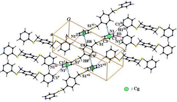
The crystal packing (Fig. 2) is stabilized by C—H···π interactions between a benzene H atom and the pyridyl ring of
neighbouring molecule, with a C8—H8···Cg separation of 2.77 Å (Fig. 2 and Table 1; Cg is the centroid of
N1/C1/C2/C3/C5 pyridyl ring, symmetry code as in Fig. 2). The molecular packing (Fig. 2) is further stabilized by
intermolecular C—H···N hydrogen bonds between a pyridyl H atom and the pyridine N atom of neighbouring molecule,
with a C1—H1···N1i separation of 2.61 Å (Fig. 2 and Table 1; symmetry code as in Fig. 2).
S2. Experimental
The title compound was prepared by the reaction of α,α′-dibromo-p-xylene with 4-mercaptopyridine in acetonitrile
according to reported methods (Atherton et al., 1999; McMorran & Steel, 2003). Single crystal suitable for X-ray
analysis were obtained by evaporation of a solution of the title compound in acetonitrile.
S3. Refinement
All H-atoms were positioned geometrically and refined using a riding model with d(C—H) = 0.93 Å, Uiso =1.2Ueq(C) for
Figure 1
The molecular structure of (I), showing the atom-numbering scheme. Displacement ellipsoids are drawn at the 50%
probability level for non-H atoms. [Symmetry code: (i) -x + 1, -y + 1, -z + 1]
Figure 2
C—H···π and C—H···N interactions (dotted lines) in the title compound. Cg denotes the ring centroid. [Symmetry codes:
(i) x+1, y+1, z+1; (ii) x1, y+1/2, z+3/2; (iii) x1, y1/2, z+3/2; (iv) x+2, y+1/2, z1/2; (v) x+2, y+3/2, z1/2; (vi)
-x, -y, -z+1;(vii) x+1, y+1, z.]
1,4-Bis(4-pyridylsulfanylmethyl)benzene
Crystal data
C18H16N2S2 Mr = 324.45 Monoclinic, P21/c
Hall symbol: -P 2ybc
a = 7.145 (1) Å
b = 6.1667 (8) Å
β = 90.391 (3)°
V = 791.03 (18) Å3 Z = 2
F(000) = 340
Dx = 1.362 Mg m−3
[image:3.610.133.481.320.520.2]θ = 2.3–27.0°
µ = 0.33 mm−1 T = 298 K
Plate, colourless 0.35 × 0.20 × 0.15 mm
Data collection
Bruker SMART CCD area-detector diffractometer
Radiation source: fine-focus sealed tube Graphite monochromator
φ and ω scans
4706 measured reflections 1717 independent reflections
893 reflections with I > 2σ(I)
Rint = 0.074
θmax = 27.0°, θmin = 2.3° h = −9→7
k = −7→7
l = −21→22
Refinement
Refinement on F2
Least-squares matrix: full
R[F2 > 2σ(F2)] = 0.052 wR(F2) = 0.121 S = 0.96 1717 reflections 100 parameters 0 restraints
Primary atom site location: structure-invariant direct methods
Secondary atom site location: difference Fourier map
Hydrogen site location: inferred from neighbouring sites
H-atom parameters constrained
w = 1/[σ2(F
o2) + (0.0516P)2]
where P = (Fo2 + 2Fc2)/3
(Δ/σ)max = 0.001
Δρmax = 0.32 e Å−3
Δρmin = −0.17 e Å−3
Special details
Geometry. All e.s.d.'s (except the e.s.d. in the dihedral angle between two l.s. planes) are estimated using the full
covariance matrix. The cell e.s.d.'s are taken into account individually in the estimation of e.s.d.'s in distances, angles and torsion angles; correlations between e.s.d.'s in cell parameters are only used when they are defined by crystal symmetry. An approximate (isotropic) treatment of cell e.s.d.'s is used for estimating e.s.d.'s involving l.s. planes.
Refinement. Refinement of F2 against ALL reflections. The weighted R-factor wR and goodness of fit S are based on F2,
conventional R-factors R are based on F, with F set to zero for negative F2. The threshold expression of F2 > σ(F2) is used
only for calculating R-factors(gt) etc. and is not relevant to the choice of reflections for refinement. R-factors based on F2
are statistically about twice as large as those based on F, and R- factors based on ALL data will be even larger.
Fractional atomic coordinates and isotropic or equivalent isotropic displacement parameters (Å2)
x y z Uiso*/Ueq
S1 0.01648 (12) 0.27398 (12) 0.56971 (5) 0.0651 (3)
N1 −0.2740 (4) −0.1917 (5) 0.72963 (13) 0.0677 (8)
C1 −0.3414 (4) 0.0057 (6) 0.71296 (17) 0.0632 (9)
H1 −0.4533 0.0486 0.7346 0.076*
C2 −0.2540 (4) 0.1478 (5) 0.66562 (16) 0.0587 (8)
H2 −0.3052 0.2842 0.6570 0.070*
C3 −0.0897 (4) 0.0876 (4) 0.63081 (15) 0.0501 (7)
C4 −0.0215 (4) −0.1175 (5) 0.64684 (15) 0.0561 (8)
H4 0.0875 −0.1672 0.6245 0.067*
C5 −0.1161 (5) −0.2463 (5) 0.69596 (15) 0.0593 (8)
H5 −0.0660 −0.3822 0.7065 0.071*
C6 0.2555 (4) 0.1735 (4) 0.56468 (16) 0.0620 (9)
H6A 0.3003 0.1373 0.6143 0.074*
C7 0.3805 (4) 0.3443 (4) 0.53083 (15) 0.0493 (7)
C8 0.4517 (4) 0.3178 (4) 0.46027 (16) 0.0537 (8)
H8 0.4190 0.1961 0.4325 0.064*
C9 0.4290 (4) 0.5300 (5) 0.56966 (15) 0.0554 (8)
H9 0.3801 0.5526 0.6169 0.066*
Atomic displacement parameters (Å2)
U11 U22 U33 U12 U13 U23
S1 0.0593 (5) 0.0556 (5) 0.0806 (6) 0.0050 (4) 0.0101 (4) 0.0205 (4) N1 0.0699 (19) 0.0749 (19) 0.0584 (16) −0.0087 (15) 0.0095 (14) 0.0062 (14) C1 0.051 (2) 0.079 (2) 0.059 (2) −0.0008 (18) 0.0091 (16) −0.0047 (18) C2 0.057 (2) 0.0542 (18) 0.065 (2) 0.0081 (16) −0.0015 (16) −0.0020 (17) C3 0.0483 (19) 0.0475 (17) 0.0546 (17) −0.0018 (14) −0.0016 (14) −0.0025 (14) C4 0.059 (2) 0.0474 (17) 0.0616 (19) 0.0032 (15) 0.0121 (16) −0.0005 (16) C5 0.066 (2) 0.0527 (18) 0.0595 (19) 0.0022 (17) −0.0004 (17) 0.0065 (15) C6 0.056 (2) 0.0492 (17) 0.081 (2) 0.0074 (14) 0.0188 (17) 0.0112 (16) C7 0.0475 (18) 0.0459 (17) 0.0546 (18) 0.0039 (13) 0.0049 (15) 0.0074 (14) C8 0.0600 (19) 0.0493 (17) 0.0519 (18) −0.0006 (15) 0.0007 (15) −0.0040 (15) C9 0.060 (2) 0.0618 (19) 0.0443 (17) 0.0053 (17) 0.0105 (15) 0.0009 (15)
Geometric parameters (Å, º)
S1—C3 1.764 (3) C5—H5 0.9300
S1—C6 1.820 (3) C6—C7 1.511 (4)
N1—C5 1.327 (4) C6—H6A 0.9700
N1—C1 1.342 (4) C6—H6B 0.9700
C1—C2 1.374 (4) C7—C8 1.378 (3)
C1—H1 0.9300 C7—C9 1.383 (4)
C2—C3 1.385 (4) C8—C9i 1.379 (4)
C2—H2 0.9300 C8—H8 0.9300
C3—C4 1.385 (4) C9—C8i 1.379 (4)
C4—C5 1.369 (4) C9—H9 0.9300
C4—H4 0.9300
C3—S1—C6 102.52 (13) C4—C5—H5 117.6
C5—N1—C1 115.7 (3) C7—C6—S1 109.87 (18)
N1—C1—C2 123.6 (3) C7—C6—H6A 109.7
N1—C1—H1 118.2 S1—C6—H6A 109.7
C2—C1—H1 118.2 C7—C6—H6B 109.7
C1—C2—C3 119.9 (3) S1—C6—H6B 109.7
C1—C2—H2 120.1 H6A—C6—H6B 108.2
C3—C2—H2 120.1 C8—C7—C9 117.9 (3)
C2—C3—C4 116.7 (3) C8—C7—C6 120.7 (3)
C2—C3—S1 118.4 (2) C9—C7—C6 121.4 (3)
C4—C3—S1 124.9 (2) C7—C8—C9i 120.7 (3)
C5—C4—C3 119.3 (3) C7—C8—H8 119.7
C3—C4—H4 120.4 C8i—C9—C7 121.3 (3)
N1—C5—C4 124.8 (3) C8i—C9—H9 119.3
N1—C5—H5 117.6 C7—C9—H9 119.3
C5—N1—C1—C2 −1.4 (4) C3—C4—C5—N1 1.2 (5)
N1—C1—C2—C3 1.6 (5) C3—S1—C6—C7 −165.4 (2)
C1—C2—C3—C4 −0.4 (4) S1—C6—C7—C8 −109.1 (3)
C1—C2—C3—S1 179.4 (2) S1—C6—C7—C9 71.7 (3)
C6—S1—C3—C2 160.6 (2) C9—C7—C8—C9i 1.5 (5)
C6—S1—C3—C4 −19.7 (3) C6—C7—C8—C9i −177.7 (2)
C2—C3—C4—C5 −0.9 (4) C8—C7—C9—C8i −1.5 (5)
S1—C3—C4—C5 179.3 (2) C6—C7—C9—C8i 177.7 (3)
C1—N1—C5—C4 −0.1 (5)
Symmetry code: (i) −x+1, −y+1, −z+1.
Hydrogen-bond geometry (Å, º)
D—H···A D—H H···A D···A D—H···A
C1—H1···N1ii 0.93 2.61 3.484 (4) 158
C8—H8···Cgiii 0.93 2.77 3.560 (4) 143