EVALUATION OF CONVENTIONAL AND 16S r DNA IDENTIFICATION METHODS FOR DETECTION
OF XANTHOMONAS CAUSING LEAF BLIGHT IN RICE
1*
Pramod, D.,
2Kishore
1
Bioserve Biotechnolo
2
University College, Palamuru University, Mahabubnagar, India
ARTICLE INFO ABSTRACT
Leaf blight of rice is a disease of major concern in rice cultivation. This disease caused by bacterial pathogen
material between countries. World loss due to this disease i
pathologists and food technologists. Conventional methods used for determining the disease consume time, not viable for accurate pathogen identification and thereby hinder the implementation of apt control measures. Molecular
less time consuming and flexible for easy usage. The regulatory measures levied for controlling the movement of pathogen from one country to other country can be addressed by molecula identification methods determining pathogenic and non
paper an attempt was made to identify wild xanthomonads using conventional and molecular methods (16S r DNA).
Copyright © 2014 Pramod et al. This is an open access article distributed under the Creative Commons Attribution License, which permits unrestricted use, distribution, and reproduction in any medium, provided the original work is properly cited.
INTRODUCTION
Rice (Oryza sativa) is consumed worldwide and mostly in Asia as staple food. It gains importance being supplier of 1/5 th the total calorie consumed by humans (Smith, 1988). Rice cultivation is severely affected particularly in tropical Asia by bacterial leaf blight (BB) caused by Xanthomonas oryzae oryzae (XOO) leading to yield losses of up to 50% (Adhikari
al., 1994). The disease leads to poor grain development, lower
quality / under developed grains with reduced weight resulting in high proportion of broken rice (Ou, 1985). BB disease management concentrates on methods that reduce the initial inoculums and subsequent development of the pathogen on host plants and this can be accomplished through chemical protection, host plant resistance, and biological control. The presence of the pathogen in infected seeds and disease transmission from these infected seeds has be
(Mew, 1987; Hsieh et al., 2001). Importation and movement of this pathogen infected plant material is regulated by plant protection and quarantine program of USDA and several other countries. In view of global seed trade, phyto
regulations enforced by several countries entail need for reliable, sensitive and less time consuming diagnostic tools for XOO. Traditional methods employed for detection and identification of xanthomonads, such as biochemical (Mooter and Swing, 1990) serological and pathogenecity tests (Benedict
et al., 1990; Berthier et al., 1993) are been extended by
*Corresponding author: Pramod, D.
Bioserve Biotechnologies Pvt. Ltd. Hyderabad, India
ISSN: 0975-833X
Article History:
Received 16th April, 2014 Received in revised form 04th May, 2014
Accepted 05th June, 2014
Published online 20th July,2014
Key words:
Xanthomonas oryzae pv oryzae,
blight of rice, 16S r DNA.
RESEARCH ARTICLE
EVALUATION OF CONVENTIONAL AND 16S r DNA IDENTIFICATION METHODS FOR DETECTION
OF XANTHOMONAS CAUSING LEAF BLIGHT IN RICE
Kishore, N.,
2Vijender Reddy, A. and
1Nasaruddin
Bioserve Biotechnologies Pvt. Ltd. Hyderabad, India
University College, Palamuru University, Mahabubnagar, India
ABSTRACT
Leaf blight of rice is a disease of major concern in rice cultivation. This disease caused by bacterial pathogen Xanthomonas oryzae pv oryzae creates major issues during export and import of food material between countries. World loss due to this disease i
pathologists and food technologists. Conventional methods used for determining the disease consume time, not viable for accurate pathogen identification and thereby hinder the implementation of apt control measures. Molecular methods for identification of pathogen are often reliable, econometric, less time consuming and flexible for easy usage. The regulatory measures levied for controlling the movement of pathogen from one country to other country can be addressed by molecula identification methods determining pathogenic and non-pathogenic Xanthomonads. In the present paper an attempt was made to identify wild xanthomonads using conventional and molecular methods (16S r DNA).
access article distributed under the Creative Commons Attribution License, which permits unrestricted use, distribution, and reproduction in any medium, provided the original work is properly cited.
) is consumed worldwide and mostly in Asia as staple food. It gains importance being supplier of 1/5 th of the total calorie consumed by humans (Smith, 1988). Rice cultivation is severely affected particularly in tropical Asia by
Xanthomonas oryzae pv
(XOO) leading to yield losses of up to 50% (Adhikari et 94). The disease leads to poor grain development, lower quality / under developed grains with reduced weight resulting in high proportion of broken rice (Ou, 1985). BB disease management concentrates on methods that reduce the initial ent development of the pathogen on host plants and this can be accomplished through chemical protection, host plant resistance, and biological control. The presence of the pathogen in infected seeds and disease transmission from these infected seeds has been demonstrated Importation and movement of this pathogen infected plant material is regulated by plant protection and quarantine program of USDA and several other countries. In view of global seed trade, phyto-sanitary ulations enforced by several countries entail need for suming diagnostic tools for Traditional methods employed for detection and identification of xanthomonads, such as biochemical (Mooter ical and pathogenecity tests (Benedict
1993) are been extended by
Bioserve Biotechnologies Pvt. Ltd. Hyderabad, India.
molecular methods (Donald and Graham, 1989).Traditional methods are tedious, time consuming and sometimes with non reproducible results. While molecular methods are less time
consuming, accurate and with reproducible results.
Accordingly an attempt was made identif
Xanthomonads isolated from rice plant materials of different locations using conventional and molecular methods.
MATERIALS AND METHODS
Plant Samples
Forty leaf and seed samples of rice from different locations, representing most of the Mahabubnagar, Warangal and Krishna districts of Andhrapradesh were obtained. Plants of age group of near seeding plants were observed for presence of pathogenic lesions, carefully separated by a sharp blade and scissors and carried to laboratory in an ic
refrigerator. Isolation of Xanthomonads was completed within 12 hrs of sample collection.
Isolation of Xanthomonads
From Seeds: Sixty grams of rice seeds were blended in sterile salt solution (Phosphate-Buffered Saline pH 7.2) and
at 50 C for 2 hrs. From this 25 ml of soaking suspension was
centrifuged (10,000 RPM, 10min) and the precipitate obtained is again resuspended in salt solution (1/10 in 2.5 ml). Three
different concentrations (10-1, 10
solution was inoculated (100 micro
Available online at http://www.journalcra.com
International Journal of Current Research
Vol. 6, Issue, 07, pp.7409-7413, July,2014
INTERNATIONAL
z
EVALUATION OF CONVENTIONAL AND 16S r DNA IDENTIFICATION METHODS FOR DETECTION
OF XANTHOMONAS CAUSING LEAF BLIGHT IN RICE
Nasaruddin
University College, Palamuru University, Mahabubnagar, India
Leaf blight of rice is a disease of major concern in rice cultivation. This disease caused by bacterial creates major issues during export and import of food material between countries. World loss due to this disease is the major concern of today’s pathologists and food technologists. Conventional methods used for determining the disease consume time, not viable for accurate pathogen identification and thereby hinder the implementation of apt methods for identification of pathogen are often reliable, econometric, less time consuming and flexible for easy usage. The regulatory measures levied for controlling the movement of pathogen from one country to other country can be addressed by molecular pathogenic Xanthomonads. In the present paper an attempt was made to identify wild xanthomonads using conventional and molecular methods
access article distributed under the Creative Commons Attribution License, which permits unrestricted use,
methods (Donald and Graham, 1989).Traditional methods are tedious, time consuming and sometimes with non reproducible results. While molecular methods are less time
consuming, accurate and with reproducible results.
Accordingly an attempt was made identify and analyze wild Xanthomonads isolated from rice plant materials of different locations using conventional and molecular methods.
MATERIALS AND METHODS
Forty leaf and seed samples of rice from different locations, e Mahabubnagar, Warangal and Krishna districts of Andhrapradesh were obtained. Plants of age group of near seeding plants were observed for presence of pathogenic lesions, carefully separated by a sharp blade and scissors and carried to laboratory in an ice-box and stored in a refrigerator. Isolation of Xanthomonads was completed within
From Seeds: Sixty grams of rice seeds were blended in sterile Buffered Saline pH 7.2) and incubated C for 2 hrs. From this 25 ml of soaking suspension was centrifuged (10,000 RPM, 10min) and the precipitate obtained is again resuspended in salt solution (1/10 in 2.5 ml). Three
, 10-2, 10-3) of the above salt
micro l/plate) on to nutrient agar
+ cycloheximide (semi-selective media) and Xanthomonas
oryzae selective medium (XOS)(Sucrose 20g, Sodium
glutamate 5g, Ca(NO3) 0.2g, K2HPO4 2g, Fe-EDTA 1mg,
Agar 17g, Tetrazolium chloride 10mg, cycloheximide 100mg, kasugamycin 20mg, Cephalirin 20mg, methyl violet 0.3mg,
water 1lt, pH 6.8-7.0). Plates were incubated at 280C for 5-7
days. Appearance of straw yellow coloured colonies of 1-2 mm diameter on nutrient agar and rose pink, mucoid raised colonies on XOS agar indicate Xanthomonads and Xanthomonas oryzae respectively. These were picked, isolated on to peptone sucrose broth for maintaining.
Pathogenecity Test
Isolates are tested on susceptible rice cultivars. For X. oryzae pv. oryzae 30-45 days old TN1 (Taichung native 1) rice plants were used. Local popular rice varieties from the Andrapradesh region with known susceptibility to the disease viz., Sonamasuri, Tellahamsa, Vijeta MTU 1001, Swarna MTU 7029 and Samba masuri BPT 5204 are included in the tests.
The clip-method (Kauffman et al., 1973)
The tips of 30-40 leaves of rice plants were cut (2-3 cm) with a
scissors immersed in the bacterial suspension (108 CFU mL−1).
The inoculated plants were covered for 24 h with a polythene bag, and incubated at 30°C with 12 h light cycle. Plants were observed for symptoms after 48-72 h up to 14 days ie., checked for water-soaked areas in the inoculated leaves, usually beginning from the inoculated ends as water-soaked stripes, which is an indication of X. oryzae pv. oryzae. Plants are observed for milky exudates, extension of wavy lesions (red stripes) symptoms. The rice plants were grown in potting
mixture with a weekly fertilization of 1–2 g urea L-1.
Inoculated plants were kept under high moisture conditions with 12 h light/dark cycle at an optimum temperature of 28– 32°C. A negative control was maintained (plants inoculated with sterile saline solution alone) to monitor false positive reactions caused.
Identification
Conventional methods (Biochemical tests)
All the Forty isolates of Xanthomonas were identified using conventional biochemical methods. Strains were identified basing the tests- Grams reaction, Indole-Methyl red-Voges proskauer-Citrate tests (IMVIC), Oxidase, Nitrate reduction, Arginine hydrolase, Pectinase, Catalase and Urease. Standard methods were followed as described by Bergey’s manual for determinative bacteriology (Bradbury, 1984). Further they were checked for their ability to utilize different sugars Viz., Arabinose, L- Arabinose, Galactose, Glucose, D-Fructose, Cellobiose, Adonitol, Dulcitol, Inositol, Mannitol, Salicin, Sorbitol, Lactose, Manose, Maltose, Melibiose, Rhammose, Raffinose, Sucrose, Inulin, Xylose, and Trehalose. Readymade sugar discs (Himedia) were incorporated in to the
sterile medium (NH4H2PO4-0.5g, K2HPO4- 0.5g,
MgSO4.7H2O- 0.2g, NaCl- 5g, Yeast extract-1g, Distilled
water- 1 lt, Bromocresol purple- 0.7ml (1.5%alcohol sol) containing test tubes which were aseptically inoculated with
respective bacterial isolate. Tubes incubated for 48 hrs at 28+2o
C were checked for presence of growth and formation of yellow color indicating acid production (Schaad, 1980).
Molecular analysis
DNA extraction
Overnight grown culture (1.5ml) was centrifuged at 13000 rpm for 2 minutes. Supernatant was discarded and pellet was
obtained. To the pellet 100 µl of 1x TE buffer was added and mixed properly so that pellet comes into solution.
This suspension was added to enzyme coated tubes (lysozyme 5µl (10mg/ml) + lyticase100 units/sample) and incubated in
water bath at 370 C for 30 minutes. To this 100 µl of 2% SDS
and 5 µl (20 mg/ml) of proteinase K was added. The tubes
were incubated at 550 C for 1 and 1/2 hours. To this 200 µl of
Guanidium HCl (PH-5.2) solution was added and centrifuged at 10000 rpm for 10 min. The pellet was discarded and to the supernatant equal volume of isopropanol was added and loaded on to the column. The elute obtained by centrifugation at 10,000 rpm for 2 min was discarded. To the column 500 µl of 70% alcohol was added and centrifuged at 10,000 rpm for 2 minutes, elute discarded. The column was spun with caps open at 10000rpm for 3 min (drying). The column was placed in eppendorf tube and 100 µl of Elution buffer was added
Quantitation of DNA by Spectrophotometric method
10ml of DNA solution was diluted with 990ml of TE. Mixed well and absorbance at 260nm and 280nm was measured. The absorbance at 260nm can be used to calculate the concentration of DNA as follows:
Calculations
OD260 of 1= 50 micro g/ml DNA; Dilution factor =100 Concentration of DNA in a given solution (micro g/ml) =50 X OD X Dilution factor / 1 X 1000
PCR amplification (16s rDNA)
PCR Set up
A 1.0µl of Sample DNA (approximately 100 ng/µl) was added to PCR Mixture containing 100mM Tris HCl (pH 8.3), 500mM
KCl (pH 8.3), 2.5µl MgCl2 (25mM), 2.0µl dNTP’s (2.5mM),
1.0µl Primer Forward & Reverse (each of 10pm/µl) and 1u /µl of Taq Polymerase (Bioserve Make) and the final volume made to 25 µl with nuclease free water. The primer set FD1 Fw and RP2 Rw designed in the conserved region was used for the amplification of the 16S region of the test organisms and the primer sequences are
FD1 Fw: 5’- AGAGTTTGATCCTGGCTCAG -3’ RP2 Rw: 5’-ACGGCTACCTTGTTACGACTT-3’
PCR condition
The amplification conditions for amplification of bacterial 16S
followed by 35 cycles of denaturation at 950C for 30 Sec,
annealing at 520C for 90 Sec; extension at 720C for 60Sec and
final extension at 720C for 10 min and held at 40C
Electrophoresing the amplicons
The PCR Products (6 - 10µl) were separated by electrophoresis
in 2% Agarose gels containing ethidium bromide (1µg ml
-1
).The electropherogram obtained after electrophoresis of the PCR amplicons. In the electropherogram the bands of the size ~1500bp (for Xoo samples 1-40) was observed against 1Kb DNA ladder (Fermentas make). About 3µl Ethidium Bromide was used as staining dye and 5µl of Bromothymol blue was used as tracking dye. About 100V DC was maintained between the electrodes of gel apparatus for 20 minutes. Following electrophoresis the gel was analyzed and pictured in gel doc system (BIOSERVE Hyderabad).
Gel Elution
DNA band of ~1500bp was cut from gel with sterile blade and taken in 1.5 ml eppendorf tube; elution was done using Bioserve gel extraction kit following the manufacturer’s instructions.
DNA sequencing
QIAGEN QIA quick TM kit was used for sequencing reaction. The sequencing PCR was done to amplify one strand of gene employing the primers under standard PCR conditions. Primers used are: 16 Seq 4 R- TGCTGCCTCCCGTAGGAGT; 16 Seq 2 R- TACGCATTTCACCGCTACAC; 16 Seq 2 R 2- TACGCATTTCACTGCTACAC; 16 Seq 4 F- TTAGATAC CCTGGTAGTCCA; 16 Seq 3F- AAGTCCCGCAACGAGC GC; 16S 1 REV- TGCGCTCGTTGCGGGACTTAACC. The samples were precipitated and suspended in 40µl of loading solution provided with the kit. Sequencing was done with Mega Bace sequencer- Bioserve India, Hyderabad. Sequences thus obtained were edited and aligned using commercial Gene tool Lite (version 1.1) software
RESULTS
Selective isolation of Xanthomonas resulted in 40
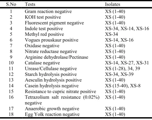
[image:3.595.301.565.119.216.2](Xanthomonas strains (XS)) isolates. All these isolates were initially checked for pathogenecity on different rice varieties (Table 1). Taichung native (TN-1) rice variety was observed to be susceptible to all the 40 isolates (XS 1-40). Strains which were strongly positive /negative are represented in the Table 1. Among the 40 isolates of Xanthomonas, that was found positive in causing infection in at least one of the rice variety were further evaluated for biochemical tests in order to evaluate them on physiological basis. From Table 1 it can be inferred that all the isolates were able to cause the infection in one or more rice varieties. A critical perusal of the Table 2 indicates that all the isolates were gram negative. They were negative also for oxidase test, egg yolk reaction, tetrazolium salt resistance, fluorescent pigment production, nitrate reductase, arginine hydrolase, pectinase and growth under anaerobic conditions. For KOH test, aesculin hydrolysis and resistance to cupric nitrate (0.001%) all the tested isolates were found positive. Remaining tests shown in the Table 2
represented varied results (indole, MR, VP, catalase, starch hydrolysis, casein hydrolysis)
Table 1. Test for pathogenecity of 40 isolates, XS (1-40) on Rice plants
S.No Rice variety Negative Isolates
1 TN 1 XS (1-40)
2 Sonamasuri XS-13, XS-17, XS-27, XS-28, XS-30,
XS-31, XS-38, XS-39
3 Tellahamsa XS-19, XS-21, XS-23, XS-30, XS-37,
XS-38, XS-39
4 Vijetha MTU 1001 XS-17, XS-31, XS-36, XS-40
5 Swarna MTU 7029 XS-17, XS-19, XS-25, XS-27, XS-40
[image:3.595.311.555.247.440.2]6 Sambamasuri BPT 5204 XS-13, XS-17, XS-28, XS-30, XS-36, XS-37
Table 2. Biochemical tests pertaining to 40 Xanthomonas isolates
S.No Tests Isolates
1 Gram reaction negative XS (1-40)
2 KOH test positive XS (1-40)
3 Fluorescent pigment negative XS (1-40)
4 Indole test positive XS-34, XS-14, XS-16
5 Methyl red positive XS-34
6 Vogues prouskaur positive XS-14, XS-16
7 Oxidase negative XS (1-40)
8 Nitrate reductase negative XS (1-40)
9 Arginine dehydrolase/Pectinase XS (1-40)
10 Catalase negative XS-14, XS-27, XS-31
11 Urease/Cellulase negative XS (1-28), 34, 39
12 Starch hydrolysis positive XS-34, XS-39
13 Aesculin hydrolysis positive XS (1-40)
14 Casein hydrolysis negative XS (15-40), XS-8
15 Resistance to cupric nitrate positive XS (1-40) 16 Tetrazolium salt resistance (0.02%)
negative
XS (1-40)
17 Anaerobic growth negative XS (1-40)
[image:3.595.310.554.466.666.2]18 Egg Yolk reaction negative XS (1-40)
Table 3. Utilization of sugars by the 40 Xanthonmonas isolates
S.No Acid Production from Isolate
1 D-Arabinose Positive XS (1-40)
2 L- Arabinose Negative XS (1-40)
3 D-Galactose Positive XS (1-40)
4 D-Glucose Positive XS (1-40)
5 D-Fructose Positive XS (1-40)
6 Cellobiose Positive XS (1-40)
7 Adonitol Negative XS- 13, XS-16
8 Inositol Negative XS-31, XS-40,
9 Salicin Negative XS-34,XS-40
10 Sorbitol Negative XS-40
11 Lactose Negative XS-40
12 Manose Positive XS (1-40)
13 Maltose Negative XS (1-40)
14 Melibiose Negative XS-40
15 Rahmmose Positive XS (1-40)
16 Raffinose Negative XS-13, XS-40
17 Sucrose Negative XS-34,XS-37,XS-40
18 Inulin Negative XS-34, XS-40
19 Xylose Positive XS (1-40)
20 Trehalose Negative XS (1-40)
Table 3 depicts the ability of the 40 isolates of xanthomonas in utilizing various sugars for acid production by fermentation. Most of the isolates were able to use all the sugars tested. All the 40 isolates were producing acid in the media containing L- Arabinose, D-Galctose, D-Fructose, Cellobiose, Adonitol, Mannose, Maltose, and Rhamnose. While, all isolates were negative for acid production with L-Arabinose, Maltose and
Trehalose as media component. All the 40 isolates showed varied results for biochemical properties representing Xanthomonas (Schaad et al., 2001). These isolates however could not be differentiated from other Xanthomonas sp. The pathogenecity assay and biochemical characteristics confirm them as Xanthomonas pathogens (Nayudu, 1972)
Fig. 1. Amplification of Xanthomonas 16s rDNA region recognized by band in 1500bp region
The DNA isolation from all the 40 isolates yielded sufficient and ample amount of DNA in terms of purity as evidenced
from A260/A280 between 1.80-1.90. Molecular confirmation of
the isolates using PCR p
(AGAGTTTGATCATGGCTCAG) and RP1
(ACGGTTACCTTGTTACGACTT) resulted a 1500 bp band (targeted to 16s rDNA region) on 1.5% agarose gel on electrophoresis. Carefully eluted bands were subjected to sequencing using sequencing primers (16 seq 2R, 16 seq seq 4F, 16 seq 4R and 16s1Rev) resulting in complete 1500 bp 16 s rDNA sequence. NCBI BLAST performed with the sequences confirmed the isolates as Xanthomonas
Further, few of these sequences were deposited in NCBI gene bank and the accession numbers are HM 747116, HM 747117, HM 747118, HM 747119, HM 125569 and HM 125570.
DISCUSSION
In the present investigation, biochemical tests for identification of the Xanthomonas pathogen gave varied results indicating existence of genetic variability. For instance, starch hydrolysis has been reported by Swings et al., (1990) which was contrasting to the results presented by Guvera and Marsella (1999) who did find this character in their isolates. Our isolates varied, as some of the isolates were positive
negative. Overlapping results and cross contamination with other similar isolates for identification on the basis of biochemical characterization is a problem needed to be answered. Traditional methods used for identification of pathogen leaves vague results. Labor intensive, time taking biochemical tests identification of pathogen are needed to be replaced by molecular based methods. Accurate, reliable and Trehalose as media component. All the 40 isolates showed cal properties representing 2001). These isolates however could not be differentiated from other Xanthomonas sp. The pathogenecity assay and biochemical characteristics confirm them as Xanthomonas pathogens (Nayudu, 1972)
16s rDNA region recognized by band in 1500bp region
The DNA isolation from all the 40 isolates yielded sufficient and ample amount of DNA in terms of purity as evidenced 1.90. Molecular confirmation of
the isolates using PCR primers FD2
(AGAGTTTGATCATGGCTCAG) and RP1
(ACGGTTACCTTGTTACGACTT) resulted a 1500 bp band (targeted to 16s rDNA region) on 1.5% agarose gel on electrophoresis. Carefully eluted bands were subjected to sequencing using sequencing primers (16 seq 2R, 16 seq 3F, 16 seq 4F, 16 seq 4R and 16s1Rev) resulting in complete 1500 bp 16 s rDNA sequence. NCBI BLAST performed with the
Xanthomonas oryzae.
Further, few of these sequences were deposited in NCBI gene numbers are HM 747116, HM 747117, HM 747118, HM 747119, HM 125569 and HM 125570.
In the present investigation, biochemical tests for identification of the Xanthomonas pathogen gave varied results indicating or instance, starch hydrolysis (1990) which was contrasting to the results presented by Guvera and Marsella (1999) who did find this character in their isolates. Our isolates varied, as some of the isolates were positive and other was negative. Overlapping results and cross contamination with other similar isolates for identification on the basis of biochemical characterization is a problem needed to be answered. Traditional methods used for identification of ves vague results. Labor intensive, time taking biochemical tests identification of pathogen are needed to be replaced by molecular based methods. Accurate, reliable and
econometric molecular methods help in answering the regulatory affairs levied by vario
import of food materials. Molecular methods increase the ease of identification of the pathogen
oryzae. Many molecular based methods are recently been
explored largely but, there still remains many unoccupie lacuna even in this area. Still investigations are under way for accurate differentiation between pathogenic and non pathogenic strains of Xanthomonas.
REFERENCES
Adhikari, T. B., Mew T. W., and Teng, P. S. 1994.
bacterial blight on rice cultivars carrying different Xa genes for resistance in the field. Plant Dis, 78: 73
Benedict, A. A., Alvarez, A. M., and Pollard, L. W. 1990. Pathovar-specific antigens of
begoniae and X. campestris
monoclonal antibodies. Appl. Environ. Microbiol 574.
Berthier, Y., Verdier, V., Guesdon, J., Chevrier, D., Denis, J., Decoux, G., and Lemattre, M. 1993. Characterization of
Xanthomonas campestris
restriction patterns. Appl. Environ. Microbiol., 59, 851 Bradbury, J. F. 1984. Xanthomonas
210.In N. R. Krieg and J. G. Holt (ed.), Bergey's manual of systematic bacteriology, vol. 1. The Williams & Wilkins Co., Baltimore.
Donald, A. C., and Graham, J. H. 1989. Genomic fingerprinting of two pathovars of phytopathogenic bacteria by rare-cutting restriction enzymes and field inversion gel electrophoresis. Phytopathology, 79, 745
Guvera, Y., and Marsella, A. 1999. Ric Venezuela. Agri. Trop. Maracay 49, 505
Hsieh, S. P., Buddenhagen, I. W., and Kauffman, H. E. 2001. An improved method for detecting the presence of
Xanthomonas oryzae pv.
Phytopathology, 64, 273–274.
Kauffman, H. E., Reddy, A. P. K., Hsieh, S. P. Y., and Merca, S. D. 1973. An improved technique for evaluating resistance of rice varieties to
Disease Reporter, 57, 537–541.
Mew, T.W. 1987. Current status and future prospects of research on bacterial blight of rice. Annual Review of Phytopathology, 25, 359–382.
Nayudu, M. V. 1972. Pseudomonas
anew bacterial disease of grapevine. Phytopathologische Zeitschrift, 73, 183-186.
Ou, S. H. 1985. Rice Diseases, Vol 2.
Biology, Surrey, UK Porter B, Chittoor, J., Yano, M., Sasaki, T., White, F. F. 2003. Development and mapping of markers linked to the rice bacterial blight resistance gene Xa7. Crop Sci, 43, 1484–1492.
Schaad, N. W., Jones, J. B., and
Guide for identification of Plant Pathogenic Bacteria, 3rd edition. APS Press, St Paul (US).
Schaad, N.W. 1980. Laboratory guide for identification of plant pathogenic bacteria. American Phytopathological Society, Minnesota
econometric molecular methods help in answering the regulatory affairs levied by various countries in export and import of food materials. Molecular methods increase the ease of identification of the pathogen-Xanthomonas oryzae pv . Many molecular based methods are recently been explored largely but, there still remains many unoccupied lacuna even in this area. Still investigations are under way for accurate differentiation between pathogenic and non-pathogenic strains of Xanthomonas.
Adhikari, T. B., Mew T. W., and Teng, P. S. 1994. Progress of bacterial blight on rice cultivars carrying different Xa– genes for resistance in the field. Plant Dis, 78: 73–77 Benedict, A. A., Alvarez, A. M., and Pollard, L. W. 1990.
specific antigens of Xanthomonas campestris pv.
campestris pv. pelargonii detected with Appl. Environ. Microbiol., 56,
572-Berthier, Y., Verdier, V., Guesdon, J., Chevrier, D., Denis, J., Decoux, G., and Lemattre, M. 1993. Characterization of pathovars by rRNA gene restriction patterns. Appl. Environ. Microbiol., 59, 851-859.
Xanthomonas Dawson 1939, p.
199-210.In N. R. Krieg and J. G. Holt (ed.), Bergey's manual of systematic bacteriology, vol. 1. The Williams & Wilkins
Donald, A. C., and Graham, J. H. 1989. Genomic fingerprinting of two pathovars of phytopathogenic bacteria cutting restriction enzymes and field inversion gel electrophoresis. Phytopathology, 79, 745-750.
Guvera, Y., and Marsella, A. 1999. Rice bacterial blight in Venezuela. Agri. Trop. Maracay 49, 505-516.
Hsieh, S. P., Buddenhagen, I. W., and Kauffman, H. E. 2001. An improved method for detecting the presence of
pv. oryzae in rice seed.
274.
, H. E., Reddy, A. P. K., Hsieh, S. P. Y., and Merca, S. D. 1973. An improved technique for evaluating resistance of rice varieties to Xanthomonas oryzae. Plant
541.
Mew, T.W. 1987. Current status and future prospects of on bacterial blight of rice. Annual Review of
382.
Pseudomonas viticola sp nov., incitant of
anew bacterial disease of grapevine. Phytopathologische
Ou, S. H. 1985. Rice Diseases, Vol 2. Association Applied Biology, Surrey, UK Porter B, Chittoor, J., Yano, M., Sasaki, T., White, F. F. 2003. Development and mapping of markers linked to the rice bacterial blight resistance gene
1492.
Schaad, N. W., Jones, J. B., and Chun, W. 2001. Laboratory Guide for identification of Plant Pathogenic Bacteria, 3rd edition. APS Press, St Paul (US).
Smith, Bruce D. 1998. The Emergence of Agriculture. Scientific American Library, A Division of HPHLP, New York, ISBN 0-7167-6030-4.
Swings, J., Van Den Mooter, M., Vauterin, L., Hoste, B., Gillis, M., Mew, T. W. 1990. Reclassification of the causal agents of bacterial blight (Xanthomonas campestris pv.
oryzae) and bacterial leaf streak (Xanthomonas campestris
pv. oryzicola) of rice as pathovars of Xanthomonas oryzae (ex Isiyama 1922) sp. nov., nom. rev. International Journal
of Systematic Bacteriology, 40, 301–311.
Van den Mooter, M., and Swings, J. 1990. Numerical analysis of 295 phenotypic features of 266 Xanthomonas strains and related strains and an improved taxonomy of the genus. Int.
J. Syst. Bacteriol., 40, 348-369.