SPLUNC1 degradation by the cystic
fibrosis mucosal environment drives
airway surface liquid dehydration
Megan J. Webster
1, Boris Reidel
1, Chong D. Tan
1, Arunava Ghosh
1,
Neil E. Alexis
2, Scott H. Donaldson
1,3, Mehmet Kesimer
1, Carla M.P. Ribeiro
1,4and Robert Tarran
1,4Affiliations: 1Marsico Lung Institute, The University of North Carolina, Chapel Hill, NC, USA. 2Center for Asthma and Lung Biology, The University of North Carolina, Chapel Hill, NC, USA.3Division of Pulmonary and Critical Care Medicine, The University of North Carolina, Chapel Hill, NC, USA.4Dept of Cell Biology and Physiology, The University of North Carolina, Chapel Hill, NC, USA.
Correspondence: Robert Tarran, 7102 Marsico Hall, 125 Mason Farm Road, University of North Carolina, Chapel Hill, NC, 27599, USA. E-mail: [email protected]
The extracellular cystic fibrosis (CF) environment plays a key role in regulating transepithelial ion transport. CF secretions degrade SPLUNC1 and activate ENaC, causing airway dehydration. http://ow.ly/6WJA30lnDyC
Cite this article as: Webster MJ, Reidel B, Tan CD,et al. SPLUNC1 degradationby the cystic fibrosis mucosalenvironmentdrivesairwaysurfaceliquiddehydration.EurRespirJ2018;52:1800668[https://doi. org/10.1183/13993003.00668-2018].
ABSTRACT The multi-organ disease cystic fibrosis (CF) is caused by mutations in the cystic fibrosis
transmembrane regulator gene (CFTR) that lead to diminished transepithelial anion transport. CF lungs are characterised by airway surface liquid (ASL) dehydration, chronic infection/inflammation and neutrophilia. Dysfunctional CFTR may upregulate the epithelial Na+channel (ENaC), further exacerbating dehydration. We previously demonstrated that short palate lung and nasal epithelial clone 1 (SPLUNC1) negatively regulates ENaC in normal airway epithelia.
Here, we used pulmonary tissue samples, sputum and human bronchial epithelial cells (HBECs) to determine whether SPLUNC1 could regulate ENaC in a CF-like environment.
We found reduced endogenous SPLUNC1 in CF secretions, and rapid degradation of recombinant SPLUNC1 (rSPLUNC1) by CF secretions. Normal sputum, containing SPLUNC1 and SPLUNC1-derived peptides, inhibited ENaC in both normal and CF HBECs. Conversely, CF sputum activated ENaC, and rSPLUNC1 could not reverse this phenomenon. Additionally, we observed upregulation of ENaC protein levels in human CF bronchi. Unlike SPLUNC1, the novel SPLUNC1-derived peptide SPX-101 resisted protease degradation, bound apically to HBECs, inhibited ENaC and prevented ASL dehydration following extended pre-incubation with CF sputum.
Introduction
Cystic fibrosis (CF) is a genetic, multi-organ disease caused by mutations in the cystic fibrosis transmembrane conductance regulator gene (CFTR) that result in defective transepithelial anion transport [1]. In the airways, the epithelial Na+channel (ENaC) is the rate-limiting step that governs transepithelial Na+ absorption [2]. The absence of CFTR leads to increased ENaC activity and Na+ hyperabsorption, which in combination with anion hyposecretion causes ASL dehydration, thus reducing mucociliary clearance [3–5]. Together, these changes are predicted to cause the accumulation of dehydrated intraluminal mucus plugs that serve as a nidus for subsequent bacterial infections. A failure to resolve these infections causes chronic inflammation/neutrophilia and elevated lung luminal protease levels. These proteases, including neutrophil elastase (NE) and cathepsins B and S, then cause bronchiectasis and ultimately destroy the lung [6, 7]. A correlation between NE activity and CF lung disease severity has recently been reported [8].
Short palate lung and nasal epithelial clone 1 (SPLUNC1) is a multifunctional 25 kDa innate defence protein that is secreted into the airway surface liquid (ASL) [9–12]. SPLUNC1 is an allosteric regulator that binds extracellularly, causing a conformational change to ENaC. This binding event is followed by NEDD4.2-dependent ubiquitination of α-ENaC and subsequent internalisation of the channel, which serves to reduce Na+and ASL absorption [11–13]. Resolution of SPLUNC1’s crystal structure has allowed its functions to be assigned to distinct regions. For example, the N-terminal “S18” region inhibits the ENaC to modulate ASL volume [11, 12].
Interactions between CFTR and ENaC have been reported in heterologous expression systems [4, 14, 15]. However, the nature of these interactions and the underlying mechanism of Na+ hyperabsorption are unknown. As such, the role of ENaC in CF lung pathophysiology remains controversial [16, 17]. SPLUNC1 inhibits ENaC in normal ASL. However, due to a series of pH-sensitive salt bridges, SPLUNC1 fails to function in moderately acidic CF ASL (∼pH 6.5) [11]. Thus, under thin film conditions where native ASL has accumulated, SPLUNC1 can spontaneously inhibit ENaC in normal but not CF epithelia. Because they lack the pH-sensitive salt bridges, peptides of SPLUNC1’s ENaC-inhibitory domain are pH independent and inhibit ENaC in CF-affected epithelia [12]. For example, a SPLUNC1 peptidomimetic, SPX-101, is currently undergoing clinical trials for the treatment of CF lung disease [18]. We have previously focused on the initiating events in CF [5, 11]. However, given the high levels of neutrophilia and protease activity within CF airways, we postulated that endogenous SPLUNC1 was susceptible to non-pH-related degradation in CF lungs, which, if it occurred within the S18 region, would prevent ENaC inhibition. Thus, using patient-derived samples, we tested the hypotheses that 1) SPLUNC1 is degraded in CF airways, leading to increased ENaC activity; and 2) SPX-101 can overcome this deficiency to induce ENaC inhibition and rehydrate CF ASL.
Methods
A detailed description of the methods used is provided in the supplementary material.
Normal/CF sputum and supernatant of mucopurulent material
Induced and spontaneous sputum samples were obtained as described [9]. Supernatant of mucopurulent material (SMM) was harvested from the airways of excised human CF lungs as described [19]. Donor demographics are shown in supplementary tables S1, S2 and S3.
Proteins and peptides
SPLUNC1 was expressed and purified as described previously [11]. SPX-101 (aaLPIPLDQTaa) was prepared by solid-state fluorenylmethyloxycarbonyl (Fmoc) synthesis as described previously [18].
Western blot
Endogenous NE and SPLUNC1 protein levels were determined using neat sputum samples from 18 donors. For degradation experiments, PBS, NE, normal or CF sputa ( pooled from n=6 donors per group) and SMM ( pooled from n=3 donors) were co-incubated with 10 µM rSPLUNC1 at 37°C. Where applicable, inhibitors were pre-incubated with sputa for 1 h prior to rSPLUNC1 incubation.
NE activity assay
NE activity levels were determined using the NE-sensitive peptide Suc-Ala-Ala-Ala-7-amino-4-methylcoumarin (MCA) as described in the supplementary material.
ENaC and Na+/K+-ATPase expression in human bronchi
regions. Tissues were rinsed using a lactated Ringer’s solution and proteins extracted using lysis buffer containing NP40 [20].
Determination of rSPLUNC1 and SPX-101 cleavage by mass spectrometry
10 µM of rSPLUNC1 and SPX-101 were incubated with pooled normal or CF sputa (n=6 donors per group). Samples were snap-frozen at −80°C and prepared by filter-aided sample preparation for proteomics [21]. Solubilised peptide material was injected for label-free quantitative proteomic analysis utilising a Q Exactive (Thermo Scientific, Waltham, MA, USA) mass spectrometer coupled to an UltiMate 3000 (Thermo Scientific) nanoHPLC system, and data acquisition was performed as described [22].
HBEC culture
Cells were harvested from human lungs deemed unsuitable for transplantation (non-CF donors) or post-transplantation (CF donors) as per University of North Carolina (UNC) protocol #03-1396. They were cultured as described on permeable supports and maintained at the air–liquid interface in a modified bronchial epithelial growth medium [20]. Cells were studied 21–28 days after seeding.
ASL height measurements
30 µM of rSPLUNC1 or SPX-101 were co-incubated with PBS or pooled normal/CF sputa (n=6 donors per group) overnight at 37°C with or without sivelestat before being added to HBEC mucosal surfaces. ASL heights were measured as described previously [11].
Binding of SPLUNC1 and SPX-101 to HBECs
Cells were exposed apically to either amine-reactive Dylight-633 rSPLUNC1 or 5-TAMRA-SPX-101 pre-incubated with PBS, normal or CF pooled sputa. Cells were counterstained using calcein-acetoxymethyl (AM) and imaged using an SP8 confocal microscope (Leica Microsystems, Wetzlar, Germany).
Transepithelial potential difference
A single-barrelled transepithelial potential difference (Vt)-sensing microelectrode was positioned in the ASL by a micromanipulator and used in conjunction with a macroelectrode in the serosal solution to measure Vtusing a voltmeter (World Precision Instruments, Sarasota, FL, USA) as described [11, 23].
Statistics
Normally distributed data were analysed using ANOVA followed by Tukey’s test or a t-test. Non-parametric equivalents (Mann–Whitney U test, Kruskal–Wallis test with Dunn’s multiple comparisons test) were used when data were not normally distributed. For SPLUNC1 degradation over time, curves were fit with single exponentials and analysed using the extra sum of squares F test. Data analysis was performed using GraphPad Prism 7.0 (GraphPad Software, La Jolla, CA, USA).
25
25
* ****
20000
15000
10000
5000 25000
0
SPLUNC1 SPLUNC1
NE NE
Normal sputum CF sputum
Densitometry A/U
1 2 3 4 5 6
1 2 3 4 5 6 Donor
Normal sputum
CF sputum
Donor kDa
25
25 kDa
a) b)
Normal sputum
CF sputum
FIGURE 1 SPLUNC1 protein levels are significantly reduced in cystic fibrosis (CF) sputum.a)Representative
Results
SPLUNC1 protein levels are decreased in CF sputum
Consistent with previous studies, we detected SPLUNC1 levels by Western blot analysis in normal sputum samples [9, 24]. We detected a significant reduction in endogenous SPLUNC1 protein levels in CF sputum (figure 1). Because NE can degrade SPLUNC1 [24], we next determined NE protein levels. In agreement with previous studies [25], we found increased NE levels in CF sputum (figure 1). We next co-incubated rSPLUNC1 with pooled sputa from normal and CF donors. Full-length rSPLUNC1 was exponentially degraded by CF sputum (t1/2=26 min), but remained stable following incubation with normal sputum
(figure 2a, b). Because CF lung disease increases the ASL protein content, we co-incubated rSPLUNC1 with volume-adjusted normal and CF sputa to test for rSPLUNC1 degradation independently of protein concentrations. rSPLUNC1 levels were significantly reduced in the presence of CF sputum only (figure 2c, d). To confirm that the degradation of SPLUNC1 was protein mediated, sputum samples were incubated with normal or CF sputum at 4°C or in the presence of heat-denatured normal and CF sputum (95°C/ β-mercaptoethanol). Both of these manoeuvres attenuated rSPLUNC1 degradation in CF sputum (supplementary figure S1a, b). Given that collection methods differed for normal and CF sputa (i.e. inducedversusspontaneous collection, respectively), we confirmed that rSPLUNC1 stability was unaffected by the collection method and we found no difference in SPLUNC1 stability in spontaneousversusinduced sputum (supplementary figure S2).
NE in CF sputum is partially responsible for SPLUNC1 degradation
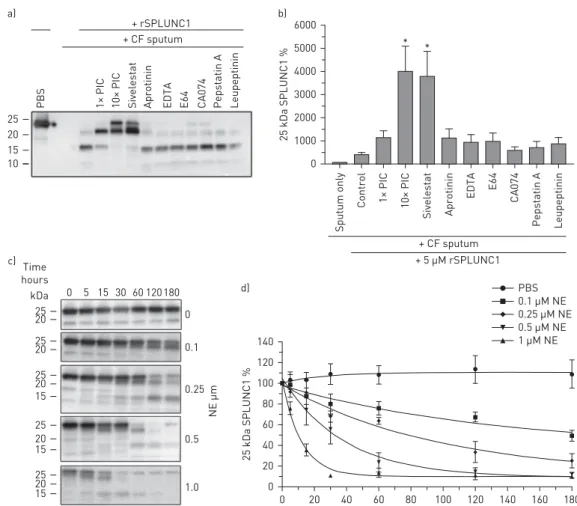
We co-incubated rSPLUNC1 and CF sputum with inhibitors of several proteases, their respective inhibitors and an EDTA-free protease inhibitor cocktail (PIC), used to inhibit multiple proteases [19]. Only sivelestat and 10× PIC significantly attenuated rSPLUNC1 degradation (figure 3a, b). Because NE was elevated in CF sputum, and SPLUNC1’s degradation was sivelestat sensitive, we co-incubated rSPLUNC1 with NE, which degraded rSPLUNC1 in a concentration-dependent manner (figure 3c, d). Owing to the potential importance of these findings, we exposed rSPLUNC1 to normal or CF sputa in the presence of protease inhibitors for extended times. Surprisingly, sivelestat was unable to prevent rSPLUNC1 degradation after extended incubation periods (figure 4a, b).
0 2 4 6 8 10 12
0 0.25 0.5 1 2 4 6 12 Time hours
Time hours
Time hours
1 sputum: 1 PBS PBS
kDa
kDa 25
25 20 15 10
25 20 15 10
Normal sputum
Normal sputum
Normal sputum
CF
sputum
PBS
CF sputum
CF sputum
0 6 0 6 0 6
140
120
100
80
60
40
20
0
CF Normal
120
100
80
60
40
20
0 PBS
25 kDa SPLUNC1 %
25 kDa SPLUNC1 %
0 hours 6 hours **
***
a) b)
c) d)
FIGURE 2 rSPLUNC1 is rapidly degraded in cystic fibrosis (CF) sputum. a) Representative Western blots
To confirm the translation of high NE protein levels into elevated NE activity, we added a NE-sensitive substrate to sputum±sivelestat, and measured the formation of the fluorescent product AMC, which was significantly elevated after exposure to CF but not normal sputum (figure 4c). Sivelestat was unable to reduce AMC formation following 12 h of exposure to CF sputum (figure 4d) and failed to recover SPLUNC1’s ability to regulate ASL height (supplementary figure S3a, b). Additionally, we tested whether cathepsins B, K, S and L were active in CF sputum. Using protease-specific fluorogenic substrates, we observed significantly greater cathepsin B, cathepsin K and cathepsin S/L activity in CF relative to normal sputum (supplementary figure S4a–c). We also examined MMP-2/9 activity, which was significantly elevated in CF sputum (supplementary figure S4d). Because the inhibitors of these enzymes failed to attenuate rSPLUNC1 degradation, we conclude that whilst a number of proteases are functionally active in CF sputum, NE is predominantly responsible for rSPLUNC1 degradation.
Upregulation ofα-,β- andγ-ENaC and Na+/K+ATPaseα1 protein expression in CF bronchi indicates increased ENaC activity
SPLUNC1 was undetectable in SMM collected from excised CF lungs (figure 5a). Similarly, co-incubation of SMM and rSPLUNC1 resulted in a rapid diminution of rSPLUNC1 (figure 5b, c). Because SPLUNC1 affects ENaC proteostasis [13], we measured ENaC levels. Bronchi were collected from non-CF and relatively disease-free areas of CF lungs and we measured protein levels by Western blot [11]. We observed a significant increase in all ENaC subunits in CF bronchi (figure 5d, e). α- and γ-ENaC must be proteolytically cleaved before ENaC can conduct Na+[26]. Interestingly, we detected significant increases
+ rSPLUNC1 + CF sputum
+ 5 μM rSPLUNC1 + CF sputum
1× PIC
PBS 10× PIC Sivelestat Aprotinin EDTA E64 CA074 Pepstatin A Leupeptinin
1× PIC
Control
Sputum only
10× PIC
Sivelestat Aprotinin
EDTA E64 CA074
Pepstatin A Leupeptinin 25
20 15 10
6000
5000
4000
3000
2000
1000
0
* *
PBS 0.1 μM NE 0.25 μM NE 0.5 μM NE 1 μM NE 140
120
100
80
60
40
20
0
0 20 40 60 80 100 120 140 160 180
25 kDa SPLUNC1 %
25 kDa SPLUNC1 %
Time hours
0 5 15 30 60 120 180 kDa
25 20
25 20
25 20 15
25 20 15
25 20 15
0
0.1
0.25
0.5
1.0
NE μm
a)
c)
d) b)
FIGURE 3 Sivelestat partially attenuates rSPLUNC1 degradation. a) Representative Western blot showing
in cleaved ENaC subunits in CF bronchi, suggesting that ENaC activity is upregulated (figure 5d, e). Consistent with increased ENaC levels, the Na+/K+ATPaseα1 subunit was also increased in CF bronchi (figure 5d, e).
CF sputum cleaves the ENaC-inhibitory domain of SPLUNC1
NE predominantly cleaves valine and alanine residues. Indeed, co-incubation of rSPLUNC1 with CF but not normal sputum generated elastase-specific peptides (figure 6a). As a control, we performed a tryptic digest, and detected peptides regardless of genotype (figure 6b). Coverage analysis revealed valine-specific cleavage of SPLUNC1’s S18 region (GGLPVPLDQTLPLNVPA) and the formation of PLDQTLPLNV, which is likely unable to regulate ENaC (figure 6c). We recently developed a novel, NE-resistant, SPLUNC1 peptidomimetic called SPX-101 [18]. The ENaC interacting region of the SPX-101 peptide remained intact following exposure to CF sputum (figure 6c). To determine if rSPLUNC1 and SPX-101 remained active in the presence of CF sputum, we co-incubated PBS or sputum with rSPLUNC1 or SPX-101 overnight and added this apically to HBECs from normal donors. rSPLUNC1 and SPX-101 increased ASL height in the presence of PBS and normal sputum (figure 6d, e). Consistent with mass spectrometry data, only SPX-101 was capable of regulating ASL height in the presence of CF sputum (figure 6d, e).
CF sputum prevents SPLUNC1 binding and elevates ENaC activity
Because ENaC is apically expressed, SPLUNC1 must also bind apically as part of the inhibition process [13]. We therefore co-incubated fluorescently labelled rSPLUNC1 and SPX-101 with PBS or sputum,
3000
2500
2000
1500
1000
500
0
25 kDa SPL
UNC1 A/U
* **
*
* **
Contr
ol
1× PIC 10× PIC
Siv
el
es
tat
Apr
otinin
Contr
ol
1× PIC 10× PIC Siv
el
es
tat
Apr
otinin
+ CF sputum and rSPLUNC1 + CF sputum
250
200
150
100
50
0
6 hours 12 hours
AMC pmol
AMC pmol
Normal sputum Normal
Normal + Silvelestat
CF
CF + Silvelestat
CF sputum 2000
1500
1000
500
0
+ rSPLUNC1
kDa
25 15 10
a) b)
c) d)
FIGURE 4 Sivelestat fails to prevent rSPLUNC1 degradation by cystic fibrosis (CF) sputum over extended
before apical addition to HBECs. We observed apical binding of Dylight633-rSPLUNC1 and 5-TAMRA-SPX-101 in both PBS- and normal sputa-treated HBECs (figure 7a). After exposure to CF sputum, SPX-101 but not SPLUNC1 was able to bind apically (figure 7a). Furthermore, SPX-101 but not SPLUNC1 remained functionally active and internalised α-ENaC-GFP following exposure to NE (supplementary figure S5).
We next determined whether SPLUNC1 degradation by CF secretions contributed to ENaC hyperactivity. We have previously reported increases in ENaC activity in primary CF HBECs [23]. However, passaged HBECs do not display this phenomenon. To start with similar transepithelial voltages (Vt), we exposed passaged normal and CF HBECs to normal or CF sputum, or PBS (control), and measured the resultant amiloride-sensitive Vt as a marker of ENaC activity. Baseline amiloride-sensitive Vts were not significantly different between normal and CF HBECs (figure 7b, c). However, adding normal sputum reduced Vtwhilst CF sputum significantly elevated Vt, independently of HBEC genotype. Addition of rSPLUNC1 in PBS significantly reduced Vt in normal but not CF HBECs, consistent with our previous findings that SPLUNC1 cannot function in CF HBECs [11]. rSPLUNC1 had no further effect on the already reduced Vt in HBECs exposed to normal sputum, suggesting that endogenous S18-like peptides were present in sufficient quantities to inhibit ENaC as described [12]. Normal but not CF sputum was able to significantly decrease the CF Vt (figure 7c), suggesting that spontaneously produced bioactive peptides are present in normal but not CF sputum. To further examine this effect, we passed normal sputum through a size-exclusion column to remove
a) b) c)
d) e)
rSPLUNC1 SMM donor 1 SMM donor 2 SMM donor 3 SMM donor 4
Time hours
0 0.5 1 1.5 3 6 12 24 kDa
25
25
10
PBS
PBS
1 SMM:2 PBS
1 SMM:2 PBS 140
120
100
80
60
40
20
0
25 kDa SPLUNC1 %
0 2 4 6 8 10 12 14 16 18 20 22 24
***
Time hours
Non-CF Non-CF
CF CF
10000
1000
100
10
1
Protein abundance AU
** ** **
***
* *
FL Clv FL Clv Na+/K+
α1 ATPase 1 2 3 4 5 6 7 1 2 3 4 5 6 7 Donor
kDa kDa
25
10
α-ENaC
α-ENaC
β-ENaC
β-ENaC
γ-ENaC
γ-ENaC Na+/K+
ATPase
Pan-cytokeratin 150
100
75
100 75
100 75
50
FIGURE 5 SPLUNC1 is degraded and α-, β- and γ-epithelial Na+ channel (ENaC) subunits are upregulated in cystic fibrosis (CF) bronchi.
peptides and proteins ⩾10 kDa. We then added this filtrate to normal HBECs and measured ASL height 6 h later. This filtrate significantly increased ASL height and adding 30 µM rSPLUNC1 to the filtrate had no further effect (supplementary figure S6). Consistent with our previous observation that full-length SPLUNC1 is inactive in CF airways, rSPLUNC1 failed to reduce amiloride-sensitive Vt in the presence of CF sputum. SPX-101 was able to significantly inhibit the amiloride-sensitive Vtin both normal and CF HBECs after exposure to either PBS or CF sputum (figure 7b, c).
Elastase-specific rSPLUNC1 peptides
6 h 0
24 h 6 h 24 h
CF sputum Normal sputum
6 h 24 h 6 h 24 h
CF sputum Normal sputum Trypsin-specific rSPLUNC1 peptides
Total ion curr
ent
Total ion curr
ent
10×1010 4×10¹⁰
3×10¹⁰
2×10¹⁰
1×10¹⁰
8×10⁹
6×10⁹
4×10⁹
2×10⁹
0
rSPLUNC1 CF coverage
P L S P T G L A G S L T N A L S N G L L L L G K V T S V I P V T I P L G I K L Q E R I H L V L G D C L N K V L P Q F V I K V
A A L P I P L D Q T A A E L V Q
Q F G G L P V P L D Q T L P L N V N P A L L P L L D I L K P G T D P Q L L E L G L L R L A V K L D I T L L D G L G P L P I L R G L D I T L V H S G G L L G I L E N
G L N N I I D I K V V N T P L V G A S L T H S P G S L Q I S G N V C P L V N E V G G T S G G L L G G
V Q S P D G H R L Y A E I L A V R D K Q Q G L L D S L T G I D I V NM L I H G L
SPX-101 CF coverage
PBS Normal sputum CF sputum
PBS
rSPLUNC1
SPX-101
PBS
PBS
rSPLUNC1 SPX-101
15
10
5
0
Normal sputum
CF sputum
NS NS
NS
** **
**
* ***
***
ASL height μm
a)
c)
b)
d)
e)
FIGURE 6Cystic fibrosis (CF) sputum cleaves SPLUNC1’s epithelial Na+channel (ENaC) inhibitory region.a, b)Total ion chromatogram intensities
for elastase-specific (a) and trypsin-specific (b) SPLUNC1 peptides following incubation with CF and normal sputum at 37°C. c) SPLUNC1 coverage after proteomic analysis of elastase-specific peptides. The peptide identified as PLDQTLPLNV indicating disruption of SPLUNC1’s S18 region is highlighted in red.d)Representative airway surface liquid (ASL) heights following a 2 h incubation of human bronchial epithelial cells (HBECs) with PBS, normal sputum or CF sputum ( pooled n=6 donors) ±30 µM rSPLUNC1 or 30 µM SPX-101.e)ASL height measurements from n=12–15 transwells cultured from n=4–5 individual donors, presented as mean±SEM. Data were analysed by two-way ANOVA analysis with Tukey’s
Discussion
The lung mucosal environment contains∼1000 proteins, of which∼5% are proteases and their inhibitors [27]. Under normal conditions, anti-proteases prevent excessive NE activity in the airways [28]. In general, SPLUNC1 was relatively stable in normal sputum (figure 1), although in some cases we did observe some degradation (e.g. supplementary figure S2). However, the degradation product was close in size to full-length SPLUNC1 (>20 kDa) and was similar in size to a SPLUNC1-degradation product that we had previously observed after exposing SPLUNC1 to trypsin [29]. Thus, we speculate that serine proteases present in normal ASL or sputum may post-translationally affect SPLUNC1. However, chronic
CF sputum Normal sputum
rSPLUNC1
SPX-101
** **
**
** *
* ****
****
****
****
**** ****
*** PBS
CF sputum Normal sputum
PBS
rSPLUNC1 SPX-101
PBS
rSPLUNC1 SPX-101
PBS 10
8
6
4
2
0
10
8
6
4
2
0
ΔVt amil
oride mV
ΔVt amil
oride mV
CF sputum Normal sputum
PBS a)
b)
c)
FIGURE 7Cystic fibrosis (CF) sputum prevents SPLUNC1 binding to human bronchial epithelial cell (HBEC)
neutrophilia in the CF lung lumen significantly increases free NE levels [8, 28, 30]. Similarly, we found significantly higher NE protein levels and activity in CF sputum compared with sputum from normal healthy donors (figure 1). We also observed that SPLUNC1 protein levels were significantly reduced in CF sputum and that rSPLUNC1 was rapidly degraded in CF sputum (figures 1 and 2). To confirm that SPLUNC1 was degraded, we performed proteomic analysis. NE-specific SPLUNC1 coverage was markedly increased in CF sputum, indicating inflammation-induced degradation. Crucially, intact S18-like peptides that can inhibit ENaC were absent in CF sputum. Despite being cleaved by NE, SPLUNC1 remains aggregated for several hours before dissociating [31]. However, the lack of SPLUNC1 coverage in CF sputum and the relatively short half-life of rSPLUNC1 in CF sputum suggest that at steady state SPLUNC1 does not exist as a functional entity in CF airway secretions.
NE inhibitors have previously been tested in CF patients, with little effect. For example, AZD9668 was tested in CF patients and had no effect on inflammation or lung function [32]. Similarly, recombinantα1 anti-trypsin also failed to affect lung inflammation [6]. Our data indicate that sivelestat was only partially effective at preventing SPLUNC1 degradation in CF sputum and this effect was lost over time. ENaC is highly promiscuous and is cleaved by multiple proteases. Thus, there are two possible contributors to the failure of NE inhibitors in the clinic: 1) SPLUNC1 and other proteins are still degraded by CF secretions in the absence of NE; and 2) ENaC can still be activated by other proteases in the CF lung leading to persistent ASL dehydration. Thus, whilst newer broad-spectrum anti-proteases such as QUB-TL1 may be more effective at preventing ENaC cleavage [33], whether or not they are fully effective in the CF lung remains to be determined.
Our working hypothesis is that SPLUNC1 causes removal of ENaC from the plasma membrane, preventing its cleavage and activation. We have recently developed SPX-101, a size-optimised peptide that can inhibit ENaC in CF HBECs and CF-like animal models [18]. Because SPX-101 is currently undergoing clinical trials for the treatment of CF, we tested whether SPX-101 was stable and efficacious in CF sputum samples. Both SPLUNC1 and SPX-101 increased ASL height after overnight incubation in normal sputum. However, after exposure to CF sputum, SPLUNC1 was degraded and failed to regulate ASL height, whereas SPX-101 remained intact and active. As a control, we also incubated SPLUNC1 and SPX-101 overnight with NE and probed forα-ENaC-GFP diminution, as an indication of internalisation. Whilst SPLUNC1 lost the ability to inhibit ENaC, SPX-101 again remained active even after prolonged exposure to NE (supplementary material).
SPLUNC1 was degraded in CF sputum from the central airways. However, by utilising SMM, we also observed SPLUNC1 degradation in more distal regions of CF lungs. Consistent with the lack of SPLUNC1, we observed a significant increase in full-lengthα-,β- andγ-ENaC subunits, suggesting that SPLUNC1 had failed to internalise ENaC. Crucially,α- and/orγ-ENaC must be proteolytically cleaved to be active [26, 34, 35] and we also detected significant increases in cleavedα- andγ-ENaC subunits in CF airways. The basolateral Na+/K+-ATPase serves to pump out Na+that enters the cell viaENaC in order to keep intracellular Na+at∼23 mM [36]. This pump has previously been shown to be functionally upregulated in CF airways [37]. Similarly, we observed a significant increase in Na+/K+-ATPase protein levels in CF airways, consistent with the increase in cleavedα- andγ-ENaC subunits. Welsh and colleagues [38, 39] have proposed that the increase in ENaC activity detected by electrophysiology is an artefact caused by amiloride-induced apical membrane hyperpolarisation. However, our biochemical data, which demonstrate that cleaved (i.e. active) ENaC as well as the necessary Na+/K+-ATPase are upregulated in CF epithelia, suggest that this is not the case.
Whilst defective ion transport is widely accepted to be the initial event in the pathogenesis of CF lung disease, the role of ENaC remains highly controversial [17]. A number of studies have demonstrated increased electrical Na+ flux and fluid changes in CF airway epithelia that were attributable to increased ENaC activity [3, 40]. In contrast, others failed to find differences in CF piglets and in cultured airway epithelia [38, 39, 41]. Our data revealed that rSPLUNC1 failed to bind to HBEC apical membranes after pre-incubation with CF sputum, whereas N-terminally labelled SPX-101 bound equally well after exposure to normal or CF sputum. rSPLUNC1 was amine-labelled with DyLight633, suggesting that multiple residues were labelled. Thus, whilst it is possible that CF proteases may have removed some dye-labelled residues, we limited our SPLUNC1/CF sputum pre-incubation to 2 h, at which point∼20% of the protein should have remained.
of pH-sensitive salt bridges in the tertiary structure of SPLUNC1, which render it inactive below pH 7. Conversely, isolated ENaC-inhibitory peptides, such as SPX-101, lack the salt bridges and are pH independent [11]. Surprisingly, both non-CF and CF HBECs showed a significant decrease in the amiloride-sensitive Vtafter being exposed to normal sputum for∼2 h. This is likely due to the presence of endogenous S18-like ENaC-inhibitory peptides [12]. Indeed, after passing normal sputum through a size-exclusion column to remove proteins⩾10 kDa, including full-length SPLUNC1 which is∼25 kDa, the filtrate was able to significantly increase ASL height. Adding rSPLUNC1 to the filtrate had no further effect (supplementary figure S5), suggesting that the filtrate and SPLUNC1 have a similar target (i.e. ENaC). Thus, whilst we cannot state that SPLUNC1/S18 is the only ENaC-regulatory peptide in the ASL, our data indicate that small, ASL-regulatory peptides are present in normal sputum. In contrast, adding CF sputum significantly elevated the amiloride-sensitive Vt in both non-CF and CF HBECs relative to normal sputum. As with the ASL height and binding experiments, SPLUNC1 was ineffective at ameliorating these effects, due to excessive degradation. However, SPX-101 remained active and could significantly reduce the amiloride-sensitive Vt. These changes are likely due to an abundance of proteases, many of which can cleave and activate ENaC, as well as a lack of functional SPLUNC1 or SPLUNC1-derived peptides in CF sputum. Taken together, these findings demonstrate that the CF mucosal environment (i.e. the ASL secretions) influences ion transport in established CF lung disease. Previous studies have demonstrated cellular interactions between CFTR and ENaC [4, 14, 15]. However, our study is the first to test the effects of CF airways secretions on ENaC activity and our data strongly indicate that the extracellular environment drives ENaC hyperactivity in CF airways.
In conclusion, we have shown that SPLUNC1 is absent or markedly reduced in CF airway secretions, which contributes to increased ENaC activity and ASL dehydration in CF HBECs. These data suggest that studying CF HBECs in the presence of CF sputum will serve as a more realistic model of CF airways and also indicate that novel therapies should be tested for efficacy in the presence of CF secretions.
Acknowledgements: We thank the University of Carolina (UNC) Cystic Fibrosis Center Tissue Cores for providing cells and tissue, Colin Bingle (University of Sheffield, Sheffield) for providing the SPLUNC1 construct, Debbie Baines (St. George’s, University of London) for providing the α-ENaC-GFP construct, Thomas Kleyman (University of Pittsburgh) and M. Jackson Stutts (UNC) for theβ- andγ-ENaC constructs, and Michael Miley and Ricard Feng (UNC) for purifying SPLUNC1. Finally, we would like to thank Brian Button (UNC) for allowing use of his equipment for this project.
Author contributions: M.J. Webster, B. Reidel, C.D. Tan, A. Ghosh and R. Tarran designed and performed research and analysed data. N.E. Alexis, S.H. Donaldson and C.M.P. Ribeiro provided sputum and SMM samples, designed experiments and interpreted data. M. Kesimer designed experiments and analysed data. M. J. Webster, B. Reidel and R. Tarran wrote the manuscript. All other authors edited and approved the manuscript.
Support statement: This study was funded by National Institutes of Health R01HL108927, P30DK065988, the UK Cystic Fibrosis Trust (INOVCF), Cystic Fibrosis Foundation R026-CR11 and TARRAN17G0. Funding information for this article has been deposited with the Crossref Funder Registry.
Conflict of interest: M.J. Webster has nothing to disclose. B. Reidel has nothing to disclose. C.D. Tan has nothing to disclose. A. Ghosh has nothing to disclose. N.E. Alexis has nothing to disclose. S.H. Donaldson reports grants from Vertex Pharmaceuticals and Parion Sciences, grants and personal fees from Nivalis Pharmaceuticals, and personal fees from Pulmatrix, Novartis Pharmaceuticals and PTC Therapeutics, outside the submitted work; in addition, S.H. Donaldson has a patent“Regulation of sodium channels by PLUNC proteins”with royalties paid. M. Kesimer has nothing to disclose. C.M.P. Ribeiro is founder of and has equity in Irex Pharma. R. Tarran is founder of and on the board of directors of Spyryx Biosciences, outside the submitted work; in addition, R. Tarran has a patent“Regulation of sodium channels by PLUNC proteins”with royalties paid.
References
1 Quinton PM. Chloride impermeability in cystic fibrosis.Nature1983; 301: 421–422.
2 Matalon S, Bartoszewski R, Collawn JF. Role of epithelial sodium channels in the regulation of lung fluid homeostasis.Am J Physiol Lung Cell Mol Physiol2015; 309: L1229–L1238.
3 Boucher RC, Stutts MJ, Knowles MR,et al.Na+ transport in cystic fibrosis respiratory epithelia. Abnormal basal rate and response to adenylate cyclase activation.J Clin Invest1986; 78: 1245–1252.
4 Stutts MJ, Canessa CM, Olsen JC,et al.CFTR as a cAMP-dependent regulator of sodium channels.Science1995; 269: 847–850.
5 Matsui H, Grubb BR, Tarran R, et al. Evidence for periciliary liquid layer depletion, not abnormal ion composition, in the pathogenesis of cystic fibrosis airways disease.Cell1998; 95: 1005–1015.
6 Martin SL, Moffitt KL, McDowell A,et al.Association of airway cathepsin B and S with inflammation in cystic fibrosis.Pediatr Pulmonol2010; 45: 860–868.
7 Chalmers JD, Moffitt KL, Suarez-Cuartin G,et al.Neutrophil elastase activity is associated with exacerbations and lung function decline in bronchiectasis.Am J Respir Crit Care Med2017; 195: 1384–1393.
9 Wu T, Huang J, Moore PJ,et al.Identification of BPIFA1/SPLUNC1 as an epithelium-derived smooth muscle relaxing factor.Nat Commun2017; 8: 14118.
10 Bartlett J, Gakhar L, Penterman J,et al.PLUNC: a multifunctional surfactant of the airways.Biochem Soc Trans 2011; 39: 1012–1026.
11 Garland AL, Walton WG, Coakley RD,et al.Molecular basis for pH-dependent mucosal dehydration in cystic fibrosis airways.Proc Natl Acad Sci USA2013; 110: 15973–15978.
12 Hobbs CA, Blanchard MG, Alijevic O,et al.Identification of the SPLUNC1 ENaC-inhibitory domain yields novel strategies to treat sodium hyperabsorption in cystic fibrosis airway epithelial cultures.Am J Physiol Lung Cell Mol Physiol2013; 305: L990–L1001.
13 Kim CS, Ahmad S, Wu T,et al.SPLUNC1 is an allosteric modulator of the epithelial sodium channel.FASEB J 2018; 32: 2478–2491.
14 Schreiber R, Hopf A, Mall M,et al.The first-nucleotide binding domain of the cystic-fibrosis transmembrane conductance regulator is important for inhibition of the epithelial Na+ channel.Proc Natl Acad Sci USA1999; 96: 5310–5315.
15 Rubenstein RC, Lockwood SR, Lide E,et al.Regulation of endogenous ENaC functional expression by CFTR and ΔF508-CFTR in airway epithelial cells.Am J Physiol Lung Cell Mol Physiol2011; 300: L88–L101.
16 Stoltz DA, Meyerholz DK, Pezzulo AA,et al. Cystic fibrosis pigs develop lung disease and exhibit defective bacterial eradication at birth.Sci Transl Med2010; 2: 29ra31.
17 Stoltz DA, Meyerholz DK, Welsh MJ. Origins of cystic fibrosis lung disease.N Engl J Med2015; 372: 1574–1575.
18 Scott DW, Walker MP, Sesma J,et al.SPX-101 is a novel epithelial sodium channel-targeted therapeutic for cystic fibrosis that restores mucus transport.Am J Respir Crit Care Med2017; 196: 734–744.
19 Abdullah LH, Coakley R, Webster MJ, et al. Mucin production and hydration responses to mucopurulent materials in normalversusCF airway epithelia.Am J Respir Crit Care Med2017; 197: 481–491.
20 Fulcher ML, Randell SH. Human nasal and tracheo-bronchial respiratory epithelial cell culture.Methods Mol Biol 2013; 945: 109–121.
21 Wisniewski JR, Zougman A, Nagaraj N,et al.Universal sample preparation method for proteome analysis.Nat Methods2009; 6: 359–362.
22 Kesimer M, Cullen J, Cao R,et al.Excess secretion of gel-forming mucins and associated innate defense proteins with defective mucin un-packaging underpin gallbladder mucocele formation in dogs. PLoS One 2015; 10: e0138988.
23 Tarran R, Trout L, Donaldson SH,et al.Soluble mediators, not cilia, determine airway surface liquid volume in normal and cystic fibrosis superficial airway epithelia.J Gen Physiol2006; 127: 591–604.
24 Jiang D, Wenzel SE, Wu Q,et al.Human neutrophil elastase degrades SPLUNC1 and impairs airway epithelial defense against bacteria.PLoS One2013; 8: e64689.
25 Sagel SD, Sontag MK, Wagener JS,et al.Induced sputum inflammatory measures correlate with lung function in children with cystic fibrosis.J Pediatr2002; 141: 811–817.
26 Caldwell RA, Boucher RC, Stutts MJ. Neutrophil elastase activates near-silent epithelial Na+ channels and increases airway epithelial Na+transport.Am J Physiol Lung Cell Mol Physiol2005; 288: L813–L8L9.
27 Kesimer M, Kirkham S, Pickles RJ,et al.Tracheobronchial air-liquid interface cell culture: a model for innate mucosal defense of the upper airways?Am J Physiol Lung Cell Mol Physiol2009; 296: L92–L100.
28 Birrer P, McElvaney NG, Rüdeberg A,et al.Protease-antiprotease imbalance in the lungs of children with cystic fibrosis.Am J Respir Crit Care Med1994; 150: 207–213.
29 Garcia-Caballero A, Rasmussen JE, Gaillard E, et al. SPLUNC1 regulates airway surface liquid volume by protecting ENaC from proteolytic cleavage.Proc Natl Acad Sci2009; 106: 11412–11417.
30 Armstrong DS, Grimwood K, Carlin JB,et al.Lower airway inflammation in infants and young children with cystic fibrosis.Am J Respir Crit Care Med1997; 156: 1197–1204.
31 Ahmad S, Tyrrell J, Walton WG,et al.Short Palate, Lung, and Nasal Epithelial Clone 1 has antimicrobial and antibiofilm activities against the Burkholderia cepacia complex. Antimicrob Agents Chemother 2016; 60: 6003–6012.
32 Elborn JS, Perrett J, Forsman-Semb K,et al. Efficacy, safety and effect on biomarkers of AZD9668 in cystic fibrosis.Eur Respir J2012; 40: 969–976.
33 Reihill JA, Walker B, Hamilton RA,et al. Inhibition of protease-epithelial sodium channel signaling improves mucociliary function in cystic fibrosis airways.Am J Respir Crit Care Med2016; 194: 701–710.
34 Vallet V, Chraibi A, Gaeggeler HP,et al. An epithelial serine protease activates the amiloride-sensitive sodium channel.Nature1997; 389: 607–610.
35 Kleyman TR, Kashlan OB, Hughey RP. Epithelial Na+channel regulation by extracellular and intracellular factors. Annu Rev Physiol2018; 80: 263–281.
36 Boucher RC. Human airway ion transport. Part one.Am J Respir Crit Care Med1994; 150: 271–281.
37 Stutts MJ, Knowles MR, Gatzy JT,et al.Oxygen consumption and ouabain binding sites in cystic fibrosis nasal epithelium.Pediatr Res1986; 20: 1316–1320.
38 Itani OA, Chen JH, Karp PH,et al.Human cystic fibrosis airway epithelia have reduced Cl- conductance but not increased Na+conductance.Proc Natl Acad Sci USA2011; 108: 10260–10265.
39 Chen JH, Stoltz DA, Karp PH,et al.Loss of anion transport without increased sodium absorption characterizes newborn porcine cystic fibrosis airway epithelia.Cell2010; 143: 911–923.
40 Knowles M, Gatzy J, Boucher R. Increased bioelectric potential difference across respiratory epithelia in cystic fibrosis.N Engl J Med1981; 305: 1489–1495.