Original Article
Regulation of apoptosis by long non-coding RNA
HIF1A-AS1 in VSMCs: implications for TAA pathogenesis
Yongbo Zhao1, Guangxing Feng1, Yanzhi Wang1, Yuehong Yue2, Weichao Zhao1
1Department of Cardiovascular Surgery, Fourth Hospital of Hebei Medical University, Shijiazhuang, China; 2Department of Neurology, Hebei General Hospital, Shijiazhuang, China
Received September 20, 2014; Accepted November 8, 2014; Epub October 15, 2014; Published November 1, 2014
Abstract: Objective: Long non-coding RNAs (lncRNAs) play important roles in diverse biological processes, such as transcriptional regulation, cell growth and tumorigenesis. However, little was known about whether lncRNA HIF 1 alpha-antisense RNA 1 (HIF1a-AS1) in regulating the proliferation and apoptosis of VSMCs in vitro and the expres-sion of HIF1a-AS1 in serum of TAA patients. Methods: The cell viability was detected by the CCK8 assay. The cell apoptosis was assessed by annexin V-PI double-labeling staining. Expression of genes and proteins were analyzed by real-time PCR and western blotting respectively. Cells were transfected with siRNAs as a gene silencing methods.
Results: In serum of TAA patients, the expression of HIF1a-AS1 was significantly increased (superior to 6 folds) compared to the normal control. Moreover, PA induced cell apoptosis in VSMCs in a time- and dose-dependent manner, and the proportion of the apoptotic cells had gained as compared to untreatment group. PA also induced upregulation expression of HIF1a-AS1. We also found that transfection of cells with HIF1a-AS1 siRNA decreased the expression of caspase3 and caspase8 and increased the expression of Bcl2, and protected PA-induced cell apoptosis in VSMCs. Conclusions: HIF1a-AS1 was overexpressed in the thoracoabdominal aorta aneurysm and the interaction between HIF1a-AS1 and apoptotic proteins plays a key role in the proliferation and apoptosis of VSMCs in vitro, which may contribute to the pathogenesis of thoracoabdominal aorta aneurysm.
Keywords: Thoracoabdominal aorta aneurysm, HIF 1alpha-antisense RNA 1, vascular smooth muscle cells, long non-coding RNA
Introduction
Aneurysm of the thoracoabdominal aorta (TAA) is relatively uncommon in the spectrum of aneurysmal disease, accounting for only 3% of diagnosed aneurysms in the United States [1]. Currently, the incidence and operations of
tho-racoabdominal aortic aneurysms have signifi -cantly increased. The indications for repair are considered to be a diameter of 6 cm or more and 5.5 cm for patient groups with increased risk of rupture [2]. Complex open surgical repair
is associated with significant mortality and
complication rates. The endovascular approach has evolved to be a good and predominant alternative to open repair of these aneurysms for older and high-risk patients as well as for aneurysms with optimal morphological suitabil-ity [3]. Nevertheless, the advances in effective therapy for TAA have been limited because the pathological mechanisms causing tumor are
not known. Therefore, revealing the molecular mechanism for the TAA is indispensable for developing effective treatment.
Table 1. Disease-related LncRNA in human
Symbol Cancers Chromosome location Start End Strand Species Alias NCBI No.
7SK Cancer chr6 52860418 52860749 + Human RN7SK; 7SK NR_001445.2 22377309
BCAR4 Breast cancer chr16 11913687 11922689 - Human BCAR4 NR_024049.1 21506106
BCYRN1 Breast cancer chr2 47562454 47562653 + Human BCYRN1; BC200; BC200a; LINC00004; NCRNA00004 NR_001568.1 15240511 BOK-AS1 Testicular cancer chr2 242483799 242498558 - Human BOK-AS1; BOKAS; NAToB; BOK-AS; NCRNA00151 NR_033346.1 19287972
C1QTNF9B-AS1 Prostate cancer chr13 24463028 24466242 + Human C1QTNF9B-AS1; PCOTH BC073902 15930275
CASC2 Endometrial cancer chr10 119806332 119969665 + Human CASC2; C10orf5 NR_026939.1 15024726
CBR3-AS1 Prostate cancer chr21 37504065 37528606 - Human CBR3-AS1; PlncRNA-1 NR_038892.1 22264502
CCAT1 Colorectal cancer chr8 128219629 128231724 - Human CCAT1 XR_108886.3 23416875
CDKN2B-AS1 Breast cancer chr9 21994790 22121096 + Human CDKN2B-AS1; ANRIL; p15AS; CDKN2BAS; CDKN2B-AS;
NCRNA00089; RP11-145E5.21 NR_003529.20 17440112
DNM3OS Ovarian cancer chr1 172107724 172113975 - Human DNM3OS; DNM3-AS1; MIR199A2HG NR_038397.1 20400975
DSCAM-AS1 Breast cancer chr21 41755010 41757285 + Human DSCAM-AS1; M41 NR_038896.1 12177779
EPB41L4A-AS1 Cancer chr5 111496223 111498198 + Human EPB41L4A-AS1; TIGA1; C5orf26; NCRNA00219 NR_015370.2 16973895
GAS5 Breast cancer chr1 173833039 173837125 - Human GAS5; SNHG2; NCRNA00035 NR_002578.7 20673990
H19 Bladder cancer chr11 2016406 2019065 - Human H19; ASM; BWS; WT2; ASM1; PRO2605; D11S813E; LINC00008; NCRNA00012
NR_002196.5 11193051
HIF1A-AS1 Kidney cancer chr14 62147759 62162536 - Human HIF1A-AS1; 5’aHIF-1A 21897117
HOTAIR Breast cancer chr12 54356096 54362515 - Human HOTAIR; HOXAS; HOXC-AS4; HOXC11-AS1; NCRNA00086 NR_047517.15 19182780
IGF2-AS Prostate cancer chr11 2161758 2169896 + Human IGF2-AS; PEG8; IGF2AS; IGF2-AS1 NR_028044.1 19767753
KCNQ1OT1 Colorectal cancer chr11 2661768 2721228 - Human “KCNQ1OT1; LIT1; KvDMR1; KCNQ10T1; KCNQ1-AS2; KvLQT1-AS; NCRNA00016
16965397
lincRNAp21 Lung cancer N/A N/A N/A N/A Human N/A N/A 22535282
LSINCT5 Breast cancer N/A N/A N/A N/A Human N/A N/A 21532345
MALAT1 Cancer chr11 65265233 65273940 + Human MALAT1; HCN; NEAT2; MALAT-1; PRO2853; LINC00047; NCRNA00051
NR_002819.6 20711585
MEG3 Bladder cancer chr14 101292445 101327363 + Human MEG3; GTL2; FP504; prebp1; PRO0518; PRO2160; LINC00023;
NCRNA00028 NR_002766.7 14602737
MIR31HG Breast cancer chr9 21454267 21559697 - Human MIR31HG NR_027054.1 22289355
PCA3 Prostate cancer chr9 79379354 79402465 + Human PCA3; DD3; NCRNA00030 NR_015342.12 18602209
PCAT1 Prostate cancer chr8 128025399 128033259 + Human PCAT1; PCAT-1 NR_045262.1 21804560
PCGEM1 Prostate cancer chr2 193614571 193641625 + Human PCGEM1; LINC00071; NCRNA00072 NR_002769.2 16515751
PCNCR1 Prostate cancer N/A N/A N/A N/A Human N/A N/A 22535282
PVT1 Breast cancer chr8 128806779 129113499 + Human PVT1; LINC00079; NCRNA00083 NR_003367.6 17908964
RRP1B Cancer chr21 45079432 45115960 + Human RRP1B; Nnp1; RRP1; NNP1L; KIAA0179 NM_015056.2 19710015
SRA1 Breast cancer chr5 139929653 139937678 - Human SRA1; SRA; SRAP; STRAA1; pp7687 NM_001035235.6 20079837
TDRG1 Testicular cancer chr6 40346163 40347631 + Human TDRG1; LINC00532 NR_024015.1 21243750
UCA1 Bladder cancer chr19 15939757 15946230 + Human UCA1; CUDR; LINC00178; NCRNA00178 NR_015379.3 20117985
WRAP53 Cancer chr17 7589389 7606820 + Human WRAP53; DKCB3; TCAB1; WDR79 NM_001143990.1 21441950
XIST Breast cancer chrX 73040495 73072588 - Human XIST; SXI1; swd66; DXS1089; DXS399E; LINC00001; NCRNA00002 NR_001564.3 17545591
Yiya Cancer chr1 214098092 214099997 + Human LINC00538; Yiya NR_046189.1 22258142
above research results show that the function-al dysfunction of VSMCs may be correlated with cardiovascular diseases and cancers.
Eukaryotic genomes encode numerous long
non coding RNAs (LncRNAs), which is defined
as endogenous cellular RNAs with length longer than 200 nucleotides, but lack open reading
frames of significant length [7]. Within 4 years, the number of identified lncRNA genes increase
more than 8000 [8]. Although the function of most lncRNAs is still unknown, their increasing numbers and the accumulating evidence for their involvement in many biologic processes provide compelling arguments in support of the
dysregulation of lncRNAs has been correlated to cancer development, invasion and metastasis in the malignant cell [8-10] (Table 1). To date, the underlying mechanisms for lcnRNAs regulation VSMCs proliferation and apoptosis are quite limited.
In this study, we performed a hierarchical cluster analysis of the differentially expressed Lnc-RNA in the serum of TAA patients to identify the lncRNA HIF 1 alpha-antisense RNA 1 (HIF1A-AS1) that associated with Lnc-RNA expression. We also investigated the role of HIF1A-AS1 in vitro in regulating the proliferation and apoptosis of aortic VSMCs.
Materials and methods
Serum samples and cell cul-ture
[image:3.612.92.377.84.545.2]Human serum samples were obtained with written informed consent from The Fourth Ho- spital of Hebei Medical Uni- versity. The study was app- roved by the Ethics Committee of The Fourth Hospital of Hebei Medical University. 50 serum samples of TAA patients and 50 cases of normal control group were collected between 02/2010 and 12/2014. Table 2. PCR primers used in this study
Symbol Forward Reverse
7SK AAACAAGCTCTCAAGGTC CCTCATTTGGATGTGTCT BCAR4 GGACTCATTGTTGTTCTAC ACCTATGGCTATCATTGTT BCYRN1 CTGGGCAATATAGCGAGAC TGCTTTGAGGGAAGTTACG BOK-AS1 CTTGGCAGTTCTGATTGTG TTGTCCGCTGTGGATAAG C1QTNF9B-AS1 AGACACCTGAACATTCCT CTGAGCAAGTTTCCTTCTT CASC2 CTATTCCGAGTAAGAAGTG TCTGTGTTGATGTTGATT CBR3-AS1 CTTCTGGTTACAATGATTCTC CACTTACTGCCTACATTAGA CDKN2B-AS1 TCATCATCATCATCATCATC TGCTTCTGTCTCTTCATA DNM3OS ATAGAGCAAGTCTGGATT GGATGAGGCAATAACATT DSCAM-AS1 ACTAGCACAGATGGCATTC CAGGAAGCATCGTGAACA EPB41L4A-AS1 TAAGACAGTGAGGATGTGAAT ATTATGGTGACAGCAGTGA GAS5 CACAGGCATTAGACAGAA AGGAGCAGAACCATTAAG H19 CTCCACGACTCTGTTTCC TCTCCACAACTCCAACCA HIF1A-AS1 AATGTGTTCCTTGCTCTT GTATGTCTCAGTTATCTTCCT HOTAIR AATAGACATAGGAGAACACTT AATCTTAATAGCAGGAGGAA IGF2-AS CGCCACTGTGTTACCATT TTGCCCATCCCAGATAGAA KCNQ1OT1 GCATATCTGTCTTCCGTAT CCTCTTCCTTCGTTCAAT LSINCT5 TAGACAACTTACTTAACCTCAT TCCTTATCCACCTTATCCA MALAT1 CCGCTGCTATTAGAATGC CTTCAACAATCACTACTCCAA MEG3 TGGCATAGAGGAGGTGAT AGACAAGTAAGACAAGCAAGA MIR31HG ACTTCCACGATAGCAATG GAATGAATCCTCTGTCCTC PCA3 AATCATACTGGTCACTTATCT TTAACAACTGGTCCTGAG PCAT1 TAGAGCCTTGAAGATGAG TCGTGTAGTTGTAAGATGA PCGEM1 TAGTTAAGCAGATTCATAGA GATGTCATAGTCCTCTTC PVT1 CTTGAGAACTGTCCTTACG CAGATGAACCAGGTGAAC RRP1B CAGTATATCTCAACTCAGT TTCTTCTTCCTTCTTCTC SRA1 TTACAGAGATTAGAACCACATT GGCAAGTCAGAGTTACAAT TDRG1 GATTCGTCTGGTTCCTTA TTCCTCTTGACTGATTCTAA UCA1 TTCCTTATTATCTCTTCTG TCCATCATACGAATAGTA WRAP53 CAATAGTGCTGATAACAT CAGTAATCATAGATGGTAT XIST GAACCACCTACACTTGAG TGCTATGCGTTATCTGAG Yiya TATCCTATTCTTAGCAACTG ACATACCTGGCATATAGT ZNFX1-AS1 CCAGTTCCACAAGGTTAC GCAGGTAGGCAGTTAGAA U6 CTCGCTTTGGCAGCACA AACGCTTCACGAATTTGCGT
The vascular smooth muscle cells (VSMCs) were maintained in RPMI-1640 (Invitrogen, USA) supplemented with 10% FBS (Invitrogen,
USA) at 37°C in a humidified incubator (Thermo,
USA), 5% CO2, 95% air atmosphere. The
medi-um was replenished every day. Confluent cells
were treated with various concentrations of pal-mitic acid (Sigma, USA).
Cell viability detection by CCK8
respec-tively. 10 μL of CCK8 (Beyotime, China) was
added to the cells, and the OD value of the cells was measured at 450 nm using an ELISA read-er (BioTek, USA) according to the manufactur-er’s instructions.
Quantification of apoptosis by flow cytometry
Apoptosis was assessed using annexin V, a pro-tein that binds to phosphatidylserine (PS)
resi-protocol (Invitrogen, Carlsbad, CA, USA). Synthesis of cDNAs was performed by reverse
transcription reactions with 2 μg of total RNA
using moloney murine leukemia virus reverse transcriptase (Promega, Switzerland) with oligo dT (15) primers (Fermentas) as described by
the manufacturer. The first strand cDNAs
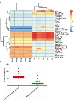
[image:4.612.93.376.67.447.2]served as the template for the regular poly-merase chain reaction (PCR) performed using a DNA Engine (ABI 9700). The cycling conditions Figure 1. Hierarchical cluster analysis of the differentially expressed long
non-coding RNAs (LncRNA) and sHIF1a-AS1 expression in serum of TAA
pa-tients. The figure is drawn by MeV software (version 4.2.6). A. Differentially
expressed LncRNAs chosen from lncRNA and disease database. Correlation similarity matrix and average linkage algorithms are used in the cluster analy-sis. Each row represents an individual LncRNA, and each column represents a sample. The dendrogram at the left side and the top displays similarity of expression among LncRNAs and samples individually. The color legend at the right represents the level of mRNA expression, with red indicating high expression levels and blue indicating low expression levels; B. The expres-sion of HIF 1alpha-antisense RNA 1 (HIF1a-AS1) in serum of TAA patients is measured by Quantitative real-time PCR, 50 serum samples of TAA patients and 50 cases of normal control group were collected. Values are expressed as mean ± SEM, n=50 in each group.
dues which are exposed on the cell surface of apoptotic cells. VSMCs (5.0×10⁵/well, 1 ml) were plated and treated in 6-well plates (three wells per group). After treatment, cells were washed twice with PBS (pH=7.4), and re-suspended in staining buffer containing 10
μl PI and 5 μl annexin V-FITC.
Double-labeling was per-formed at room temperature for 15 min in the dark before
the flow cytometric analysis.
Cells were immediately ana-lyzed using FACScan and the Cellquest program (Becton Dickinson). Quantitative assessment of apoptotic cells was also assessed by the ter-minal deoxynucleotidyl trans-ferase-mediated deoxyuridine triphosphate nick end-labeling (TUNEL) method, which exam-ines DNA-strand breaks during apoptosis by using BD ApoAlertTM DNA Fragmentation Assay Kit. The cells were
tryp-sinized, fixed with 4% parafor -maldehyde, and permeabilized with 0.1% Triton-X-100 in 0.1% sodiumcitrate. After being washed, the cells were incu-bated with the reaction mix-ture for 60 min at 37°C. The stained cells were then
ana-lyzed with flow cytometer.
Quantitative real-time PCR
were 2-min polymerase activation at 95°C fol-lowed by 40 cycles at 95°C for 15 s and 55°C for 60 s. PCR with the following primers: as shown in Table 2. U6 as an internal control was used to normalize the data to determine the relative expression of the target genes. The reaction conditions were set according to the kit instructions. After completion of the
reac-tion, the amplification curve and melting curve
were analyzed. Gene expression values are rep-resented using the 2-ΔΔct method.
Western blotting
The VSMCs were homogenized and extracted in NP-40 buffer, followed by 5-10 min boiling and
centrifugation to obtain the supernatant.
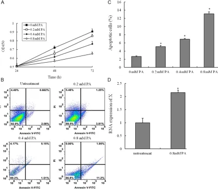
[image:5.612.96.520.73.441.2]Samples containing 50 μg of protein were sepa -rated on 10% SDS-PAGE gel, transferred to PVDF Transfer Membrane (Millipore). After sat-uration with 5% (w/v) non-fat dry milk in TBS and 0.1% (w/v) Tween 20 (TBST), the mem-branes were incubated with the following anti-bodies, caspase3, caspase8 and Bcl-2 (Santa Cruz, USA), at dilutions ranging from 1:500 to 1:2,000 at 4°C over-night. After three washes with TBST, membranes were incubated with secondary immunoglobulins (Igs) conjugated to IRDye 800CW Infrared Dye (LI-COR), including donkey anti-goat IgG and donkey anti-mouse IgG at a dilution of 1:10,000-1:20,000. After 1 Figure 2. Palmitic acid-induced the apoptosis of vascular smooth muscle cells (VSMCs). VSMCs are incubated with various concentrations of palmitic acid (PA) for 24 h, 48 h or 72 h, and the cell viability was examined by CCK8 as-say. (A) Cells are treated with vehicle, 0.2 mM PA, 0.4 mM PA or 0.8 mM PA for 48 h; (B) The percentage of apoptotic
cells is also analyzed by flow cytometric analysis of annexin V/PI double staining and (C) the percentage of apoptotic
hour incubation at 37°C, membranes were washed three times with TBST. Blots were visu-alized by the Odyssey Infrared Imaging Sy- stem (LI-COR Biotechnology). Signals were den-sitometrically assessed (Odyssey Application Software version 3.0) and normalized to the
β-actin signals to correct for unequal loading using the monoclonal anti-β-actin antibody
(Bioworld Technology, USA).
RNA interference
The small interfering (si) RNA for human PTX3 or scramble siRNA was obtained from Dhar- macon (Lafayette, USA). The small interfering with the following primers: siHIF1A-AS1-1, For-
ward 5’-GAGUCUGUGUGGGACAAGCACUUCA-3’ and Reverse 5’-AGUAGAGGAUGUGACUCACUG- UCUG-3’; siHIF1A-AS1-2, Forward 5’-GCUAAC- ACUGGUCUGAGCAAGGU-3’ and Reverse 5’-UC- CUCAAGGAGAGAGGACUAAGC-3’, siHIF1A-AS1- 3, Forward 5’-GCACAGGAUUCAGUCCACUGUC- UU-3’ and Reverse 5’-GACACAGGACACUGAA- AGCUUGG-3’; scramble, Forward 5’-CACCAGU- GGCUAUCACACGUGUGA-3’ and Reverse 5’-UCA- AGAGGAGUGUAACCCACACGU-3’. The siRNA
oli-gonucleotides (at a final concentration of 100
[image:6.612.94.521.66.427.2]nM) were transfected into human umbilical vein endothelial cells using Lipofectamine 2000 (Invitrogen, USA) according to the manufactur-er’s instructions.
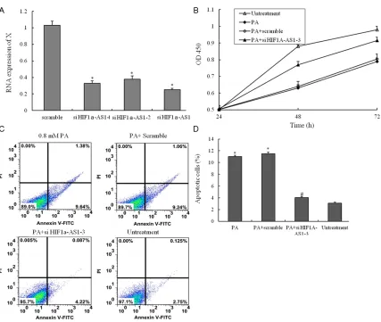
Figure 3. The small interfering RNA for suppressing the function of HIF1a-AS1. (A) Three different small interfering RNA were transfected into VSMCs suppressing the RNA expression of HIF1a-AS1; (B) VSMCs are treated with un-treatment, 0.8 mM PA only, 0.8 mM PA plus scramble si-RNA and 0.8 mM PA plus si- HIF1a-AS1-3 for 48 h, and the cell viability was examined by CCK8 assay; (C) VSMCs are treated with untreatment, 0.8 mM PA only, 0.8 mM PA plus scramble si-RNA and 0.8 mM PA plus si- HIF1a-AS1-3 for 48 h, the percentage of apoptotic cells is also analyzed
by flow cytometric analysis of annexin V/PI double staining and (D) the percentage of apoptotic cells (at the right
of pictures). Values are expressed as mean ± SEM, n=3 in each group. *P < 0.05, versus untreatment group; #P <
Statistical analysis
The data from these experiments were report-ed as mean ± standard errors of mean (SEM) for each group. All statistical analyses were performed by using PRISM version 4.0 (GraphPad). Inter-group differences were ana-lyzed by one-way ANOVA, and followed by Tukey’s multiple comparison test as a post test to compare the group means if overall P < 0.05. Differences with P value of < 0.05 were
consid-ered statistically significant. Results
Hierarchical cluster analysis and HIF1a-AS1 expression in vivo
HIF 1alpha-antisense RNA 1 (HIF1a-AS1) plays a key role in the proliferation and apoptosis of
PA-induced cell apoptosis and LncRNA HIF1a-AS1 expression in VSMCs
To evaluate the potential cell apoptosis of PA in VSMCs, we analyzed the effect of PA on cell survival in VSMCs. The CCK8 assay was used to measure cell viability. The viabilities of
HUVECs treated with PA were significantly lower
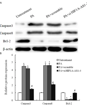
[image:7.612.92.371.68.405.2]than those of untreatment group. Treatment of HUVECs with PA induced cell death in a time and dose-dependent manner by using CCK8 assay (Figure 2A). We next investigated wheth-er PA induces cell death through an apoptotic mechanism. Annexin V-PI double-labeling was used for the detection of PS externalization, a hallmark of early phase of apoptosis. Consistent with the CCK8 assay, the results showed that the proportion of the apoptotic cells had gained as compared to untreatment group (Figure 2B Figure 4. PA-mediated regulation of apoptosis-related proteins in VSMCs. A.
VSMCs are treated with untreatment, 0.8 mM PA only, 0.8 mM PA plus scram-ble si-RNA and 0.8 mM PA plus si-HIF1a-AS1-3 for 48 h, and the expression of caspase3, caspase8 and Bcl2 are analyzed by western blotting; B. These
results are confirmed by densitometric analyses. Values are expressed as
mean ± SEM, n=3 in each group. *P < 0.05, versus untreatment group; #P <
0.05, versus PA group.
vascular smooth muscle cells in vitro, which may contribute to the pathogenesis of thoracic aortic aneurysms. We then investigated the possible mechanisms that Lnc-RNA reg-ulates the thoracoabdominal aorta tumorigenesis. We per-formed a hierarchical cluster analysis of the differentially expressed Lnc-RNA in the serum of TAA patients that associated with Lnc-RNA expression. After the removal of redundant and unannotated sequences, 10 genes were
found to be significantly
up-regulated and 15 genes to be
significantly down-regulated in
the TAA group compared to the normal control group. The results showed that the overex-pression of HIF1a-AS1 was associated with TAA, the ex- pression of which was at the highest levels in all 33 Lnc-RNAs in vivo (Figure 1A). To fur-ther validated the interaction between the TAA and HIF1a-AS1, large sample statistics results showed that compared to the normal control the expression of HIF1a-AS1 was
and 2C). Moreover, the percentage of the apop-totic cells in a dose-dependent manner. LncRNA HIF1a-AS1 is highly associated with CVD, and HIF1a-AS1 is highly expressed in advanced ath-erosclerosis tissues. The current study sug-gested that HIF1a-AS1 was associated with PA-induced dysfunction of VSMCs. The RNA
expression of HIF1a-AS1 was significantly high -er in VSMCs with PA (0.8 mM) than those of untreatment group (Figure 2D). Therefore, our data suggest that up-regulation the expression of HIF1a-AS1 was involved in PA-induced cell death.
Identification of HIF1a-AS1 in the regulation of VSMCs dysfunction
In this work, knock-out of endogenous HIF1a-AS1 with small-interfering RNA (siRNA), the expression of HIF1a-AS1 was down-regulated (Figure 3A). To evaluate the potential protective mechanisms of inhibition the function of HIF1a-AS1 in VSMCs, the CCK8 assay was used to measure cell viability. The viabilities of VSMCs inhibited with PA were protected by si- HIF1a-AS1 (Figure 3B). Consistent with the CCK8 assay, the Annexin V-PI double-labeling results showed that inhibition the function of HIF1a-AS1 with si-RNA could decrease the proportion of the apoptosis cells inducing by PA treatment (Figure 3C and 3D).
PA-mediated regulation of apoptosis-related proteins
The apoptotic response was further investigat-ed by measuring caspase-3 and caspase-8 activity and apoptosis-related proteins with Western blot techniques. PA administration caused 4.5- and 4.4- fold increases in cas-pase-3 and caspase activity respectively. However, the combination PA with si-HIF1a-AS1
induced strong and specific suppression of pro -tein expression of caspase-3 and caspase-8 Figure 4 expression, and expression of which was statistically up-regulated in the PA combi-nation with si-HIF1a-AS1-treated group as com-pared to PA single treatment group. Therefore, our data suggest that up-regulation the expres-sion of si-HIF1a-AS1 was involved in PA-induced VSMCs death.
Discussion
Research into the pathogenesis of
thoracoab-dominal aortic aneurysms (TAA) is difficult,
because this disease is caused by multiple fac-tors such as hemodynamics, metabolism, in-
flammation and genetic influences [11]. In addi
-tion to activa-tion of proteolysis and inflamma -tion, apoptosis of smooth muscle cells and oxi-dative stress have been suggested by several clinicopathological studies, and these factors together with many others seem to be intricate-ly interwoven to produce aneurysms [12-14].
Another difficulty is that suitable animal mod -els are not available for the study of aortic aneurysms. In this study, apoptosis of VSMCs were considered to approximately represent the TAA cell injury model. The exposure of PA to VSMCs has been demonstrated to cause a series of dysfunction, and trends to ma- ke as the TAA cell injury model. There were two
significant findings in this report: (1) patients
with TAA were increased HIF1a-AS1 in the serum; (2) we found that PA could induce VSMCs apoptosis in a dose dependent manner and increase the expression of HIF1a-AS1 in VSMCs. VSMCs apoptosis was thought to be involved in TAA [12]. Thus, apoptosis was
mea-sured in the present study to better confirm
and to analyze the VSMCs injury by PA. We found that the proportion of the apoptotic cells was increased.
In this study, we performed a LncRNA microar-ray technique using human samples, which can evaluate thousands genes simultaneously, and hierarchical cluster analysis of the differentially expressed Lnc-RNA in the serum of TAA patients
to identify the lncRNA HIF1A-AS1 was signifi -cantly up-regulated. Our results is consistent with other studies that reported that HIF1A-AS1
is identified through BRG1 knock-down VSMCs,
and the expression of HIF1A-AS1 is found to be regulated by BRG1 in VSMCs [4]. These studies will have particular relevance in the future, as the role of lncRNA in cardiovascular disease states becomes increasingly recognized. More- over, PA dose-dependently decreased the cell viability and increased the apoptosis of VSMCs, and down-regulated the expression of Bcl2 and up-regulated the expression of caspase3 and caspase8. Interestingly, LncRNA HIF1A-AS1 kn- ock-down could suppress PA-induced dysfunc-tion of VSMCs in vitro. Therefore, our data sug-gest that up-regulation the expression of HIF1a-AS1 was involved in PA-induced cell apoptosis. In conclusion, our results demonstrate that HIF1A-AS1 was overexpressed in the TAA pa- tients. LncRNA HIF1A-AS1 knock-down could suppress PA-induced apoptosis of VSMCs in vitro, which may contribute to the pathogenesis of thoracoabdominal aorta aneurysm.
Disclosure of conflict of interest
None.
Address correspondence to: Dr. Guangxing Feng, Department of Cardiovascular Surgery, The Fourth Hospital of Hebei Medical University, 12 Jiankang Road, Shijiazhuang 050011, Hebei, P.R. China. Tel: (86) 311-86095348; Fax: (86) 311-86095348; E-mail: [email protected]
References
[1] Orr N, Minion D and Bobadilla JL. Thoracoa- bdominal aortic aneurysm repair: current en-dovascular perspectives. Vasc Health Risk Manag 2014; 10: 493-505.
[2] Zanow J and Settmacher U. [Aneurysms of the thoracic and thoracoabdominal aorta]. Chirurg 2014; 85: 767-73.
[3] Watanabe Y, Kuratani T, Shirakawa Y, Torikai K, Shimamura K and Sawa Y. Hybrid endovascu-lar repair of a dissecting thoracoabdominal aortic aneurysm with stent graft implantation through the false lumen. J Vasc Surg 2014; 59: 264-267.
[4] Wang S, Zhang X, Yuan Y, Tan M, Zhang L, Xue X, Yan Y, Han L and Xu Z. BRG1 expression is increased in thoracic aortic aneurysms and regulates proliferation and apoptosis of vascu-lar smooth muscle cells through the long non-coding RNA HIF1A-AS1 in vitro. Eur J Cardio- thorac Surg 2014; [Epub ahead of print]. [5] Durdu S, Deniz GC, Balci D, Zaim C, Dogan A,
Can A, Akcali KC and Akar AR. Apoptotic vascu-lar smooth muscle cell depletion via BCL2 fam-ily of proteins in human ascending aortic aneu-rysm and dissection. Cardiovasc Ther 2012; 30: 308-316.
[6] Jiang L, Wang M, Zhang J, Monticone RE, Te- lljohann R, Spinetti G, Pintus G and Lakatta EG. Increased aortic calpain-1 activity medi-ates age-associated angiotensin II signaling of vascular smooth muscle cells. PLoS One 2008; 3: e2231.
[7] Qiao HP, Gao WS, Huo JX and Yang ZS. Long non-coding RNA GAS5 functions as a tumor suppressor in renal cell carcinoma. Asian Pac J Cancer Prev 2013; 14: 1077-1082.
[8] Martens-Uzunova ES, Bottcher R, Croce CM, Jenster G, Visakorpi T and Calin GA. Long non-coding RNA in prostate, bladder, and kidney cancer. Eur Urol 2014; 65: 1140-1151. [9] Gibb EA, Brown CJ and Lam WL. The functional
role of long non-coding RNA in human carcino-mas. Mol Cancer 2011; 10: 38.
[10] Gutschner T and Diederichs S. The hallmarks of cancer: a long non-coding RNA point of view. RNA Biol 2012; 9: 703-719.
[11] Taketani T, Imai Y, Morota T, Maemura K, Mo- rita H, Hayashi D, Yamazaki T, Nagai R and Ta- kamoto S. Altered patterns of gene expression
specific to thoracic aortic aneurysms: microar -ray analysis of surgically resected specimens. Int Heart J 2005; 46: 265-277.
[12] Rowe VL, Stevens SL, Reddick TT, Freeman MB, Donnell R, Carroll RC and Goldman MH. Vascular smooth muscle cell apoptosis in an-eurysmal, occlusive, and normal human aor-tas. J Vasc Surg 2000; 31: 567-576.
[13] Miller FJ Jr, Sharp WJ, Fang X, Oberley LW, Ob- erley TD and Weintraub NL. Oxidative stress in human abdominal aortic aneurysms: a poten-tial mediator of aneurysmal remodeling. Ar- terioscler Thromb Vasc Biol 2002; 22: 560-565.
[14] Yajima N, Masuda M, Miyazaki M, Nakajima N, Chien S and Shyy JY. Oxidative stress is invo- lved in the development of experimental ab-dominal aortic aneurysm: a study of the
tran-scription profile with complementary DNA mi -croarray. J Vasc Surg 2002; 36: 379-385. [15] Wang L, Zhi H, Li Y, Ma G, Ye X, Yu X, Yang T, Jin
prognosis of coronary artery disease. Int J Clin Exp Pathol 2014; 7: 5093-5102.
[16] Choe N, Kwon JS, Kim JR, Eom GH, Kim Y, Nam KI, Ahn Y, Kee HJ and Kook H. The microRNA
miR-132 targets Lrrfip1 to block vascular
smooth muscle cell proliferation and neointi-mal hyperplasia. Atherosclerosis 2013; 229: 348-355.
[17] Liu X, Cheng Y, Yang J, Qin S, Chen X, Tang X, Zhou X, Krall TJ and Zhang C. Flank sequences of miR-145/143 and their aberrant expression in vascular disease: mechanism and therapeu-tic application. J Am Heart Assoc 2013; 2: e000407.
[18] Kataoka M and Wang DZ. Non-Coding RNAs Including miRNAs and lncRNAs in Cardiova- scular Biology and Disease. Cells 2014; 3: 883-898.
[19] Wang L, Fu D, Qiu Y, Xing X, Xu F, Han C, Xu X, Wei Z, Zhang Z, Ge J, Cheng W and Xie HL.
Genome-wide screening and identification of
long noncoding RNAs and their interaction with protein coding RNAs in bladder urothelial cell carcinoma. Cancer Lett 2014; 349: 77-86. [20] Rinn JL and Chang HY. Genome regulation by
long noncoding RNAs. Annu Rev Biochem 2012; 81: 145-166.
[21] Rinn JL, Kertesz M, Wang JK, Squazzo SL, Xu X, Brugmann SA, Goodnough LH, Helms JA, Far- nham PJ, Segal E and Chang HY. Functional de-marcation of active and silent chromatin do-mains in human HOX loci by noncoding RNAs. Cell 2007; 129: 1311-1323.
[22] Chu C, Qu K, Zhong FL, Artandi SE and Chang HY. Genomic maps of long noncoding RNA oc-cupancy reveal principles of RNA-chromatin interactions. Mol Cell 2011; 44: 667-678. [23] Yan B, Gu W, Yang Z, Gu Z, Yue X, Gu Q and Liu
L. Downregulation of a long noncoding RNA-ncRuPAR contributes to tumor inhibition in colorectal cancer. Tumour Biol 2014; [Epub ahead of print].
[24] Arab K, Park YJ, Lindroth AM, Schafer A, Oakes C, Weichenhan D, Lukanova A, Lundin E, Risch A, Meister M, Dienemann H, Dyckhoff G, He- rold-Mende C, Grummt I, Niehrs C and Plass C. Long Noncoding RNA TARID Directs Deme- thylation and Activation of the Tumor Sup- pressor TCF21 via GADD45A. Mol Cell 2014; 55: 604-614.