Dr R. Muthukrishnan, MD et al JMSCR Volume 06 Issue 10 October 2018 Page 24
Analysis of Patterns of Mucosal and Histopathological Changes in Cases of
Acute Appendicitis
Authors
Dr R. Muthukrishnan, MD
1, Dr M. Malathi, MD
2, Dr S. Ravi, MD
3,
M. Kishore Kumar
41
Associate Professor of Pathology, ACS Medical College, Chennai Email: [email protected], 9940074977
2
Assistant Professor of Pathology, Chengalpattu Government Medical College, Chengalpattu
3
Professor of Pathology, Chengalpattu Government Medical College, Chengalpattu, 942866270
4
3rd MBBS, Chengalpattu Government Medical College, Chengalpattu Corresponding Author
Dr M. Malathi, Md
Email: [email protected], 9965981739 .
Introduction
Acute appendicitis is the most common general surgical emergency. The lifetime risk for appendicitis is 7%, commonly occurring in adolescent and young adults[13]. In England, 42,526 patients underwent appendectomies in the year 2004–5, with a mean age of 28 years[10]. The rate of acute appendicitis varies among countries. In USA and Europe there is a declining rates of acute appendicitis[13]. In developing countries due to changes in life style there is increasing incidence in most urban centers[14]. Despite of advances in technology and imaging modalities, there is dilemma in the diagnosis of acute appendicitis, however histopathological examination still remains the gold standard for the confirmation of appendicitis[15], whether it is acute or chronic.
Acute appendicitis is defined as an inflammation of the inner lining of the appendix vermiformis, which then spreads to other parts of the organ.
Various etiologies for this clinic-pathologic condition have been identified, but luminal obstruction is considered the most critical factor, as it triggers the inflammatory process. When lumen obstruction occurs, intraluminal pressure surpasses that in the appendiceal veins, causing venous outflow obstruction. Finally, ischemia develops in the appendiceal wall, which weakens the epithelial integrity and increases the organ's risk of bacterial invasion.
Although lymphoid hyperplasia and fecoliths are the most common causative factors of luminal obstruction, other less frequent factors have been associated with the condition, including entero-biasis, endometriosis, tuberculosis, amebiasis, granulomatous diseases, polyp, mucocele, as well as appendiceal malignancies, such as carcinoid tumor[7]. Intestinal parasites can proliferate in the appendix and occlude the lumen[2]
Clinical findings form the basis for diagnosis which may be consolidated by blood tests such as
www.jmscr.igmpublication.org Impact Factor (SJIF): 6.379
Index Copernicus Value: 79.54 ISSN (e)-2347-176x ISSN (p) 2455-0450
Dr R. Muthukrishnan, MD et al JMSCR Volume 06 Issue 10 October 2018 Page 25 C-reactive protein and white cell count, and
management is early appendicectomy[1].
Specimens obtained after appendectomy in acute appendicitis patients may be macroscopically normal but histological examination may reveal the precise underlying pathology. Neutrophil lymphocyte ratio of greater than 6.35 were statistically associated with severe acute appendicitis, with a median of one extra hospital day admission[11]. Mucosal ulceration, inflammatory cells involvement and their types, their level of infiltration into the layers of appendix, serosal congestion, ganglionic and neuronal hyperplasia, lymphoid hyperplasia are analysed carefully to find the etiology upto the cellular level.
This study aimed to determine the various histological diagnoses of all surgically removed appendices and to find out the age and sex related occurrence of appendicitis, and to find some unusual factors. Based on the outcomes, better conclusions can be arrived which may be useful for the management of post operated patients, newer therapeutic targets and can be treated in a better way.
Review of Literature
Several studies have been conducted across the globe to evaluate the histopathology of acute appendicitis.
An analysis by Marudhanayagam et al, Department of General Surgery, University Hospital of Wales, Cardiff, UK, datas shows that Of the 2660 appendicectomy specimens, acute appendicitis was seen in 1718 patients (64.58%), with a peak in patients in their second decade (35.09% of cases of acute appendicitis). The perforation rate was 13.9% and was significantly higher in patients aged 70 years or more. The negative appendicectomy rate was 28.8%, and was significantly higher in female patients and in the 11-30 year age group. Other pathologies include carcinoid (0.52%), adenocarcinoma (0.39%), and mucinous cystadenoma (0.60%)[4]
Another study by Gorter et al, Pediatric Surgical Centre of Amsterdam, Emma Children's Hospital AMC and VU University Medical Centre, Amsterdam. More neutrophils and less lymphocytic cells were identified in complex appendicitis comparatively with simple appendicitis. The increase in proinflammatory cells and decrease of adaptive cells in patients with complex appendicitis suggest potential aggravating processes in complex appendicitis[6] In a study conducted by Alun E Jones et al, Department of General Surgery, Norfolk and Norwich University Hospital NHS Trust, England, 77 percent (941/1225) of the specimens reported changes consistent with acute inflammation of appendix. Out of which, 4.8%(46) were found with incidental abnormal diagnosis, 2 with carcinoids, 11 with intra luminal parasites, 3 with endometriosis and 6 with crohn’s disease.[1]
A study by Emre et al, by the department of surgery, Malatya state hospital, turkey showed that a total of 1255 patients met the inclusion criteria, including 712(56.7%) and 546(43.3%) females. The mean age was 30 and the majority of the patients (61.7%) were <30 years old, with only 7% of patients> 50 years old[7]
A study by Mohammad Ayub Jat et al showed that histopathological diagnosis include Acute appendicitis (52%), Suppurative appendicitis (28%), Acute gangrenous appendicitis(12.5%), Perforated appendicitis (2%), chronic appendicitis (2.5%)[13]
In a study by W chan et al, university of hongkong, Queen mary hospital, the diagnosis made by routine pathological examination of 180 specimens showed 9 cases with granulomas mainly tuberculosis, 55 with parasitic infections predominantly enterobiasis, 16 with benign tumours of appendix especially mucocele, 2 malignant lesions and 3 endometriosis[5].
Dr R. Muthukrishnan, MD et al JMSCR Volume 06 Issue 10 October 2018 Page 26 A study by khan OA et al Department of surgery,
St Mary’s hospital, UK showed that examination of the specimens revealed inflammation or necrosis in 74% of the cases- However unexpected histological findings were seen in 4.2 % of cases. 2.1 % resultedin a change in medical therapy[3]
Aims and Objectives
To analyse the demographic and histopathologic data of patients who underwent appendectomy to treat an initial diagnosis of acute appendicitis.
To detect mucosal changes in the specimen.
To detect any unusual findings which may be the underlying cause.
Materials and Methods
The ethical committee clearance for this study was obtained from the Institutional Ethical Committee in the month of June 2017.
Study Design
Prospective analytical study was conducted in Chengalpattu Medical College Hospital, Chengalpattu during the period -Aug 2017 – Sept 2017, in collaboration with ACS Medical College, Chennai.
In this study, 90 specimens of appendix with initial diagnosis of Acute appendicitis were included analytically to evaluate the histopathological changes taking place.
Patients who underwent appendectomy for chronic appendicitis and incidental appendectomy during other surgeries were excluded from the study.
The detailed procedure is explained in the following sections.
Step 1- Record Details
Relevant clinical data retrieved included patients ‘age, gender, pre-operative clinical presentation and operative findings.
Step 2- Collection and Processing
The appendix specimens were obtained from the Surgery Department, Government Chengalpattu Medical College Hospital All the surgically resected specimens of acute appendicitis, submitted to Department of Pathology were included in this study.
The appendix specimen obtained at surgery were fixed in formalin.
Bits are taken from the tip, central area and base, then processed and the sections were cut at 5 micron thickness using microtome and stained with Hematoxylin and Eosin.
Step 3- Microscopic Examination
The specimens were examined under microscopy for the histopathological evidence of Acute appendicitis and unusual findings.
The specimens were analysed under the following headings.
Mucosal ulceration.
Type of inflammatory cells and its level of infiltration
Serosal congestion
Ganglion and Neuronal hyperplasia.
Luminal contents such as faecolith, worms.
Lymphoid hyperplasia
Any other unusual findings
In acute appendicitis there are neutrophils in mucosa, muscularis propria of appendix and congested blood vessel in the serosa.
The collected data were analysed.
Observations and Results
Dr R. Muthukrishnan, MD et al JMSCR Volume 06 Issue 10 October 2018 Page 27
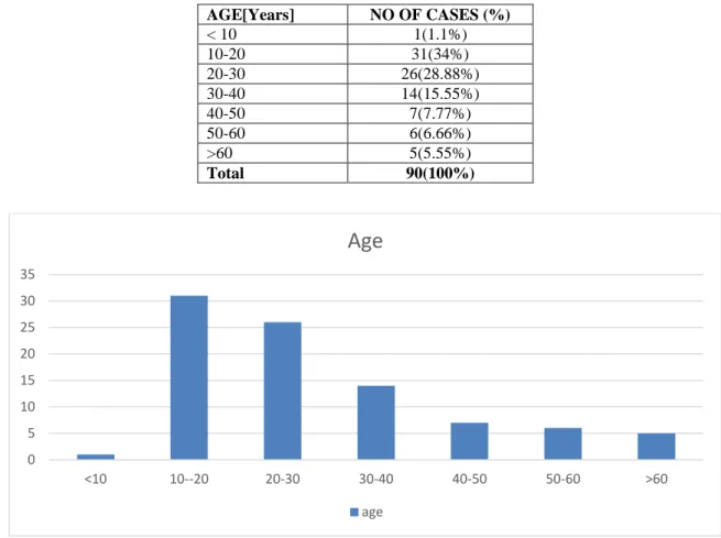
Table-1: Distribution of Patients with Age
Figure: 1
About 34 percent of the acute appendicitis cases were detected in the second decade of life. Only 1 out of 90 were detected in children less than 10 years. About 5 cases were detected in people greater than 60 years of age.
Table-2: Distribution of Patients with Sex
SEX NO OF CASES(%)
MALE 56(62%)
FEMALE 34(38%)
Total 90(100%)
Figure: 2
About 62 % of the patients diagnosed and resected for acute appendicitis are males.
Table 3 Histopathological Analysis And Their Distribution
HISTOPATHOLOGICAL FEATURES
NO OF CASES(%)
Mucosal ulceration 90(100%) Lymphoid hyperplasia 2(2.22%) Ganglional hyperplasia 3(3.33%) Serosal congestion 66(73.33%) Fibrinous exudate 36(40%)
Total 90(100%)
0 5 10 15 20 25 30 35
<10 10--20 20-30 30-40 40-50 50-60 >60
Age
age
AGE[Years] NO OF CASES (%)
< 10 1(1.1%)
10-20 31(34%)
20-30 26(28.88%)
30-40 14(15.55%)
40-50 7(7.77%)
50-60 6(6.66%)
>60 5(5.55%)
Dr R. Muthukrishnan, MD et al JMSCR Volume 06 Issue 10 October 2018 Page 28
Figure 3
Almost all the appendix specimens are positive for mucosal ulceration. Lymphoid hyperplasia is seen only in 2 cases out of 90 (Table 3 & Figure 3).
Table 3 Luminal Contents
LUMINAL CONTENTS NO OF CASES(%)
Faecoliths 19(21.11)
Parasites 0
Total 90(100%)
Most of the cases of Appendiceal lumen shows necrotic material, 19 cases shows fecolith. None of the cases showing parasites in the lumen. (Table 3)
Table – 4: Types of Inflammatory Cells and Their Distribution
Types Of Predominant
Inflammatory Cells
No Of Cases
Neutrophils 75(83.33%) Eosinophils 7(7.77%) Lymphocytes 8(8.88%)
Total 90(100%)
Neutrophils were present in about 82 % of the cases. About 7 out of 90 cases were positive with eosinophils which are also presented with neutrophilic infiltration (Table 4).
About 48 (53 %) of the specimens were infiltrated by neutrophils up to the serosal layer, 20 specimens (22 %) up to muscularis mucosa. About 10 and 2 specimens were infiltrated up to mucosa and submucosa respectively.
Table-5: Type of Appendicitis and Their Distribution
TYPE OF APPENDICITIS NO OF CASES(%)
Acute suppurative appendicitis 8(8.88%) Gangrenous appendicitis 1(1.11%) Perforating appendicitis 2(2.22%) Acute appendicitis 79(87.77%)
Total 90(100%)
About 8 of the total 90 cases were found with acute suppurative appendicitis. Gangrenous appendicitis was noticed in one case, 2 cases with perforation were seen and the remaining 79 showed simple acute appendicitis.
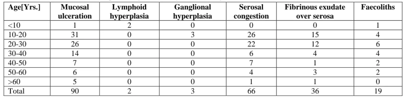
Table 6 Distribution of Histopathological Analysis with Age
Age[Yrs.] Mucosal
ulceration
Lymphoid hyperplasia
Ganglional hyperplasia
Serosal congestion
Fibrinous exudate over serosa
Faecoliths
<10 1 2 0 0 0 1
10-20 31 0 3 26 15 4
20-30 26 0 0 22 12 6
30-40 14 0 0 6 4 4
40-50 7 0 0 7 1 2
50-60 6 0 0 4 3 2
>60 5 0 0 1 1 0
Dr R. Muthukrishnan, MD et al JMSCR Volume 06 Issue 10 October 2018 Page 29 Table 6 shows that most of the histopathological
changes were seen in second and third decade of life.
Unusual Findings
There is no histological evidence of any Granulomatous inflammation, Mucocele, Carcinoid, Adenocarcinoma, Hyperplastic polyp etc.
Discussion
Acute appendicitis has been the most common surgical emergency and appendectomy is the most frequently performed abdominal operation. In our study acute appendicitis was higher [34%] in the age group of second and third decade and about 74 % of appendicitis occurring below 40 year of age. This finding is in correlation with many other studies. A study by Alun E Jones university of wales, Cardiff UK In which it showed that the peak incidence consists of about 35 % of the cases seen in the second decade of life[1]. Another study showed about 64% of the patient were diagnosed with acute appendicitis in the second decade[4]. In the year 2004-5, about 42,000 patients with the mean age of 28 years underwent appendectomy[10].
When considering the sex of the patients, the study consisted of about 62% of males and 38% of females with male female ratio of 1.6:1. This is in correlation with a study in which 62% were males (719/1159)[14].This incidence is in contrast with a study in which the female male ratio was 3:1[3]. The present study showed that frequency of histopathological diagnosis was acute appendicitis (52.%) followed by acute suppurative (28%), acute gangrenous appendicitis (12.5%), acute perforated appendicitis (2%), resolving or chronic appendicitis (2.5%) similar results were also showed in other studies done by Shrestha R and Zulfikar et al. The perforation rate of appendix in this study was low (2%) similar to that shown by other studies. The low perforation rate might be due to early visit in the surgical clinic and the prompt decision to operate for suspected appendicitis by the surgeons. Low perforation rate
indicate a better prospective regarding morbidity and mortality[25]
There were no parasitic infection manifested in these 90 cases. A study showed that eleven out of 1225 (0.8%) revealed intraluminal parasites (10
Enterobius and 1 Schist soma)[1]. Parasitic
infection was demonstrated in 1.4 percent out of 1159 patients[10]. They were seen rarely constituting less than 1 percent of incidence. In a study, 33 percent of the cases showed marked neuronal hyperplasia in histologically positive acute appendicitis[12]. In this present study only 21 percent were positive for ganglionic and neuronal hyperplasia.
When considering lymphoid hyperplasia, only 6 out of 90 cases were positive. In a study by the department of surgery, Baskent University, only 10 out of 1600 cases were found positive with lymphoid hyperplasia. This shows the low prevalence of that finding.
Another study showed that the increased neutrophilic infiltration in the muscularis propria was statistically higher in patients with severe appendicitis when compared to simple appendicitis (7.29)[18]. In this present study, about 82 percent of the cases were infiltrated by neutrophils and about 7 percent were found positive for eosinophils. Acute eosinophilic appendicitis was not seen our study, in contrast to a study done by Aravindhan et al[9].
Summary
This study aims at analysing the demographic and histopathologic data of patients who underwent appendectomy to treat an initial diagnosis of acute appendicitis.
This analytical study was conducted in ACS Medical college Hospital, Chennai.
In this study, 90 specimens of appendix with initial diagnosis of Acute appendicitis were included to evaluate the histopathological changes taking place.
Dr R. Muthukrishnan, MD et al JMSCR Volume 06 Issue 10 October 2018 Page 30 The present study showed a high number of
appendicitis in adolescents and young adults. Males are more commonly affected.
Complications seen in about 10% of patients. Lymphoid hyperplasia seen in 2nd decade of life. Fecolith is seen in 21% of patients. Most of them are in third decade of life. Most of the appendicectomy specimes showed fibrinous exudate in the serosa. There was no cases of eosinophilic appendicitis. There was no unusual findings like parasites and tumour.
This study supports the sending of all appendicectomy specimens for routine histopathological examination to confirm Appendicitis and to find out complications and unusual findings.
Conclusion
The unusual findings, in cases of Appendicitis were not suspected on macroscopic examination at the time of surgery. These would have been missed, if we had the specimens not been examined microscopically. The intra-operative diagnosis of the surgeon is therefore unreliable in detecting abnormalities of the appendix. This study supports routine histological examination of all the appendicectomy specimens to avoid missing of any clinically important and treatable condition.
Since our study did not establish any unusual findings, it is recommended to continue the routine histopathological examination of all appendicitis cases. Our study was restricted in terms of period of 2 months. It is suggested to have a deeper study covering larger sample size and duration.
References
1. The value of routine histopathological examination of appendicectomy specimens Alun E Jones*, Alexander W Phillips, John R Jarvis and Kevin Sargen
2. Short practice of surgery by Bailey &Love 3. Routine pathological analysis of
appendicectomy specimens--is it justified?
Khan OA1, Morhan A, Jegatheeswaran S, Jackson E, Pelikan A.Department of Surgery, St. Mary's Hospital, Isle of Wight, UK.
4. Review of the pathological results of 2660 appendicectomy specimens. Marudana-yagam R1, Williams GT, Rees BI. Department of General Surgery, University Hospital of Wales, Cardiff, UK. 5. Value of routine histopathological examination of appendices in Hong Kong W CHAN, K H
FU From the Department of Pathology, University of Hong Kong, Queen Mary Hospital, Hong Kong
6. Composition of the cellular infiltrate in patients with simple and complex appendi-citis. Gorter RR1, Wassenaar ECE2, de Boer OJ3, Bakx R2, Roelofs JJTH3 Bunders MJ4, van Heurn LWE2, Heij HA 7. Routine histopathologic examination of
appendectomy specimens: retrospective analysis of 1255 patients.Emre A1, Akbulut S, Bozdag Z, Yilmaz M, Kanlioz M, Emre R, Sahin N..
8. Unusual histopathological findings in appendectomy specimens from patients with suspected acute appendicitis. Yilmaz M1, Akbulut S, Kutluturk K, Sahin N, Arabaci E, Ara C, Yilmaz S
9. Acute eosinophilicappendicitis and the significance of eosinophil - Edema lesion. Aravindan KP1, Vijayaraghavan D, Manipadam MT. Department of Pathology, Medical College, Calicut - 673 008, India.
10.Hospital Episode Statistics Department of Health:
[http:www.hesonline.nhs.uk/Ease/servlet/ Content Server? siteID=1937 & category ID=204].
Dr R. Muthukrishnan, MD et al JMSCR Volume 06 Issue 10 October 2018 Page 31 12.Neuronal hypertrophy and mast cells in
histologically negative, clinically diagnosed acute appendicitis: a quantitative immunophenotypical analysis. Amber S, et al. Indian J Gastroenterol. 2010, Department of pathological, Kasturba medical college, mangalore, Karnataka, in
13.Histopathological examination of appendicectomy specimens at a district hospital of Saudi Arabia, Mohammad Ayub Jat, Farhan Khashim Alswailmi, and shehabalanazi, Dr Mohammad A, yub Jat, FCPS. Assistant professor of surgery, college of medicine, Northern Border University, Arar, Saudia Arabia
14.Role of eosinophils in acute appendicitis, Shrestha R et al. JNMA J Nepal Med Asso 2015