Niyatha Balakrishnan et al JMSCR Volume 06 Issue 09 September 2018 Page 1022
Immunohistochemical Profile in Lung Carcinoma
Authors
Niyatha Balakrishnan
1, C. R. Jyothi
2 1Senior Resident, Department of Pathology, Medical College, Thrissur, Kerala, India Mobile no: 9495238241 Email: [email protected]
2
Associate Professor, Department of Pathology, Government Medical College, Thrissur, Kerala, India Corresponding Author
C. R. Jyothi
Mobile no: 9495635733. Email: [email protected]
Abstract
Introduction: Small biopsies obtained by bronchoscopy are commonly used for diagnosis of lung malignancies. The emergence of treatments for lung carcinoma with differential efficacy and toxicity between subtypes has highlighted the importance of specific pathologic subtyping. The use of immunohistochemistry (IHC) has been well documented as an important ancillary tool in sub classifying lung carcinomas.
Objectives: This study is aimed to assess the expression of a panel 3 IHC markers - CK5/6, p63 and TTF-1, in 39 cases of lung carcinomas
Materials & Methods: This was a cross sectional study done over a period of 18 months in bronchial biopsy specimens of histologically proven lung carcinoma
Observations: It was observed that CK5/6 and p63 were 100% senisitive and specific for squamous cell carcinomas, if unquantified reactivities were consiered the specificity of p63 reduced to 83% and TTF-1 had 100% sensitivity and specificity in diagnosing adenocarcinoma, but if diffuse TTF-1 positivity was considered the sensitivity reduced to 66.6%.
TTF-1/p63 immuno profile showed no overlap when squamous cell carcinomas and small cell lung carcinomas were compared.
Conclusion: 3 panel IHC markers, CK5/6, p63 and TTF-1 when used together and interpreted correctly can be of help for the final diagnosis even in cases in which the morphology is unclear.
Keywords: Immunohistochemistry; CK5/6; p63; TTF-1; Squamous cell carcinoma; Adenocarcinoma; Small cell carcinoma.
Introduction
Lung cancer is the leading cause of cancer related mortality both in the United States and worldwide(1). Overall 2,22,520 new cases of lung cancer and 1,57,300 deaths were estimated for 2010(2).
Clinically, lung carcinomas were classified as non-small cell lung cancer (NSCLC) or non-small cell lung
cancer. According to the current World Health Organization classification, Epithelial tumors includes the following histological types: Squamous cell carcinoma (SCC); Adenocarcinoma (ADC), Neuroendocrine tumors, Large cell carcinoma (LCC) etc. In the same classification, small cell carcinomas (SCLC) are included under neuroendocrine tumors(3). Clinical staging and identification of the
www.jmscr.igmpublication.org Impact Factor (SJIF): 6.379
Niyatha Balakrishnan et al JMSCR Volume 06 Issue 09 September 2018 Page 1023 histological type are fundamental in establishing the
therapeutic strategy for treating such patients.
The diagnostic reliability among experienced pathologists is 90%. In this context, immunohistochemistry represents a tool that can provide a clear distinction among the various types TTF-1, CK 5/6 and p63 seem to be useful for differentiating adenocarcinoma from squamous cell carcinoma. If they are used together and interpreted correctly, they can be of help for the final diagnosis even in cases in which the morphology is unclear(4).
Objectives
This study was aimed to assess the expression of a panel of 3 IHC markers - CK5/6, p63 and TTF-1, in bronchial biopsy specimens of lung carcinomas
Procedure
The study design was hospital based cross sectional study done over a period of18 months (1-1-2015 to 31-7-2016) on histologically proven 39 cases of lung carcinomas in which differentiation could be established. Biopsy specimens for non malignant lung pathology, non epithelialtumors and metastatic tumors were excluded from study.
The formalin fixed bronchial biopsy of all the cases after processing and embedding in paraffin, multiple 3–5 mm sections were taken and each biopsies were stained with hematoxylin and eosin (H&E).
Subtyping of lung carcinomas on H & E slides were done according to the WHO classification of lung tumors(3).
The specimens were processed for immunohistochemical examination using an immunoenzymatic soluble compex method.4µm thick sections were made on poly L lysine coated slide, kept at 370C overnight & 600C for 1 hour, dewaxing was done in xylene. Hydration was done followed by antigen retrieval & staining.
Antibodies used for immunohistochemistry were: 1) Rabbit Monoclonal Antibody CK 5/6 2) Mouse Monoclonal Antibody p63 3) Mouse Monoclonal Antibody TTF-1
Immunohistochemically, cytoplasmic staining was considered positive for cytokeratin 5/6 and nuclear staining was considered positive for TTF-1 and p63. Immunoreactivity was scored semi quantitatively by recording percentage of reactive tumor cells. Diffuse reactivity was defined as labeling of >50% tumor cells, and focal reactivity as 1–49%. In addition, intensity of reactivity was recorded as weak (1+, less than normal cells); moderate (2+, same as normal cells); and strong (3+, stronger than normal cells). Pneumocytes served as internal controls for TTF-1 reactivity, and bronchial basal cells for p63 & CK5/6. Data analysis was done using SPSS software, version 16.
Observations
Out of the 39 cases, 27 cases (45%) were Squamous cell carcinoma, 6 cases (10%) were Adenocar-cinoma, 6 cases (10%) were Small cell carcinoma
Single Marker Expression Profile in Lung Carcinoma
Table 1 Expression of CK5/6 in each histological subtype of lung carcinoma
Tumor type CK5/6 (Percentage of immunoreactive cells)
0 1-49 % >/=50%
Squamous cell carcinoma (27)
0 0 27
Adenocarcinoma(6) 6 0 0
Small cell carcinoma(6) 6 0 0
Table 2 Expression of p63 in each histological subtype of lung carcinoma
Tumor type
p63 (Percentage of immunoreactive cells)
0 1-49 % >/=50% Squamous cell
carcinoma(27) 0 0 27
Adenocarcinoma(6) 5 1 0
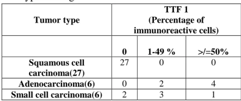
Niyatha Balakrishnan et al JMSCR Volume 06 Issue 09 September 2018 Page 1024 Table 3 Expression of TTF-1 in each histological
subtype of lung carcinoma
Tumor type
TTF 1 (Percentage of immunoreactive cells)
0 1-49 % >/=50% Squamous cell
carcinoma(27)
27 0 0
Adenocarcinoma(6) 0 2 4
Small cell carcinoma(6) 2 3 1
Comparison of Intensity of Reactivity of Individual Markers in Histological Subtypes of Lung Carcinoma
Squamous cell carcinoma had strong positivity with CK5/6.
Staining intensity of p63 was observed to be strong in all cases of squamous cell carcinoma. However, one case of adenocarcinoma which showed positivity for p63 was weak. It was also observed that intensity of staining of TTF-1 varied from weak to strong in both adenocacinomas and small cell carcinomas
Single MarkerSensitivity & Specificity for Squamous Cell Carcinoma v/s Adenocarcinoma Analysis of one-marker sensitivity and specificity showed that immunohistochemical markers CK5/6 and p63 were 100% senisitive and specific for squamous cell carcinomas. However. if unquantified reactivities were considered (i.e, any reactivity considered positive) the specificity of p63 reduced to 83%.This study also showed 100% sensitivity and specificity for TTF-1 in diagnosing adenocarcinoma. But, when diffuse positivity was considered the sensitivity reduced to 66.6%.
Sensitivity & Specificity of IHC Marker Combinations For Differentiating Squamous cell carcinoma & Adenocarcinoma
Analysis of coexpression profiles showed that coexpression of p63 and CK5/6 irrespective of TTF-1 and coexpression of p63 and CK5/6 in TTF-TTF-1 negative tumor both were 100% sensitive and specific for squamous cell carcinomas. Further it was also observed that TTF-1 positive, CK5/6 and p63 negative coexpression profile showed 100% specificity in diagnosing adenocarcinomas
One marker expression profile in our cases revealed that squamous cell carcinoma displayed a highly consistent immunoprofile (TTF-1 negative, CK5/6 and p63 positive). In particular, all squamous cell carcinomas were consistently immunoreactive for CK5/6 and p63 with strong and diffuse positivity. In contrast to homogenous immunoprofile in squamous cell carcinoma, adenocarcinoma showed mild heterogeneity for one of the squamous cell marker, p63. However, the positivity observed with p63 was only focal and weak. All adenocarcinomas showed consistent positivity for TTF-1, although the reactivity varied from focal to diffuse and weak to strong.
In this study it was also observed that majority of the cases of small cell carcinoma (67%) showed positivity for TTF-1, and the immunoreactivity showed variation in staining pattern.
In this study it was noticed that TTF-1/p63 immunoprofile showed no overlap when squamous cell carcinomas and small cell lung carcinomas were compared.
Discussion
Several recent studies have addressed the issue of using immunohistochemistry panels to correctly subclassify lung carcinomas. Two recent articles on this subject are the studies of Mukhopadhyay and Katzenstein(5) and Terry et al(6). One observation regarding the use of IHC to sub-classify lung carcinomas has been that the clinical trials validating the various histologic-specific treatment modalities were based on H&E classification. In the current study a panel of three immunohistochemical markers, namely CK5/6, p63 and TTF-1 were used for analysing the immunoprofile in lung carcinomas.
Niyatha Balakrishnan et al JMSCR Volume 06 Issue 09 September 2018 Page 1025 CK5/6 showed 100% sensitivity and specificity in
squamous cell carcinomas. Similar results were observed in studies conducted by Wei Zhao et al(7). In this study it was observed that all cases of squamous cell carcinomas showed diffuse strong positivity for p63. It was also observed that p63 had 100% and 83% sensitivity and specificity respectively in predicting squamous cell carcinomas. The specificity was decreased due to the focal positivity noted in adenocarcinoma. However, when diffuse positivity was considered the specificity rose to 100%.
In this study all cases of adenocarcinomas were positive for TTF-1. Of the six cases, four showed diffuse positivity and two were focal and the intensity varied from weak to strong. Similar study by Argon et al, observed TTF-1 positivity in all of the adenocarcinomas(4).This study also observed 100% sensitivity and specificity for TTF-1 in diagnosing adenocarcinoma. But, when diffuse positivity was considered the sensitivity reduced to 66.6%. Furthermore, study by Argon et al, also observed that the sensitivity and specificity of TTF-1 in adenocarcinomas rose when the focal staining was considered positive(8).
In the present study it was also observed that TTF-1 was positive in 4 (67%) out of 6 small cell carcinoma cases, and the staining was diffuse only in one case rest three showed only focal positivity. Folpe et al and Kaufmann et al also observed TTF-1 expression in 90–100% of small cell lung carcinomas(9,10).
Analysis of coexpression profiles showed that coexpression of p63 and CK5/6 irrespective of TTF-1 and coexpression of p63 and CK5/6 in TTF-TTF-1 negative tumor both were 100% sensitive and specific for squamous cell carcinomas. Further it was also observed that TTF-1 positive, CK5/6 and p63 negative coexpression profile showed 100% specificity in diagnosing adenocarcinomas.
Conclusion
Of the total cases included in the study 45% were Squamous cell carcinomas, 10% were
Adenocarcinomas, 10% were Small cell carcinomas
CK5/6 and p63 were 100% senisitive and specific for squamous cell carcinomas. If unquantified reactivities were considered the specificity of p63 reduced to 83%.
TTF-1 had 100% sensitivity and specificity in diagnosing adenocarcinoma. If diffuse TTF-1 positivity was considered the sensitivity reduced to 66.6%.
Squamous cell carcinoma displayed a highly consistent immunoprofile (TTF-1 negative, CK5/6 and p63 positive).
Coexpression of p63 and CK5/6 irrespective of 1 and that of p63 and CK5/6 in TTF-1 negative tumor both were TTF-100% sensitive and specific for squamous cell carcinomas.
67% of small cell carcinoma showed positivity for TTF-1 with varying immunoreactivity.
TTF-1/p63 immunoprofile showed no overlap when squamous cell carcinomas and small cell lung carcinomas were compared.
References
1. Ueno T, Linder S, Elmberger G. Aspartic proteinase napsin is a useful marker for diagnosis of primary lung adenocarcinoma. Br J Cancer. 2003;88 (8):1229–1233.
2. Jemal A, Siegel R, Xu J, Ward E. Cancer statistics, 2010. CA Cancer J Clin.2010;60(5):277–300.
3. Travis WD, Brambilla E, Burke AP, Marx A, Nicholson AG. WHO Classification of Tumours of the Lung, Pleura, Thymus and Heart. Lyon: International Agency for Research on Cancer,2015
4. Argon A et al.The Value of Cytokeratin 5/6, p63 and Thyroid Transcription Factor-1 in Adenocarcinoma, Squamous Cell Carcinoma and Non-Small-Cell Lung Cancer of the Lung.Turk Patoloji Derg 2015;31:81-88. 5. Mukhopadhyay S, Katzenstein AL.
Niyatha Balakrishnan et al JMSCR Volume 06 Issue 09 September 2018 Page 1026 differentiation on biopsy specimens: utility
of animmunohistochemical panel containing TTF-1, napsin A, p63 and CK5/6. Am J Surg Pathol 2011;35:15–25.
6. Terry J, Leung S, Laskin J, Leslie K, Gown A, Ionescu D. Optimal immunohisto-chemical markers for distinguishing lung adenocarcinomas from squamous cell carcinomas in small tumor samples. Am J Surg Pathol. 2010; 34(12):1805–1811. 7. Wei Zhao et al. ΔNp63, CK5/6, TTF-1 and
napsin A, a reliable panel to subtype non-small cell lung cancer in biopsy specimens. Int J ClinExpPathol 2014;7(7):4247-4253. 8. Kargi A, Gurel D, Tuna B. The diagnostic
value of TTF-1, CK5/6, and p63 immunostaining in classification of lung carcinomas. Appl Immunohistochem Mol Morphol 2007;15:415–20.
9. Folpe AL, Gown AM, Lamps LW, et al. Thyroid transcription factor-1: immunohistochemical evaluation in pulmonary neuroendocrine tumors. Mod Pathol 1999;12:5–8.