Macrophage A2A Adenosinergic Receptor Modulates
Oxygen-Induced Augmentation of Murine Lung Injury
Neil R. Aggarwal1, Franco R. D’Alessio1, Yoshiki Eto1, Eric Chau1, Claudia Avalos1,
Adam T. Waickman2, Brian T. Garibaldi1, Jason R. Mock1, Daniel C. Files1,3, Venkataramana Sidhaye1, Vsevolod Y. Polotsky1, Jonathan Powell2, Maureen Horton1, and Landon S. King1
1Division of Pulmonary and Critical Care Medicine, Johns Hopkins Asthma and Allergy Center, and2Department of Hematology–Oncology, School of Medicine, Johns Hopkins University, Baltimore, Maryland; and3Division of Internal Medicine-Pulmonary, Critical Care, Allergy, and Immunologic Disease, School of Medicine, Wake Forest University, Winston-Salem, North Carolina
Acute respiratory distress syndrome (ARDS) causes significant morbid-ity and mortalmorbid-ity. Exacerbating factors increasing the risk of ARDS remain unknown. Supplemental oxygen is often necessary in both mild and severe lung disease. The potential effects of supplemental oxygen may include augmentation of lung inflammation by inhibiting anti-inflammatory pathways in alveolar macrophages. We sought to deter-mine oxygen-derived effects on the anti-inflammatory A2A adenosinergic (ADORA2A) receptor in macrophages, and the role of the ADORA2A receptor in lung injury. Wild-type (WT) and ADORA2A2/2mice re-ceived intratracheal lipopolysaccharide (IT LPS), followed 12 hours later by continuous exposure to 21% oxygen (control mice) or 60% oxygen for 1 to 3 days. We measured the phenotypic endpoints of lung injury and the alveolar macrophage inflammatory state. We tested an ADORA2A-specific agonist, CGS-21680 hydrochloride, in LPS plus oxygen-exposed WT and ADORA2A2/2mice. We determined the specific effects of myeloid ADORA2A, using chimera experiments. Compared with WT mice, ADORA2A2/2mice exposed to IT LPS and 60% oxygen demonstrated significantly more histologic lung injury, alveolar neutrophils, and protein. Macrophages from ADORA2A2/2 mice exposed to LPS plus oxygen expressed higher concentrations of proinflammatory cytokines and cosignaling molecules. CGS-21680 prevented the oxygen-induced augmentation of lung injury after LPS only in WT mice. Chimera experiments demonstrated that the transfer of WT but not ADORA2A2/2 bone marrow cells into irradiated ADORA2A2/2mice reduced lung injury after LPS plus ox-ygen, demonstrating myeloid ADORA2A protection. ADORA2A is protective against lung injury after LPS and oxygen. Oxygen after LPS increases macrophage activation to augment lung injury by inhibiting the ADORA2A pathway.
Keywords: acute lung injury; oxygen; A2A adenosinergic receptor; lung injury resolution; ARDS modifiable risk factors
Acute lung injury (ALI) and its more severe form, acute respira-tory distress syndrome (ARDS), have an annual incidence of more
than 195,000 cases. Despite 35–40% mortality even in tertiary-care center intensive care units, few interventions improve survival (1). Because pulmonary inflammation precedes the onset of clinically defined ARDS, the identification of factors that can accelerate or augment inflammation in the pathogenesis of ARDS could lead to new therapeutic approaches to limit disease incidence and reduce mortality. Patients frequently develop ALI after admission to the hospital (2–5), implying a possibility that potentially modifiable in-hospital exposures could contribute to the development of ALI in predisposed hosts. Supplemental oxygen (40–60%) comprises one such exposure (4–6).
We have shown that the delivery of supplemental oxygen (60%) 12 hours after a direct instillation of low-dose intratra-cheal lipopolysaccharide (IT LPS) augments murine lung injury (7). Alveolar macrophages exposed to LPS plus 60% oxygen exhibited a proinflammatory phenotype, with an increased ex-pression of cosignaling molecules and reactive oxygen species (ROS) and an increased secretion of proinflammatory cyto-kines, including neutrophil-recruiting chemokines. We and others have demonstrated the importance of neutrophil alveo-litis in experimental models of lung injury (7–9), but factors conducive to a macrophage inflammatory state and that state’s direct influence on the influx of neutrophils also comprise an important, yet understudied, area (10). Because of their ubiq-uitous tissue distribution, macrophages can generate a prompt, robust inflammatory response to a variety of stimuli. However, when inflammation is unregulated, excessive alveolar neutrophil accumulation and lung injury may occur (11–14).
Alveolar macrophages are activated by the engagement of pattern-recognition receptors (15, 16). Classically activated M1-type alveolar macrophages are an important component of the initial innate immune response, and they secrete the proinflam-matory cytokines TNF-aand macrophage inflammatory protein 2 (MIP-2) to augment that response. They also express CD86 and CD40, costimulatory molecules capable of regulating inflamma-tion in innate immunity via communicainflamma-tion with lymphocytes or neutrophils (17, 18). Janssen and colleagues demonstrated that resident alveolar macrophages demonstrate plasticity through a cycle of inflammation and resolution (10) in a lung injury
(Received in original form September 6, 2012 and in final form December 26, 2012)
This work was supported by American Heart Association grant 11FTF7280014 (N.R.A.), Flight Attendant Medical Research Institute Young Clinical Scientist Award (N.R.A.), National Institutes of Health grants K99HL103973 (F.R.D.), R01-HL80105 (V.Y.P.), P50-HL084945 (V.Y.P.), and R01HL089346 (L.S.K.), and the Johns Hopkins Bayview Scholars Program (L.S.K.).
Author Contributions: N.R.A., F.R.D., V.S., and L.S.K. conceived and designed the experiments. N.R.A., B.T.G., F.R.D., D.C.F., J.R.M., Y.E., C.A., and E.C. performed the experiments and the analysis. N.R.A., F.R.D., V.S., and L.S.K. wrote the man-uscript and provided creative input
Correspondence and requests for reprints should be addressed to Neil R. Aggarwal, M.D., Division of Pulmonary and Critical Care Medicine, Johns Hopkins Asthma and Allergy Center, School of Medicine, Johns Hopkins University, 5501 Hopkins Bayview Circle, 5B-77, Baltimore, MD 21224. E-mail: [email protected]
This article has an online supplement, which is accessible from this issue’s table of contents at www.atsjournals.org
Am J Respir Cell Mol Biol Vol 48, Iss. 5, pp 635–646, May 2013 Copyrightª2013 by the American Thoracic Society
Originally Published in Press as DOI: 10.1165/rcmb.2012-0351OC on January 24, 2013 Internet address: www.atsjournals.org
CLINICAL RELEVANCE
model, first manifesting a proinflammatory phenotype critical to the propagation of lung inflammation, but subsequently develop-ing an anti-inflammatory profile crucial for resolution (10, 13, 14, 19, 20). The macrophage response must be tightly controlled, because an excess release of M1-type mediators can lead to host-tissue damage (21, 22). Therefore, the induction of anti-inflammatory macrophage pathways likely occurs soon after the initial inflammatory burst, as a necessary mechanism to counter-act and regulate inflammation.
Adenosine released by cells in response to local hypoxia and in-flammation binds to adenosine receptors (8, 23–25). The activation of adenosinergic receptors, particularly A2A (ADORA2A) and A2B (ADORA2B), can modulate inflammation and promote tissue protection (8, 25). ADORA2A is one of four G-protein– coupled receptors widely expressed in leukocytes and nonhe-matopoietic cells, and has the highest affinity for adenosine binding (23–25). Adenosine binding to its receptor increases cyclic adenosine monophosphate (cAMP), leading to an in-crease in protein kinase A, which inhibits NF-kB through its scaffold A kinase-anchoring protein 95 (26, 27). Previous work in lung-injury models focused on the protective role of neutrophil ADORA2A (8, 24, 25, 28). Thiel and colleagues demonstrated that supplemental oxygen blunts the hypoxia-induced up-regulation of neutrophil ADORA2A to propagate inflammation (8), but the role of macrophage ADORA2A in modulation of lung injury has not been investigated.
We sought to identify alveolar macrophage–associated mecha-nisms that regulate the intensity and duration of inflammation. We have shown that supplemental oxygen augments the macrophage M1, proinflammatory state present in LPS-stimulated macrophages. We also know that regulation of the macrophage inflammatory state is critical to controlling tissue inflammation. Because ADORA2A can prevent excessive tissue inflammation, we examine the effects of macrophage ADORA2A in regulating the oxygen-induced aug-mentation of the macrophage inflammatory state and subsequent lung injury. In this study, we demonstrate that macrophage ADORA2A modulates the oxygen-induced augmentation of lung injury after LPS. Exposure to continuous supplemental oxygen decreased LPS-mediated macrophage ADORA2A activa-tion to promote a proinflammatory M1 phenotype, and to augment lung injury. Mice deficient in ADORA2A demonstrate an exagger-ated oxygen-mediexagger-ated effect, further demonstrating the importance of ADORA2A in regulating the macrophage inflammatory state.
MATERIALS AND METHODS
Animals
Male C57BL/6 wild-type (WT), ADORA2A2/2, and B6.SJL-Ptprca Pep3b/BoyJ (CD45.1) male mice (aged 8–10 wk) were purchased from Jackson Laboratories (Bar Harbor, ME) and housed at the Asthma and Allergy Center of Johns Hopkins University. Experiments were performed according to a protocol approved by the Animal Care and Use Committee of Johns Hopkins University.
Animal Injection and Processing
We anesthetized and injected mice as previously described (7, 17). We
usedEscherichia coli LPS (O55:B5, catalogue number L2880; Sigma
Chemical Company, St. Louis, MO) at 0.3mg/g mouse or sterile water (vehicle control). We processed mice on Days 1 to 3 after LPS expo-sure, as described elsewhere (7, 17) and in the online supplement.
Oxygen Exposure
Twelve hours after exposure to IT LPS or water, mice were placed in customized, sealed cages with food and waterad libitum, and continu-ous 60% oxygen was delivered as described elsewhere (7) and in the online supplement.
Systemic Delivery of Products (Anti–Myeloid Differentiation Antigen–1 Antibody and CGS-21680)
Neutrophil depletion was performed as previously described (7). WT and ADORA2A2/2animals received 125mg/dose/mouse of intraperito-neally injected CGS-21680 hydrochloride (Tocris Bioscience, Bristol, UK) or DMSO at 12 hours (concurrent with the initiation of oxygen exposure), 36 hours, and 60 hours after IT LPS.
Analysis of Bronchoalveolar Lavage
Bronchoalveolar lavage (BAL) was obtained, total and differential cells were counted, and BAL protein was assessed as described elsewhere (7) and in the online supplement. TNF-a, keratinocyte-derived chemokine (KC), MIP-2, IL-17, and LPS-induced chemokine (LIX) were mea-sured in BAL and the culture medium by ELISA. ROS were meamea-sured as described in the online supplement.
Histology and Lung-Injury Scoring
Lungs were processed and assessed for histologic evaluation and scor-ing, as described elsewhere (7) and in the online supplement.
Macrophage Isolation and Culture
Alveolar or peritoneal macrophages or transformed murine alveolar macrophages (MH-S) cells (CRL-2019; American Type Culture Collec-tion, Manassas, VA) were exposed to LPS (100 ng/ml) and/or 60% ox-ygen, as described elsewhere (1) and in the online supplement. NF-kB cell-permeable inhibitor synthetic peptide (SN-50) (P-600, 50 mg/ml; Enzo, Farmingdale, NY) or control (P-601; Enzo), ADORA2A agonist CGS-21680 (5mM; Tocris Bioscience), nonselective adenosine receptor agonist (NECA, 3mM; Tocris Bioscience), or dimethyloxallyl glycine (sham) were used.
Intracellular cAMP and NF-kB Activity
We assessed intracellular cAMP according to the cAMP enzyme immu-noassay protocol (RPN225; GE Healthcare, Piscataway, NJ), and NF-kB activity according to the Active Motif protocol (p65, 40096; Active Motif, Carlsbad, CA).
Quantitative RT-PCR
Total RNA was isolated and processed for RT-PCR using ADORA2A and ADORA2B primers, as described elsewhere (26, 29) and in the online supplement.
Bone Marrow Isolation, Adoptive Transfer, and Chimeras
We harvested bone marrow (BM) cells from ADORA2A2/2and CD45.1
mice, and recipient mice (5–6 wk) were irradiated and housed as described elsewhere (17) and in the online supplement. Two hours after irradiation, we injected 1.53106BM cells intravenously (17).
Flow Cytometry
Cells were processed for flow cytometry and analyzed as described else-where (7) and in the online supplement, using the antibodies (or relevant isotypes) anti–annexin V–PE, anti–7-AAD, anti–LY6G-FITC, anti–IA-IE-PE, anti–CD11b-APCe780, anti–CD45.1-PE, anti–CD45.2-PerCy5.5, anti–CD14-FITC, anti–CD86-APC, anti–CD80-PE, and anti-F4/80-APC.
Statistical Analysis
All values are reported as means6 SEMs. Markers of injury were compared using the Studentt test or Mann-Whitney rank sum test. Multiple groups were compared using one-way ANOVA or one-way ANOVA on ranks. Pairwise comparisons were performed using the Studentttest with Bonferroni correction. Statistical analysis was per-formed using Sigmaplot version 11.0 (Systat Software, Chicago, IL). The survival curve was established with Kaplan-Meier survival analysis.
RESULTS
The Absence of ADORA2A Augments Early Lung Injury after LPS and 60% Oxygen
WT and ADORA2A2/2mice were exposed to IT LPS (0.3 mg/kg/ mouse) or water, followed 12 hours later by exposure to supple-mental oxygen (60%) or room air, which was continued until the mice were killed. The time was defined by days after IT LPS or IT water. Lung injury at early time points was significantly increased in LPS plus oxygen-exposed ADORA2A2/2mice, compared with
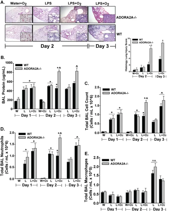
similarly treated WT mice and all other treatment groups (Figure 1). In this group, we observed a significant increase in histologic damage (Figure 1A), BAL protein (Figure 1B), and BAL neutro-phils (Figure 1C), compared with all other groups. Differences appeared on Day 2, and persisted on Day 3 for BAL protein and histologic injury after LPS instillation. BAL macrophage num-bers appeared to increase in all groups on Day 3, but with no differences between those groups (Figure 1E). The addition of 60% oxygen to IT water did not significantly increase the mea-sured lung injury parameters in WT or ADORA2A2/2mice. On
Figure 1. Anti-inflammatory A2A adenosinergic receptor–deficient (ADORA2A2/2) mice exposed to LPS plus oxygen demonstrated a significant increase in lung injury compared with ADORA2A2/2 mice exposed to LPS alone, and compared with wild-type (WT) mice exposed to LPS plus oxygen. Statistics were performed by com-paring all groups at the same time point. (A) Histology. Representa-tive low-power (34) and high-power (inset,340) hematoxylin and eosin–stained lung sections on Days 2 and 3 reveal increased alveolar consolidation and intersti-tial infiltration in ADORA2A2/2 mice exposed to LPS plus oxygen. Quantitative assessment using a lung-injury scoring system dem-onstrated a significant increase in lung injury in ADORA2A2/2mice exposed to LPS plus oxygen, be-ginning on Day 2 and persisting until Day 3. *On Day 2, significant compared with the WT water (W)1O2group. ^On Day 2, sig-nificant compared with all other groups. ^On Day 3, significant compared with the WT LPS (L)1 O2group. (B) Bronchoalveolar la-vage (BAL) protein was notable for an early, pronounced increase in ADORA2A2/2mice exposed to LPS plus oxygen. *On Day 1, sig-nificant compared with the WT W and ADORA2A2/2W groups. *On Day 2, significant compared with the WT W1O2and ADORA2A2/2 W1O2groups. ^On Day 2, sig-nificant compared with the WT LPS, WT L1O2, and ADORA2A2/2LPS groups. ^On Day 3, significant compared with the WT LPS, WT L 1 O2, and ADORA2A2/2 LPS groups. (C) The BAL total cell count was increased in ADORA2A2/2 mice exposed to LPS plus oxygen, compared with all other exposure groups on Day 2. *On Day 1, significant compared with the WT W and ADORA2A2/2W groups. *On Day 2, significant compared with the WT W1O2and ADORA2A2/2W1O2groups. ^On Day 2, significant compared with the WT LPS, WT L1O2, and ADORA2A2/2LPS groups. ^On Day 3, significant compared with the WT LPS and ADORA2A2/2LPS groups. (D) BAL Neutrophils were also increased in ADORA2A2/2mice exposed to LPS plus oxygen on Day 2. *On Day 1, significant compared with the WT W and ADORA2A2/2W groups. *On Day 2, significant compared with the WT W1O2and ADORA2A2/2W1O2groups. ^On Day 2, significant compared with the WT LPS, WT L1O2, and ADORA2A2/2LPS groups. ^On Day 3, significant compared with the WT LPS and ADORA2A2/2LPS groups. (E) BAL macrophages did not increase in ADORA2A2/2mice exposed to LPS plus oxygen, compared with all other groups at each time point (n¼4 in the W and W1O
Days 1 and 2, lung water accumulation was no different in WT and ADORA2A2/2mice. We previously observed an oxygen-induced
augmentation of lung water content no earlier than Day 4 (7). ADORA2A2/2 mice demonstrated a significant increase in both lung injury and mortality when treated with higher doses of LPS (1–3 mg/kg/mouse; no supplemental oxygen) (Figure E1A in the online supplement), but not at the primary study dose (0.3 mg/kg/ mouse). When we depleted neutrophils in ADORA2A2/2 mice exposed to LPS plus oxygen, lung injury was significantly abro-gated (Figure E1B), and the expression of alveolar macrophage CD86 was reduced (Figure E1C).
ADORA2A-Deficient Alveolar Macrophages Are Proinflammatory after LPS and Oxygen
To begin assessing ADORA2A-mediated differences in alveolar inflammation, we measured proinflammatory cytokines at early time points (Figure 2). BAL TNF-a and MIP-2 were signifi-cantly increased in ADORA2A2/2 mice exposed to LPS plus oxygen compared with WT mice exposed to LPS plus oxygen, and in comparison with other experimental groups on Day 1
(Figure 2A). MIP-2 is a potent neutrophil chemoattractant pri-marily secreted by macrophages. In ADORA2A2/2 mice
ex-posed to LPS plus oxygen, increased BAL MIP-2 secretion occurred before the increase in alveolar neutrophils observed on Day 2. No difference in BAL MIP-2 or TNF-awas evident between WT and ADORA2A2/2mice exposed to IT water plus 60% oxygen. Other neutrophil chemokines, including IL-17, KC, and LIX, were not increased in ADORA2A2/2 mice ex-posed to LPS plus oxygen; they are primarily secreted by other cell-types, and not macrophages (Figure 2B).
We also assessed ADORA2A-mediated differences in neutro-phil and macrophage life spans in the inflammatory milieu of the alveolar space by measuring cell apoptosis, a process known to be crucial in the modulation of inflammation (10, 19). Compared with other groups, BAL neutrophils from ADORA2A2/2mice
ex-posed to LPS plus oxygen exhibited similar apoptotic rates on Days 2 or 3, as assessed by annexin V staining (Figure 2C). In contrast, alveolar macrophages from ADORA2A2/2 mice ex-posed to LPS plus oxygen produced significantly less activated caspase-3, a marker of macrophage apoptosis (10), compared with WT macrophages exposed to LPS plus oxygen (Figure 2D).
Figure 2. Macrophages from ADORA2A2/2 mice secrete more neutrophil-recruiting chemokines and are less apo-ptotic after exposure to LPS plus oxygen, compared with WT mice. Statistics were determined by comparing all groups at the same time point. (A) BAL concentrations of macrophage inflammatory protein–2 (MIP-2) and TNF-a were increased in ADORA2A2/2mice exposed to LPS plus oxygen on Day 1. ^Significant compared with the WT LPS, WT L1O2, and ADORA2A2/2LPS groups. (B) Other che-mokines, including keratinocyte-derived chemokine (KC), IL-17, and LPS-induced chemokine (LIX), were not in-creased in ADORA2A2/2mice exposed to LPS plus oxy-gen, compared with WT mice. ^Significant compared with all other groups on BAL KC Day 1. *Significant com-pared with the WT W 1 O2 group on BAL KC Day 1. *Significant compared with the WT W 1 O2 group on BAL KC Day 2. ^Significant compared with all other groups on BAL KC Day 2. For BAL IL-17, no difference was evident between the assessed groups. *For BAL LIX, significant compared with the WT and ADORA2A2/2W1O
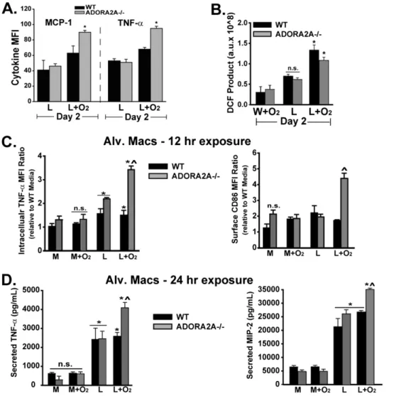
To assess the macrophage inflammatory state, we measured the intracellular mean fluorescence intensity of proinflammatory cytokines in CD11b1 alveolar macrophages (defined by high forward–side scatter and F4-80 positivity; see Figure E2A and Table 1). Others have demonstrated F4-80 to be a reliable al-veolar macrophage marker, and CD11b to be a marker of a recruited or exudative (in contrast to resident) macrophages (10, 30, 31). We found that the percentage of F4-80 positivity according to flow cytometry correlates with BAL macrophages identified by cytospin and Diff-Quik staining (Figure 1E). The exposure of WT or ADORA2A2/2 mice to LPS plus oxygen increased the percentage of CD11b1expression among F4-801 alveolar macrophages, compared with LPS plus room air mac-rophages (Table 1). BAL macmac-rophages (F4-801CD11b1) from ADORA2A2/2 mice exposed to LPS plus oxygen exhibited
more monocyte chemoattractant protein-1 (MCP-1) and TNF-a than did WT mice exposed to LPS plus oxygen, or ADOR-A2A2/2mice exposed to LPS plus room air (Table 1 and Figure 3A; a representative histogram is presented in Figure E2B). Add-ing 60% oxygen to LPS similarly increased alveolar macrophage ROS production in WT and ADORA2A2/2mice (Figure 3B).
In the alveolar microenvironment, macrophage communica-tion with other cells, including alveolar epithelial cells, recruited monocytes, or neutrophils, may influence the macrophage phe-notype (18, 30). To examine specific macrophage effects, we isolated alveolar macrophages (AMs) from unchallenged WT
or ADORA2A2/2 mice, and then exposed them to LPS, oxy-gen, or a combination of both in culture for 12 to 24 hours. At 12 hours, LPS-exposed alveolar macrophages (AMs) from WT and ADORA2A2/2mice produced more TNF-athan did con-trol WT AMs (Figure 3C). The addition of oxygen 3 hours after LPS further increased TNF-a in AMs from ADORA2A2/2 TABLE 1. EXUDATIVE MACROPHAGES
BAL Cells
F4-801 F4-80
1CD11b1
Day 2 Exposure Percent F4-801 Percent CD11b1 MCP-1 MFI TNF-aMFI
WT LPS (L) 13 (1.4) 33 (5.2) 41 (12.7) 53 (2.6)
A2AR2/2LPS 12 (2.8) 38 (6.3) 46 (3.1) 51 (3.9)
WT L1O2 10 (0.7)* 51 (1.7)* 63 (9.3) 68 (2.4)
A2AR2/2L1O
2 8 (3.6)* 51 (3.1)* 90 (2.4)† 95 (2.9)†
Definition of abbreviations: ADORA2A, anti-inflammatory A2A adenosinergic receptor; A2AR2/2, ADORA2A-deficient; BAL, bronchoalveolar lavage; L,
lipo-polysaccharide; MCP, monocyte chemoattractant protein; MFI, mean fluores-cence intensity; WT, wild-type.
Day 2 BAL exudative macrophages (F4-801CD11b1) from ADORA2A2/2mice
exposed to LPS plus oxygen produced more TNF-aand MCP-1, as determined by intracellular staining using flow cytometry. Statistics were determined by com-paring all groups with one-way ANOVA. Values are listed as averages (SEMs) (n¼
4–6 in each group).
* Significant compared with the WT LPS and ADORA2A2/2LPS groups. ySignificant compared with the WT LPS, ADORA2A2/2LPS, and WT L1O
2groups.
Figure 3. ADORA2A-deficient macrophages exposed to LPS plus oxygen demonstrate increased cytokine production as well as cosignaling molecule expression, com-pared with WT macrophages. Statistics were performed by comparing all groups at the same time point. (A) MCP-1 and TNF-a were increased in macrophages (F4-801 CD11b1) from ADORA2A2/2 mice exposed to LPS plus oxygen. *Sig-nificant compared with the WT LPS, ADORA2A2/2 LPS, and WT L 1 O
mice, but not in AMs from WT mice. Cosignaling molecule CD86 expression at 12 hours after LPS plus oxygen was in-creased only in AMs from ADORA2A2/2 mice (Figure 3C). Supplemental oxygen did not increase TNF-a or CD86 in media-exposed macrophages from WT or ADORA2A2/2mice. Exposing AMs from WT or ADORA2A2/2 mice to LPS for 24 hours significantly increased both TNF-aand MIP-2, com-pared with media exposure. Adding oxygen to LPS further increased TNF-aand MIP-2 only in AMs from ADORA2A2/2 mice (Figure 3D).
Oxygen Suppresses LPS-Induced Augmentation of ADORA2A to Promote Inflammation and Lung Injury
In ADORA2A2/2mice, LPS plus oxygen augments lung injury and the M1 macrophage proinflammatory state. Therefore, we wanted to determine whether ADORA2A manipulation in WT mice might alter lung inflammation. CGS-21680 is a specific ADORA2A agonist that increases ADORA2A pathway activ-ity (32). A daily systemic delivery of CGS-21680 (125 mg in
100ml PBS) beginning at the time of oxygen exposure signifi-cantly blunted the oxygen-induced augmentation of lung injury on Day 3 after LPS (Figure 4). We assessed the effects of CGS-21680 on Day 3, when an oxygen-mediated increase in lung injury was most pronounced in WT mice (7). A marked decrease in histologic injury was evident (Figure 4A), as well as a decrease in BAL protein (Figure 4B), and the total alveolar cell count, including both neutrophils and macrophages (Figure 4C). CGS-21680 did not exert a significant effect in LPS-exposed WT mice, or in LPS plus oxygen–exposed ADORA2A2/2mice. The amount of BAL MIP-2 was lower in LPS plus oxygen–exposed WT mice treated with CGS-21680 (Figure 4D), but CGS-21680 did not alter BAL KC secretion (not shown).
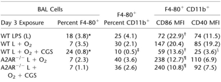
We isolated BAL cells to determine the importance of ADORA2A pathway activation in macrophage phenotype. Add-ing oxygen to LPS decreased the overall percentage of macro-phages (F4-801) on Day 3, an effect that was reversed with the addition of CGS-21680 (Table 2). The addition of CGS-21680 also decreased CD11b1expression among F4-801macrophages (Figure E2A). Furthermore, CGS-21680 induced a significant
reduction in surface expression of cosignaling molecules CD86 and CD40 (Table 2 and Figure E2C) among F4-801 CD11b1 cells from LPS plus oxygen–exposed mice. In isolated naive al-veolar macrophages stimulated for 24 hours, adding CGS-21680 to LPS plus oxygen–treated cells significantly reduced TNF-aand MIP-2 concentrations, compared with sham treatment in the LPS plus oxygen group (Figure 4E). Therefore, CGS-21680, a specific ADORA2A agonist, significantly reduced the expression of proinflammatory markers in macrophages from WT mice ex-posed to LPS plus oxygen.
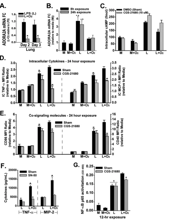
In WT mice, adding 60% oxygen to LPS decreased lung ADORA2A mRNA on Days 2 and 3 after IT LPS (Figure 5A). We also wanted to assess the effects of oxygen on ADORA2B in response to oxygen-mediated effects on ADORA2A. However, no differences in lung ADORA2B mRNA were evident between WT or ADORA2A2/2mice exposed to LPS and oxygen for 2 or 3 days
(Figure E2D). To determine whether oxygen may specifically de-crease macrophage ADORA2A, we used both a macrophage cell line (MH-S) andex vivoalveolar and peritoneal macrophages. LPS induced a 3- to 4-fold increase in ADORA2A mRNA in MH-S cells at 8 hours, and this increase was sustained at 24 hours (Figure 5B). Adding oxygen to LPS-treated macrophages significantly de-creased ADORA2A mRNA compared with LPS alone, as we observed in lung tissue. Adding oxygen to media-treated MH-S cells did not alter ADORA2A mRNA concentrations. To deter-mine whether an oxygen-induced decrease in ADORA2A mRNA translated to a functional effect on the macrophage ADORA2A pathway, we measured intracellular cAMP. ADORA2A activation increases cAMP, and receptor-specific manipulation alters cAMP concentrations (33). LPS significantly increased cAMP in MH-S cells (Figure 5C). Exposing LPS-stimulated macrophages to supple-mental oxygen significantly reduced cAMP compared with LPS treatment alone, an effect that was negated by pretreatment with the ADORA2A agonist CGS-21680. Adding supplemental oxygen to media-exposed macrophages did not alter cAMP concentrations, and CGS-21680 exerted no effect. To assess the impact of the manipulation of ADORA2A signaling on macrophage phenotype and function, we measured cytokines (TNF-a and MCP-1) and cosignaling molecules (CD86 and CD40) in macrophages by flow cytometry. When added to LPS-exposed peritoneal macrophages, 60% oxygen significantly increased TNF-aand MCP-1 at 24 hours,
an effect blocked by pretreatment with CGS-21680 (Figure 5D). CGS-21680 also reduced the abundance of cosignaling molecules CD86 and CD40 in LPS plus oxygen–treated macrophages (Figure 5E). Adding supplemental oxygen to media-exposed peritoneal macrophages did not increase TNF-a, MCP-1, CD86, or CD40 after 24 hours. When we pretreated macrophages with a nonspecific adenosine receptor agonist, NECA (3mM), no additional reduction was observed in cytokines or cosignaling molecule expression com-pared with CGS-21680 treatment, indicating a primary role for ADORA2A signaling in our model (data not shown).
NF-kB mediates the expression of several proinflammatory molecules, including TNF-aand MIP-2 (34, 35), and may regulate the expression of cosignaling molecules CD86 and CD40 (36). We previously observed an LPS plus oxygen–mediated increase in macrophage NF-kB p65 activity in comparison to LPS exposure alone. Oxygen alone exerted no effect on cells exposed to media (7). In alveolar macrophages exposed to LPS, NF-kB blockade with SN-50 (50 mg/ml) mildly reduced TNF-a, but exerted no effect on MIP-2 (Figure 5F). In alveolar macrophages exposed to LPS plus oxygen, NF-kB blockade using SN-50 administered at the time of oxygen exposure led to a significant, greater than 50% reduction in alveolar macrophage TNF-a and MIP-2 pro-duction. In peritoneal macrophages, the addition of oxygen to LPS increased p65 activity by 50% compared with LPS alone. Pretreatment with CGS-21680 in the group with LPS plus oxygen reduced total NF-kB p65 activity by greater than 50% of the amount induced by oxygen plus LPS. CGS-21680 exerted no ef-fect on cells exposed to LPS alone (Figure 5G).
Both Myeloid and Nonmyeloid ADORA2A Modulate LPS plus Oxygen–Induced Lung Injury
To determine the specific contribution of inflammatory-cell ADORA2A in modulating lung injury, we performed chimera experiments by irradiating mice and transferring bone marrow– derived cells to designated groups. We used CD45.1 (WT) mice to differentiate leukocytes from the CD45.2 leukocyte common an-tigen found in ADORA2A2/2 mice, allowing us to distinguish
donor from recipient cells. Eight weeks after bone marrow cell transfer, we determined chimerism to be greater than 90% in all groups of mice (flow cytometry; data not shown). We exposed irradiated and nonirradiated age-matched WT and ADORA2A2/2 mice to LPS plus oxygen, and assessed the response in groups of chimeras. After 2 days of LPS plus oxygen (the earliest time point when ADORA2A2/2mice first demonstrate an appreciable aug-mentation of lung injury compared with WT mice), injury param-eters were evaluated (Figure 6). Histologic injury (Figure 6A) was most severe in ADORA2A2/2mice that received ADORA2A2/2 myeloid cells. WT mice that received ADORA2A2/2 myeloid cells demonstrated the second most severe histologic injury. His-tologic injury was significantly milder in ADORA2A2/2mice that
received WT myeloid cells and in WT mice that received WT myeloid cells. The concentration of BAL protein (Figure 6B) was higher in both chimera groups that received ADORA2A2/2
myeloid cells, compared with both groups that received WT my-eloid cells. BAL total cells (Figure 6C) were significantly increased only in ADORA2A2/2mice that received ADORA2A2/2cells, compared with WT mice that received WT cells. BAL neutrophils (Figure 6D) were significantly increased only in both chimera groups that received ADORA2A2/2 myeloid cells. Although BAL macrophage numbers were no different between treatment groups (Figure 6D), both F4-801 CD11c1and F4-801 CD11b1 macrophages from WT mice that received ADORA2A2/2 mye-loid cells expressed significantly more TNF-a than did macro-phages from both chimera groups that received WT myeloid cells (Figure 6E).
TABLE 2. EXUDATIVE MACROPHAGES BAL Cells
F4-801 F4-80
1CD11b1
Day 3 Exposure Percent F4-801 Percent CD11b1 CD86 MFI CD40 MFI
WT LPS (L) 18 (3.8)* 25 (4.1) 72 (22.9)† 74 (11.5)
WT L1O2 7 (3.5) 30 (2.1) 147 (20.4) 85 (19.2)
WT L1O21CGS 24 (0.8)* 10 (0.5)‡ 59 (13.6)† 25 (3.6)x
A2AR2/2L1O
2 7 (2.3) 40 (3.6) 238 (12.7)¶ 110 (6.9)
A2AR2/2L1 O21CGS
7 (1.1) 36 (2.6) 240 (10.8)¶ 92 (7.5)
Definition of abbreviations: ADORA2A, anti-inflammatory A2A adenosinergic recep-tor; A2AR2/2, ADORA2A-deficient; BAL, bronchoalveolar lavage; CGS, CGS-21680
hydrochloride; L, lipopolysaccharide; MFI, mean fluorescence intensity; WT, wild-type. Day 3 BAL exudative macrophages (F4-801CD11b1) from WT mice treated with the ADORA2A agonist CGS-21680 expressed significantly less of the M1 inflammatory markers CD86 and CD40 according to flow cytometry analysis. Statistics were determined by comparing all groups with one-way ANOVA. Val-ues are listed as averages (SEMs) (n¼4–6 in each group).
* In the Percent F4-801column, significant compared with the WT L1O2,
ADORA2A2/2L1O
2, and ADORA2A2/2L1O21CGS-21680 groups. ySignificant compared with the WT L1O
2group.
zSignificant compared with all other groups, among F4-801CD11b1
inflam-matory macrophages.
xSignificant compared with all groups.
DISCUSSION
We conclude that macrophage ADORA2A is a critical modifier of the oxygen-induced augmentation of inflammation and lung injury after intratracheal LPS. In the absence of ADORA2A, mice ex-posed to LPS plus oxygen exhibit more severe lung injury, and alveolar macrophages from these mice demonstrate an augmented and sustained proinflammatory M1-phenotype, known to be asso-ciated with poor outcomes (21, 22). Furthermore, supplemental oxygen delivery after LPS augments the macrophage proinflam-matory state even in WT mice, at least in part by attenuating the LPS-induced increase in anti-inflammatory ADORA2A expres-sion. Delivery of the ADORA2A-specific agonist CGS-21680
increased ADORA2A signaling, to reduce lung injury signifi-cantly and dampen the macrophage proinflammatory state in WT mice exposed to LPS and oxygen.
space. In ADORA2A-deficient mice, the alveolar recruitment of neutrophils is further enhanced after LPS plus oxygen exposure because of increased chemokine secretion, predominantly from macrophages. Alveolar macrophages from ADORA2A2/2mice
exposed to LPS plus oxygen exhibit an increased production of MCP-1, a potent monocyte chemoattractant, indicating that
macrophages from ADORA2A2/2mice promote both monocyte
and neutrophil recruitment to the alveolar space.
the LPS-mediated increase in ADORA2A mRNA. The tran-scription factor hypoxia-inducible factor–1 (HIF-1) is increased after LPS and stabilized under hypoxic conditions, and is known to induce the transcription of ADORA2A (37, 38). The degra-dation of HIF-1 requires the activity of prolyl hydroxylases (PHDs), which use oxygen as a substrate with a Michaelis-Menten constant known to demonstrate peak activity at 45% oxygen (39). One possibility, as Chandel and Budinger pro-posed, involves supranormal oxygen (60%) maximizing PHD activity in excess of that which is present in ambient air (21%) (40). If this is the case, then macrophages exposed to oxygen after LPS in our model would manifest relatively lower HIF-1 concen-trations compared with macrophages exposed to LPS alone, which could decrease ADORA2A mRNA.
Adding 60% oxygen also decreased the LPS-induced increase in macrophage cAMP, indicative of functional ADORA2A pathway blockade. This may result from reduced ADORA2A total protein or reduced ADORA2A surface receptor protein. Collins and colleagues recently showed that hyaluronic acid (HA) prevented the cycling of ADORA2A to the cell mem-brane, but exerted no effect on total ADORA2A protein con-centrations (29). The mechanisms by which oxygen alters ADORA2A signaling after LPS exposure remain unknown, but possibilities include the production of reactive oxygen spe-cies (41), direct binding to a cell surface receptor/coreceptor, or indirect interaction through HA/Toll-like receptor (TLR) sig-naling pathways (42–44).
ADORA2A is an important regulator of inflammation in other models of injury, yet the actions of ADORA2A in macrophages re-main largely unknown (25, 29, 45–48). Because CGS-21680 does express weak affinity for another adenosine receptor, ADORA2B (1/100 potency compared with ADORA2A), we used NECA as a nonselective AR agonist to assess the potential additional contri-bution of ADORA2B to the observed response in our model (11). We observed no additional anti-inflammatory benefit of NECA over CGS-21680, reinforcing the principal role of ADORA2A.
The inhibitory effect of ADORA2A in macrophages has been identified in models of inflammation involving signaling through several TLRs, including 2, 3, 7, and 9 (49). Therefore, ADORA2A binding likely inhibits a common proinflammatory activator (11, 26). Multiple mechanisms surely exist, but fundamental to the anti-inflammatory effects of the ADORA2A pathway is its inhibition of the transcription factor NF-kB (50, 51). CGS-21680, a specific ADORA2A agonist, blocked 50% of the oxygen-induced increase in NF-kB activity in macrophages exposed to LPS. However, as others have observed with adenosine (52), CGS-21680 exerted no effect on NF-kB activity in macrophages exposed to LPS alone, suggesting distinct oxygen-mediated effects on NF-kB compared
with LPS. One possibility involves a differential effect on interleukin-1 receptor-associated kinase M, a known repressor of NK-kB sig-naling that directly associates with CD86 according to coimmuno-precipitation experiments (53). CD86 expression in macrophages was also down-regulated with CGS-21680 treatment.
Macrophages from ADORA2A2/2mice may promote inflam-mation by interactions with other cell types, including neutrophils and epithelial cells. Others have shown that the neutrophil acti-vation of macrophages occurs via CD86 (36, 48). When we sys-temically depleted neutrophils, the expression of CD86 in F4-801 CD11b1alveolar macrophages was reduced, suggesting a possible neutrophil-mediated effect on macrophage CD86 expression (Fig-ure E1C). In isolated macrophages, the antibody-mediated block-ade of CD86 did not alter the abundance of other cosignaling molecules or intracellular cytokines in the context of LPS, oxygen, or the two in combination (data not shown). However, we did not test this effect in the presence of neutrophils.
Epithelial ADORA2A has been shown to modify lung water ac-cumulation (54, 55) and prevent allograft airway rejection (49, 56). Our chimera experiments demonstrated that the absence of either myeloid or epithelial/endothelial ADORA2A augments lung injury after LPS plus oxygen exposure, and the absence of both exerts an additive effect on lung injury. In contrast to our findings, Reutershan and colleagues concluded that epithelial or endothelial ADORA2A may be proinflammatory (25). The harmful effect of lacking epithe-lial ADORA2A may be particularly revealing in our lung injury model, in which lung epithelial cells are constantly exposed to ele-vated oxygen tensions. Therefore, the addition of supplemental ox-ygen may constitute a pertinent difference in our model. Adding 60% oxygen to LPS-treated alveolar epithelial cells (murine lung epithelial-12 cell line) in contact with macrophages synergistically increased the secretion of neutrophil-recruiting chemokines, and subsequently enhanced neutrophil migration (unpublished observa-tions by Neil R. Aggarwal and Landon S. King). In the absence of epithelial ADORA2A, the synergistic proinflammatory effect of this cell–cell interaction may be even more pronounced.
Macrophage ADORA2A prevents the augmentation of lung injury after LPS and oxygen. Without ADORA2A, macrophages exposed to LPS plus oxygen displayed both an increased and sus-tained proinflammatory M1 phenotype, leading to an enhanced recruitment of alveolar neutrophils and the augmentation of lung injury. In WT mice, supplemental oxygen appears to blunt LPS-induced increases in cAMP, consistent with an ADORA2A path-way blockade. The effects of oxygen in macrophages are clearly multifactorial. Our experiments demonstrate that supplemental oxygen, as a modifiable exposure, augments lung inflammation through the down-regulation of the ADORA2A pathway. Al-though we do not understand the specific oxygen-induced effects
;
in humans with acute lung inflammation and injury, the protective effects of the ADORA2A agonist CGS-21680 on the oxygen-mediated augmentation of LPS-induced murine lung injury may provide an avenue for investigation in human trials of ARDS.
Author disclosuresare available with the text of this article at www.atsjournals.org.
References
1. Matthay M, Zimmerman G. Acute lung injury and the acute respiratory distress syndrome: four decades of inquiry into pathogenesis and ra-tional management.Am J Respir Cell Mol Biol2005;33:319–327. 2. Gajic O, Dabbagh O, Park PK, Adesanya A, Chang SY, Hou P, Anderson
H, Hoth JJ, Mikkelsen ME, Gentile NT,et al. Early identification of patients at risk of acute lung injury: evaluation of lung injury prediction score in a multicenter cohort study.Am J Respir Crit Care Med2011; 183:462–470.
3. Matute-Bello G, Frevert CW, Martin TR. Animal models of acute lung injury.Am J Physiol Lung Cell Mol Physiol2008;295:L379–L399. 4. Bertolini G, Lewandowski K, Bion J, Romand JA, Villar J, Thorsteinsson A,
Damas P, Armaganidis A, Lemaire F, Minelli C,et al. Epidemiology and outcome of acute lung injury in European intensive care units.Intensive
Care Med2004;30:51–61.
5. Pepe PE, Potkin RT, Reus DH, Hudson LD, Carrico CJ. Clinical pre-dictors of the adult respiratory distress syndrome.Am J Surg1982; 144:124–130.
6. Rachmale S, Li G, Gregory W, Malinchoc M, Gajic O. Practice of ex-cessive inspired oxygen supplementation and effect on pulmonary outcomes in mechanically ventilated patients with acute lung injury.
Respir Care2012;57:1887–1893.
7. Aggarwal NR, D’Alessio FR, Tsushima K, Files DC, Damarla M, Sidhaye VK, Fraig MM, Polotsky VY, King LS. Moderate oxygen augments lipopolysaccharide-induced lung injury in mice. Am J
Physiol Lung Cell Mol Physiol2010;298:371–381.
8. Thiel M, Chouker A, Ohta A, Jackson E, Caldwell C, Smith P, Lukashev D, Bittmann I, Sitkovsky MV. Oxygenation inhibits the physiological tissue-protecting mechanism and thereby exacerbates acute inflam-matory lung injury.PLoS Biol2005;3:e174.
9. Sue RD, Belperio JA, Burdick MD, Murray LA, Xue YY, Dy MC, Kwon JJ, Keane MP, Strieter RM. CXCR2 is critical to hyperoxia-induced lung injury.J Immunol2004;172:3860–3868.
10. Janssen WJ, Barthel L, Muldrow A, Oberley-Deegan RE, Kearns MT, Jakubzick C, Henson PM. Fas determines differential fates of resident and recruited macrophages during resolution of acute lung injury.Am
J Respir Crit Care Med2011;184:547–560.
11. HaskóG, Pacher P, Deitch E, Vizi ES. Shaping of monocyte and macro-phage function by adenosine receptors.Pharmacol Ther2007;113:264–275. 12. Nathan C. Points of control in inflammation.Nature2002;420:8946–8952. 13. Gordon S, Taylor PR. Monocyte and macrophage heterogeneity.Nat
Rev Immunol2005;5:953–964.
14. Gordon S, Martinez FO. Alternative activation of macrophages: mech-anism and functions.Immunity2010;32:593–604.
15. Maus UA, Janzen S, Wall G, Srivastava M, Blackwell TS, Christman JW, Seeger W, Welte T, Lohmeyer J. Resident alveolar macrophages are replaced by recruited monocytes in response to endotoxin-induced lung inflammation.Am J Respir Cell Mol Biol2006;35:227–235. 16. Maus UA, Koay MA, Delbeck T, Mack M, Ermert M, Ermert L,
Blackwell TS, Christman JW, Schlo¨ndorff D, Seeger W,et al. Role of resident alveolar macrophages in leukocyte traffic into the alveolar air space of intact mice.Am J Physiol Lung Cell Mol Physiol2002;282: L1245–L1252.
17. D’Alessio FR, Tsushima K, Aggarwal NR, Mock JR, Eto Y, Garibaldi BT, Files DC, Avalos CR, Rodriguez JV, Waickman AT,et al. Resolution of experimental lung injury by monocyte-derived inducible nitric oxide synthase.J Immunol2012;189:2234–2245.
18. Nolan A, Weiden M, Kelly A, Hoshino Y, Hoshino S, Mehta N, Gold JA. CD40 and CD80/86 act synergistically to regulate inflammation and mortality in polymicrobial sepsis.Am J Respir Crit Care Med
2008;177:301–308.
19. Matute-Bello G, Liles WC, Radella F, Steinberg KP, Ruzinski JT, Jonas M, Chi EY, Hudson LD, Martin TR. Neutrophil apoptosis in the acute
respiratory distress syndrome.Am J Respir Crit Care Med1997;156: 1969–1977.
20. Rosseau S, Hammerl P, Maus U, Walmrath H, Schu¨tte H, Grimminger F, Seeger W, Lohmeyer J. Phenotypic characterization of alveolar monocyte recruitment in acute respiratory distress syndrome. Am
J Physiol Lung Cell Mol Physiol2000;279:L25–L35.
21. Murray PJ, Wynn TA. Protective and pathogenic functions of macro-phage subsets.Nat Rev Immunol2011;11:723–737.
22. Sindrilaru A, Peters T, Wieschalka S, Baican C, Baican A, Peter H, Hainzl A, Schatz Susanne, Qi Y, Schlecht A,et al. An unrestrained proinflammatory M1 macrophage population induced by iron impairs wound healing in humans and mice.J Clin Invest2011;121:985–997. 23. Sitkovsky MV, Lukashev D, Apasov S, Kojima H, Koshiba M, Caldwell
C, Ohta A, Thiel M. Physiological control of immune response and inflammatory tissue damage by hypoxia-inducible factors and aden-osine A2A receptors.Annu Rev Immunol2004;22:657–682. 24. Reutershan J, Vollmer I, Stark S, Wagner R, Ngamsri K, Eltzschig HK.
Adenosine and inflammation: CD39 and CD73 are critical mediators in LPS-induced PMN trafficking into the lungs.FASEB J2009;23:473–482. 25. Reutershan J, Cagnina RE, Chang D. Therapeutic anti-inflammatory effects of myeloid cell adenosine receptor A2A stimulation in lipopolysaccharide-induced lung injury.J Immunol2007;179:1254–1263.
26. Scheibner KA, Boodoo S, Collins S, Black KE, Chan-Li Y, Zarek P, Powell JD, Horton MR. The adenosine A2A receptor inhibits matrix-induced inflammation in a novel fashion.Am J Respir Cell Mol Biol
2009;40:251–259.
27. Barnholt KE, Kota RS, Aung HH, Rutledge JC. Adenosine blocks IFN-gamma–induced phosphorylation of STAT1 on serine 727 to reduce macrophage activation.J Immunol2009;183:6767–6777.
28. Barletta KE, Ley K, Mehrad B. Regulation of neutrophil function by adenosine.Arterioscler Thromb Vasc Biol2012;32:856–864. 29. Collins SL, Black KE, Chan-Li Y, Ahn Y, Cole PA, Powell JD, Horton MR.
Hyaluronan fragments promote inflammation by down-regulating the anti-inflammatory A2A receptor.Am J Respir Cell Mol Biol2011;45:675–683. 30. Johnston LK, Rims CR, Gill SE, McGuire J, Manicone AM. Pulmonary macrophage subpopulations in induction and resolution of acute lung injury.Am J Respir Cell Mol Biol2012;47:417–426.
31. Tighe RM, Liang J, Liu N, Jung Y, Jiang D, Gunn MD, Noble PW. Recruited exudative macrophages selectively produce CXCL10 after noninfectious lung injury.Am J Respir Cell Mol Biol2011;45:781–788. 32. Jarvis MF, Schulz R, Hutchison AJ, Do UH, Sills MA, Williams M. [3H]CGS 21680, a selective A2 adenosine receptor agonist directly labels A2 receptors in rat brain.J Pharmacol Exp Ther1989;251:888–893. 33. HaskóG, Linden J, Cronstein B, Pacher P. Adenosine receptors:
ther-apeutic aspects for inflammatory and immune diseases.Nat Rev Drug
Discov2008;7:759–770.
34. Pahl HL. Activators and target genes of Rel/NF-kappaB transcription factors.Oncogene1999;18:6853–6866.
35. Chandel NS, Trzyna WC, Mcclintock DS, Schumacker PT. Role of oxidants in NF-B activation and TNF-agene transcription induced by hypoxia and endotoxin 1.J Immunol2000;165:1013–1021.
36. Hoebe K, Janssen EM, Kim SO, Alexopoulou L, Flavell RA, Han J, Beutler B. Upregulation of costimulatory molecules induced by lipo-polysaccharide and double-stranded RNA occurs by TRIF-dependent and TRIF-independent pathways.Nat Immunol2003;4:1223–1229. 37. Manalo DJ, Rowan A, Lavoie T, Natarajan L, Kelly BD, Ye SQ, Garcia
JG, Semenza GL. Transcriptional regulation of vascular endothelial cell responses to hypoxia by HIF-1.Blood2005;105:659–669. 38. Fang HY, Hughes R, Murdoch C, Coffelt SB, Biswas SK, Harris AL,
Johnson RS, Imityaz HZ, Simon MC, Fredlund E, et al. Hypoxia-inducible factors 1 and 2 are important transcriptional effectors in primary macrophages experiencing hypoxia.Blood2009;114:844–859. 39. HirsiläM, Koivunen P, Günzler V, Kivirikko KI, Myllyharju J. Char-acterization of the human prolyl 4-hydroxylases that modify the hypoxia-inducible factor.J Biol Chem2003;278:30772–30780. 40. Chandel NS, Budinger GRS. The cellular basis for diverse responses to
oxygen.Free Radic Biol Med2007;42:165–174.
41. Forman HJ, Torres M. Reactive oxygen species and cell signaling: re-spiratory burst in macrophage signaling.Am J Respir Crit Care Med
42. Asehnoune K, Strassheim D, Mitra S, Kim JY, Abraham E. Involvement of reactive oxygen species in Toll-like receptor 4–dependent activa-tion of NF-kappa B.J Immunol2004;172:2522–2529.
43. Zhang X, Shan P, Qureshi S, Homer R, Medzhitov R, Noble PW, Lee PJ. Cutting edge: TLR4 deficiency confers susceptibility to lethal oxidant lung injury.J Immunol2005;175:4834–4838.
44. Jiang D, Liang J, Fan J, Yu S, Chen S, Luo Y, Prestwich GD, Mascarenhas MM, Garg HG, Quinn DA,et al. Regulation of lung injury and repair by Toll-like receptors and hyaluronan. Nat Med
2005;11:1173–9.
45. Tang LM, Zhu JF, Wang F, Qian J, Zhu J, Mo Q, Lu HH, Li GQ, Wang XH. Activation of adenosine A2A receptor attenuates inflammatory response in a rat model of small-for-size liver transplantation.
Transplant Proc2010;42:1915–1920.
46. Hamano R, Takahashi H, Iwagaki H, Kanke T. Stimulation of adenosine A2A receptor inhibits LPS-induced expression of intercellular adhe-sion molecule 1 and production of TNF-ain human peripheral blood mononuclear cells.Shock2008;29:2–5.
47. Belikoff B, Hatfield S, Sitkovsky M, Remick DG. Adenosine negative feedback on A2A adenosine receptors mediates hyporesponsiveness in chronically septic mice.Shock2011;35:382–387.
48. Bystrom J, Evans I, Newson J, Stables M, Toor I, van Rooijen N, Crawford M, Colville-Nash P, Farrow S, Gilroy DW. Resolution-phase macrophages possess a unique inflammatory phenotype that is controlled by cAMP.
Blood2008;112:4117–4127.
49. Power Coombs MR, Belderbos ME, Gallington LC, Bont L, Levy O. Adenosine modulates Toll-like receptor function: basic mechanisms and translational opportunities. Expert Rev Antiinfect Ther 2011;9: 261–269.
50. Sands WA, Martin AF, Strong EW, Palmer TM. Specific inhibition of nuclear factor–B–dependent inflammatory responses by cell type– specific mechanisms upon A2a adenosine receptor gene transfer.Mol
Pharmacol2004;66:1147–1159.
51. Majumdar S, Aggarwal BB. Adenosine suppresses activation of nuclear factor–kappaB selectively induced by tumor necrosis factor in dif-ferent cell types.Oncogene2003;22:1206–1218.
52. Hoshino Y, Hoshino S, Gold JA, Raju B, Prabhakar S, Pine R, Rom WN, Nakata K, Weiden M. Mechanisms of polymorphonuclear neutrophil– mediated induction of HIV-1 replication in macrophages during pulmo-nary tuberculosis.J Infect Dis2007;195:1303–1310.
53. Nolan A, Kobayashi H, Naveed B, Kelly A, Hoshino Y, Hoshino S, Karulf MR, Rom WN, Weiden MD, Gold JA. Differential role for CD80 and CD86 in the regulation of the innate immune response in murine polymicrobial sepsis.PloS One2009;4:e6600.
54. Murphree LJ, Sullivan GW, Marshall MA, Linden J. Lipopolysaccharide rapidly modifies adenosine receptor transcripts in murine and human macrophages: role of NF-kB in A2a adenosine receptor induction.
Biochem J2005;580:575–580.
55. Factor P, Mutlu GM, Chen L, Mohameed J, Akhmedov AT, Meng FJ, Jilling T, Lewis ER, Johnson MD, Xu A,et al. Adenosine regulation of alveolar fluid clearance.Proc Natl Acad Sci USA2007;104:4083– 4088.