2 ABSTRACT
Elizabeth Goodall Rossitch
The Effect of COPD on the Immune Response to Influenza Virus Vaccination (Under Direction of Melinda A. Beck)
Chronic Obstructive Pulmonary Disease (COPD) is a health concern worldwide and particularly in the United States. It is the currently the fourth leading cause of death in the world and is predicted to become the third leading cause of death by 2030. People with COPD often experience exacerbations, an acute onset of more severe symptoms, caused by infections-bacterial and viral. These exacerbations decrease quality of life and further disease progression. Not to mention, while healthy individuals can easily recover from an influenza, it could be deadly in COPD patients. Thus, it is important to ensure that individuals with COPD are adequately covered by influenza vaccination. Currently, little is known about how COPD impacts the cell-mediated immune response, but gaining this knowledge would help determine if individuals with COPD are effectively covered by the vaccine.
The purpose of this pilot study was to determine if COPD affects the cell-mediated immune response post influenza vaccination. Specifically, it aimed to
3 A/Victoria/361/2011. The cells were stained for CD3, CD8, TNFα, and IFNγ using
human fluorochrome-conjugated human antibodies.
4 ACKNOWLEDGEMENTS
5 TABLE OF CONTENTS
LIST OF TABLES……….7
LIST OF FIGURES………...8
Chapter 1. INTRODUCTION………10
1.1 Chronic Obstructive Pulmonary Disease (COPD)………10
1.2 Influenza Infection……….11
1.3 Cellular Immune Response to Influenza and Importance of Vaccination.12 1.4 COPD and Influenza Virus………...13
2. SPECIFIC AIM AND HYPOTHESIS………14
3. METHODS………...15
3.1 Study Population and Samples………...15
3.2 Peripheral Blood Mononuclear Cell Collection……….16
3.3 PBMC Thawing, Plating, and Stimulation………..17
3.4 PBMC Staining and Flow Cytometry………..18
3.5 Statistical Analysis……….19
4. RESULTS………20
4.1 Demographics of the Study Population……….20
4.2 CD3+CD8+ T Cells………20
4.3 CD3+CD8+IFNγ+ T Cells……….21
4.4 CD3+CD8+TNFα+ T Cells………..….23
6
5.1 CD3+CD8+ T Cells………26
5.2 CD3+CD8+IFNγ+ T Cells……….…26
5.3 CD3+CD8+TNFα T Cells……….26
5.4 Limitations………..…27
5.5 Study Implications and Future Studies……….….28
7 LIST OF TABLES
Table
8 LIST OF FIGURES
1. There are no significant differences in the percentage of CD3+CD8+ T cells between people with COPD and people without COPD for either unstimulated or stimulated PMBCs………...…21 2. There are no significant differences in the percentage of CD3+CD8+IFNγ T cells between people with COPD and people without COPD for either unstimulated or
stimulated PMBCs………..22 3. All participants trended towards an increase in CD3+CD8+IFNγ+ T cell expression upon stimulation with H3N2, but individuals without COPD trended towards a greater expression CD3+CD8+IFNγ+ T cells after stimulation with
H3N2……….………22 4. Impaired upregulation of CD3+CD8+IFNγ+ T cells in PBMCs from individuals with COPD………...23 5. Individuals with COPD had a significantly higher expression of CD3+CD8+TNFα+ T Cells than individuals without COPD for unstimulated PMBCs………..24 6. Impaired upregulation of CD3+CD8+TNFα+ T cells in PBMCs from individuals with COPD………...24 7.All individuals without COPD trended towards an increase in CD3+CD8+TNFα+ expression upon stimulation with H3N2, but individuals with COPD had different
9 LIST OF ABBREVIATIONS
APC allophycocyanin BMI Body Mass Index
CDC Centers for Disease Control and Prevention COPD Chronic Obstructive Pulmonary Disease
CTL cytotoxic T Cell
FDA Food and Drug Administration
FITC fluorescein isothiocyanate HBSS Hank’s Balanced Salt Solution IFN interferon
MHC major histocompatibility complex NIH National Institutes of Health
PBMC peripheral blood mononuclear cells PE phycoerythrin
PerCP peridinin-chlorophyll-protein complex
10 CHAPTER 1
INTRODUCTION 1.1 Chronic Obstructive Pulmonary Disease (COPD)
Chronic Obstructive Pulmonary Disease (COPD) is predicted to be a worldwide health concern over the next few decades. Currently it is the fourth leading cause of death, but the World Health Organization projects that COPD will move to third by 2030 (3). A literature review extracting prevalence data from 80 articles found that the United States had the highest COPD prevalence of 37%, compared to Japan which had the lowest prevalence of 0.2% (4).
COPD is a life threatening lung disease that disrupts normal breathing through long-term cough with mucus build up (chronic bronchitis) or destruction of the lung tissue over time (emphysema). Symptoms of COPD develop slowly over time and include dyspnea, chronic cough, abnormal sputum, and trouble doing normal daily activities such as walking up a flight of stairs. The number one risk factor for COPD is smoking tobacco, but other risks include certain gas or fume exposures in the workplace, secondhand smoke or pollution exposure, and exposure to cooking fire without proper ventilation (5).
11 exacerbations which aggravate the lungs, increase death in COPD patients, and
decrease quality of life. An exacerbation is a period (at least 48 hours) where relatively more severe symptoms are experienced by the patient. So what causes these
exacerbations? There are several suggested causes such as non-adherence to
medication, heart failure, pneumonia, inhalation of irritants such as tobacco smoke, and most notably bacterial and viral infections (3).
1.2 Influenza Infection
The flu season typically lasts from October to March and The Centers for Disease Control and Prevention (CDC) estimates that during this time 5 to 20 percent of people in the United States are infected with influenza (6). This may not seem like an alarming statistic because many people can recover from flu without medication or complications (7). Despite this, the seasonal influenza hospitalizes more than 200,000 and kills more than 36,000 Americans each year (6). People at greater risk for hospitalization and death from influenza infection include children younger than 5 years old, older adults greater than 65 years old, pregnant women, and people with chronic illnesses such as COPD (7).
12 transmission occurs when the recipient touches virus from these droplets that remains on a surface or object and then subsequently touches their mouth, eyes or nose.
Viruses can last on surfaces between two and eight hours depending on conditions (6).
1.3 Cellular Immune Response to Influenza and Importance of Vaccination
The body has several lines of defense to protect against infection, including mucus production to prevent it in the first place, however this paper will focus on the cell
mediated response specifically for influenza. When a specific strain of influenza infects for the first time, the body depends on the innate immune system to respond. Later in the infection, the adaptive immune response responds, and unlike innate immunity, the adaptive immune response contains memory cells that can quickly respond to
reinfection with the same viral strain. The goal of vaccination is to induce the production of memory T cells and B cells, necessary for a rapid adaptive immune response. Upon creation of these cells, the body is able to respond more quickly and effectively if the same pathogen infects again. This is called a secondary immune response (9).
13 cells. They also indirectly inhibit viral replication by secreting cytokines such as
interferon-γ (IFNγ) and tumor necrosis factor-α (TNF-α) (10).
1.4 COPD and Influenza Virus
14 CHAPTER 2
SPECIFIC AIMS AND HYPOTHESIS
Specific Aim: To determine if Chronic Obstructive Pulmonary Disease affects cell-mediated immune response post influenza vaccination
15 CHAPTER 3
METHODS 3.1 Study Population and Samples:
This study population was chosen from individuals participating in a prospective observation study being carried out at the University of North Carolina at Chapel Hill Family Medicine Center. Specifically, we targeted subjects who were followed during the 2012-2013 flu season. Adults 18 years or older who were scheduled to receive the 2012-2013 trivalent influenza vaccine (TIV) at the UNC Chapel Hill Family Medicine Center were eligible for participation. Patients were excluded based on the following criteria: immunosuppression, self-reported use of immunomodulator or
immunosuppressive drugs, acute febrile illness, history of hypersensitivity to any
influenza vaccine components, history of Guilian-Barre syndrome, or use of theophylline preparations or warfarin (1).
After receiving the 2012-2013 TIV in the deltoid muscle, the subject was asked to participate in the study. Informed consent was obtained and the study nurse measured each participants’ height and weight and obtained a blood sample. The FDA approved the following influenza strains for inclusion in the 2012-2013 influenza vaccine:
A/California/7/2009 (H1N1)-like virus, A/Victoria/361/2011 (H3N2)-like virus,
16 From this larger study, 25 individuals were selected for inclusion into this pilot study. Fifteen of the samples (6 COPD and 9 non-COPD) were lost due to a staining failure on this particular plate. Of the remaining 10 samples, 1 COPD sample and 1 non-COPD sample were lost due to lack of cells remaining at the end of the staining process. Thus, of the 8 participants that remained, 3 had a diagnosis of COPD and 5 had no COPD diagnosis. The medical records of patients classified as having no COPD diagnosis were analyzed to ensure that there was no history of any respiratory difficulties. Diabetes was defined as either having a current diagnosis of diabetes or having no diabetes diagnosis. Each participant was defined as either a current smoker, past smoker, or never a smoker. Smoking status was self-reported by the patients.
3.2 Peripheral Blood Mononuclear Cell Collection:
17 at room temperature. The cells were suspended in HBSS one final time (3 total washes in HBSS).
The supernatant was discarded and the cells were suspended in 500µL of Serum Albumin Solution that was kept at 4⁰C until use. This solution was transferred to a 2mL cryogenic vial and 500µL of Freezing Medium was added dropwise. The vials were placed inside isopropanol-filled Mr. Frosty containers and stored at -80⁰C for at least four hours. The cells were then moved in their vials to the liquid nitrogen tank until thawed for specific studies.
3.3 PBMC Thawing, Plating, and Stimulation:
The PBMCs from my 8 samples and a control sample (the control had received the 2012-2013 TIV, was 25 years old, and had a healthy weight BMI) were thawed, counted, and diluted with AIM-V media. The cells were diluted to a 1X10⁶ cells/200µL media solution. From each sample, 1X10⁶ cells were plated into two separate wells on 96-well round-bottom plate by pipetting 200µL of solution to each of the two wells (one to be stimulated and one to be left unstimulated). This held true for all the samples with the exception of 04-1012 and 04-1488 because there were fewer than 2X10⁶ cells. For each of these samples 1X10⁶ cells were plated in the stimulated wells and 5.6X10⁵ and 6.87X10⁵ cells were plated into the unstimulated wells for samples 1012 and 04-1488 respectively. The cells were rested for 2 hours in the incubator at 37⁰C and 5% CO2.
18 A/Victoria/361/2011 (H3N2) virus and 80µL AIM-V media (a total dilution of 1:15), while control wells received 100µL AIM-V media. Cell were incubated at 37⁰C and 5% CO2 for 114 hours. Cells were centrifuged and stimulated wells received 79µL AIM-V media, 1µL GolgiPlug, and 20µL of the 1:3 dilution of live H3N2 virus. Unstimulated wells received 99µL AIM-V media and 1µL GolgiPlug. Cells were then incubated for 6 more hours at 37⁰C and 5% CO2.
3.4 PBMC Staining and Flow Cytometry:
PBMCs were stained extracellularly for CD3 and CD8 and intracellularly for IFNγ and TNFα. Extracellular staining was performed by spinning the cells down, removing the media and adding 100µL of extracellular master mix presented in Table 1 below. The plate was covered and incubated at 4⁰C for 30 minutes before adding 100µL of FACS buffer to each well and mixing. The cells were centrifuged at 350 x g for 5 minutes at 4⁰C and supernatant was flicked gently into the sink. These steps were repeated with 200µL of FACS buffer and the cells were scraped off the bottom of each well.
Table 1: Extracellular staining mix recipe per one sample
CD3 PerCP-cy5.5 (µL) CD8 FITC (µL) FACS Buffer/FcR block mix (µL)
Total Volume (µL)
5 5 90 100
* The FACS Buffer/ FcR block was a 1:100 dilution of FcR block
19 were centrifuged at 350 x g for 5 minutes at 4⁰C and supernatant was flicked gently into the sink. The cells were washed again with 200µL Perm/Wash, centrifuged at the previous settings, and supernatant was removed.
Next the cells were stained intracellularly by adding 100µL of the master mix presented below in Table 2. The plate was covered in foil and incubated at 4⁰C for 30 minutes before. Next 100µL of Perm/Wash was added and mixed in each well before covering the plate in foil and centrifuging it for 5 minutes at 350 x g and 4⁰C.
Supernatant was removed. Next 200µL of FACS buffer was added and mixed in each well, gently scraping the cells from the bottom of the well. The plate was covered in foil and centrifuged at 4⁰C, 350 x g, for 5 minutes. Supernatant was gently flicked off and 100µL of FACS buffer was added to each well (scraping cells gently while mixing). This FACS buffer was transferred to a dilution box and 100µL more of FACS buffer was added to each well so that any remaining cells could be scraped from the bottom of each well. The FACS buffer was transferred to the same dilution box. Each sample was vortexed before being analyzed using a BD Accuri Flow Cytometer and the data was analyzed using FlowJo.
Table 2: Intracellular master mix recipe per one sample
TNF-α PE (µL) IFNγ APC (µL) Perm wash (µL) Total Volume (µL)
5 5 90 100
3.5 Statistical Analysis:
20 CHAPTER 4
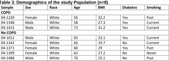
RESULTS 4.1 Demographics of the Study Population:
Participants were classified into two groups: COPD and no COPD. The COPD group is characterized by individuals with a diagnosis of COPD. The no COPD group is characterized by individuals who had no respiratory medical history. The demographics of each participant are shown in Table 3 below.
Table 3. Demographics of the study Population (n=8)
Sample Sex Race Age BMI Diabetes Smoking
COPD
04-1220 Female White 56 32.2 Yes Past
04-1348 Male White 58 27.3 Yes Current
04-1415 Male White 72 31.2 Yes Current
No COPD
04-1012 Male White 65 23.1 Yes Current
04-1342 Female White 66 19.7 No Current
04-1371 Female White 60 29 Yes Past
04-1399 Female White 62 27.2 No Never
04-1488 Male White 76 25.1 No Past
The average age in the COPD group was 62 ± 8.72, compared to the average age of 65.8 ± 6.18 in the group with no COPD. The average BMIs for the COPD group and No COPD group were 30.23 ± 2.59 and 24.82 ± 3.62 respectively.
4.2 CD3+CD8+ T Cells
21 Figure 1. There are no significant differences in the percentage of CD3+CD8+ T cells between people with COPD and people without COPD for either unstimulated or stimulated PMBCs
4.3 CD3+CD8+IFNγ+ T Cells
There were no significant differences in the percentage of CD3+CD8+IFNγ+ T cells between people with COPD and people without COPD for either unstimulated or stimulated PMBCs (Figure 2). Yet, individuals without COPD trended towards a greater expression of CD3+CD8+IFNγ+ T cells when comparing the stimulated samples
between the two groups. With the exception of one COPD participant, all individuals without COPD had greater CD3+CD8+IFNγ+ T cell expression than individuals with COPD after stimulation (Figure 3). Despite there being no statistical differences
22 percent increase between unstimulated and stimulated PBMCs from individuals with COPD was significantly lower for CD3+CD8+IFNγ+ T cells (Figure 4).
Figure 2. There are no significant differences in the percentage of CD3+CD8+IFNγ T cells between people with COPD and people without COPD for either unstimulated or stimulated PMBCs
23 Figure 4. Impaired upregulation of CD3+CD8+IFNγ+ T cells in PBMCs from individuals with COPD.
4.4 CD3+CD8+TNFα+ T Cells
Individuals with COPD had a significantly higher expression of
CD3+CD8+TNFα+ T cells than individuals without COPD for unstimulated PMBCs. There was no difference between CD3+CD8+TNFα+ T cell expression in people with COPD compared to people without COPD for PBMCs stimulated with H3N2 (Figure 5). Despite this, we did find that the percent increase between unstimulated and stimulated PBMCs from individuals with COPD was significantly lower for CD3+CD8+TNFα+ T cells (Figure 6). All participants without COPD were able to upregulate their expression of CD3+CD8+TNFα+ T cells when stimulated with H3N2, while individuals with COPD varied in their response upon stimulation. Figure 7 shows the three different responses among COPD patients. One COPD participant upregulated expression of
24 Figure 5. Individuals with COPD had a significantly higher expression of CD3+CD8+TNFα+ T Cells than individuals without COPD for unstimulated PMBCs.
26 CHAPTER 5
DISCUSSION 5.1 CD3+CD8+ T Cells
CD3+CD8+ T cell expression was similar in people with COPD and people without COPD for unstimulated PBMCs and PBMCs stimulated with H3N2. This suggests that the dendritic cells are recognizing influenza virus, migrating to the lymphatic system, and presenting influenza virus-derived antigens to T cells properly. Secondly, this indicates that CD3+CD8+ T cells are being activated appropriately, but there might be a problem in their function that causes people with COPD to be more susceptible to severe complications or even death when exposed to influenza. 5.2 CD3+CD8+IFNγ+ T Cells
PBMCs from each participant had an increased expression of CD3+CD8+IFNγ+ T cells when stimulated. In other words, PMBCs stimulated with H3N2 had more CD3+CD8+ T cells that were able to secrete IFNγ in response to influenza infection. This is important because IFNγ plays a role in inhibiting viral replication (13). But it is important to note that individuals without COPD had a significantly greater percent increase in CD3+CD8+IFNγ+ T cell expression upon stimulating PBMCs with H3N2. This indicates that there might be an impairment in the function of CD3+CD8+ T cells in people with COPD. Specifically, they could have impairments in the signaling pathway for the secretion of IFNγ in CD3+CD8+ T cells or there might be a defect in the IFNγ secretion pathway itself.
27 Individuals with COPD had a significantly higher expression of
CD3+CD8+TNFα+ T cells than individuals without COPD in unstimulated PMBCs. This was an expected finding because individuals with COPD have chronic systemic
inflammation and TNFα is an important pro-inflammatory cytokine (11). Despite this, individuals with COPD and individuals without COPD had a similar percentage of CD3+CD8+ T cells with the ability to secrete TNFα when stimulated. So even though we found that individuals without COPD had a significantly greater percent increase of expression for CD3+CD8+TNFα+ T cells between unstimulated and stimulated PBMCs, the ability of CD3+CD8+ T cells to secrete TNFα may not be impaired in people with COPD. Instead, maybe people with COPD have decreased sensitivity to the anti-viral effects of TNFα and thus TNFα is not able to contribute significantly to the inhibition of viral replication (12). Or, there is a possibility that TNFα may not play a significant enough role in inhibiting viral replication and all of the TNFα pathways are normal in people with COPD.
5.4 Limitations
This study had several limitations. One of the greatest limitations of the study was the sample size. There were 8 total participants, 3 participants with COPD and 5 participants without COPD. Thus, the groups are too small to draw true significant differences between. Though we did find some significant differences, these among other trends observed can only point towards future research studies because of the small sample size.
28 COPD, we could not assess the severity or chronicity of each individuals’ COPD.
People who were diagnosed with COPD more recently might not have the same
immune response to influenza as people who have been diagnosed for a longer period of time. In other words, it may take time for some changes in the immune response to occur. In addition, people without COPD were chosen based on their medical records indicating no history of respiratory difficulties. Despite this, we cannot be sure that this person has not been seen for respiratory difficulties somewhere other than the Family Medicine Center or is just not reporting their symptoms to the physician.
Finally, the BMIs between the group with COPD and group without COPD are also a limitation to this study. People with COPD had an average BMI of 30.23 ± 2.59 and people without COPD had an average BMI of 24.82 ± 3.62. According to the National Institutes of Health (NIH), a healthy weight BMI is 18.5-24.9, overweight is 25.0-29.9, and obese is 30.0 and above (14). This means the No COPD group is considered to have a healthy weight BMI while the COPD group is considered obese. This is problematic because previous studies show that activation and function of CD8+ T cells are impaired in PBMCs from overweight and obese individuals exposed to influenza and there is impaired upregulation of TNFα secretion in PBMC supernates from obese individuals (1). So ultimately, the trends that we see here could be due to obesity instead of COPD.
5.6 Study Implications and Future Studies
29 coverage, future studies should look at CD3+CD8+ T cells activation and function just as we did here to confirm the trends presented in this paper. If in fact there are no significant differences in the percentage of CD3+CD8+ T cells between people with COPD and people without COPD for either unstimulated or stimulated PMBCs, it would be interesting to perform a study where a lung biopsy was performed in people who were actually infected with influenza. Differences in the expression of CD3+CD8+ T cells in the lung would indicate whether there is a problem in recruiting these cells to the lung during infection.
30 References:
1. Paich, H. A., Sheridan, P. A., Handy, J., Karlsson, E. A., Schultz‐Cherry, S., Hudgens, M. G., & Beck, M. A. (2013). Overweight and obese adult humans have a defective cellular immune response to pandemic H1N1 Influenza a virus. Obesity, 21(11), 2377-2386.
2. FDA approves vaccines for the 2012-2013 influenza season [Internet]. 2012 Aug 13. Silver Spring(MD):U.S. Food and Drug Administration; [2012 Aug 13, cited 2014 Mar 17] . Available from:
http://www.fda.gov/NewsEvents/Newsroom/PressAnnouncements/ucm315365.ht m
3. Decramer, M., Janssens, W., & Miravitlles M (2012). Chronic Obstructive Pulmonary Disease. Lancet, 379, 1341-51.
4. Rycroft, C. E., Heyes, A., Lanza, L., & Becker, K. (2011). Epidemiology of chronic obstructive pulmonary disease: a literature review. International journal of chronic obstructive pulmonary disease, 7, 457-494.
5. Chronic obstructive pulmonary disease [Internet]. Bethesba(MD): National Center for Biotechnology Information, U.S. National Library of Medicine; [2011 May 1, cited 2013 March 19]. Available from:
http://www.ncbi.nlm.nih.gov/pubmedhealth/PMH0001153/
6. Flu (Influenza) [Internet]. Bethesba(MD): National Institute of Allergy and Infectious Disease; [2012 Nov 16, cited 2013 March 19]. Available from: http://www.niaid.nih.gov/topics/Flu/understandingFlu/Pages/definitionsOve rview.aspx
7. People at High Risk of Developing Flu-Related Complications [Internet]. Atlanta(GA): Centers for Disease Control and Prevention; [2013 Nov 7, cited 2013 March 19]. Available from:
http://www.cdc.gov/flu/about/disease/high_risk.htm
8. Blut, A., & Krankheitserreger, B. B. (2008). Influenza Virus. Transfusion Medicine and Hemotherapy, 35, 42-49.
9. D Medzhitov, R., & Janeway Jr, C. A. (1997). Innate immunity: impact on the adaptive immune response. Current opinion in immunology, 9(1), 4-9.
10. D Kreijtz, J. H. C. M., Fouchier, R. A. M., & Rimmelzwaan, G. F. (2011). Immune responses to influenza virus infection. Virus research, 162(1), 19-30.
11. von Haehling, S., Hopkinson, N. S., Polkey, M. I., Niethammer, M., Anker, S. D., & Genth-Zotz, S. (2009). Elevated TNFα production in whole blood in patients with severe COPD: the potential link to disease severity. Wiener klinische Wochenschrift, 121(9-10), 303-308.
12. Seo, S. H., & Webster, R. G. (2002). Tumor necrosis factor alpha exerts powerful anti-influenza virus effects in lung epithelial cells. Journal of virology, 76(3), 1071-1076.
13. Katze, M. G., He, Y., & Gale, M. (2002). Viruses and interferon: a fight for supremacy. Nature Reviews Immunology, 2(9), 675-687.
14. Assessing Your Weight and Health Risk [Internet].:National Heart, Lung, and Blood Institute; [ cited 2013 March 24] . Available from: