Comparison of Temperature Changes Through Dentin Discs with
Different Thicknesses and Different Distances of Light Curing Tip by
Two Different Light Curing Units
M. Barkatayn1, MR. Malekipoor 1, S. Alaei 2 , M. Kavoosi 31Assistant Professor, Department ofOperative Dentistry, School of Dentistry, Azad University. Khorasgan Branch, Iran 2Postgraduate Student, Department ofOperative Dentistry, School of Dentistry, Azad University. Khorasgan Branch, Iran 3Dentist
Corresponding author: S. Alaei, Postgraduate Student, Department ofOperative Denti-stry, School of DentiDenti-stry, Azad University, Khorasgan Branch, Iran
[email protected] Received: 5 Oct 2011 Accepted: 29 April 2012
Abstract
Background and Aim: Excessive heat produced while curing of light-activated dental restorations may cause irreversible damage to dental pulp. The Aims of this study wereto measure the temperature rise(TR) induced by two different light curing units and to evaluate the relathionship between the measured TR in dentin discs of different thicknesses and different distances of light guide tips from dentin disc.
Materials and Methods: In this experimental study, Quartz-Tungsten-Halogen (QTH) (400 ) and Light-Emitting Diode (LED) systems (1500 ) were used as the curing units. Dentin discs of 0.5, 1.0 and 1.5 mm thicknesses were prepared. Distances of 1,2 and 3 mm from the tip of the light guide and dentin discs were set. Temperatures were recorded using a digital laser thermometer. Data were statistically analyzed using two-way ANOVA, Duncan and T-test.
Results: Results showed that both dentin thickness and distance between tip of curing units and dentin disks had significant effects on temperature rise in both curing units (p<0.001).
Conclusion: LED produced the highest and the QTH the lowest TR for all tested conditions. Reduced thermal insults were related to increased dentin thickness, increased distance between the light guide tip and dentin, and decreased energy emitted from the light-curing unit.
Key Words: Temperature, Curing lights, Dentin
Journal of Islamic Dental Association of IRAN (JIDAI) / Autumn 2012 /24 / (4)
Introduction
Health of a tooth is indebted with health of the dental pulp as a unique tissue [1]. Surgical and res-torative treatments induce thermal, physical and mechanical insults to the dental pulp among which intrapulpal heat buildup is of paramount impor-tance [2]. It appears that there is a critical spectrum for intrapulpal temperature. Studies have shown that intrapulpal temperature increase beyond 5 to 8 degrees centigrade may easily lead to cell death [3,4]. Principal studies by Cohen and Zach
indi-cated that temperature rise beyond 5.5 degrees cen-tigrade causes irreversible cellular damage [4]. One of the most frequent factors that leads to intrapupal temperature rise is application of lght-curing devices for polymerization of tooth-colored restorative materials. It is reported that intrapulpal temperature rises up to 8 degrees centigrade during light curing a composite restoration.
Intrapulpal temperature rise is dependant upon several factors such as type of curing device used, intensity of curing, time, type of curing, filtering
quality of the curing device, distance to the light source, restoration size, presence of temperature barrier layers, thickness, color and composition of the composite used [5-9]. Another important factor is the remaining dentin thickness. Direct tempera-ture rise within the pulp chamber is minimized by the low thermal conductivity of dentin [10] Also, risk of pulpal damage while using light curing de-vices with higher output energy is increased [11]. Some light curing devices generate a considerable amount of heat while being used so that the opera-tor cannot keep his/her finger in a 2- to 3-millimeter distance from the tip of the device for 20 seconds [12]. In recent light curing devices which are LED-based, there is usually a high in-tensity of irradiation which can jeopardize pulpal health. Santini et al stated that the heat generated in pulp chamber by LED devices with 1100 mw/cm2
was significantly higher than that of QTH devices with 500 mw/cm2[11] Bagis also showed that in
comparison of QTH, LED and plasma arch (PAC) devices, in a 1-mm distance the maximal and mi-nimal temperature increase was observed in plasma arch and LED devices, respectively [13]. Dogan also showed that PAC devices induced higher tem-perature increase in comparison with LED devices in different dentin thicknesses [14].
The aim of this investigation was to compare the heat produced by LED and QTH devices and the effect of the remaining dentin thickness in cavities and the distance between the light curing device tip and remaining dentin on the resultant temperature increase.
Methods and materials
In this interventional experimental study 30 intact human intact caries-free third molars were in-cluded. The teeth were stored in 0.2% thymol solu-tion before use. Initially the whole thickness of enamel was removed from the tooth surface using a diamond bur and copious water irrigation. After preparing a relatively smooth surface from the cross section of the tooth crown, each sample was mounted in orthodontic acrylic resin blocks. The samples were randomly divided into three groups
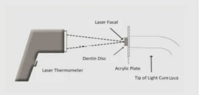
of ten each. Dentin thicknesses of interest were marked on the lateral surface of the acrylic molds (0.5, 1 and 1.5 mm). Teeth were sectioned (Non stop-Bego, Germany) so that the sectioning blade was located perpendicular to the acrylic mold and an even disc was obtained in each section. Tooth sections were measured by a gauge after section-ing. Therefore, 30 discs in different thicknesses i.e., 0.5, 1, and 1.5 mm in diameter (n=10 each) were prepared and stored in distilled water. Acryl-ic plates were prepared to establish a fixed distance between the light cure tip and dentin discs. Tem-perature changes were measured using a laser digi-tal thermometer (Dostman electronic,China- 485 Scan temp) (fig.1)
The complex was assembled in a way that the den-tin disc was fix on one side of the acrylic plate and the tip of the light curing device on the other side in predetermined distances of 1 to 3mm. The initial temperature of the dentin discs were equalized with the room temperature (27.0±0.1).
Output intensity of the QTH device (Litex 695 C– Dentamerica, USA) was 400 mw/cm2. The samples
were cured for 20 minutes. Maximal temperature was recorded. Thermal measurement was per-formed three times for each section. After light curing, the temperature of each sample was re-turned to 27 degrees centigrade.
In other experimental groups, the same procedure was followed by using an LED system (Litex 680 A- Dentamerica,USA) with an output intensity of 1500 mw/cm2 for 20 seconds. Output intensity of
each device was measured by a light meter device (LCM 1000 and CM300-1000)
Fig 1.Schematic representation of thermal measurement
Statistical analysis was carried out using t-, two-way ANOVA and Duncan tests, using SPSS soft-ware.
Results
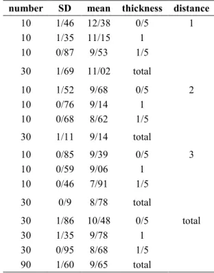
The results of this study is represented as the mean of temperature changes in degrees centigrade. Ac-cording to the presence of three dentin disc thick-nesses (0.5, 1.0, and 1.5 mm) and three light cure tip to dentin disc distances (1, 2, and 3mm) as well as two types of light-cure devices (QTH and LED), a total of 18 different conditions were evaluated using ANOVA test. The results are demonstrated in tables 1 and 2. In order to evaluate the effect of different dentin thicknesses and light cure dis-tances on temperature rise two-way ANOVA and Duncan tests were used.
The maximum temperature increase was recorded in LED group at a 1-mm distance of the light-cure tip from the 0.5-millimeter-thick dentin disc (12.38
±1.46 degrees centigrade). The minimal tempera-ture rise was recorded in the QTH device at a 3-mm distance of the light cure tip from the 1.5-millimeter-thick dentin disc (4.93±0.29 degrees centigrade). The two-way ANOVA test results in-dicated that both the distance of the light-cure tip and the dentin thickness had significant effects on both QTH and LED devices. (p<0.001) It was ob-served that the mean temperature increase in cer-tain distances increased with decreases in dentin thicknesses, regardless of the device used. (p<0.001) The LED device induced a significantly more temperature increase than the QTH device regardless of the thickness of the dentin sample and the light cure tip distance. (p<0.001) It was demonstrated that increasing the distance of the light cure device tip with the target dentin sample leads to a significant decrease in temperature rise, regardless of the device used.(p<0.001) There was a statistically significant difference among differ-ent ddiffer-entin thicknesses according to the Duncan test in both light curing devices. There was also a
sig-distance thickness mean SD number 1 0/5 12/38 1/46 10 1 11/15 1/35 10 1/5 9/53 0/87 10 total 11/02 1/69 30 2 0/5 9/68 1/52 10 1 9/14 0/76 10 1/5 8/62 0/68 10 total 9/14 1/11 30 3 0/5 9/39 0/85 10 1 9/06 0/59 10 1/5 7/91 0/46 10 total 8/78 0/9 30 total 0/5 10/48 1/86 30 1 9/78 1/35 30 1/5 8/68 0/95 30 total 9/65 1/60 90 distance thickness mean SD number 1 0/5 7/18 0/85 10 1 6/96 0/77 10 1/5 6/12 0/59 10 total 6/75 0/86 30 2 0/5 6/31 0/79 10 1 5/87 0/53 10 1/5 5/00 0/36 10 total 5/73 0/79 30 3 0/5 6/24 0/63 10 1 5/74 0/58 10 1/5 4/93 0/29 10 total 5/63 0/74 30 total 0/5 6/57 0/85 30 1 6/19 0/83 30 1/5 5/35 0/69 30 total 6/04 0/94 90
Table 1.Comparison of mean temperature changes in different light-cure tip distances and various
dentin thicknesses using the LED device
Table 2.Comparison of mean temperature changes in different light-cure tip distances and various
dentin thicknesses
nificant difference between the 1-millimeter tance of the light cure tip with the other two dis-tances.
Discussion
Use of light-activating devices play an integral part in quality of the tooth-colored restorations. Nowa-days, most of the manufacturers of the light-curing devices tend to introduce devices with intensities of more than 1000mw/cm2to the market to reduce
the curing time and increase curing depth. When high-intensity light curing devices are used, a sig-nificant temperature increase is observed in com-parison with QTH devices. There is also an in-creased risk of pulpal damage when higher intensi-ty devices are used [11]. In this study, the effect of dentin thicknesses, tip distances and two light cure devices were evaluated on the post-curing tempera-ture increase. It was shown that the temperatempera-ture rise following use of the LED device was signifi-cantly more than that of the QTH device (p<0.001). The range of the temperature changes was between 7.91 and 12.38 degrees centigrade in LED and between 4.93 and 7.18 degrees in the QTH device. Zach and Cohen demonstrated that increasing the pulpal temperature more than 5.5 degrees centigrade could cause pulpal necrosis in 15 % of the cases. Intrapulpal temperature increase up to 11.1 degress centigrade caused 60% and up to 16.6 degrees centigrade, 100% pulpal necrosis [4]. The temperature increase caused by the LED device was beyond the critical 5.5 degrees centi-grade in all distance- and thickness- conditions. On the other hand, in the QTH device, only in two out of nine conditions, the temperature increase was lower than the critical 5.5 degrees. According to the more intense output of the LED device (1500mw/cm2) compared with that of the QTH
device (400mw/cm2), higher temperature increase
was observed in the LED device. Therefore the risk of pulpal irritation is increased. This was in accordance with the results of Guiraldo et al who reported that the output light intensity and radia-tion time were the most important factors in tem-perature rise after curing [15]. Millen and
co-workers also demonstrated that LED device caused an increased temperature rise in comparison with QTH [16]. Durey et al investigated the temperature increase within the dental pulp after use of two LED and one QTH curing devices. They demon-strated that pulpal temperature increase was seen significantly more in LED compared with QTH devices, but there was no significant difference between the two LED systems [17]. On the con-trary, Dogan and colleagues reported a higher tem-perature increase after use of a QTH device in comparison with an LED system. Such incongru-ence can be attributed to the differincongru-ences in light intensity of the QTH devices used as well as va-riances in curing times.
In studies by Santini et al [11] and Yazici et al [18], temperature changes were determined by us-ing thermocouples within the pulp chamber of the teeth with class II cavity preparations. Contrarily, in the current study as well as the work done by Dogan et al [14] dentin discs were prepared which made it possible to evaluate dentin thicknesses precisely. On the other hand, for evaluation of the temperature changes, a laser beam was directed to the dentin surface, without being influenced by environmental temperature.
According to the results of our study, it can be concluded that more intensive remedies must be taken into consideration to protect pulpal tissue from thermal irritation when cavities with the re-maining dentin thicknesses of less than 1.5 mm are restored.
It can also be recommended that the light cure tip be placed at a 2 mm distance from the dentin sur-face in tooth-colored restorations to prevent exces-sive heat buildup. On the other hand, there is no need to increase the distance of the light cure tip up to 3 mm due to the decrease in depth of polymeri-zation [20].
Soh and Yap evaluated the temperature increase following use of LED and QTH devices at 3- and 6-millimeter distances. At the 3-millimeter dis-tance the temperature rise was recorded to be 4.1-12.9 and 17.4-46.4 degrees centigrade for LED and QTH devices, respectively. They found out that the
temperature rise at a 6-millimeter distance was 2.4-7.5 and 12.7-25.5 degrees centigrade for LED and QTH systems, respectively. The higher tempera-ture increase in the closer distance is in accordance with the results of our investigation [21].
Curing time for both devices were set at 20 seconds. Knezevic and colleagues stated that the most pronounced thermal increase following use of curing devices were seen within the initial 20 seconds of the curing procedure. The thermal changes between 20 and 30 seconds was consi-dered to be between 1-2 degrees and was not sig-nificant and there was no thermal changes between 30 to 40 seconds [22].
It must not be overemphasized that the results of this in vitro study cannot be extrapolated to the clinical conditions, because there are a lot of con-tributing factors that compensate such thermal changes within the pulp chamber, such as pulpal blood flow, presence and flow of the dentinal fluid and periodontal tissues [14].
Conclusion
It can be concluded that the temperature rise fol-lowing use of LED system is more than that of QTH device in different dentin thicknesses and different light cure tip distances.
References
1- Pashley DH. Dynamics of the pulpo-dentin complex. Critical reviews in oral biology and med-icine: An official publication of the Ame Asso Oral Biolog. 1996 Jan;7(2):104-33.
2- Summitt J, Robbins J. Fundamentals of opera-tive dentistry: A contemporary approach. 3rd. Chi-cago: Quintessence Publishing; 2006.
3- Zach L, Cohen G. Thermogenesis in operative techniques: Comparison of four methods. J Pros-thet Dent. 1962 Nov;12(5):977-84.
4- Zach L, Cohen G. Pulp response to externally applied heat. Oral Surg Oral Med Oral Pathol. 1965 Apr; 19(4):515-30.
5- Goodis HE, White JM, Jr. GWM, Yee K, Fuller N, Gee L, et al. Effects of Nd: And Ho: yttrium-aluminium-garnet lasers on human dentine fluid
flow and dental pulp-chamber temperature in vitro. Arch Oral Biol. 1997Dec; 42(12):845-54.
6- Karaarslan ES, Secilmis A, Bulbul M, Yildirim C, Usumez A. Temperature increase beneath etched dentin discs during composite polymeriza-tion. Photomed Laser Surg. 2011 Jan; 29(1):47-52. 7-Hannig M, Bott B. In-vitro pulp chamber tem-perature rise during composite resin polymeriza-tion with various light-curing sources. Dent Mater. 1999 Jul;15(4):275–81.
8- Jakubinek MB, O’Neill C, Felix C, Price RB, White MA. Temperature excursions at the pulp-dentin junction during the curing of light-activated dental restorations. Dent Mater. 2008 Nov; 24 (11):1468-76.
9- Shortall AC, Harrington E. Temperature rise during polymerization of light-activated resin composites. J Oral Rehabil. 1998 Dec;25(12):908-13.
10- Hargreaves KM, Goodis HE. Seltzer and Bender’s dental pulp. 3rd ed. Chicago: Quintes-sence Pub. Co. 2002.
11- Santini A, Watterson C, Miletic V. Tempera-ture rise within the pulp chamber during composite resin polymerisation using three different light sources. Open Dent J. 2008 Jan;5(2):137-41. 12- Craig RG, Ward ML. Restorative Dent Mater. España: Elsevier; 2006.
13- Bagis B, Bagis Y, Ertas E, Ustaomer S. Com-parison of the heat generation of light curing units. J Contem Dent Pract. 2008 Jan;9(2):65-72.
14- Dogan A, Hubbezoglu I, Dogan OM, Bolayir G, Demir H. Temperature rise induced by various light curing units through human dentin. Dent Ma-ter J. 2009 May;28(3):253-60.
15- Guiraldo RD, Consani S, Lympius T, Schneid-er LFJ, Sinhoreti MAC, CorrSchneid-er-Sobrinho L. Influ-ence of the light curing unit and thickness of resi-dual dentin on generation of heat during composite photoactivation. J Oral Sci. 2008 Jun; 50 (2):137-42.
16- Millen C, Ormond M, Richardson G, Santini A, Miletic V, Kew P. A study of temperature rise in the pulp chamber during composite
tion with different light-curing units. J Contemp Dent Pract. 2007 Nov1; 8(7):29-37.
17- Durey K, Santini A, Miletic V. Pulp chamber temperature rise during curing of resin-based com-posites with different light-curing units. Prim Dent Care. 2008 Jan;15(1):33-8.
18- Yazici AR, Müftü A, Kugel G, Perry RD. Comparison of temperature changes in the pulp chamber induced by various light curing units, in vitro. Oper Dent. 2006 Mar;31(2):261-5.
19- Aguiar FHB, Barros GKP, Lima DANL, Am-brosano GMB, Lovadino JR. Effect of composite resin polymerization modes on temperature rise in human dentin of different thicknesses: an in vitro study. Biomed Mater. 2006 Sep;1(3):140-3.
20- Roberson TM, Heymann H, Swift EJ. Sturde-vant’s art and science of operative dentistry. 5 th. St. Louis:Mosby; 2006.
21- Yap AUJ, Soh MS. Thermal emission by dif-ferent light-curing units. Operative Dent. 2003 May;28(3):260-6.
22- KnezeviQ A, Tarle Z, Meniga A, Sutalo J, Pichler G. Influence of light intensity from differ-ent curing units upon composite temperature rise. J Oral Rehabil. 2005 May;32(5):362-7.