Original Research Article
Reliability and validity of ICDAS II coding for occlusal caries using
magnification: an in-vitro study
Sathyanarayanan R., Carounanidy Usha*, Sudagar R.
INTRODUCTION
Dental caries is a disease that is characterized by the localized destruction of susceptible dental hard tissue by acidic by-products from bacterial fermentation of dietary carbohydrates.The process is a dynamic interaction at the biofilm-tooth interface characterised by alternating cycles
of demineralisation and remineralization. A caries/carious lesion are a detectable change in the tooth structure that results from this interaction. The changes may range from initial outer surface demineralization, at the molecular level, through subsurface demineralization producing enamel white-spot lesion formation, through macroscopic lesion cavitation, to dentine and pulpal infection, to
ABSTRACT
Background: Pit and fissure caries presents diagnostic challenges due to its anatomical complexity and fluoride exposure. ICDAS II is a coding system for caries detection using clinical visual inspection. It identifies carious lesions by the change in colour, texture and surface integrity. Magnification might facilitate better detection of the lesions by enhancing the visual acuity. Thus, the objective of this study is to compare the reliability and validity of ICDAS II in detection of occlusal caries, with and without magnification, by using histological standard.
Methods: This single blinded, randomized study included 334 unrestored extracted human premolars and molars. Two examiners independently scored pit and fissure caries status using ICDAS II criteria without magnification and later under 6 x magnification using surgical microscope. The samples were sectioned and lesions were scored using the ERK histological criteria, under Stereo microscope. The scores of the examiners were correlated with the histological scoring. Kappa statistics and Spearman correlation coefficients were performed. Optimal sensitivity, specificity of visual and enhanced visual examination was calculated by Receiver Operating Characteristic Curve (ROC). Likelihood ratios (LR) were also calculated.
Results: The kappa values for Inter examiner reproducibility of visual and enhanced visual examination under microscope were 0.638-0.694 and for histological examination it was 0.979. Intra examiner reproducibility for visual and enhanced visual examination was 0.665 – 0.594. There was a strong relationship between visual, enhanced visual and histological examinations. Spearman’s correlation coefficient of ICDAS-II visual and enhanced visual examination for each examiner, to ERK histological scores was 0.869-0.848. The sensitivity and LR+ for visual and enhanced visual examination was decreased as the ICDAS score was increased and specificity and LR- increased with increased ICDAS score.
Conclusions: Reliability and validity of ICDAS scoring in detecting occlusal caries under magnification did not differ from clinical visual inspection.
Keywords: ICDAS–II coding, Occlusal caries, Visual examination, Histological examination
Conservative Dentistry and Endodontics, Indira Gandhi Institute of Dental Sciences, Sri Balaji Vidyapeeth, Puducherry, India
Received: 18 February 2017 Accepted: 02 May 2017
*Correspondence: Dr. Carounanidy Usha E-mail: [email protected]
Copyright: © the author(s), publisher and licensee Medip Academy. This is an open-access article distributed under the terms of the Creative Commons Attribution Non-Commercial License, which permits unrestricted non-commercial use, distribution, and reproduction in any medium, provided the original work is properly cited.
complete tissue destruction.1 Incipient non-cavitated lesions at the enamel level can be managed by non-operative/ preventive treatment strategies such as remineralisation or sealants, without surgical ‘drill and fill’.2
Thus, it is imperative to detect the earliest demineralisations, which is a realisation that led to the development of various lesion detection methods. A good diagnostic aid that is sensitive enough to produce true positive results and specific enough to produce true negative results, keeping the false negatives and false positives to the minimum, is considered as valid aids/correct aids. It should also be reliable/valid, reproducible and accurate with least inter-/intra observer variation.
The traditional caries detection methods included visual inspection, use of explores and radiographs, whereas the novel methods include Quantitative Laser Fluorescence, Diagnodent and Electrical Caries meters.3,4 An International Consensus Workshop on Caries Clinical Trials (ICW-CCT) in 2002, concluded that visual diagnosis is the standard of caries diagnosis. The most recent development in this regard is the International Caries Detection and Assessment System (ICDAS), in which carries detection is based predominantly on visual criteria that describe the characteristics of the lesions.5 The initial studies on ICDAS have reported it to have good reliability and validity.6 Later in 2005, it was modified to be known as ICDAS II.7 Studies have demonstrated good sensitivity, specificity, reproducibility and diagnostic accuracy for this system.8-10
Though the occlusal pit and fissure areas are approachable and accessible for cleansing and examination, the incidence of carious lesion is more in these sites. This can be attributed to the complex invaginated anatomy of the pits and the fissures. Caries detection is challenging in these convoluted terrains of teeth. In addition, the usual fluoride exposure of the teeth renders the superficial enamel resistant to demineralisation. But caries progresses through the lateral surfaces of the fissures, under the seemingly intact enamel surface, resulting in hidden caries.11
Use of magnification, in the form of loupes to microscopes, has been emphasised in many fields of dentistry, including restorative dentistry and endodontics. Better visualisation leads to better performance, dentists’ posture and delivery of high quality dentistry.12 A concept known as Microscope Assisted Precision Dentistry was introduced.13 Detection of early carious lesions is an integral and vital part of minimal invasive dentistry, thus magnification becomes mandatory in detection.
Very few studies are available in assessing the improved effectiveness of caries detection with magnification and the results are contradictory. Few studies were done on
occlusal cares detection using magnification but without using ICDAS criteria, and few were done on proximal caries detection without ICDAS system.14-17 ICDAS II criteria for occlusal caries detection with and without magnification (high and low level) were assessed in few studies with varying results.18-20
Due to such contradictory and sparse evidence on the use of magnification in ICDAS II system, this study was done with the objective to compare the validity and reliability of ICDAS II system with and without magnification under surgical microscope at 6X magnification, for detection of occlusal caries, by using a golden standard of histological examination and scoring. A null hypothesis was generated that there will be no difference in the reliability and validity of ICDAS II system in detecting occlusal lesions with or without magnification.
METHODS
This was a simple randomized, single blinded, in-vitro study with a parallel group design and an allocation ratio of 1:1. This study was conducted in the Department of Conservative Dentistry and Endodontics, Department of Prosthodontia, Indira Gandhi Institute of Dental Sciences, Puducherry. The histological sections were prepared in the Department of Oral pathology in SRM dental college, Chennai. Ethical clearance was obtained from Institutional Review Board and Institutional Ethical Clearance Board. Extracted human premolars and molars with occlusal caries only, were collected. Informed consent was obtained from the patients before extraction. All the samples were cleaned with rotary brush, pumice and stored in distilled water until examination. The sample size of 334 was calculated based on the study done by Jabonski- Momeni et al.8
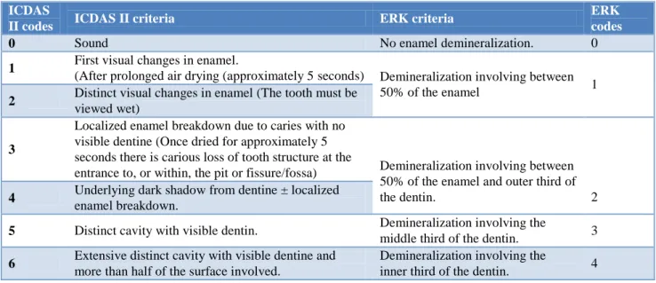
Table 1: ICDAS II scoring and corresponding scoring in ERK histological examination.
ICDAS
II codes ICDAS II criteria ERK criteria
ERK codes
0 Sound No enamel demineralization. 0
1 First visual changes in enamel.
(After prolonged air drying (approximately 5 seconds) Demineralization involving between
50% of the enamel 1
2 Distinct visual changes in enamel (The tooth must be viewed wet)
3
Localized enamel breakdown due to caries with no visible dentine (Once dried for approximately 5 seconds there is carious loss of tooth structure at the
entrance to, or within, the pit or fissure/fossa) Demineralization involving between 50% of the enamel and outer third of
the dentin. 2
4 Underlying dark shadow from dentine ± localized enamel breakdown.
5 Distinct cavity with visible dentin. Demineralization involving the
middle third of the dentin. 3
6 Extensive distinct cavity with visible dentine and more than half of the surface involved.
Demineralization involving the
inner third of the dentin. 4
Statistical analysis was done with SPSS version 14. Inter and intra examiner reproducibility was calculated by weighted kappa. Visual and enhanced visual examination under microscope was correlated with histological examination by using Spearman’s correlation coefficient. Optimal sensitivity and specificity of both methods were calculated by Receiver Operating Characteristic Curve (ROC).
RESULTS
334 teeth were sectioned for histological evaluation. 17 teeth were damaged during root resection and histological section preparation. Only 317 sections were histologically examined by both examiners. Table 2 shows the distribution of ICDAS-II coding cross tabulated with ERK histological scores for the independent investigated sections for examiner 1, who was considered as reference examiner in this study. Table 3 shows the inter examiner agreement of the visual and enhanced visual examinations of ICDAS-II. The inter examiner weighted kappa value was 0.638 (good) for visual examination and 0.694 for enhanced visual examination. Table 4 shows the intra examiner reproducibility’s (kappa values) of visual and enhanced visual examination (under magnification) of ICDAS-II. The degree of intra examiner agreement for the two examiners with and without magnification was good to moderate with kappa values ranging from 0.669 – 0.594. Table 5 shows the inter examiner agreement of histology examination. 98% agreement was there between two examiners using the Ekstrand’s classification. The inter examiner weighted kappa value was 0.979 for ERK classification, which is very good. The visual examination and enhanced visual examination was correlated with histological examination by using Spearman’s correlation coefficient. Table 6 shows the
Spearman’s correlation coefficient of ICDAS-II visual and enhanced visual examination for each examiner, to ERK histological scores. It is generally accepted that a correlation coefficient of 0.7 or above represents a strong relationship between two variables. Table 7 and 8 shows the optimum sensitivity, specificity and likelihood ratio (LR) of visual and enhanced visual examination. Each examiner and diagnostic methods’ sensitivity, specificity were calculated under receiver operating characteristic curve (ROC) as given in Figure 1.
Figure 1: ROC curve showing the optimum sensitivity, specificity of visual and enhanced visual
Table 2: distribution of ICDAS-II coding cross tabulated with ERK histological scores.
ICDAS II Code 0 1 2 3 4 5 6 Total
Visual 48 47 44 43 48 46 41 317
with magnification 32 34 52 53 51 53 42 317
ERK histological score 49 Code 0 45 Code 1 107 Code 2 78 Code 3 38 Code 4 317
Fractured samples 0 1 4 5 0 2 5 17
Total 334
Table 3: Inter examiner reproducibility’s (kappa values) for the visual and enhanced visual examinations of ICDAS-II.
Measure of agreement Kappa value No. of valid cases
Visual 1 and visual examination 2 0.638 317
Enhanced visual 1 and enhanced visual examination 2 0.694 317
Table 4: Intra examiner reproducibility’s (kappa values) for the visual and enhanced visual examinations of ICDAS-II.
Measure of agreement Kappa value No. of valid cases
Visual 1 and enhanced visual examination1 0.665 317
Visual 1 and enhanced visual examination2 0.663 317
Enhanced visual 1 and visual examination2 0.669 317
Enhanced visual 2 and visual examination2 0.594 317
Table 5: Inter examiner reproducibility’s (kappa values) of histological examination.
Measure of agreement; histological examination Kappa value No. of valid cases
Examiner 1 and Examiner 2 0.979 317
Reference value of weighted kappa= <0.20- poor; 0.21 - 0.40- fair; 0.41 - 0.60- moderate; 0.61 - 0.80- good; 0.81 - 1.00- very good.
Table 6:Spearman’s correlation coefficient of ICDAS-II visual and enhanced visual examination for each examiner, to ERK histological scores.
Measure of correlation Values No. of valid cases
Visual 1 and histological examination 1 0.869 317
Visual 2 and histological examination 1 0.859 317
Enhanced visual 1 and histological examination 1 0.851 317
Enhanced visual 2 and histological examination 1 0.848 317
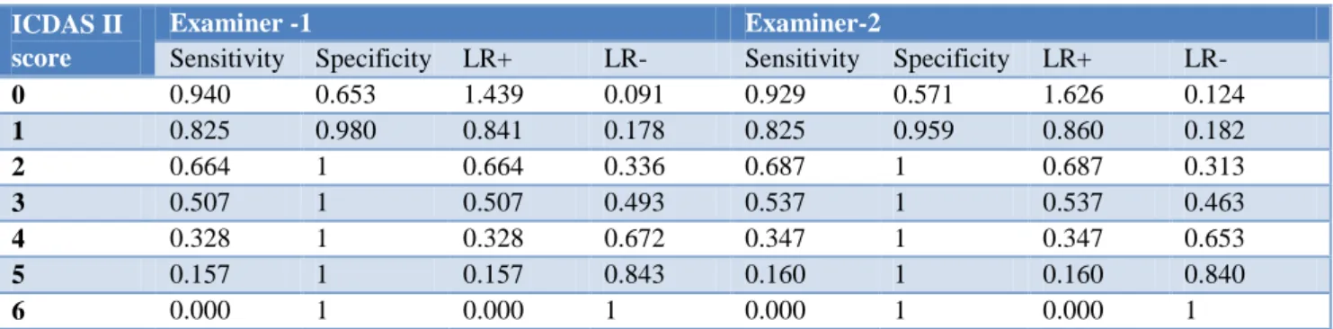
Table 7: Optimum sensitivity, specificity, LR+, LR- values of visual examination.
ICDAS II score
Examiner -1 Examiner-2
Sensitivity Specificity LR+ LR- Sensitivity Specificity LR+ LR-
0 0.940 0.653 1.439 0.091 0.929 0.571 1.626 0.124
1 0.825 0.980 0.841 0.178 0.825 0.959 0.860 0.182
2 0.664 1 0.664 0.336 0.687 1 0.687 0.313
3 0.507 1 0.507 0.493 0.537 1 0.537 0.463
4 0.328 1 0.328 0.672 0.347 1 0.347 0.653
5 0.157 1 0.157 0.843 0.160 1 0.160 0.840
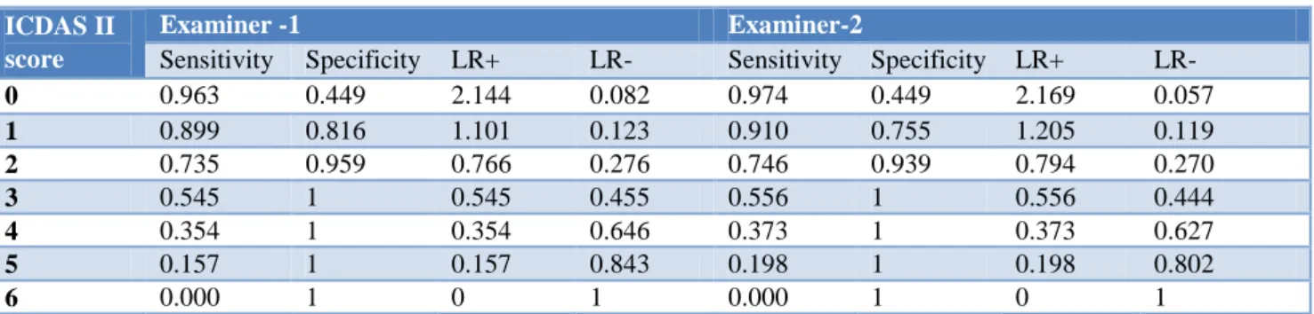
Table 8: Optimum sensitivity, specificity, LR+, LR- values of enhanced visual examination.
ICDAS II score
Examiner -1 Examiner-2
Sensitivity Specificity LR+ LR- Sensitivity Specificity LR+ LR-
0 0.963 0.449 2.144 0.082 0.974 0.449 2.169 0.057
1 0.899 0.816 1.101 0.123 0.910 0.755 1.205 0.119
2 0.735 0.959 0.766 0.276 0.746 0.939 0.794 0.270
3 0.545 1 0.545 0.455 0.556 1 0.556 0.444
4 0.354 1 0.354 0.646 0.373 1 0.373 0.627
5 0.157 1 0.157 0.843 0.198 1 0.198 0.802
6 0.000 1 0 1 0.000 1 0 1
DISCUSSION
The null hypothesis generated at the beginning of the study is accepted that there is no difference in the reliability and validity of ICDAS II in detecting occlusal caries under magnification or unaided visual examination.
The result of this study revealed inters examiner reliability values of 0.638 and 0.694 for visual and visual examination under magnification, respectively. The intra examiner values were found in the range of 0.665-0.594. Both these values are considered as moderate to substantial kappa values for reproducibility.23 But the initial study by Ismail et al in 2007, on the reproducibility of ICDAS II in occlusal caries detection by unaided visual examination has reported excellent inter examiner agreement.6 Study by Jablonski et al in 2008 has reported the same.8 However a study by Rodrigues et al in 2008 found similar inter observer reliability as our study and concluded that ICDAS II in combination with bitewing radiography performed better. 24
In our study, the agreement between the examiners between unaided and enhanced examination was also observed to be moderate to substantial. This result is in accordance to a study by Susodia et al in 2014, an in-vitro study to test the accuracy and reproducibility in detection of incipient occlusal caries and treatment decision making using unenhanced visual–tactile technique and low level magnification using loupes and surgical operating microscope (SOM). They reported that the intra-observer reproducibility for caries detection using surgical operating microscope ranged from average to good.18 Yet another study by Ari et al 2013, evaluated low power magnification with LED light for ICDAS coding of occlusal caries in primary molars, reported good to excellent reproductivity.20 On the contrary, Mitropoulos et al in 2012 compared the impact of low-powered magnification on the detection of occlusal caries. The occlusal surfaces of 38 extracted teeth were examined with and without magnification by two examiners. Inter-examiner agreement was excellent for ICDAS grades 0, 2, 3 irrespective of magnification but for code 1, magnification resulted in lower agreement than unaided vision.19 Thus, it can be inferred that
ICDAS II has acceptable reproducibility with or without magnification.
Histological validation is considered as the gold standard for caries detection methods.25 The ERK histological classification system was introduced in 1997 by Ekstrand KR.22 This present dissertation recorded the deepest part of the lesion from where it originated at the investigation site and the sections were scored as 0-4 based on ERK histological score depending on the severity of the lesion. 98% agreement was there between two examiners using the Ekstrand’s classification. The inter examiner weighted kappa value was 0.979 for ERK classification, which is very good. Based on the above result, examiner 1 was authorized as reference examiner. Spearman’s correlation coefficient is a statistical measure of the strength of a relationship between data. The result of our study shows a very strong correlation with histological examination for bot he methods and for both examiners, ranging from 0.48 to 0.869. This is a better the relationship than proven in many studies.8,26 Study by Mitropoulos et al, reported the correlation co-efficient as 0.44 to 0.51 only for both visual and enhanced visual methods with ICDAS II.19 The reason for better relation observed in our study can be attributed to the intense training of both examiners in the ICDAS II system. ICDAS II is a part of the clinical practice in both graduate and undergraduate pogroms in our institute, which can be a strong influencing factor in accurate diagnosis observed in this study. Though many authors have concluded stating that experience or training of the examiner need not influence the detection power of the visual examination tool, experience gained during training seems to be influencing the diagnostic decisions with ICDAS II.27,28
sensitivity at the D1 diagnostic threshold.8,26 Jallad et al reported equal sensitivity and specificity, while comparing ICDAS II with other detection methods.29 Interestingly Jablonski et al 2012 reported 100% sensitivity and specificity at D1 threshold.21
The result of this study shows that there was no significant difference in the sensitivity and specificity values as well as the LH+ and LH- values in ICDAS II system under magnification. This is in accordance with results from Mitropoulos et al, in which it was reported that specificity, sensitivity and LR+ and LR–values showed no significant differences between the examiners, and between unaided and magnified vision.19 Similarly, Sisodia et al, while comparing high power magnification with microscope with low powered loupes reported sensitivity and specificity that were not different from unaided visual inspection.18 Ari et al assessed the performance of ICDAS-II using low powered magnification with light emitting diode headlight and alternating current impedance spectroscopy device for detection of occlusal caries on primary molars and reported that with magnification, the sensitivity and specificity scores for examiners were 0.87–0.90 and 0.70–0.75, respectively, at the D1 diagnostic threshold, which is lesser than the values reported in our study.20 But studies that were done previously, without using ICDAS scoring reported better performance with magnification.14-17 Thus it can be inferred that the scoring criteria and the conditions of dry and illuminated field used in ICDAS criteria are robust enough to detect the physical changes on the tooth surface, just by unaided visual examination. However, it should be noted that ICDAS II has lesser specificity that may result in over diagnosis. To improve the specificity, modern diagnostic aids that have better specificity can be used as supplemental aids.30
Limitations of the study: Histological sections preparation was a technically sensitive procedure. It resulted in loss of samples. But this was anticipated and adjustments were made a priori in sample size. Yet another challenge was the 3-dimensional shape of the progress of lesion. Only one section through the carious site might not represent the entire progress of the lesion. The adjacent sections would also represent the spread of the decay. Our study chose the section showing the deepest spread of decay. One study chose the sections by randomisation to reduce this bias.8
Surface texture is an important criterion, for determining activity of the lesion to reach an appropriate treatment decision. It was observed that in the enhanced visual examination the surface texture was more evident and diagnosable. But this study did not include the assessment of activity. Thus, further research can be extrapolated with enhanced visual examination for assessing the surface texture of carious lesion for the predictability the activity of lesion.
CONCLUSION
Within the limitations of this study it can be concluded that ICDAS II is a reliable, reproducible and valid diagnostic aid even without magnification. However, it exhibits higher sensitivity and lower specificity at the D1 diagnostic threshold with a tendency towards over diagnosis. Thus, it is recommended that further exploration be done in combination with other diagnostic aids that have better specificity.
ACKNOWLEDGEMENTS
The authors acknowledge Dr. Praveen Rajesh, Senior lecturer, Conservative Dentistry and Endodontics, IGIDS, SBV, for his publication assistance.
Funding: No funding sources Conflict of interest: None declared
Ethical approval: The study was approved by the institutional ethics committee
REFERENCES
1. Longbottom CL, Huysmans MC, Pitts NB, Fontana M. Glossary of key terms. Monogr Oral Sci. 2009;21:209-16.
2. Kidd EA, Fejerskov O. The control of disease progression: Non-operative treatment. In: Fejerskov O, Kidd E, Nyvad B, Baelum V, editors. Dental caries: The disease and its clinical management. 2nd ed. San Francisco, US: Blackwell Munksgaard Ltd; 2008: 252–255.
3. Neuhaus KW, Ellwood R, Lussi A, Pitts NB. Traditional lesion detection aids. Monogr Oral Sci. 2009;21:42-51.
4. Neuhaus KW, Longbottom C, Ellwood R, Lussi A. Novel lesion detection aids. Monogr Oral Sci. 2009;21:52-62.
5. Pitts NB, Stamm JW. International Consensus Workshop on Caries Clinical Trials (ICW-CCT)--final consensus statements: agreeing where the evidence leads. J Dent Res. 2004;83:125-8.
6. Ismail A, Sohn W, Tellez M, Amaya A, Sen A, Hasson H, Pitts NB. The International Caries Detection and Assessment System (ICDAS): an integrated system for measuring dental caries. Community Dent Oral Epidemiol. 2007;35(3):170-8.
7. Ismail A. Rationale and Evidence for the International Caries Detection and Assessment System. ICDAS Coordination Committee; 2005: 1– 67.
8. Jablonski-Momeni A, Stachniss V, Ricketts DN, Heinzel-Gutenbrunner M, Pieper K. Reproducibility and accuracy of the ICDAS-II for detection of occlusal caries in vitro. Caries Res. 2008;42(2):79-87.
Impact of Scoring Single or Multiple Occlusal Lesions on Estimates of Diagnostic Accuracy of the Visual ICDAS-II System. Int J Dent. 2009;798283:1-7.
10. Jablonski-Momeni A, Ricketts DN, Weber K, Ziomek O, Heinzel-Gutenbrunner M, Schipper HM, Stoll R, Pieper K. Effect of different time intervals between examinations on the reproducibility of ICDAS-II for occlusal caries. Caries Res. 2010;44(3):267-71.
11. Trevisan TC, Andrade MC De, Presoto CD, De OB, Júnior O, Andrade MF, et al. Hidden caries: A critical review. Sci J Dentistry. 2015;2:33–6. 12. Christensen GJ. Magnification in dentistry. Useful
tool or another gimmick? J Am Dent Assoc. 2003;134(12):1647-50.
13. Friedman M, Mora AF, Schmidt R. Microscope-assisted precision dentistry. Compend Contin Educ Dent. 1999;20(8):723-8.
14. Forgie AH, Pine CM, Pitts NB. The use of magnification in a preventive approach to caries detection. Quintessence Int. 2002;33(1):13-6. 15. Zafersoy-Akarslan Z, Erten H, Uzun O, Semiz M.
Reproducibility and agreement of clinical diagnosis of occlusal caries using unaided visual examination and operating microscope. J Can Dent Assoc. 2009;75(6):455.
16. Haak R, Wicht MJ, Hellmich M, Gossmann A, Noack MJ. The validity of proximal caries detection using magnifying visual aids. Caries Res. 2002;36(4):249-55.
17. Peker I, Toraman Alkurt M, Bala O, Altunkaynak B. The efficiency of operating microscope compared with unaided visual examination, conventional and digital intraoral radiography for proximal caries detection. Int J Dent. 2009;9:1-6.
18. Sisodia N, Manjunath MK. Impact of Low Level Magnification on Incipient Occlusal Caries Diagnosis and Treatment Decision Making. J Clin Diagn Res. 2014;8(8):32-5.
19. Mitropoulos P, Rahiotis C, Kakaboura A, Vougiouklakis G. The Impact of Magnification on Occlusal Caries Diagnosis with Implementation of the ICDAS II Criteria. Caries Res. 2012;46:82–6. 20. Ari T, Ari N. The Performance of ICDAS-II Using
Low Powered Magnification with Light Emitting Diode Headlight and Alternating Current Impedance Spectroscopy Device for Detection of Occlusal Caries on Primary Molars. ISRN Dent. 2013;2013:276070.
21. Jablonski-Momeni A, Stucke J, Steinberg T, Heinzel-Gutenbrunner M. Use of ICDAS-II, Fluorescence-Based Methods, and Radiography in Detection and Treatment Decision of Occlusal Caries Lesions: An In Vitro Study. Int J Dent. 2012:371595;1-8.
22. Ekstrand KR, Ricketts DN, Kidd EA. Reproducibility and accuracy of three methods for assessment of demineralization depth on the occlusal surface: an in vitro examination. Caries Res. 1997;31:224–31.
23. Viera AJ, Garrett JM. Understanding Inter-observer Agreement: The Kappa Statistic. Fam Med. 2005;37(5):360-3.
24. Rodrigues JA, Hug I, Diniz MB, Lussi A. Performance of fluorescence methods, radiographic examination and ICDAS II on occlusal surfaces in vitro. Caries Res. 2008;42(4):297-304.
25. Le YL, Verdonschot EH. Performance of diagnostic systems in occlusal caries detection compared. Comm Dent and Oral Epidemiol. 1994;22:187–91. 26. Diniz MB, Rodrigues JA, Hug I, Cordeiro Rde C,
Lussi A. Reproducibility and accuracy of the ICDAS-II for occlusal caries detection. Community Dent Oral Epidemiol. 2009;37(5):399-404.
27. Gimenez T, Bittar DG, Piovesan C, Guglielmi CA, Fujimoto KY, Matos R, et al. Influence of examiner experience on clinical performance of visual inspection in detecting and assessing the activity status of caries lesions. Oper Dent. 2013;38(6):583-90.
28. Bussaneli DG, Boldieri T, Diniz MB, Rivera LM, Santos-Pinto L, Cordeiro Rde C. Influence of professional experience on detection and treatment decision of occlusal caries lesions in primary teeth. Int J Paediatr Dent. 2015;25(6):418-27.
29. Jallad M, Zero D, Eckert G, Zandona AF. In vitro detection of occlusal caries on permanent teeth by a visual, light-induced fluorescence and photothermal radiometry and modulated luminescence methods. Caries Res. 2015;49(5):523-30.
30. Carounanidy U, Sathyanarayanan R. Dental caries: A complete changeover (Part II)- Changeover in the diagnosis and prognosis. J Conserv Dent. 2009;12:87-100.