ARTIGO DE REVISÃO
AMP
STUDENT
RESUMO
Introdução: O papel dos eosinófilos na carcinogénese colorretal tem sido discutido em várias publicações científicas. O objetivo deste estudo foi o de rever o valor dos eosinófilos tecidulares no prognóstico do cancro colorretal, enfatizando a sua identificação, mensuração e associação com as características clinicopatológicas da doença.
Material e Métodos: Utilizámos os motores de busca PubMed e Web of Science para pesquisar estudos que associassem os eosinófilos tecidulares com o prognóstico do cancro colorretal.
Resultados: Selecionámos 15 estudos para a nossa revisão. Maioritariamente, a análise do infiltrado foi realizada através da coloração de hematoxilina-eosina, com criação de scores. A maioria dos trabalhos descreveu a eosinofilia tecidular como um fator de prognóstico favorável no cancro colorretal e estabeleceu uma associação inversa entre ela e o comportamento metastático dos tumores. A associação com outros fatores de prognóstico foi por vezes abordada, com resultados inconsistentes. A eosinofilia tecidular diminuiu na progressão adenoma-carcinoma.
Discussão: Vários mecanismos têm sido propostos para explicar a quimiotaxia de eosinófilos para os tecidos tumorais e a sua interação com as células neoplásicas, sugerindo o envolvimento dos eosinófilos na progressão do cancro colorretal. Apesar de não existir um sistema de avaliação validado, a eosinofilia tecidular associada a tumores pode constituir um parâmetro histopatológico de prognóstico no cancro colorretal.
Conclusão: As evidências disponíveis associam a presença de eosinófilos no microambiente do cancro colorretal com a modulação da sua progressão. O impacto clínico deste achado deve ser estudado no futuro.
Palavras-chave: Eosinófilos; Lesões Pré-Cancerosas; Neoplasias Colorretais; Prognóstico
New Insights Into the Role of Tissue Eosinophils in the
Progression of Colorectal Cancer: A Literature Review
Novas Perspetivas Sobre o Papel dos Eosinófilos
Tecidulares na Progressão do Cancro Colorretal: Uma
Revisão da Literatura
1. Faculdade de Medicina. Universidade do Porto. Porto. Portugal. 2. Department of Pathology. Centro Hospitalar de São João. Porto. Portugal.
3. Department of Pathology. Faculdade de Medicina. Universidade do Porto. Porto. Portugal.
4. Instituto de Patologia e Imunologia Molecular (Ipatimup)/ i3S - Instituto de Investigação e Inovação em Saúde. Universidade do Porto. Porto. Portugal.
Autor correspondente: Ana Laura Saraiva. [email protected]
Recebido: 19 de dezembro de 2017 - Aceite: 28 de maio de 2018 | Copyright © Ordem dos Médicos 2018
Ana Laura SARAIVA1, Fátima CARNEIRO2,3,4
Acta Med Port 2018 Jun;31(6):329-337 ▪ https://doi.org/10.20344/amp.10112
ABSTRACT
Introduction: Amongst the inflammatory cells implicated in the immune surveillance of colorectal cancer, a growing body of evidence suggests a role for eosinophils in carcinogenesis. We aimed to review the value of tumour-associated tissue eosinophilia (TATE) in the prognosis of colorectal cancer emphasizing the identification and measurement of tissue-infiltrating eosinophils and their association with the clinicopathological features of the disease.
Material and Methods: We used PubMed and Web of Science search engines to retrieve studies that looked at the association between tissue eosinophils and colorectal cancer prognosis.
Results: We selected 15 studies for our review. In the majority of the studies, eosinophils were identified in hematoxylin-eosin stained sections and scores were generated for analysis. Most of the studies pointed to tumour-associated tissue eosinophilia as a favourable prognostic marker in colorectal cancer and found an inverse association between eosinophil count and the metastatic potential of these neoplasms. The association between tumour-associated tissue eosinophilia and established prognostic markers of colorectal cancer was assessed in some studies, with inconsistent results. Additionally, tumour-associated tissue eosinophilia decreased with the adenoma-carcinoma progression of colorectal lesions.
Discussion: Several mechanisms have been proposed regarding eosinophil chemoatraction to tumour tissues and eosinophil-cancer cell cross-talk, suggesting that eosinophils are actively involved in colorectal cancer progression. Although a scoring system is still lacking, tumour-associated tissue eosinophilia meets the criteria of a convenient histopathological prognosticator in colorectal cancer.
Conclusion: Collectively, current evidence associates the presence of eosinophils in the colorectal cancer microenvironment with the modulation of tumour progression. The clinical impact of this finding deserves future research.
Keywords: Colorectal Neoplasms; Eosinophils; Precancerous Conditions; Prognosis
INTRODUCTION
Colorectal cancer (CRC) is a leading neoplasia and a major cause of cancer mortality worldwide,1 whose
incidence has increased in recent years. Thus, great efforts have been developed to deepen our understanding of the disease. The identification of new prognostic factors and predictors of disease progression is of particular interest,
since therapeutic options offered to CRC patients may depend on them.
The tumour, node, metastasis (TNM) stage system of the American Joint Committee on Cancer (AJCC) is the strongest prognosticator and the major determinant for
ARTIGO DE REVISÃO
AMP
STUDENT
intermediate stages of the disease, the TNM classification loses strength since for some patients with stage II CRC, complete surgical excision may be curative, but for other patients with the same stage, adjuvant therapies may be necessary.3 Therefore, additional risk assessment is of
utmost importance in these patients.3
Different histopathological prognostic markers have been assigned to CRC patients, namely histomorphological variants of CRC, tumour differentiation, lymphatic and venous invasion, perineural invasion, tumour budding,
tumour necrosis and inflammatory response.4 Importantly,
novel prognostic markers must not only be accurate, but also easily accessible on hematoxylin-eosin stained specimens and broadly applicable.4 In this context, correlates of
inflammatory responses may prove of interest.
Several authors have reviewed the value of local inflammatory reaction in CRC prognosis. Cells of both the innate and the adaptive immune systems are found in infiltrating CRCs, with a dual effect on the stimulation and inhibition of CRC growth.5 Overall tissue inflammatory
infiltration has been considered as a favourable prognostic marker.4,6 Amongst the specific types of inflammatory cells
implicated in CRC prognosis, T lymphocytes have been related both to positive and negative outcomes, depending on the subset considered.5,6 Importantly, it is well established
that CD3+ and CD8+ T cells infiltrates are related to good prognosis.5-7 Concerning macrophages, the results are
also contradictory since M1 and M2 macrophages are associated with tumour growth inhibition and stimulation,
respectively.5,8 However, CD68+ macrophages have been
linked to a favourable outcome.4,6 Neutrophils, mast cells
and dendritic cells have been also implicated in CRC prognosis to a lesser extent.5,6
Eosinophilic infiltration of tumour tissues, also called tumour-associated tissue eosinophilia (TATE), has been referred as an easily recognizable histological parameter, related with survival and recurrence of CRC.4 Several studies
reported TATE in CRC as an independent prognostic factor. However, the role of eosinophils in colorectal carcinogenesis is still not fully understood. Therefore, a review of the literature in this area of study may have a great impact on the understanding of previously published data. Furthermore, it will certainly raise awareness as to the importance of these immune cells in the context of future research studies. In the present work, we aimed to review the value of TATE in the prognosis of CRC, highlighting the identification and measurement of tissue-infiltrating eosinophils and the association of TATE with clinicopathological features and prognostic outcomes in patients diagnosed with CRC.
MATERIAL AND METHODS
We performed a literature search using PubMed and Web of Science, for articles published until June 27, 2017. Regarding the search strategy and inclusion criteria for the main purposes of our review, the search terms were ‘colorectal cancer AND eosinophils’. Only articles published in English were considered. No limits were applied
concerning the year of publication. Review articles and unavailable abstracts were excluded. After scanning the titles and abstracts of the retrieved studies, we selected the full text of 15 original research articles in accordance with the objectives established and all articles were included in our review.9-23 Data from the included studies were independently
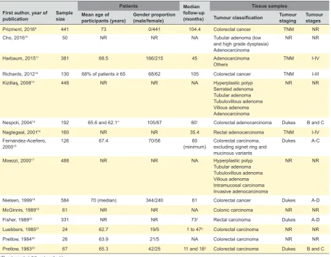
extracted from each study and comprised: author; year of publication; sample size; participants’ characteristics (mean age and gender proportion); follow-up assessment; tumour classification, stage and clinicopathological features; TATE characterization (i.e. staining, count and location); and major findings. In order to enable a better understanding of our work, we performed a broad search of the literature on the biology of eosinophils and their role in cancer, as well as on CRC up-to-date information, prognosis, microenvironment and inflammation. We included the most significant studies in the final reference list.
RESULTS
Baseline characteristics of the selected studies
We incorporated 15 original research articles in our
review, published between 1983 and 2016.9-23 Twelve
studies associated TATE with survival and/or metastatic behaviour of CRC9,11,12,14-16,18-23 and three studies associated
TATE with malignant progression of colorectal lesions.10,13,17
The first set of studies evaluated 2523 CRCs.9,11,12,14-16,18-23
The mean age of the participants was recorded in seven studies and ranged between 62.1 and 73 years.9,11,14,16,21-23
Nine studies detailed gender proportion, totalling 835 males and 1136 females.9,11,12,14,16,18,21-23 Nine studies specified the
CRC staging approach: TNM was used in four studies9,11,12,15
and Dukes staging was performed in five studies.14,16,18,20,23
The median follow-up interval was recorded in five studies and ranged from 35.4 to 105 months.9,11,12,15,18 In one study,
the average time that patients were in the study was 73 months.20 In other studies, the follow-up period was 60
months or longer.14,16 Luebbers et al assessed survival
data for periods between one and 47 months.21 Pretlow et
al recorded survival data for a period of 11 months for all patients, although for the majority of the patients included in their study, the follow-up period was18 months.23 The
second set of three studies comprised a total of 986 colorectal benign and malignant lesions.10,13,17 In these
studies, no information was recorded concerning the mean age of participants and gender proportion.10,13,17 A summary
of the selected studies is presented in Table 1.
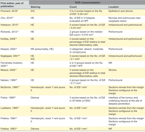
TATE characterization
In seven of the studies herein included, eosinophils
were identified in hematoxylin-eosin stained
sections.10-13,16-18 In one study, eosinophils were assessed
by immunohistochemistry for eosinophil peroxidase (clone 144B, homebrew from Dr. James Lee, Mayo Clinic, Arizona).9 In another study, the authors included both
ARTIGO DE REVISÃO
AMP
STUDENT
articles, eosinophils were identified by hematoxylin, eosin Y and azure II staining procedure.19,21,22 Also, in two articles
Giemsa staining was used for eosinophil recognition.20,23 In
one study, the staining procedure was not clearly reported,
but presumably hematoxylin-eosin staining was used.14
Regarding eosinophil counts, most of the studies established scores based on the number of eosinophils in a particular area or the percentage of eosinophils relative to total stromal inflammatory cells (seven and two studies, respectively).9,11-13,15-18,20 In five articles, scores were not
applied.10,19,21-23 In one study the authors considered three
categories for analysis.14
The location of eosinophil counts within the lesions was reported in 13 studies.9-15,17-22 In the study performed by
Prizment et al, eosinophils were counted in the epithelial and stromal compartments.9 Cho et al performed cell counts
in the mucosa and submucosa, near the neoplastic lesion.10
In three studies, intratumoural and peritumoural eosinophils were assessed.11,13,15 Richards et al, Nespoli et al and Nielsen
et al evaluated only peritumoural eosinophils.12,14,18 In other
three studies, the authors counted eosinophils in sections remote from the margin and contiguous to the margin, defined as “one cm from the border of tumour with uninvolved
mucosa” and “the border itself”, respectively.19,21,22 In one
study, the eosinophil count was performed in the stroma of the lesions; in invasive carcinoma cases, the authors assessed the transitional zone in particular (“the area between normal tissue and tumour”).17 In another article,
the authors presented eosinophil counts in the “interface of the tumour and underlying tissues at the site of deepest penetration”.20
A summary of TATE characterization described in the selected studies is presented in Table 2.
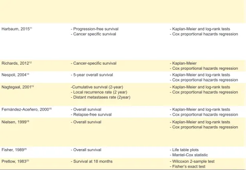
TATE as a favourable prognostic marker in CRC
Innine studiesincluded in our review, survival analyses
were performed in relation to TATE.9,11,12,14-16,18,20,23 Table 3
highlights the association between TATE and prognostic outcomes within each study.
Briefly, TATE was associated with improved overall and/or CRC-specific survival in seven studies.9,11,15,16,18,20,23
Further, TATE was inversely associated with all-cause and CRC-specific death in the study of Prizment et al.9 TATE
could also predict progression-free survival in one study11
and relapse-free survival in another study.16 Importantly, five
studies pointed TATE as an independent prognostic factor in
Table 1 – Summary of the selected studies
First author, year of
publication Sample size
Patients Median
follow-up (months)
Tissue samples Mean age of
participants (years) Gender proportion (male/female) Tumour classification Tumour staging Tumour stages
Prizment, 20169 441 73 0/441 104.4 Colorectal cancer TNM NR
Cho, 201610 50 NR NR NA Tubular adenoma (low
and high grade dysplasia) Adenocarcinoma
NR NR
Harbaum, 201511 381 68.5 166/215 45 Adenocarcinoma
Others TNM I-IV
Richards, 201212 130 68% of patients ≥ 65 68/62 105 Colorectal cancer TNM I-III
Kiziltaᶊ, 200813 448 NR NR NA Hyperplastic polyp
Serrated adenoma Tubular adenoma Tubulovillous adenoma Villous adenoma Adenocarcinoma
NR NR
Nespoli, 200414 192 65.6 and 62.1* 105/87 60† Colorectal adenocarcinoma Dukes B and C
Nagtegaal, 200115 160 NR NR 35.4 Rectal adenocarcinoma TNM I-IV
Fernández-Aceñero, 200016
126 67.4 70/56 60
(minimum) Colorectal carcinoma, excluding signet ring and mucinous variants
Dukes A-C
Moezzi, 200017 488 NR NR NA Hyperplastic polyp
Tubular adenoma Tubulovillous adenoma Villous adenoma Intramucosal carcinoma Invasive adenocarcinoma
NR NR
Nielsen, 199918 584 70 (median) 344/240 61 Colorectal cancer Dukes A-D
McGinnis, 198919 61 NR NR NA Colonic carcinoma NR NR
Fisher, 198920 331 NR NR 73‡ Rectal carcinoma Dukes A-D
Luebbers, 198521 24 62.7 19/5 1 to 47§ Colorectal carcinoma NR NR
Pretlow, 198422 26 63.9 21/5 NA Colorectal carcinoma NR NR
Pretlow, 198323 67 65.3 42/25 11 and 18§ Colorectal carcinoma Dukes B and C
NR: not reported; NA: not applicable
ARTIGO DE REVISÃO
AMP
STUDENT
CRC.9,11,15,16,18 Also significant, in four studies, the prognostic
value of TATE was maintained in intermediate stages of the disease - TNM stages II and III or Dukes stages B and C.9,11,18,23 Two studies stated a lack of association between
eosinophil count and cancer-specific or overall survival in univariate analysis.12,14
Association between TATE and metastatic behaviour of CRC
Nagtegaal et al described a significant association
between high scores of eosinophils and lower rates of distant metastases (p = 0.03).15 Additionally, an inverse relationship
was observed between peritumoural eosinophils and local recurrence and distant metastases (p = 0.007 and p = 0.009, respectively).15 Four other studies described significantly
higher concentrations of eosinophils in tumours without metastases as compared to metastatic ones (p < 0.05). 19,21-23 In one of those studies, the proportion of tumours with less
than 30 eosinophils / mm2 with metastases was significantly
higher than the proportion of tumours with more than 30 eosinophils / mm2 with metastases.23 Later on, the same
group suggested a threshold lower than 30 eosinophils / mm2.22 In another study of the same group, cut-offs
predictive of absence of metastases were proposed, namely 20 eosinophils / mm2 and 25 eosinophils / mm2, respectively
depending on whether sections remote or contiguous to the
margin of the tumour were considered.19
Association between TATE and established prognostic factors of CRC
Several items related to patients and tumour characteristics were analysed in association with TATE in the herein included studies. In this section, we summarize the association between TATE and the established prognostic factors of CRC, namely: stage, grade (differentiation), lymphatic, venous and perineural invasion, tumour budding
Table 2 – Summary of methods described in the selected studies to achieve TATE characterization
First author, year of publication
TATE characterization
Staining Count Location
Prizment, 20169 IHC 3 to 4 scores based on the No.
of EØ / 0.28 mm2 Epithelium and stroma
Cho, 201610 HE No. of EØ in 3 hotspots
evaluated at HPF Mucosa and submucosa near neoplastic lesion Harbaum, 201511 HE 4 scores based on the No. of EØ
/ 0.24 mm2 Intratumoural and peritumoural Richards, 201212 HE 2 groups based on the median
EØ count / 0.018 mm2 Peritumoural Kiziltaᶊ, 200813 HE 3 scores based on the
percentage of EØ relative to total stromal inflammatory cells
Intratumoural and peritumoural
Nespoli, 200414 NR (presumably, HE) 3 categories: absent, moderate
or conspicuous Peritumoural Nagtegaal, 200115 HE
IHC 3 scores based on the No. of EØ / 2.1 mm2 Intratumoural and peritumoural Fernández-Aceñero,
200016 HE 2 or 4 groups based on the No. of EØ / HPF NR Moezzi, 200017 HE 3 scores based on the
percentage of EØ relative to total stromal inflammatory cells
Stroma
Nielsen, 199918 HE 4 groups based on the No. of EØ
/ 0.17 mm2 Peritumoural McGinnis, 198919 Hematoxylin, eosin Y and azure
II No. of EØ / mm
2 Sections remote from the margin Sections contiguous to the margin
Fisher, 198920 Giemsa 3 scores based on the No. of EØ
in 30 fields (x1000) Interface of the tumour and underlying tissues at the site of deepest penetration
Luebbers, 198521 Hematoxylin, eosin Y and azure
II No. of EØ / mm
2 Sections remote from the margin Sections contiguous to the margin
Pretlow, 198422 Hematoxylin, eosin Y and azure
II No. of EØ / mm
2 Sections remote from the margin Sections contiguous to the margin
Pretlow, 198323 Giemsa No. of EØ / mm2 NR
ARTIGO DE REVISÃO
AMP
STUDENT
and inflammation.4
Higher eosinophil counts were significantly associated with lower CRC stage in four studies (p ≤ 0.04).9,11,15,20 In one
study, the aforementioned association was not observed.16
In two studies, TATE was associated with better tumour differentiation (p ≤ 0.05).11,20 However, these data were not
corroborated in four other studies.9,15,16,22
Increasing inflammatory reaction was positively associated with TATE in two studies (p ≤ 0.001).11,12 TATE
was significantly correlated with several inflammatory cell types in different studies, namely CD8+, CD3+, CD4+, neutrophils, macrophages and mast cells.9,15 However, in
three studies, no association was found between TATE and mast cell counts.18,20,23 The concentration of plasma cells
was significantly related to the concentration of eosinophils in one study.19
In one study, a higher eosinophil count was significantly associated with the absence of lymphatic and venous
invasion and tumour budding (p ≤ 0.02).11 However,
Fernández-Aceñero et al did not find an association
between TATE and vascular or neural invasion.16
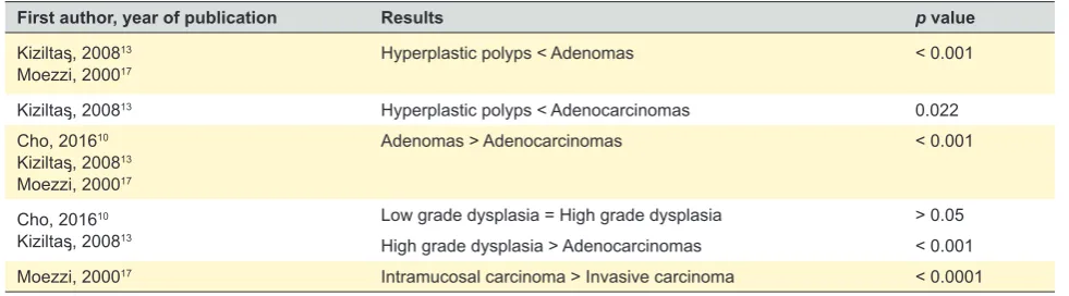
TATE in adenoma-carcinoma progression of colorectal lesions
As stated before, three studies included in our review addressed the intensity of TATE in different colonic lesions, including hyperplastic polyps, adenomas and
adenocarcinomas (Table 1).10,13,17 Table 4 summarizes the
results of these studies.
It was observed that the number of eosinophils significantly decreased throughout adenoma-carcinoma progression.10,13,17 The intensity of TATE in hyperplastic
polyps was significantly lower than in adenomatous lesions13,17 and adenocarcinomas.13 In the study of Kiziltaᶊ
et al, there was no significant difference in TATE intensity between low- and high grade dysplasia,13 in line with the
results of Cho et al.10 Still, in both studies, the intensity
of TATE was higher in high grade dysplasia compared to adenocarcinomas.10,13 Additionally, Moezzi et al described
a higher TATE in intramucosal carcinoma compared to invasive carcinoma.17 Kiziltaᶊ et al further included serrated
adenomas in their series and found that TATE was lower in the hyperplastic polyps and higher in adenomatous polyps compared to those lesions (p < 0.001).13
DISCUSSION
Tumour microenvironment refers to the malignant and tumour-associated stromal and immune cells, as well as
the cross-talk between them.24 The immune system plays
a dual action both in cancer promotion and prevention.25
Considering the critical role of the immune system in the recognition and elimination of transformed cells - cancer immunosurveillance - new insights have emerged regarding the prognostic role of immune infiltrates in tumour tissues.25
In this context, TATE has been widely studied, as previously revised.4,6,26-31 In this study, we aimed at reviewing the
prognostic value of TATE in CRC. In contrast to previous
reviews, which summarized this topic in the context of wider subjects,4,6,26,28,29,31 our review included a larger number of
studies and provided more detailed information on TATE characterization and on the association between TATE and other clinicopathological features in CRC. Moreover, we reviewed the association of TATE and adenoma-carcinoma progression. Therefore, we accomplished a more exhaustive understanding of the published data.
TATE has been associated with a favourable prognosis in several types of solid tumours, including CRC.4,6,26-29,31 Our
analysis adds to this concept. Importantly, the prognostic value of TATE was maintained when TNM stages II and III of the disease were considered.9,11 These findings support
that the evaluation of TATE in CRC specimens may be useful for individual prognosis and might be used in the selection of patients who benefit from aggressive therapies as previously suggested.4,16,18,29
Despite the association found in different studies, the mechanisms by which eosinophils are recruited to the cancer niche and influence cancer prognosis remain unclear. However, current knowledge suggests that TATE plays an active and protective role in CRC progression rather than being an occasional finding. Understanding the molecular mechanisms involved in the cross-talk between eosinophils and CRC cells would certainly contribute to improve the existing knowledge of the immunogenic characteristics of the disease and may have a major impact in the development of novel therapeutic targets.6,9,11,26,28,29,31-35
In the case of CRC, eosinophil recruitment, activation and survival was suggested to be dependent on local production of IL-5 by tissue resident eosinophils36 and
active damage associated molecular pattern (DAMP) molecules, namely high mobility group box 1 (HMGB1), in the context of tumour cell necrosis.32 Surprising findings
have been achieved on the role of eotaxins in eosinophil chemoatraction to CRCs, since low numbers of tissue eosinophils are found both in primary CRCs10,37 and liver
metastases,37 albeit not always associated with low levels of
tissue eotaxins.37,38 The contribution of neoplastic epithelial
cells for the secretion of eotaxins appears to be residual.10,39
Briefly, three mechanisms may explain the lower number of eosinophils in CRC tissues despite the often high levels of tissue eotaxins: the eosinophil chemoattractant role of eotaxins may be blocked in CRC37; lower levels of eotaxins
in plasma may have impact in eosinophil traffic to the tissues38,39; or the differential eotaxins compartmentalization
in CRC tissues influences eosinophil chemoatraction.10
Following activation, several mechanisms for CRC control by eosinophils have been proposed. After attachment to carcinoma cells, eosinophils lose their IL-5 transcripts and undergo apoptosis, releasing their cytotoxic granules.36
Eosinophil degranulation results in the release of major basic protein (MBP) and eosinophil peroxidase (EPO), which promote oxidation of cancer cell lysates, both directly and indirectly through neutrophils.32 Additionally, the cytotoxic
ARTIGO DE REVISÃO
AMP
STUDENT
assigned to eosinophil cationic protein, eosinophil derived neurotoxin (EDN), TNF-α, and granzyme A, depending on CD11a/CD18 for effector-target cell adhesion, and γδTCR/ CD3.40,41 The same group addressed IL-18 as a key mediator
of eosinophils-Colo-205 cells contact, through CD11a and ICAM-1 adhesion molecules, contributing to the pro-apoptotic action of eosinophils.33 Conversely, Taylor et al
previously stated that CD11a was not involved in eosinophil binding to MCA-38 colon adenocarcinoma cells, but the authors used mouse eosinophils.42 Still, they attributed a
direct cytotoxic effect of eosinophils against MCA-38 cells, demonstrating the role of protein tyrosine kinase and cyclic AMP in effector-target cell adhesion.42 Earlier, the local
anti-tumour effect of IL-4 (secreted by engineered colon 26 cells) was endorsed in part to eosinophils in an early stage, supporting an advantageous role of TATE in the defence against cancer.34 Recently, a protective role was also
attributed to IL-18 in a mouse model of inflammatory colon
cancer and inflammatory bowel disease (AOM/DSS mice).39
The levels of IL-18 were likely related with TATE, rather than with epithelial cells secretion.39 IL-17E (IL-25) was found to
increase TATE and inhibited tumour growth, through IL-5
induction, in a human colon tumour xenograft model.35
Thus, eosinophils may be engaged in the anti-tumour effect of IL-17E, and IL-17E was endorsed as a putative effective
therapy in colon adenocarcinoma.35
In addition to the aforementioned molecular mechanisms, the relation between eosinophils and other immune cells raises the hypotheses that eosinophils may also exert their action via other cells and vice-versa.9,15,19
The interaction between eosinophils and other immune cells has been recognized.26-28,31,43 Interestingly, Nagtegaal
et al proposed a model for protective immune responses in rectal cancer, in which T cells interact with nonspecific immune response (eosinophils, neutrophils, mast cells, macrophages and NK cells).15 All these cells have been
implicated in CRC prognosis in different extents.4-6 Recently,
Prizment et al suggested that eosinophils may act partially through cytotoxic T-cells in CRC.9
Despite the body of evidence placing eosinophils as cells with anti-tumoural activity, their therapeutic potential remains difficult to explore. This is greatly due to the lack of specific activity of eosinophils against tumoural cells.31
Indeed, attracting eosinophils to the cancer niche and
Table 3 – Association between TATE and prognostic outcomes
First author, year of publication Outcome measured Statistical methods Main findings
Prizment, 20169 - 5-year all-cause and CRC survival - 5-year all-cause and CRC-specific death - Total follow-up all-cause and CRC-specific death
- Kaplan-Meier plots and log-rank tests
- Cox proportional hazards regression - In univariate analysis, higher stromal EØ scores (survival (p = 0.0006 and p = 0.001, respectively) versus lower stromal EØ scores) associated with better 5-year all-cause and CRC - In multivariate analysis, stromal EØ scores (highest versus lowest) inversely associated with risk for 5-year all-cause and CRC death (HR 0.61; 95% CI 0.36 - 1.02; p = 0.02 and HR 0.48; 95% CI 0.24 - 0.93; p = 0.01, respectively) as with total follow-up all-cause and CRC-specific death (HR 0.72; 95% CI 0.48 - 1.08; p = 0.04 and HR 0.61; 95% CI 0.34 - 1.12; p = 0.04, respectively) - The association was maintained with statistical significance when stages II and III were combined
- In both univariate and multivariate analyses, the highest score of epithelial EØ showed a trend to better outcomes, though without statistical significance
Harbaum, 201511 - Progression-free survival
- Cancer specific survival - Kaplan-Meier and log-rank tests- Cox proportional hazards regression - Both peritumoural and intratumoural EØ associated significantly with progression-free and cancer specific survival (- Only peritumoural EØ independently associated with progression-free and cancer specific survival (HR 0.75; 95% CI 0.58 - 0.98; p < 0.001) p
= 0.04 and HR 0.7; 95% CI 0.53 - 0.93; p = 0.01, respectively)
- In patients with stage II CRC, the presence of peritumoural EØ (versus the absence of peritumoural EØ) independently associated with progression-free and cancer specific survival (HR 0.24; 95% CI 0.07 - 0.87; p = 0.03 and HR 0.25; 95% CI 0.06 - 1.02; p = 0.05, respectively)
Richards, 201212 - Cancer-specific survival - Kaplan-Meier
- Cox proportional hazards regression - In univariate analysis, EØ count did not associate with cancer-specific survival (HR 1.72; 95% CI 0.89 - 3.35; p = 0.11) Nespoli, 200414 - 5-year overall survival - Kaplan-Meier and log-rank tests
- Cox proportional hazards regression - In univariate analysis, EØ infiltration did not associated with survival (p ≥ 0.3) Nagtegaal, 200115 -Cumulative survival (2-year)
- Local recurrence rate (2 year) - Distant metastases rate (2year)
- Kaplan-Meier and log-rank tests
- Cox proportional hazards regression - In univariate analysis, peritumoural EØ directly associated with cumulative survival (recurrence and distant metastases (p = 0.007 and 0.009, respectively) p = 0.0008) and inversely associated with local - In multivariate analysis, peritumoural EØ showed independent prognostic value regarding cumulative survival, additional to TNM staging
Fernández-Aceñero, 200016 - Overall survival
- Relapse-free survival - Kaplan-Meier and log-rank tests- Cox proportional hazards regression - Higher EØ counts independently associated with longer overall survival and relapse-free survival (respectively) p = 0.0005 and p = 0.0009, Nielsen, 199918 - Overall survival - Kaplan-Meier and log-rank tests
- Cox proportional hazards regression - EØ count significantly predicted a good overall survival (improvement p < 0.0001), with a progressive effect of increasing counts on the survival - In univariate analysis, EØ count significantly associated with survival (HR 0.74; 95% CI 0.66 - 0.84; p < 0.0001)
- In multivariate analysis, EØ count independently associated with survival (HR 0.81; 95% CI 0.72 - 0.92; p = 0.001) - EØ counts directly associated with survival in Dukes B and C stages (p = 0.03 and 0.002, respectively)
Fisher, 198920 - Overall survival - Life table plots
- Mantel-Cox statistic - Overall survival was higher in patients whose tumours had 10 or more EØ- After adjustment for Dukes’ stage or treatment, the No. of EØ had no prognostic value Pretlow, 198323 - Survival at 18 months - Wilcoxon 2-sample test
- Fisher’s exact test - Survival was significantly greater when more than 30 EØ / mm2 were counted (p = 0.028)
ARTIGO DE REVISÃO
AMP
STUDENT
activating them would most likely lead to detrimental damage of the remaining tissue, in addition to cancer cells.31
Thus, important lines of research are opening for the future, mainly related to the development of strategies allowing eosinophils to specifically target cancer cells.31
Concerning the association between TATE and other prognostic markers of CRC, the results of our review were inconsistent. The limited number of the included studies
that associated TATE with overall inflammation or specific cell types and other clinicopathological prognosticators impaired a comprehensive analysis about this topic. The most reliable finding encompassed the association between higher eosinophil counts and lower CRC stage.9,11,15,20 The
association between TATE and such a strong prognostic marker might validate the impact of TATE itself in CRC prognosis. Moreover, this finding suggests that TATE,
Table 3 – Association between TATE and prognostic outcomes
First author, year of publication Outcome measured Statistical methods Main findings
Prizment, 20169 - 5-year all-cause and CRC survival - 5-year all-cause and CRC-specific death - Total follow-up all-cause and CRC-specific death
- Kaplan-Meier plots and log-rank tests
- Cox proportional hazards regression - In univariate analysis, higher stromal EØ scores (survival (p = 0.0006 and p = 0.001, respectively) versus lower stromal EØ scores) associated with better 5-year all-cause and CRC - In multivariate analysis, stromal EØ scores (highest versus lowest) inversely associated with risk for 5-year all-cause and CRC death (HR 0.61; 95% CI 0.36 - 1.02; p = 0.02 and HR 0.48; 95% CI 0.24 - 0.93; p = 0.01, respectively) as with total follow-up all-cause and CRC-specific death (HR 0.72; 95% CI 0.48 - 1.08; p = 0.04 and HR 0.61; 95% CI 0.34 - 1.12; p = 0.04, respectively) - The association was maintained with statistical significance when stages II and III were combined
- In both univariate and multivariate analyses, the highest score of epithelial EØ showed a trend to better outcomes, though without statistical significance
Harbaum, 201511 - Progression-free survival
- Cancer specific survival - Kaplan-Meier and log-rank tests- Cox proportional hazards regression - Both peritumoural and intratumoural EØ associated significantly with progression-free and cancer specific survival (- Only peritumoural EØ independently associated with progression-free and cancer specific survival (HR 0.75; 95% CI 0.58 - 0.98; p < 0.001) p
= 0.04 and HR 0.7; 95% CI 0.53 - 0.93; p = 0.01, respectively)
- In patients with stage II CRC, the presence of peritumoural EØ (versus the absence of peritumoural EØ) independently associated with progression-free and cancer specific survival (HR 0.24; 95% CI 0.07 - 0.87; p = 0.03 and HR 0.25; 95% CI 0.06 - 1.02; p = 0.05, respectively)
Richards, 201212 - Cancer-specific survival - Kaplan-Meier
- Cox proportional hazards regression - In univariate analysis, EØ count did not associate with cancer-specific survival (HR 1.72; 95% CI 0.89 - 3.35; p = 0.11) Nespoli, 200414 - 5-year overall survival - Kaplan-Meier and log-rank tests
- Cox proportional hazards regression - In univariate analysis, EØ infiltration did not associated with survival (p ≥ 0.3) Nagtegaal, 200115 -Cumulative survival (2-year)
- Local recurrence rate (2 year) - Distant metastases rate (2year)
- Kaplan-Meier and log-rank tests
- Cox proportional hazards regression - In univariate analysis, peritumoural EØ directly associated with cumulative survival (recurrence and distant metastases (p = 0.007 and 0.009, respectively) p = 0.0008) and inversely associated with local - In multivariate analysis, peritumoural EØ showed independent prognostic value regarding cumulative survival, additional to TNM staging
Fernández-Aceñero, 200016 - Overall survival
- Relapse-free survival - Kaplan-Meier and log-rank tests- Cox proportional hazards regression - Higher EØ counts independently associated with longer overall survival and relapse-free survival (respectively) p = 0.0005 and p = 0.0009, Nielsen, 199918 - Overall survival - Kaplan-Meier and log-rank tests
- Cox proportional hazards regression - EØ count significantly predicted a good overall survival (improvement p < 0.0001), with a progressive effect of increasing counts on the survival - In univariate analysis, EØ count significantly associated with survival (HR 0.74; 95% CI 0.66 - 0.84; p < 0.0001)
- In multivariate analysis, EØ count independently associated with survival (HR 0.81; 95% CI 0.72 - 0.92; p = 0.001) - EØ counts directly associated with survival in Dukes B and C stages (p = 0.03 and 0.002, respectively)
Fisher, 198920 - Overall survival - Life table plots
- Mantel-Cox statistic - Overall survival was higher in patients whose tumours had 10 or more EØ- After adjustment for Dukes’ stage or treatment, the No. of EØ had no prognostic value Pretlow, 198323 - Survival at 18 months - Wilcoxon 2-sample test
- Fisher’s exact test - Survival was significantly greater when more than 30 EØ / mm2 were counted (p = 0.028)
TATE: tumour associated tissue eosinophilia; CRC: colorectal cancer; EØ: eosinophil(s); HR: hazard ratio; CI: confidence interval; No(s).: number(s)
Table 4 – Association between TATE and colorectal adenoma-carcinoma progression
First author, year of publication Results p value
Kiziltaᶊ, 200813
Moezzi, 200017 Hyperplastic polyps < Adenomas < 0.001 Kiziltaᶊ, 200813 Hyperplastic polyps < Adenocarcinomas 0.022 Cho, 201610
Kiziltaᶊ, 200813 Moezzi, 200017
Adenomas > Adenocarcinomas < 0.001
Cho, 201610 Kiziltaᶊ, 200813
ARTIGO DE REVISÃO
AMP
STUDENT
namely stromal eosinophils, may prevent CRC progression as stated by Prizment et al,9 which is in line with the decrease
of TATE in adenoma-carcinoma progression of colorectal lesions.10,13,17 In conclusion, these results also point to
TATE as a potential immune-evading strategy of CRC.10
Therefore, in addition to investigating the mechanisms of action of TATE in the context of CRC, it is very important to continue research on the power of TATE in the clinical management of CRC patients. For example, given the decrease of TATE in adenoma-carcinoma progression, TATE has been proposed as a marker of risk for the development of CRC, as well as an indicator for more and tighter surveillance schemes.13,17 Additionally, TATE may
be used as a diagnostic tool to differentiate hyperplastic polyps, adenomatous polyps and serrated adenomas.13,17
In our review, we only found three articles concerning this topic,10,13,17 so, clearly, more research is needed in this area.
A major limitation to our review, like in previous ones,6,28 was the variability between methodologies used
in the original research manuscripts which impaired a more meaningful interpretation of the results and the establishment of a threshold above which TATE could predict CRC progression. While the use of immunohistochemistry technique strengthens the identification of tissue eosinophils, an easy, standardized and cost-effective measurement system would be of benefit to enhance
future research.4,9 Furthermore, immunohistochemistry
is not free from faults.29 In fact, TATE was assessed in
hematoxylin-eosin stained sections in the majority of the included studies, without compromising the results.10-13,15-18
Nevertheless, how TATE should be evaluated in terms of location and scoring system, remains unclear. Klintrup et al developed an easy and highly reproductive score system – the Klintrup-Mäkinen score – to assess peritumoural overall inflammatory reaction in routinely stained CRC specimens, which proved to be an independent favourable predictor of survival in CRC patients.44 In that study, higher
eosinophilic grade at the invasive margin showed a trend
towards a better prognosis.44 Accordingly, several studies
in our series highlighted peritumoural location for TATE evaluation.11,15,18,20 TATE can be easily evaluated in routinely
processed tissues and peritumoural / invasive margins appear to be the most reliable areas for TATE evaluation. Because of all this, TATE seems to meet the standards of a good histopathological prognostic marker as established by
Schneider and Langner.4
CONCLUSION
According to published studies, we concluded that: 1) current evidence points to TATE as a promising independent prognostic marker in CRC; 2) TATE is inversely associated with the metastatic behaviour of CRC; 3) TATE decreases with adenoma-carcinoma progression of colorectal lesions; and 4) TATE evaluation may have implications on the surveillance schedule and treatments offered to patients diagnosed with CRC. These exciting and promising data call for future research on the role of eosinophils in CRC.
PROTECTION OF HUMANS AND ANIMALS
The authors declare that the procedures were followed according to the regulations established by the Clinical Research and Ethics Committee and to the Helsinki Declaration of the World Medical Association.
DATA CONFIDENTIALITY
The authors declare having followed the protocols in use at their working center regarding patients’ data publication. Patient consent obtained.
CONFLICTS OF INTEREST
All authors report no conflict of interest.
FUNDING SOURCES
This research received no specific grant from any funding agency in the public, commercial, or not-for-profit sectors.
REFERENCES
1. Ferlay J, Soerjomataram I, Dikshit R, Eser S, Mathers C, Rebelo M, et al. Cancer incidence and mortality worldwide: sources, methods and major patterns in GLOBOCAN 2012. Int J Cancer. 2015;136:E359-86. 2. Amin MB, Edge SB, Greene F, Byrd DR, Brookland RK, Washington MK,
et al, editors. AJCC Cancer Staging Manual. 8th ed. New York: Springer International Publishing; 2017.
3. Compton CC. Optimal pathologic staging: defining stage II disease. Clin Cancer Res. 2007;13:6862s-70s.
4. Schneider NI, Langner C. Prognostic stratification of colorectal cancer patients: current perspectives. Cancer Manag Res. 2014;6:291-300. 5. Grizzi F, Bianchi P, Malesci A, Laghi L. Prognostic value of innate
and adaptive immunity in colorectal cancer. World J Gastroenterol. 2013;19:174-84.
6. Roxburgh CS, McMillan DC. The role of the in situ local inflammatory response in predicting recurrence and survival in patients with primary operable colorectal cancer. Cancer Treat Rev. 2012;38:451-66. 7. Hendry S, Salgado R, Gevaert T, Russell PA, John T, Thapa B, et al.
Assessing Tumor-Infiltrating Lymphocytes in Solid Tumors: A Practical Review for Pathologists and Proposal for a Standardized Method from the International Immuno-Oncology Biomarkers Working Group: Part 2: TILs in Melanoma, Gastrointestinal Tract Carcinomas, Non-Small Cell Lung Carcinoma and Mesothelioma, Endometrial and Ovarian
Carcinomas, Squamous Cell Carcinoma of the Head and Neck, Genitourinary Carcinomas, and Primary Brain Tumors. Adv Anat Pathol. 2017;24:311-35.
8. Pinto ML, Rios E, Silva AC, Neves SC, Caires HR, Pinto AT, et al. Decellularized human colorectal cancer matrices polarize macrophages towards an anti-inflammatory phenotype promoting cancer cell invasion via CCL18. Biomaterials. 2017;124:211-24.
9. Prizment AE, Vierkant RA, Smyrk TC, Tillmans LS, Lee JJ, Sriramarao P, et al. Tumor eosinophil infiltration and improved survival of colorectal cancer patients: Iowa Women’s Health Study. Mod Pathol. 2016;29:516-27.
10. Cho H, Lim SJ, Won KY, Bae GE, Kim GY, Min JW, et al. Eosinophils in colorectal neoplasms associated with expression of CCL11 and CCL24. J Pathol Transl Med. 2016;50:45-51.
11. Harbaum L, Pollheimer MJ, Kornprat P, Lindtner RA, Bokemeyer C, Langner C. Peritumoral eosinophils predict recurrence in colorectal cancer. Mod Pathol. 2015;28:403-13.
ARTIGO DE REVISÃO
AMP
STUDENT
13. Kiziltaş S, Sezgin Ramadan S, Topuzoglu A, Kullu S. Does the severity of tissue eosinophilia of colonic neoplasms reflect their malignancy potential? Turk J Gastroenterol. 2008;19:239-44.
14. Nespoli A, Gianotti L, Totis M, Bovo G, Nespoli L, Chiodini P, et al. Correlation between postoperative infections and long-term survival after colorectal resection for cancer. Tumori. 2004;90:485-90. 15. Nagtegaal ID, Marijnen CA, Kranenbarg EK, Mulder-Stapel A, Hermans
J, van de Velde CJ, et al. Local and distant recurrences in rectal cancer patients are predicted by the nonspecific immune response; specific immune response has only a systemic effect--a histopathological and immunohistochemical study. BMC Cancer. 2001;1:7.
16. Fernández-Aceñero MJ, Galindo-Gallego M, Sanz J, Aljama A. Prognostic influence of tumor-associated eosinophilic infiltrate in colorectal carcinoma. Cancer. 2000;88:1544-8.
17. Moezzi J, Gopalswamy N, Haas RJ, Jr., Markert RJ, Suryaprasad S, Bhutani MS. Stromal eosinophilia in colonic epithelial neoplasms. Am J Gastroenterol. 2000;95:520-3.
18. Nielsen HJ, Hansen U, Christensen IJ, Reimert CM, Brunner N, Moesgaard F. Independent prognostic value of eosinophil and mast cell infiltration in colorectal cancer tissue. J Pathol. 1999;189:487-95. 19. McGinnis MC, Bradley EL, Jr., Pretlow TP, Ortiz-Reyes R, Bowden CJ,
Stellato TA, et al. Correlation of stromal cells by morphometric analysis with metastatic behavior of human colonic carcinoma. Cancer Res. 1989;49:5989-93.
20. Fisher ER, Paik SM, Rockette H, Jones J, Caplan R, Fisher B. Prognostic significance of eosinophils and mast cells in rectal cancer: findings from the National Surgical Adjuvant Breast and Bowel Project (protocol R-01). Hum Pathol. 1989;20:159-63.
21. Luebbers EL, Pretlow TP, Emancipator SN, Boohaker EA, Pitts AM, Macfadyen AJ, et al. Heterogeneity and prognostic significance of macrophages in human colonic carcinomas. Cancer Res. 1985;45:5196-200.
22. Pretlow TP, Boohaker EA, Pitts AM, Macfadyen AJ, Bradley EL, Jr., Pretlow TG, 2nd. Heterogeneity and subcompartmentalization in the distribution of eosinophils in human colonic carcinomas. Am J Pathol. 1984;116:207-13.
23. Pretlow TP, Keith EF, Cryar AK, Bartolucci AA, Pitts AM, Pretlow TG, 2nd, et al. Eosinophil infiltration of human colonic carcinomas as a prognostic indicator. Cancer Res. 1983;43:2997-3000.
24. Balkwill FR, Capasso M, Hagemann T. The tumor microenvironment at a glance. J Cell Sci. 2012;125:5591-6.
25. Mittal D, Gubin MM, Schreiber RD, Smyth MJ. New insights into cancer immunoediting and its three component phases-elimination, equilibrium and escape. Curr Opin Immunol. 2014;27:16-25.
26. Sakkal S, Miller S, Apostolopoulos V, Nurgali K. Eosinophils in cancer: favourable or unfavourable? Curr Med Chem. 2016;23:650-66. 27. Davis BP, Rothenberg ME. Eosinophils and cancer. Cancer Immunol
Res. 2014;2:1-8.
28. Gatault S, Legrand F, Delbeke M, Loiseau S, Capron M. Involvement of eosinophils in the anti-tumor response. Cancer Immunol Immunother.
2012;61:1527-34.
29. Shunyakov L, Ryan CK, Sahasrabudhe DM, Khorana AA. The influence of host response on colorectal cancer prognosis. Clin Colorectal Cancer. 2004;4:38-45.
30. Lowe D, Jorizzo J, Hutt MS. Tumour-associated eosinophilia: a review. J Clin Pathol. 1981;34:1343-8.
31. Reichman H, Karo-Atar D, Munitz A. Emerging roles for eosinophils in the tumor microenvironment. Trends Cancer. 2016;2:664-75.
32. Lotfi R, Herzog GI, DeMarco RA, Beer-Stolz D, Lee JJ, Rubartelli A, et al. Eosinophils oxidize damage-associated molecular pattern molecules derived from stressed cells. J Immunol. 2009;183:5023-31.
33. Gatault S, Delbeke M, Driss V, Sarazin A, Dendooven A, Kahn JE, et al. IL-18 is involved in eosinophil-mediated tumoricidal activity against a colon carcinoma cell line by upregulating LFA-1 and ICAM-1. J Immunol. 2015;195:2483-92.
34. Itsuki Y, Suzuki S. Expression of interleukin-4 in colon 26 cells induces both eosinophil mediated local tumor killing and T-cell mediated systemic immunity in vivo. Nihon Ika Daigaku Zasshi. 1996;63:275-85.
35. Benatar T, Cao MY, Lee Y, Lightfoot J, Feng N, Gu X, et al. IL-17E, a proinflammatory cytokine, has antitumor efficacy against several tumor types in vivo. Cancer Immunol Immunother. 2010;59:805-17.
36. Tajima K, Yamakawa M, Inaba Y, Katagiri T, Sasaki H. Cellular localization of interleukin-5 expression in rectal carcinoma with eosinophilia. Hum Pathol. 1998;29:1024-8.
37. Cheadle EJ, Riyad K, Subar D, Rothwell DG, Ashton G, Batha H, et al. Eotaxin-2 and colorectal cancer: a potential target for immune therapy. Clin Cancer Res. 2007;13:5719-28.
38. Wågsäter D, Lofgren S, Hugander A, Dienus O, Dimberg J. Analysis of single nucleotide polymorphism in the promoter and protein expression of the chemokine eotaxin-1 in colorectal cancer patients. World J Surg Oncol. 2007;5:84.
39. Lang M, Berry D, Passecker K, Mesteri I, Bhuju S, Ebner F, et al. HuR small-molecule inhibitor elicits differential effects in adenomatosis polyposis and colorectal carcinogenesis. Cancer Res. 2017;77:2424-38. 40. Legrand F, Driss V, Delbeke M, Loiseau S, Hermann E, Dombrowicz D,
et al. Human eosinophils exert TNF-alpha and granzyme A-mediated tumoricidal activity toward colon carcinoma cells. J Immunol. 2010;185:7443-51.
41. Legrand F, Driss V, Woerly G, Loiseau S, Hermann E, Fournie JJ, et al. A functional gammadeltaTCR/CD3 complex distinct from gammadeltaT cells is expressed by human eosinophils. PLoS One. 2009;4:e5926. 42. Taylor R, Lee TD, Hoskin DW. Adhesion of tumoricidal eosinophils to
MCA-38 colon adenocarcinoma cells involves protein tyrosine kinase activation and is diminished by elevated cyclic AMP in the effector cell. Int J Oncol. 1998;13:1305-11.
43. Blanchard C, Rothenberg ME. Biology of the eosinophil. Adv Immunol. 2009;101:81-121.