Thromboxane A2 Activates YAP/TAZ Protein to Induce
Vascular Smooth Muscle Cell Proliferation and Migration
*
□SReceived for publication, May 23, 2016, and in revised form, June 30, 2016 Published, JBC Papers in Press, July 5, 2016, DOI 10.1074/jbc.M116.739722
Xu Feng‡, Peng Liu‡, Xin Zhou‡, Meng-Tian Li‡, Fu-Long Li‡, Zhen Wang‡, Zhipeng Meng§, Yi-Ping Sun‡, Ying Yu¶, Yue Xiong‡储1, Hai-Xin Yuan‡2, and Kun-Liang Guan‡§3
From the‡Key Laboratory of Molecular Medicine of the Ministry of Education and Institutes of Biomedical Sciences, Shanghai Medical College, Fudan University, Shanghai 200032, China, the§Department of Pharmacology and Moores Cancer Center, University of California San Diego, La Jolla, California 92130, the¶Key Laboratory of Food Safety Research, Chinese Academy of Sciences Center for Excellence in Molecular Cell Science, Institute for Nutritional Sciences, Shanghai Institutes for Biological Sciences, Chinese Academy of Sciences, Shanghai 200031, China, and the储Department of Biochemistry and Biophysics, Lineberger Comprehensive Cancer Center, University of North Carolina, Chapel Hill, North Carolina 27599
The thromboxane A2 receptor (TP) has been implicated in restenosis after vascular injury, which induces vascular smooth muscle cell (VSMC) migration and proliferation. However, the mechanism for this process is largely unknown. In this study, we report that TP signaling induces VSMC migration and prolifer-ation through activating YAP/TAZ, two major downstream effectors of the Hippo signaling pathway. The TP-specific agonists [1S-[1␣,2␣(Z),3(1E,3S*),4 ␣ ]]-7-[3-[3-hydroxy-4-(4- iodophenoxy)-1-butenyl]-7-oxabicyclo[2.2.1]hept-2-yl]-5-hep-tenoic acid (I-BOP) and 9,11-dideoxy-9␣,11␣ -methanoepoxy-prosta-5Z,13E-dien-1-oic acid (U-46619) induce YAP/TAZ activation in multiple cell lines, including VSMCs. YAP/TAZ activation induced by I-BOP is blocked by knockout of the receptor TP or knockdown of the downstream G proteins G␣12/13. Moreover, Rho inhibition or actin cytoskeleton disrup-tion prevents I-BOP-induced YAP/TAZ activadisrup-tion. Impor-tantly, TP activation promotes DNA synthesis and cell migra-tion in VSMCs in a manner dependent on YAP/TAZ. Taken together, thromboxane A2 signaling activates YAP/TAZ to pro-mote VSMC migration and proliferation, indicating YAP/TAZ as potential therapeutic targets for cardiovascular diseases.
Thromboxane A2 (TxA2)4is produced in many cells/tissues,
particularly in platelets (1), and plays an important role in
plate-let activation and aggregation in vascular injury and atheroscle-rosis (2). In patients with atheroscleatheroscle-rosis or undergoing percu-taneous transluminal coronary angioplasty (a non-surgical procedure used to treat the narrowed coronary arteries in car-diovascular disease), the concentration of TxA2 is significantly increased (3–5). A similar phenomenon has also observed in the corresponding mouse models (6 – 8). TxA2 and thrombox-ane A2 receptor (TP) are believed to contribute to restenosis after vascular injury (6, 9), which remains a challenging clinical problem, although drug-eluting stents can reduce the risk of restenosis (10).
TxA2 is a type of prostanoid; prostanoids are a family of lipid mediators generated by cyclooxygenase. TxA2 is unsta-ble, with a half-life of about 30 s, and is non-enzymatically degraded into the biologically inactive form thromboxane B2 (TxB2) (1). TxA2 exerts its biological activity through its cognate TP receptor, a G protein-coupled receptor (GPCR) that couples with G␣12/13, G␣q/11, and other trimeric G pro-teins to regulate downstream effectors (11). TP is expressed
as two isoforms in humans: TP␣and TP. Besides TxA2,
prostaglandin H2, isoprostanes (such as 8-iso-prostaglandin F2␣), and hydroxyeicosatetraenoic acids can also activate TP receptors (12–14).
In addition to platelet activation, TxA2 or TP receptor is also known to promote cell migration and proliferation of vascular smooth muscle cells (VSMCs) (15–20), an important process that is involved in a number of vascular diseases, such as post-angioplasty restenosis and atherosclerosis (21). The prolifera-tive response of VSMCs to vascular injury is markedly exagger-ated in transgenic mice with vascular overexpression of the human TP␣receptor, which can be inhibited by the TP-specific antagonist S18886 (6). Moreover, injury-induced vascular pro-liferation and platelet activation are suppressed in mice genet-ically deficient in TP receptor (6). In mouse models of athero-sclerosis, both pharmacological antagonism and TP receptor deletion delay lesion development (22–24). Taken together, these previous studies demonstrate that TxA2 and TP receptor contribute to VSMCs mediating vascular disease, although the molecular mechanism is largely unknown.
*This work was supported by National Natural Science Foundation of China Grant 31570784 (to H. X. Y.). K. L. G. is a cofounder of Vivace Therapeutics Inc. The content is solely the responsibility of the authors and does not necessarily represent the official views of the National Institutes of Health.
□S This article containssupplemental Figs. 1– 8.
1Supported by National Institutes of Health Grants RO1GM067113, RO1CA068377, and RO1CA163834. To whom correspondence may be addressed: E-mail: [email protected].
2To whom correspondence may be addressed. E-mail: yuanhaixin@ fudan.edu.cn.
3Supported by National Institutes of Health Grants R35CA196878, RO1GM51586, and RO1EY022611. To whom correspondence may be addressed. E-mail: [email protected].
4The abbreviations used are: TxA2, thromboxane A2; TP, thromboxane A2 recep-tor; GPCR, G protein-coupled receprecep-tor; VSMC, vascular smooth muscle cell; CTGF, connective tissue growth factor; MAVSMC, mouse aortic vascular smooth muscle cell; dKO, double knockout; U-46619, 9,11-dideoxy-9␣,11␣ -methanoepoxy-prosta-5Z,13E-dien-1-oic acid; SQ-29548, [1S-[1␣,2␣(Z), 3␣,4␣ ]]-7-[3-[[2-[(phenylamino)carbonyl]hydrazino]methyl]-7-oxabicyc-lo[2.2.1]hept-2-yl]-5-heptenoic acid; I-BOP, [1S-[1␣,2␣(Z),3(1E,3S*),4␣ ]]-7-[3- [3-hydroxy-4-(4-iodophenoxy)-1-butenyl]-7-oxabicyclo[2.2.1]hept-2-yl]-5-heptenoic acid; EdU, 5-ethynyl-2⬘-deoxyuridine; LATS, large tumor
suppressor kinase; MAP4K, mitogen-activated protein kinase kinase kinase kinase; TEAD, TEA domain transcription factor; CRISPR, clustered regularly interspaced short palindromic repeats.
crossmark
The Hippo signaling pathway plays a key role in the regula-tion of organ size and tissue homeostasis (25). Core compo-nents of the mammalian Hippo pathway include MST1/2 and their adaptor protein SAV1, LATS1/2 and their adaptor pro-teins MOB1A/1B, and two downstream transcriptional effec-tors, YAP/TAZ (25). MST1/2 phosphorylate and activate LATS1/2 kinases, which in turn phosphorylate and inhibit YAP/TAZ. Recently, MAP4Ks have been shown to be core components of the Hippo pathway, and they function in paral-lel to MST1/2 to phosphorylate and activate LATS1/2 (26, 27). Phosphorylation of YAP at Ser-127 results in cytoplasmic sequestration because of 14-3-3 binding (28, 29). The dephos-phorylated YAP/TAZ translocate into the nucleus and interact with the TEAD family transcription factors to induce target genes, such as connective tissue growth factor (CTGF) and cys-teine-rich angiogenic inducer 61 (CYR61), thereby promoting cell proliferation, migration, and survival (30 –32). Deregula-tion of the Hippo pathway has been observed in various human cancers and is often correlated with a poor prognosis (33). Upstream signals of the Hippo pathway had been elusive until recent studies established that GPCRs relay extracellular sig-nals to the Hippo pathway (34 –38). Ligands signaling through GPCRs coupled to G␣12/13, G␣q/11, or G␣i/oactivate YAP/TAZ,
whereas ligands signaling through G␣s-coupled GPCRs
sup-press YAP/TAZ activity (36). Therefore, stimulation of differ-ent GPCRs can result in either activation or inhibition of YAP/TAZ.
Interestingly, emerging evidence shows that YAP is induced after arterial injury and that its activation promotes smooth muscle phenotypic switching and neointima formation (39, 40). This led us to investigate the function of YAP/TAZ in TxA2-and TP receptor-induced cellular signaling TxA2-and VSMC migra-tion and proliferamigra-tion. In this study, we show that TP activamigra-tion increases YAP/TAZ activity in VSMCs and other cell types via G␣12/13. Importantly, YAP/TAZ are essential for TP-induced VSMC proliferation and migration, providing a plausible mechanism for VSMC-mediated vascular diseases.
Results
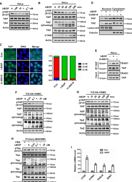
Stimulation of TP Induces YAP/TAZ Dephosphorylation and Nuclear Accumulation—To study TP regulation on YAP/TAZ activity, we treated cells with the TP agonist I-BOP because the physiological TP ligand TxA2 is extremely unstable. In serum-starved HeLa cells that express TP, YAP was highly phosphor-ylated. Addition of I-BOP resulted in a significant decrease in YAP phosphorylation, as determined by immunoblotting with a phospho-YAP antibody (Ser-127) (Fig. 1,AandB). I-BOP also induced TAZ dephosphorylation, as indicated by the differen-tial migration on phos-tag gels (Fig. 1B). Moreover, I-BOP-in-duced YAP/TAZ dephosphorylation was rapid and transient, as the YAP/TAZ dephosphorylation was visible 15 min after stim-ulation and partially recovered 4 h after I-BOP treatment (Fig. 1B). In addition, U-46619, another TP-specific agonist, could also induce YAP/TAZ dephosphorylation in a dose- and
time-dependent manner (supplemental Fig. 1,AandB).
Phosphorylation of YAP at Ser-127 by LATS1/2 leads to binding with 14-3-3 and cytoplasmic sequestration of YAP, and dephosphorylated YAP accumulates in the nucleus and induces
gene expression by interacting with the transcription factors TEAD1– 4 (29, 31). Similarly, phosphorylation of TAZ at Ser-89 by LATS1/2 also induces cytoplasmic localization (30). As expected, I-BOP treatment caused significant nuclear accu-mulation of YAP and TAZ (Fig. 1,CandD;supplemental Fig. 1
C) and enhanced the interaction between YAP and TEAD1 (Fig. 1E). A similar effect was also observed when cells were treated with U-46619 (Fig. 1,CandD;supplemental Fig. 1C). To con-firm the functional activation of YAP/TAZ upon TP stimula-tion, we examined the expression of well established YAP/TAZ target genes. The mRNA levels of CTGF and CYR61 were significantly induced by I-BOP or U-46619 treatment (supplemental Fig. 1,DandE). Accordingly, the CYR61 protein level was also increased upon I-BOP stimulation (Fig. 1B).
The effect of TP activation on YAP/TAZ was consistently observed in multiple cell lines, including MDA-MB-231,
SW480, and HEK293A (supplemental Fig. 1, F–I). Notably,
I-BOP also induces YAP/TAZ dephosphorylation in VSMCs, such as the T/G HA-VSMC cell line and primary mouse aortic
VSMCs (MAVSMC) (Fig. 1,F–H). The effect of I-BOP on YAP/
TAZ phosphorylation was dose-dependent, and YAP/TAZ dephosphorylation was evident when as little as 0.1 nmol/liter
I-BOP was added to primary MAVSMCs (Fig. 1H). Similarly,
the expression of YAP/TAZ target genes, such as CTGF, CYR61, TAGLN, and EDN1, was significantly induced by I-BOP in T/G HA-VSMCs (Fig. 1I). Based on the above data, we conclude that stimulation of TP activates YAP/TAZ by induc-ing their dephosphorylation and nuclear translocation.
I-BOP Acts through TP and G␣12/13to Activate YAP/TAZ— Because of the unstable property of TxA2, I-BOP and U-46619 were used to treat cells. To exclude that these chemicals have an unexpected effect on YAP/TAZ independent of TP, we pre-treated cells with the TP-specific antagonist SQ-29548 fol-lowed by I-BOP treatment. As shown in Fig. 2A, I-BOP-induced YAP/TAZ dephosphorylation was blocked by SQ-29548 in T/G HA-VSMCs. HEK293A cells are not very sensitive to I-BOP stimulation, likely because of the low level of TP expression.
Ectopic expression of TP␣receptor rendered YAP/TAZ more
sensitive to 1 nmol/liter I-BOP treatment, a concentration that had a minor effect on YAP/TAZ phosphorylation in the control
HEK293A cells (Fig. 2B). A similar phenomenon was observed
when TPwas overexpressed in U2OS cells (supplemental Fig.
2). These data indicate that I-BOP acts through both isoforms of TP receptor to activate YAP/TAZ.
To further confirm the role of endogenous TP in YAP/TAZ
regulation, we generatedTPKO cells using the CRISPR/Cas9
genome editing system. Two independent TP KO cell lines
were generated, and the TP deletion was verified by Sanger
sequencing (supplemental Fig. 3). TP knockout completely
blocked I-BOP-induced YAP/TAZ dephosphorylation and
YAP nuclear accumulation (Fig. 2, C and D). Consistently,
I-BOP was unable to induce the expression of YAP target genes inTPKO cells (Fig. 2E). These data show that I-BOP stimulates TP receptor to induce YAP/TAZ activation.
TP receptor activates several trimeric G␣proteins, including G␣q/11and G␣12/13, to initiate intracellular signaling pathways
(11). To determine which G␣proteins are involved in YAP/
RNAi in HEK293A cells (Fig. 2F). Knockdown of G␣12/13 strongly blocked YAP/TAZ dephosphorylation in response to
I-BOP, whereas knockdown of G␣q/11 had little effect on
I-BOP-induced YAP/TAZ dephosphorylation (Fig. 2F).
Con-sistently, I-BOP induced YAP nuclear accumulation in control siRNA- and G␣q/11siRNA-transfected cells but not in G␣12/13
FIGURE 1.Agonists of TP receptor activate YAP and TAZ.AandB, I-BOP induces YAP and TAZ dephosphorylation in a dose- and time-dependent manner in Hela cells. Cells were serum-starved for 16 h and then stimulated with I-BOP for the indicated concentration (A) or time (B). Immunoblotting was performed with the indicated antibodies. Phos-tag gels were used for assessment of TAZ phosphorylation status.C, I-BOP and U-46619 induce YAP nuclear accumulation in HeLa cells. HeLa cells were seeded with a density of 6⫻104cells/cm2for 12 h, starved in serum-free medium for 16 h, and stimulated with 1 nmol/liter I-BOP or 10 nmol/liter U-46619 for 1 h. YAP subcellular localization was determined by immunofluorescence staining for endogenous YAP (green). DAPI (blue) was used for staining cell nuclei. Representative images are shown.Scale bars⫽20m. Quantifications are shown in theright panel.Con, control.D, I-BOP and U-46619 induce YAP/TAZ nuclear accumulation in HeLa cells. Stimulation conditions were same as those inC. Subcellular fractionation was performed with NE-PERTMnuclear and cytoplasmic extrac-tion reagent (Thermo Fisher) according to the instrucextrac-tions of the manufacturer. Both fracextrac-tions were analyzed by Western blotting with indicated antibodies.E, I-BOP and U-46619 enhance YAP interaction with TEAD1. Stimulation conditions were the same as inC. Cell lysates were subjected to immunoprecipitation (IP) with YAP antibody. The coimmunoprecipitated TEAD1 was detected by immunoblotting.FandG, I-BOP induces YAP/TAZ dephosphorylation in a dose- and time-dependent manner in T/G HA-VSMCs. Cells were serum-starved for 16 h and then stimulated with I-BOP for the indicated concentration (F) or time (G). Immunoblotting was performed with the indicated antibodies. Phos-tag gels were used for assessment of YAP/TAZ phosphorylation status.H, I-BOP induces Yap/Taz dephosphorylation in a dose-dependent manner in primary MAVSMCs. Cells were serum-starved for 16 h and then stimulated with I-BOP for the concentration as indicated. FBS was included as a positive control. Immunoblotting was performed with the indicated antibodies. Phos-tag gels were used for assessment of Yap and Taz phosphorylation status.I, I-BOP induces expression of YAP target genes. T/G HA-VSMCs were treated with 1 nmol/liter I-BOP for 2 h after serum-starved for 16 h. mRNA levels of CTGF, CYR61, TAGLN, and EDN1 were measured by quantitative PCR.
siRNA-transfected cells (Fig. 2G,supplemental Fig. 4). Taken
together, we conclude that TP signals through G␣12/13 to
induce YAP/TAZ dephosphorylation and activation.
I-BOP Modulates YAP/TAZ Dephosphorylation via Rho GTPase and the Actin Cytoskeleton—Rho GTPase is a known downstream signaling module of G␣12/13, which directly inter-acts and activates the Rho guanine nucleotide exchange factor. We therefore tested the role of Rho GTPase in I-BOP-induced YAP/TAZ dephosphorylation. Expression of Rho GDP dissoci-ation inhibitor, which binds to Rho GTPase and inhibits GTPase cycling, suppressed I-BOP-induced YAP/TAZ dephos-phorylation (Fig. 3A). Likewise, botulinum toxin C3, a specific inhibitor of Rho GTPase, strongly blocked YAP/TAZ dephos-phorylation in response to I-BOP or U-46619 treatment (Fig. 3B;supplemental Fig. 5,AandB). Consistently, C3 treatment blocked I-BOP- or U-46619-induced YAP nuclear transloca-tion (Fig. 3C,supplemental Fig. 5C). These data indicate that Rho GTPase is required for TP to activate YAP/TAZ.
The major function of Rho GTPase is to modulate the actin cytoskeleton, particularly stress fiber formation. Recently stud-ies have shown that the actin cytoskeleton plays an important role in the Hippo pathway (41– 45). We therefore tested whether cytoskeletal reorganization contributes to YAP/TAZ activation by TP agonists. Latrunculin B, an F-actin-disrupting reagent, blocked I-BOP- or U-46619-induced YAP/TAZ dephosphorylation (Fig. 3D,supplemental Fig. 5D). Consistent
with changes in YAP phosphorylation, Latrunculin B treatment also prevented YAP nuclear accumulation in response to I-BOP or U-46619 (Fig. 3E,supplemental Fig. 5E). Moreover, I-BOP or U-46619 induced actin stress fiber and YAP nuclear transloca-tion (Figs. 2Gand 3,CandE;supplemental Fig. 5,CandE).
Knockdown ofG␣12/13or treatment with C3 or Latrunculin B
disrupted F-actin formation and YAP nuclear accumulation (Figs. 2Gand 3,CandE;supplemental Fig. 5,CandE). These results indicate that TP acts through Rho GTPase and the actin cytoskeleton to affect YAP/TAZ phosphorylation.
I-BOP Inhibits LATS—LATS1/2 are the kinases directly responsible for YAP/TAZ phosphorylation. The phosphoryla-tion of the activaphosphoryla-tion loop (Ser-909/Ser-872 for LATS1/2) and hydrophobic motif (Thr-1079/Thr-1041 for LATS1/2) corre-lates with LATS1/2 kinase activity (46). To test whether LATS1/2 kinases are involved in I-BOP-induced YAP/TAZ activation, we measured LATS1 kinase activityin vitro. LATS1 was potently inhibited by I-BOP treatment, as determined by its autophosphorylation at Ser-909 andin vitrophosphorylation of
the purified GST-YAP (Fig. 4A). Consistent with the results
described in Fig. 3,BandC, inhibition of Rho GTPase by C3
blocked the inhibitory effect of I-BOP on LATS1 kinase activity (Fig. 4A), indicating that Rho GTPase activation is required for I-BOP-induced LATS1 inhibition. To further determine the
role of LATS1/2 in YAP regulation by I-BOP, we usedLATS1/2
double knockout (LATS1/2 dKO) HEK293A cells. As expected,
FIGURE 3.I-BOP activates YAP/TAZ through Rho and cytoskeletons.A, I-BOP-induced YAP/TAZ dephosphorylation is blocked by Rho-GDP dissociation inhibitor expression. HEK293A cells were transiently transfected with the indicated plasmids and incubated with 10 nmol/liter I-BOP for 1 h. Immunoblotting was performed with the indicated antibodies.B, inactivation of Rho by C3 prevents YAP/TAZ dephosphorylation. Serum-starved T/G HA-VSMCs were pre-treated with C3 (4g/ml) for 3 h and then stimulated with I-BOP (1 nmol/liter) for 1 h. The cell lysates were subjected to immunoblotting analysis with the indicated antibodies.C, inactivation of Rho by C3 prevents YAP nuclear accumulation. Serum-starved HEK293A cells were pretreated with C3 (2g/ml) for 3 h and then stimulated with I-BOP (10 nmol/liter) for 1 h. After fixation, immunofluorescence staining was performed for endogenous YAP (green) and F-actin (red). DAPI (blue) was used for staining cell nuclei.Scale bars⫽20m.Con, control.D, disruption of the actin cytoskeleton blocks I-BOP-induced YAP/TAZ dephosphorylation. Serum-starved T/G HA-VSMCs were pretreated with Latrunculin B (LatB, 1g/ml) for 10 min and then stimulated with I-BOP (1 nmol/liter) for the indicated time. The cell lysates were subjected to immunoblotting analysis with the indicated antibodies.E, disruption of the actin cytoskeleton blocks I-BOP-induced YAP nuclear localization. HEK293A cells were pretreated with Latrunculin B (1g/ml) for 10 min and then stimulated with I-BOP (10 nmol/liter) for 1 h. The immunofluorescence staining is similar as inC.Scale bars⫽20m.
I-BOP could not affect YAP/TAZ phosphorylation in LATS1/ 2-dKO cells (supplemental Fig. 6), suggesting that LATS1/2 are required for I-BOP-induced YAP/TAZ dephosphorylation.
MST1/2 and MAP4Ks are responsible for LATS kinase acti-vation in response to upstream signals (26, 27, 46). To test whether MST1/2 or MAP4Ks are involved in I-BOP-induced
YAP/TAZ dephosphorylation, we usedMST1/2double
knock-out (MST1/2 dKO) and combined deletion of MST1/2 and
MAP4K1/2/3/4/5/6/7 (MM-9KO) HEK293A cells ( supple-mental Fig. 7). I-BOP-induced YAP/TAZ dephosphorylation was largely unaffected in MST1/2 dKO cells (Fig. 4B). However, I-BOP had no effect on YAP/TAZ phosphorylation in MM-9KO cells (Fig. 4C). In addition, re-expression of MST2 or MAP4K2 could restore the effect of I-BOP on YAP
phosphor-ylation in MM-9KO cells (Fig. 4D). I-BOP-induced LATS
inhi-bition was abolished in MM-9KO cells, as shown byin vitro
kinase assay (Fig. 4E). Taken together, these results suggest that MAP4Ks play a major role in I-BOP-induced YAP/TAZ
dephosphorylation, although both MST1/2 and MAP4Ks are involved.
YAP/TAZ Are Required for TP to Stimulate VSMC DNA Syn-thesis and Cell Migration—TP has been shown to promote neointima formation, which is caused by VSMC migration and proliferation (6, 9, 47). Because YAP/TAZ activity is signifi-cantly activated upon TP stimulation in VSMCs, we investi-gated whether YAP/TAZ activation is involved in the
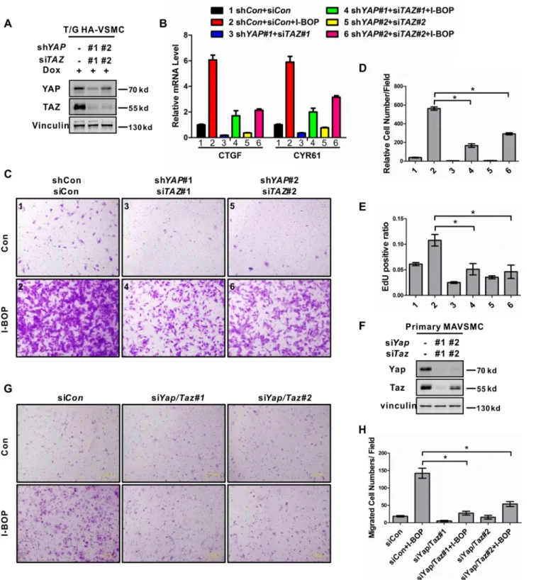
prolifer-ation and migrprolifer-ation of VSMCs.YAP/TAZwere knocked down
in T/G HA-VSMCs by inducible shRNA and siRNA, respec-tively. The knockdown efficiency was confirmed by
immuno-blotting of protein levels (Fig. 5A). Knockdown of YAP/TAZ
significantly suppressed the mRNA induction of CTGF and CYR61 in response to I-BOP (Fig. 5B). I-BOP strongly induced cell migration in control cells, whereas this effect was
signifi-cantly suppressed inYAP/TAZdouble knockdown cells (Fig. 5,
CandD). In addition, I-BOP-induced VSMC DNA synthesis
was also suppressed byYAP/TAZdouble knockdown, as
FIGURE 5.YAP/TAZ mediate the effect of TP in gene induction, DNA synthesis, and cell migration in VSMCs.A, knockdown of YAP/TAZ by shRNAs and siRNAs in T/G HA-VSMCs. Cells were transfected with the indicated siRNAs. 48 h after transfection and doxycycline (Dox) induction, cells were lysed and subjected to immunoblotting with the indicated antibodies.B, YAP/TAZ are required for I-BOP-induced gene expression. T/G HA-VSMCs were transfected with the indicated siRNAs and serum-starved for 24h in the presence of doxycycline. After treatment with I-BOP (1 nmol/liter) for 1 h, mRNA levels of CTGF and CYR61 were measured by real-time PCR. The numbers next to each treatment conditions will be used to labelC–E.C, YAP/TAZ are required for I-BOP-induced cell migration. The treatment conditions for each panel are the same as inB. After stimulation with I-BOP (1 nmol/liter) for 4 h, cell migration was performed by transwell cell migration assay. Representative images are shown.Con, control.D, quantification result of the data inC. *,p⬍0.05. Statistical analysis is described under “Experimental Procedures.”E, YAP/TAZ are required for I-BOP-induced DNA synthesis. T/G HA-VSMCs were treated as indicated inBand then subjected to an EdU incorporation assay as described under “Experimental Procedures.” About 600 –1000 randomly selected cells are quantified and shown. *,p⬍0.05.
F, Yap/Taz knockdown by siRNAs in primary MAVSMCs. Cells were transfected with the indicated siRNAs. After 48-h transfection, cells were lysed and subjected to immunoblotting with the indicated antibodies.G, Yap/Taz are required for primary MAVSMCs migration induced by I-BOP. Primary MAVSMCs were transfected with the indicated siRNAs and serum-starved for 20 h. After stimulation with I-BOP (1 nmol/liter) for 4 h, cell migration was determined by transwell cell migration assay. Representative images are shown.H, quantification result of the data inG. *,p⬍0.05.
mined by EdU incorporation (Fig. 5E,supplemental Fig. 8A). To further support the important role of the TP-YAP/TAZ axis in VSMCs, we isolated and analyzed primary MAVSMCs. Similar
Yap/Tazknockdown experiments were performed in primary
MAVSMCs. Consistently, knockdown ofYap/Tazin primary
MAVSMCs also inhibited cell migration induced by I-BOP
(Fig. 5,F–H). In addition, we observed that YAP/TAZ
knock-down suppressed I-BOP-induced gene expression and cell
migration in HeLa cells (supplemental Fig. 8, B–D). Taken
together, these results demonstrate that YAP/TAZ play an important role in TP-mediated gene induction, cell prolifera-tion, and migration in VSMCs.
Discussion
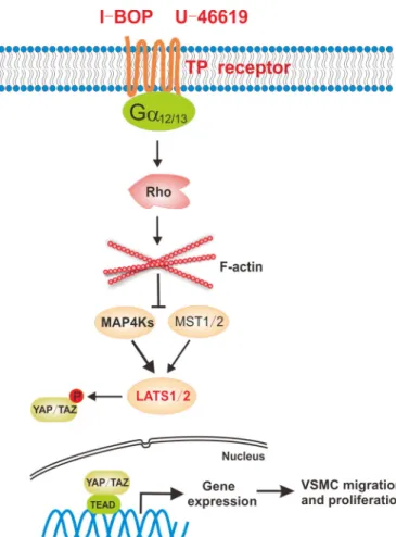
TxA2 is involved in multiple physiological and pathophysio-logical processes, including thrombosis, asthma, myocardial infarction, inflammation, atherosclerosis, and the response to vascular injury (11). TxA2 exerts its biological activity via its cognate TP receptor. In this study, we demonstrate that the Hippo pathway is a crucial downstream signaling module of TP receptor, a classical GPCR. TP agonists significantly activate YAP/TAZ in multiple cells lines, including VSMCs. Our data
also demonstrate that activation of TP couples to G␣12/13to
trigger the activation of Rho GTPase, which modulates the actin cytoskeleton to inhibit LATS1/2 kinase activity, resulting in YAP/TAZ dephosphorylation and activation (Fig. 6). In this signaling cascade, both MST1/2 and MAP4Ks, the major
kinases for LATS1/2, are involved in Hippo pathway regulation by TP. Our studies indicate a functional role of the Hippo path-way and YAP/TAZ in mediating the physiological and patho-logical functions of thromboxane and its receptor TP.
In addition to TxA2, there are four other major prostaglan-dins generated from arachidonic acidin vivo, including prosta-glandin D2, prostaglandin E2, prostaglandin F2␣, and
prostacy-clin (48). They all exert their effects via corresponding GPCRs. It is interesting to speculate that YAP/TAZ may play a similar role in physiological and disease processes that are regulated by prostaglandins, such as inflammation, atherosclerosis, and can-cer (49). For instance, the prostacyclin receptor couples to G␣s and stimulates cAMP production and PKA activation (50). Pre-viously we reported that cAMP acts through PKA to stimulate LATS kinases and inhibit YAP (51). Given that TxA2 and pros-tacyclin are antagonistic in their actions in cardiovascular dis-ease (6), it will be interesting to determine whether prostacyclin inhibits YAP/TAZ, thereby antagonizing the effect of TxA2, which activates YAP/TAZ, as shown in this report, in cardio-vascular disease.
Isoprostanes are prostaglandin-like compounds formed from the free radical-catalyzed peroxidation of unsaturated fatty acids, such as arachidonic acid, that are recognized not only as reliable markers of oxidative stress but also as important medi-ators of various diseases (52). Increased formation of isopros-tanes has been observed in diseases that are linked to oxidative stress, such as cardiovascular disease and cancer (52). As men-tioned above, isoprostanes act, at least partially, via TP to exert their physiological effects. So one may speculate that YAP/TAZ also play a role in isoprostane-mediated pathophysiological effects. Indeed, we observed that 8-iso-prostaglandin F2␣, a type
of isoprostanes, could induce YAP/TAZ dephosphorylation in
VSMCs.5Future studies are needed to delineate the
involve-ment and potential role of YAP/TAZ in isoprostane-induced physiological processes.
Our study shows that YAP/TAZ are required for TP-stimu-lated VSMC DNA synthesis and cell migration. The connection between TP receptor and the Hippo pathway in vascular smooth muscle cells has important physiological implications. During vascular injury, activated platelets or other cells pro-duce TxA2, which in turn promotes platelet activation and aggregation (4, 53). Besides TxA2, isoprostanes may also act through the TP receptor on the surface of smooth muscle cells and initiate the migration from media to intima via YAP/TAZ activation. Previous studies have reported that YAP is activated in VSMCs of the neointima (39). We propose that thrombox-ane acts through TP to induce YAP/TAZ activation to promote normal physiological wound healing in response to vascular injury. However, too much YAP/TAZ activation by TP under pathological conditions may lead to VSMC overgrowth, thereby contributing to neointima formation and restenosis. Notably, CTGF and CYR61 are strongly induced upon TP stim-ulation in a manner dependent on YAP/TAZ (Figs. 2Eand 5B). Both CTGF and CYR61 have been shown to promote athero-sclerotic lesion development and neointimal hyperplasia (54 –
5X. Feng, P. Liu, X. Zhou, M. T. Li, F. L. Li, Z. Wang, Z. Meng, Y. P. Sun, Y. Yu, Y. Xiong, H. X. Yuan, and K. L. Guan, unpublished data.
56); therefore, uncontrolled expression of these proteins may contribute to pathology associated with TP hyperactivation. No TP antagonist has been used in clinics because of efficacy and toxicity issues (2). Our work reveals new insights into the role of the Hippo pathway and YAP/TAZ in thromboxane pathophys-iology and suggests YAP/TAZ as potential therapeutic targets for vascular diseases. Actually, a small molecule, verteporfin (trade name Visudyne), a clinical drug for neovascular macular degeneration (57), has been shown to efficiently inhibit TEAD-YAP interaction and suppress YAP-induced liver over-growth (58). The data in this report suggest that YAP/TAZ inhibitors could be a potential treatment for vascular diseases caused by VSMC overgrowth.
Experimental Procedures
Plasmids—The plasmids pCMV-HA-YAP2, FLAG-LATS1, FLAG-MST2, and FLAG-MAP4K2 and GFP-GDP dissociation inhibitor were described before (26, 51, 59). TP␣/were ampli-fied from cDNA and then subcloned into pRK7-N-FLAG and pQCXIH vectors with the restriction enzymes BamHI/EcoRI.
Antibodies—Antibodies for YAP (4912), phospho-YAP (Ser-127, 4911), MST1 (3682), MST2 (3952), CYR61 (11952), phos-pho-MST1 (Thr-183)/MST2 (Thr-180, 3681), phospho-LATS1 (Ser-909, 9157), and phospho-phospho-LATS1 (Thr-1079, 8654) were purchased from Cell Signaling Technology. The LATS1 (A300-477A) and YAP (A302-308A) antibodies used for immu-noprecipitation and LATS2 (A300-479A) antibody were from Bethyl Laboratories. TAZ (HPA007415) and Vinculin (V9264) antibodies were obtained from Sigma-Aldrich. Anti-TEAD1 (610922) was purchased from BD Transduction Laboratories.
Antibodies for G␣q/11 (Cys-19, sc-392) and G␣12 (Ser-20,
sc-409) and the HA-probe antibody (sc-7392) were from Santa Cruz Biotechnology. The tubulin antibody (581P) was
pur-chased from NeoMarkers, and anti--actin (A00702), Lamin
A/C (A01455), and anti-FLAG antibodies were purchased from GeneScript. The GFP tag (7G9, M20004) was from Abmart.
The anti-G protein ␣13 antibody (EPR5436, ab128900) was
obtained from Abcam.
Chemicals—U-46619 (9,11-dideoxy-9␣,11␣ -methanoepoxy-prosta-5Z,13E-dien-1-oic acid) and SQ-29548 ([1S-[1␣,2␣(Z),3␣,4␣ ]]-7-[3-[[2-[(phenylamino)carbonyl]hydrazino]-methyl]-7-oxabicyclo[2.2.1]hept-2-yl]-5-heptenoic acid) were purchased from Santa Cruz Biotechnology. I-BOP ([1S-[1␣,2␣(Z),3(1E,3S*),4␣ ]]-7-[3-[3-hydroxy-4-(4-iodophenoxy)-1-butenyl]-7-oxabicyclo[2.2.1]hept-2-yl]-5-heptenoic acid) was purchased from Cayman Chemical. Latrunculin B and tetra-methylrhodamine B isothiocyanate-conjugated Phalloidin were purchased from Sigma-Aldrich. C3 was from Cytoskele-ton Inc. Phos-tag was purchased from Wako chemicals. DAPI was purchased from Invitrogen.
Generation of Knockout Cells Using CRISPR/Cas9 Genome Editing—To generate TP knockout HEK293A cells, the
follow-ing guide sequence targetfollow-ing the human TBXA2R gene was
used: 5⬘-GCTGGTGACCGGTACCATCG-3⬘. The detail
pro-tocol is described elsewhere (26). Two independent clones with
TBXA2Rgene deletion were used for experiments. MST1/2 dKO and LATS1/2 dKO HEK293A cells were described previ-ously (26). MM-9KO HEK293A cells were generated based on
MM-8KO cells (26). The following guide sequence targeting
the human MAP4K5 gene was used: 5⬘
-CACCTAC-GGGGACGTCTATA-3⬘. Gene deletion was verified by Sanger
sequencing of genomic DNA (supplemental Figs. S3 and S7).
Cell Culture—HEK239A and HeLa cells were cultured in DMEM (Invitrogen). U2OS cells were cultured in RPMI 1640 medium (Invitrogen). All of the above cells were supplemented
with 10% FBS (Gibco) and 50g/ml penicillin/streptomycin.
T/G HA-VSMCs were cultured in DMEM/F12 medium (Invit-rogen) and supplemented with 0.05 mg/ml ascorbic acid, 0.01 mg/ml insulin, 0.01 mg/ml transferrin, 10 ng/ml sodium sele-nite, 0.03 mg/ml endothelial cell growth supplement, and 50
g/ml penicillin/streptomycin. Mouse primary aortic VSMCs
were isolated from 4- to 8-week-old C57BL/6 male mice using a standard protocol (60) and incubated in DMEM supplemented
with 20% FBS and 50g/ml penicillin/streptomycin. Cells at
passages 3–5 were used. For serum starvation, cells were incu-bated in DMEM or DMEM/F12 without serum. All cell lines were maintained at 37 °C with 5% CO2.
Transfection and Lentiviral and Retroviral Infection—Cells were transfected with plasmid DNA using PEI. To generate
U2OS cells stably expressing TP␣/, retroviruses carrying
pQCXIH empty vector or pQCXIH-TP␣/were produced in
HEK293T cells using vesicular stomatitis virus G and GAG as packaging plasmids. The virus supernatant was filtered through a 0.45-m filter and used to infect targeting cells in the pres-ence of 8g/ml Polybrene. Stable cell pools were selected with 50g/ml hygromycin B (Amresco) for 5 days. For tetracycline-inducible shRNA expression, a lentivirus containing shRNAs in the pTRIPZ vector was made in HEK293T cells using pMD2.g and psPAX2 as packaging plasmids. Virus infection was
per-formed as described above except for selection with 1g/ml
puromycin (Amresco) for 5 days. Expression of shRNA was
induced by adding 1g/ml doxycycline for 48 h. The shRNA
sequences against YAP were as follows: YAP#1, TTCTTTATCT-AGCTTGGTGGC; YAP#2, TGGTCAGAGATACTTCTTAAA.
RNA Interference—siRNAs targetingGNAQ,GNA11,GNA12,
GNA13, YAP1/Yap1, and WWTR1/Wwtr1 were from Gene-pharma and were delivered into cells using RNAiMAX (Invitro-gen) according to the instructions of the manufacturer. The sequences of all siRNAs used in this study were as follows: siGNAQ
#1, GACACCGAGAATATCCGCTTT; siGNAQ #2,
CTAT-GATAGACGACGAGAATA; siGNA11 #1,
GCTCAAGATC-CTCTACAAGTA; siGNA11#2,
GCTCAACCTCAAGGAGTA-CAA; siGNA12#1, GCGACACCATCTTCGACAA; siGNA12#2,
GGATGTTCCTGATGGCCTT; siGNA13#1,
GCTCGAGAG-AAGCTTCATA; siGNA13#2, CCTGCTATAAGAGCATTAT;
siYAP1#1, CCCAGTTAAATGTTCACCAAT; siYAP1#2,
CAG-GTGATACTATCAACCAAA; siTAZ#1,
CAGCCAAAT-CTCGTGATGAAT; siTAZ#2,
GCGATGAATCAGCCTCT-GAAT; siYap1#1, GAAGCGCTGAGTTCCGAAATC; siYap1#2,
TGAGAACAATGACAACCAATA; siTaz#1,
CAGCCGAAT-CTCGCAATGAAT; and siTaz#2,
CCATGAGCACAGATAT-GAGAT.
ers (Transgene). cDNA was then diluted and subjected to real-time PCR with gene-specific primers using SYBR Premix Ex Taq (TaKaRa) and the 7500 real-time PCR system (Applied Biosystems). The primer pairs used in this study were as follows: Actin, GCCGACAGGATGCAGAAGGAGATCA/AA-GCATTTGCGGTGGACGATGGA; CTGF, CCAATGACAAC-GCCTCCTG/TGGTGCAGCCAGAAAGCTC; CYR61, AGCC-TCGCATCCTATACAACC/TTCTTTCACAAGGCGGCACTC; EDN1, TGTGTCTACTTCTGCCACCT/CCCTGAGTTCTTT-TCCTGCTT; ANKRD1, CACTTCTAGCCCACCCTGTGA/C-CACAGGTTCCGTAATGATTT; TAGLN,
CCCGAGAACCC-ACCCTCCA/AAAGCCATCAGGGTCCTCTGC; TP␣,
CCT-TCTGGTCTTCATCGCCC/CTGGAGGGACAGCGACCT;
and TP,
ACCCGGCCCAGACGGAGT/GGACAGAGCC-TTCCCTGTTGG.
Immunoprecipitation—Cells were lysed using lysis buffer (50
mMTris (pH 7.5), 150 mMNaCl, 0.5% Nonidet P-40, 50 mM
NaF, 1.5 mMNa3VO4, 1 mMPMSF, and protease inhibitors). Cell lysates were centrifuged at 12,000⫻gfor 15 min at 4 °C. The supernatants were incubated with YAP antibody (Bethyl Laboratories) for 2 h, followed by protein A-agarose bead incu-bation for 1 h. Immunoprecipitates were washed three times with lysis buffer, and proteins were boiled with SDS-PAGE sample buffer.
Immunoblotting—Cells were lysed in SDS sample buffer and denatured by heating on 99 °C for 10 min. Immunoblotting was performed in 8% or 10% BisTris polyacrylamide gel according to the standard protocol. The phos-tag reagents were pur-chased from Wako Chemicals, and gels containing phos-tag were prepared following the instructions of the manufacturer. YAP and TAZ proteins can be separated into multiple bands in phos-tag gels depending on differential phosphorylation levels, with phosphorylated YAP/TAZ migrating more slowly.
Immunofluorescence Staining—Cells seeded in 6-well plates were treated as indicated in specific experiments. After treat-ment, they were fixed immediately with 4% paraformaldehyde for 20 min and then permeabilized with 0.5% Triton X-100 in PBS for 5 min on ice. After blocking in 5% BSA for 30 min, cells were incubated with primary antibodies overnight at 4 °C. Cells were washed with PBS three times for 5 min, and Alexa Fluor 488-conjugated secondary antibodies were added for 1 h at room temperature. Tetramethylrhodamine B isothiocyanate-conjugated Phalloidin was used to stain actin filaments, and DAPI was used for cell nuclei. Photos were taken by an Olym-pus IX81 inverted research microscope with appropriate fluo-rescence filters.
Cell Fractionation—HeLa cells were seeded with a density of 6⫻104cells/cm2for 12 h, starved in serum-free medium for 16 h, and stimulated with 1 nmol/liter I-BOP or 10 nmol/liter U-46619 for 1 h. Subcellular fractionation was performed
with NE-PERTMnuclear and cytoplasmic extraction reagent
(Thermo Fisher) according to the instructions of the manufac-turer. Both fractions were analyzed by Western blotting.
In Vitro Kinase Assay—LATS1 kinase assay was performed as described before (38). Briefly, LATS1 was immunoprecipitated from cells and subjected to anin vitrokinase assay using GST-YAP as a substrate. The phosphorylation levels of GST-GST-YAP at
Ser-127 were determined by immunoblotting using phospho-YAP (Ser-127) antibody.
Transwell Migration Assay—Cell migration assays were per-formed using BD Falcon Cell culture inserts for 24-well plates with 8.0-m pore filters. Cells were serum-starved for 20 h and then stimulated with I-BOP or vehicle for 4 h. Cells (6⫻104
T/G HA-VSMCs, 5⫻104primary aortic VSMCs, and 2⫻105
HeLa cells) were seeded into the upper chamber of the insert for each well in DMEM/F-12 without other supplements, and the lower chamber of the plate was filled with DMEM/F-12 supple-mented with 10% FBS. After 6 h for T/G HA-VSMC (3 h for primary MAVSMC and 24 h for HeLa), cells were fixed using 4% paraformaldehyde and stained with 0.05% crystal violet. Cells in the upper chamber were removed by scrubbing with a cotton swab, and cells migrated through the filter were photo-graphed and quantified.
EdU Incorporation Assay—T/G HA-VSMCs grown on 24-well plates were serum-starved for 24 h. I-BOP (1 nmol/
liter) was added every 8 h for 3 times. 10mol/liter EdU
(5-ethynyl-2⬘-deoxyuridine) was added to the culture
medium for 4 h. After labeling, cells were fixed with 4% para-formaldehyde for 20 min, followed by permeabilization with 0.5% Triton X-100 in PBS for 20 min at room temperature. Cells were rinsed once with PBS and incubated with mixture
reaction buffer (100 mmol/liter sodium ascorbate, 4.8mol/
liter Alexa Fluor 488 azide, and 4 mmol/liter CuSO4) for 30
min at room temperature. After staining, cells were washed three times with 0.5% Triton X-100 in PBS, and DAPI was used for cell nuclei. Photos were taken by an Olympus IX81 inverted research microscope.
Statistical Analysis—All data are expressed as mean⫾S.E.
Two-group comparison was analyzed by Student’sttest.p⬍
0.05 was considered significant.
Author Contributions—X. F., Y. X., H. X. Y., and K. L. G designed the research, analyzed the data, and wrote the manuscript. X. F. per-formed the experiments with assistance from P. L., X. Z., M. T. L, F. L. L, Z. W., Z. P. M., and Y. P. S. Y. Y. provided technical and intel-lectual support. All authors discussed the results and commented on the manuscript.
Acknowledgments—We thank Dan Ye and the members of the Fudan MCB laboratory for support throughout the course of this study.
References
1. Hamberg, M., Svensson, J., and Samuelsson, B. (1975) Thromboxanes: a new group of biologically active compounds derived from prostaglandin endoperoxides.Proc. Natl. Acad. Sci. U.S.A.72,2994 –2998
2. Ting, H. J., Murad, J. P., Espinosa, E. V., and Khasawneh, F. T. (2012) Thromboxane A2 receptor: biology and function of a peculiar receptor that remains resistant for therapeutic targeting.J. Cardiovasc. Pharmacol. Ther.17,248 –259
3. Braden, G. A., Knapp, H. R., and FitzGerald, G. A. (1991) Suppression of eicosanoid biosynthesis during coronary angioplasty by fish oil and aspi-rin.Circulation84,679 – 685
4. Byrne, A., Moran, N., Maher, M., Walsh, N., Crean, P., and Fitzgerald, D. J. (1997) Continued thromboxane A2 formation despite administration of a platelet glycoprotein IIb/IIIa antagonist in patients undergoing coronary angioplasty.Arterioscler. Thromb. Vasc. Biol.17,3224 –3229
Cyclooxygenase-1 and -2-dependent prostacyclin formation in patients with atherosclerosis.Circulation102,840 – 845
6. Cheng, Y., Austin, S. C., Rocca, B., Koller, B. H., Coffman, T. M., Grosser, T., Lawson, J. A., and FitzGerald, G. A. (2002) Role of prostacyclin in the cardiovascular response to thromboxane A2.Science296,539 –541 7. Belton, O. A., Duffy, A., Toomey, S., and Fitzgerald, D. J. (2003)
Cyclooxy-genase isoforms and platelet vessel wall interactions in the apolipoprotein E knockout mouse model of atherosclerosis.Circulation108,3017–3023 8. Praticò, D., Cyrus, T., Li, H., and FitzGerald, G. A. (2000) Endogenous biosynthesis of thromboxane and prostacyclin in 2 distinct murine models of atherosclerosis.Blood96,3823–3826
9. Yamagami, S., Miyauchi, K., Kimura, T., Goh, Y., Daida, H., and Yamagu-chi, H. (1999) Effects of the thromboxane A2 receptor antagonist on plate-let deposition and intimal hyperplasia after balloon injury.Jpn. Heart J.40,
791– 802
10. Inoue, T., and Node, K. (2009) Molecular basis of restenosis and novel issues of drug-eluting stents.Circ. J.73,615– 621
11. Nakahata, N. (2008) Thromboxane A2: physiology/pathophysiology, cellular signal transduction and pharmacology.Pharmacol. Ther.118,
18 –35
12. Montuschi, P., Barnes, P. J., and Roberts, L. J., 2nd. (2004) Isoprostanes: markers and mediators of oxidative stress.FASEB J.18,1791–1800 13. Gluais, P., Lonchampt, M., Morrow, J. D., Vanhoutte, P. M., and Feletou,
M. (2005) Acetylcholine-induced endothelium-dependent contractions in the SHR aorta: the Janus face of prostacyclin.Br. J. Pharmacol.146,
834 – 845
14. Audoly, L. P., Rocca, B., Fabre, J. E., Koller, B. H., Thomas, D., Loeb, A. L., Coffman, T. M., and FitzGerald, G. A. (2000) Cardiovascular responses to the isoprostanes iPF(2␣)-III and iPE(2)-III are mediated via the thrombox-ane A(2) receptorin vivo.Circulation101,2833–2840
15. Ishimitsu, T., Uehara, Y., Ishii, M., Ikeda, T., Matsuoka, H., and Sugimoto, T. (1988) Thromboxane and vascular smooth muscle cell growth in ge-netically hypertensive rats.Hypertension12,46 –51
16. Ishimitsu, T., Uehara, Y., Ishii, M., and Sugimoto, T. (1988) Enhanced generation of vascular thromboxane A2 in spontaneously hypertensive rats and its role in the rapid proliferation of vascular smooth muscle cells.
Am. J. Hypertens.1,38S– 40S
17. Hanasaki, K., Nakano, T., and Arita, H. (1990) Receptor-mediated mito-genic effect of thromboxane A2 in vascular smooth muscle cells.Biochem. Pharmacol.40,2535–2542
18. Pakala, R., Willerson, J. T., and Benedict, C. R. (1997) Effect of serotonin, thromboxane A2, and specific receptor antagonists on vascular smooth muscle cell proliferation.Circulation96,2280 –2286
19. Pakala, R. (2004) Serotonin and thromboxane A2 stimulate platelet-de-rived microparticle-induced smooth muscle cell proliferation. Cardio-vasc. Radiat. Med.5,20 –26
20. Yokota, T., Shiraishi, R., Aida, T., Iwai, K., Liu, N. M., Yokoyama, U., and Minamisawa, S. (2014) Thromboxane A(2) receptor stimulation pro-motes closure of the rat ductus arteriosus through enhancing neointima formation.PLoS ONE9,e94895
21. Owens, G. K., Kumar, M. S., and Wamhoff, B. R. (2004) Molecular regu-lation of vascular smooth muscle cell differentiation in development and disease.Physiol. Rev.84,767– 801
22. Cayatte, A. J., Du, Y., Oliver-Krasinski, J., Lavielle, G., Verbeuren, T. J., and Cohen, R. A. (2000) The thromboxane receptor antagonist S18886 but not aspirin inhibits atherogenesis in apo E-deficient mice: evidence that eico-sanoids other than thromboxane contribute to atherosclerosis. Arterio-scler. Thromb. Vasc. Biol.20,1724 –1728
23. Kobayashi, T., Tahara, Y., Matsumoto, M., Iguchi, M., Sano, H., Mu-rayama, T., Arai, H., Oida, H., Yurugi-Kobayashi, T., Yamashita, J. K., Katagiri, H., Majima, M., Yokode, M., Kita, T., and Narumiya, S. (2004) Roles of thromboxane A(2) and prostacyclin in the development of ather-osclerosis in apoE-deficient mice.J. Clin. Invest.114,784 –794
24. Egan, K. M., Wang, M., Fries, S., Lucitt, M. B., Zukas, A. M., Puré, E., Lawson, J. A., and FitzGerald, G. A. (2005) Cyclooxygenases, thrombox-ane, and atherosclerosis: plaque destabilization by cyclooxygenase-2 inhi-bition combined with thromboxane receptor antagonism. Circulation
111,334 –342
25. Yu, F. X., Zhao, B., and Guan, K. L. (2015) Hippo pathway in organ size control, tissue homeostasis, and cancer.Cell163,811– 828
26. Meng, Z., Moroishi, T., Mottier-Pavie, V., Plouffe, S. W., Hansen, C. G., Hong, A. W., Park, H. W., Mo, J.-S., Lu, W., Lu, S., Flores, F., Yu, F.-X., Halder, G., and Guan, K.-L. (2015) MAP4K family kinases act in parallel to MST1/2 to activate LATS1/2 in the Hippo pathway.Nat. Commun.6,
8357
27. Zheng, Y., Wang, W., Liu, B., Deng, H., Uster, E., and Pan, D. (2015) Identification of Happyhour/MAP4K as alternative Hpo/Mst-like kinases in the Hippo kinase cascade.Dev. Cell34,642– 655
28. Dong, J., Feldmann, G., Huang, J., Wu, S., Zhang, N., Comerford, S. A., Gayyed, M. F., Anders, R. A., Maitra, A., and Pan, D. (2007) Elucidation of a universal size-control mechanism inDrosophilaand mammals.Cell
130,1120 –1133
29. Zhao, B., Wei, X., Li, W., Udan, R. S., Yang, Q., Kim, J., Xie, J., Ikenoue, T., Yu, J., Li, L., Zheng, P., Ye, K., Chinnaiyan, A., Halder, G., Lai, Z. C., and Guan, K. L. (2007) Inactivation of YAP oncoprotein by the Hippo pathway is involved in cell contact inhibition and tissue growth control.Genes Dev.
21,2747–2761
30. Lei, Q. Y., Zhang, H., Zhao, B., Zha, Z. Y., Bai, F., Pei, X. H., Zhao, S., Xiong, Y., and Guan, K. L. (2008) TAZ promotes cell proliferation and epithelial-mesenchymal transition and is inhibited by the Hippo pathway.Mol. Cell. Biol.28,2426 –2436
31. Zhao, B., Ye, X., Yu, J., Li, L., Li, W., Li, S., Yu, J., Lin, J. D., Wang, C. Y., Chinnaiyan, A. M., Lai, Z. C., and Guan, K. L. (2008) TEAD mediates YAP-dependent gene induction and growth control. Genes Dev. 22,
1962–1971
32. Zhao, B., Li, L., Tumaneng, K., Wang, C. Y., and Guan, K. L. (2010) A coordinated phosphorylation by Lats and CK1 regulates YAP stability through SCF-TRCP.Genes Dev.24,72– 85
33. Harvey, K. F., Zhang, X., and Thomas, D. M. (2013) The Hippo pathway and human cancer.Nat. Rev. Cancer13,246 –257
34. Miller, E., Yang, J., DeRan, M., Wu, C., Su, A. I., Bonamy, G. M., Liu, J., Peters, E. C., and Wu, X. (2012) Identification of serum-derived sphingo-sine-1-phosphate as a small molecule regulator of YAP.Chem. Biol.19,
955–962
35. Mo, J. S., Yu, F. X., Gong, R., Brown, J. H., and Guan, K. L. (2012) Regula-tion of the Hippo-YAP pathway by protease-activated receptors (PARs).
Genes Dev.26,2138 –2143
36. Yu, F. X., Zhao, B., Panupinthu, N., Jewell, J. L., Lian, I., Wang, L. H., Zhao, J., Yuan, H., Tumaneng, K., Li, H., Fu, X. D., Mills, G. B., and Guan, K. L. (2012) Regulation of the Hippo-YAP pathway by G-protein-coupled re-ceptor signaling.Cell150,780 –791
37. Wennmann, D. O., Vollenbröker, B., Eckart, A. K., Bonse, J., Erdmann, F., Wolters, D. A., Schenk, L. K., Schulze, U., Kremerskothen, J., Weide, T., and Pavenstädt, H. (2014) The Hippo pathway is controlled by angiotensin II signaling and its reactivation induces apoptosis in podocytes.Cell Death Dis.5,e1519
38. Zhou, X., Wang, S., Wang, Z., Feng, X., Liu, P., Lv, X. B., Li, F., Yu, F. X., Sun, Y., Yuan, H., Zhu, H., Xiong, Y., Lei, Q. Y., and Guan, K. L. (2015) Estrogen regulates Hippo signaling via GPER in breast cancer.J. Clin. Invest.125,2123–2135
39. Wang, X., Hu, G., Gao, X., Wang, Y., Zhang, W., Harmon, E. Y., Zhi, X., Xu, Z., Lennartz, M. R., Barroso, M., Trebak, M., Chen, C., and Zhou, J. (2012) The induction of yes-associated protein expression after arterial injury is crucial for smooth muscle phenotypic modulation and neointima formation.Arterioscler. Thromb. Vasc. Biol.32,2662–2669
40. Xie, C., Guo, Y., Zhu, T., Zhang, J., Ma, P. X., and Chen, Y. E. (2012) Yap1 protein regulates vascular smooth muscle cell phenotypic switch by inter-action with myocardin.J. Biol. Chem.287,14598 –14605
41. Dupont, S., Morsut, L., Aragona, M., Enzo, E., Giulitti, S., Cordenonsi, M., Zanconato, F., Le Digabel, J., Forcato, M., Bicciato, S., Elvassore, N., and Piccolo, S. (2011) Role of YAP/TAZ in mechanotransduction.Nature474,
179 –183
42. Fernández, B. G., Gaspar, P., Brás-Pereira, C., Jezowska, B., Rebelo, S. R., and Janody, F. (2011) Actin-capping protein and the Hippo pathway reg-ulate F-actin and tissue growth in Drosophila. Development 138,
2337–2346
43. Rauskolb, C., Pan, G., Reddy, B. V., Oh, H., and Irvine, K. D. (2011) Zyxin links fat signaling to the hippo pathway.PLoS Biol.9,e1000624 44. Sansores-Garcia, L., Bossuyt, W., Wada, K., Yonemura, S., Tao, C., Sasaki,
H., and Halder, G. (2011) Modulating F-actin organization induces organ growth by affecting the Hippo pathway.EMBO J.30,2325–2335 45. Zhao, B., Li, L., Wang, L., Wang, C. Y., Yu, J., and Guan, K. L. (2012) Cell
detachment activates the Hippo pathway via cytoskeleton reorganization to induce anoikis.Genes Dev.26,54 – 68
46. Chan, E. H., Nousiainen, M., Chalamalasetty, R. B., Schäfer, A., Nigg, E. A., and Silljé, H. H. (2005) The Ste20-like kinase Mst2 activates the human large tumor suppressor kinase Lats1.Oncogene24,2076 –2086 47. Marx, S. O., Totary-Jain, H., and Marks, A. R. (2011) Vascular smooth
muscle cell proliferation in restenosis.Circ. Cardiovasc. Interv.4,104 –111 48. Ricciotti, E., and FitzGerald, G. A. (2011) Prostaglandins and
inflamma-tion.Arterioscler. Thromb. Vasc. Biol.31,986 –1000
49. Smyth, E. M., Grosser, T., Wang, M., Yu, Y., and FitzGerald, G. A. (2009) Prostanoids in health and disease.J. Lipid Res.50,S423–S428
50. Fetalvero, K. M., Shyu, M., Nomikos, A. P., Chiu, Y. F., Wagner, R. J., Powell, R. J., Hwa, J., and Martin, K. A. (2006) The prostacyclin receptor induces human vascular smooth muscle cell differentiation via the protein kinase A pathway.Am. J. Physiol. Heart Circ. Physiol.290,H1337–H1346 51. Yu, F. X., Zhang, Y., Park, H. W., Jewell, J. L., Chen, Q., Deng, Y., Pan, D., Taylor, S. S., Lai, Z. C., and Guan, K. L. (2013) Protein kinase A activates the Hippo pathway to modulate cell proliferation and differentiation.
Genes Dev.27,1223–1232
52. Bauer, J., Ripperger, A., Frantz, S., Ergün, S., Schwedhelm, E., and Benndorf, R. A. (2014) Pathophysiology of isoprostanes in the cardiovas-cular system: implications of isoprostane-mediated thromboxane A2 re-ceptor activation.Br. J. Pharmacol.171,3115–3131
53. Peterson, M. B., Machaj, V., Block, P. C., Palacios, I., Philbin, D., and
Watkins, W. D. (1986) Thromboxane release during percutaneous transluminal coronary angioplasty.Am. Heart J.111,1– 6
54. Cicha, I., Yilmaz, A., Klein, M., Raithel, D., Brigstock, D. R., Daniel, W. G., Goppelt-Struebe, M., and Garlichs, C. D. (2005) Connective tissue growth factor is overexpressed in complicated atherosclerotic plaques and in-duces mononuclear cell chemotaxisin vitro.Arterioscler. Thromb. Vasc. Biol.25,1008 –1013
55. Matsumae, H., Yoshida, Y., Ono, K., Togi, K., Inoue, K., Furukawa, Y., Nakashima, Y., Kojima, Y., Nobuyoshi, M., Kita, T., and Tanaka, M. (2008) CCN1 knockdown suppresses neointimal hyperplasia in a rat artery bal-loon injury model.Arterioscler. Thromb. Vasc. Biol.28,1077–1083 56. Lee, H. Y., Chung, J. W., Youn, S. W., Kim, J. Y., Park, K. W., Koo, B. K., Oh,
B. H., Park, Y. B., Chaqour, B., Walsh, K., and Kim, H. S. (2007) Forkhead transcription factor FOXO3a is a negative regulator of angiogenic imme-diate early gene CYR61, leading to inhibition of vascular smooth muscle cell proliferation and neointimal hyperplasia.Circ. Res.100,372–380 57. Michels, S., and Schmidt-Erfurth, U. (2001) Photodynamic therapy with
verteporfin: a new treatment in ophthalmology.Semin. Ophthalmol.16,
201–206
58. Liu-Chittenden, Y., Huang, B., Shim, J. S., Chen, Q., Lee, S. J., Anders, R. A., Liu, J. O., and Pan, D. (2012) Genetic and pharmacological disruption of the TEAD-YAP complex suppresses the oncogenic activity of YAP.Genes Dev.26,1300 –1305
59. Lv, X. B., Liu, C. Y., Wang, Z., Sun, Y. P., Xiong, Y., Lei, Q. Y., and Guan, K. L. (2015) PARD3 induces TAZ activation and cell growth by promoting LATS1 and PP1 interaction.EMBO Rep.16,975–985
60. Ray, J. L., Leach, R., Herbert, J. M., and Benson, M. (2001) Isolation of vascular smooth muscle cells from a single murine aorta.Methods Cell Sci.