THE MOLECULAR INTERSECTION BETWEEN AXON-SPECIFIC PRUNING AND NEURONAL APOPTOSIS
Corey Leigh Cusack
A dissertation submitted to the faculty of the University of North Carolina at Chapel Hill in partial fulfillment of the requirements for the degree of Doctor of Philosophy
in the Curriculum of Neurobiology
Chapel Hill 2014
Approved by:
Mohanish Deshmukh Ken D. McCarthy Eva Anton
Sally Kornbluth
iii ABSTRACT
COREY LEIGH CUSACK: The Molecular Intersection between Axon-Specific Pruning and Neuronal Apoptosis
(Under the direction of Dr. Mohanish Deshmukh)
Neurons can activate pathways to either destroy the whole cell via apoptosis or specifically degenerate only the axon. Axon-specific degeneration, also known as pruning, is required to establish and refine neuronal connections during development and to permit plasticity in the adult nervous system. Aberrant axon degeneration is also observed in several neurodegenerative diseases and can long precede cell body death in neurons. However, despite its biological importance and clinical relevance, the exact mechanism underlying axon degeneration remains unclear.
Apoptosis and axon degeneration pathways were widely recognized to be distinct based on the prevalent axotomy-induced model of axon removal called Wallerian
degeneration. Five years ago, however, it was discovered that developmental axon pruning induced by axon-specific trophic factor deprivation required the key apoptotic protein Bax as well as a caspase, caspase-6. Several studies, including work described here, also
demonstrate the requirement of the classically apoptotic caspases-9 and -3 in axon
iv
apoptosis? What is the exact mechanism for activating caspase-6 during axon degeneration?
By applying the lab’s expertise in neuronal apoptosis and adapting microfluidic technology to study axon-specific degeneration, I have uncovered several novel aspects about the molecular points of overlap and distinction between neuronal apoptosis (where both axons and somata degenerate) and axon pruning (where only axons degenerate). This work is the first to distinguish that caspase-6 is essential for axon degeneration that occurs during pruning but not apoptosis. Second, while apoptosis requires both Apaf-1 and
vi
ACKNOWLEDGEMENTS
The work I describe here was made possible by the support, generosity and encouragement of many people. First and foremost, I would like to thank my advisor, Dr. Mohanish Deshmukh. Six years ago, Mohanish said to me, “There’s an expression about learning how to walk before you can run. You are trying to drive a Ferrari, and it’s my job to teach you how to first ride a bike.” Thank you, Mohanish, for giving me my “bike” - for always encouraging me to think critically, for helping me discover my own scientific voice, and for creating an environment that enabled me to constantly reach higher.
I would also like to deeply thank my thesis committee members for their sound advice and support. Ken MCarthy, Sally Kornbluth, Eva Anton, Mark Zylka, and Rob Nicholas, you each brought a unique and invaluable perspective to this project and have shown me how science can be approached in limitless ways. Thank you for being such wonderful advocates and role models, both in science and in life.
vii
To everyone who served as a scientific collaborator, teacher, career counselor, mentor and mentee – thank you so much for your sincere efforts, guidance and enthusiasm.
I am honored to have had the opportunity to work with such an amazing group of dedicated people, my fellow lab mates. I have learned so much from each one of you, from every conversation, every three-hour-long lab meeting, every coffee run and every
weekend late night lab visit. Thank you for sharing your advice and encouragement, your creative and fun ideas, and your own stories with me. Most importantly, thank you for making the lab a place to laugh, dance victoriously, eat lots of chocolate, and be silly – I could not have gotten through the times of struggle without your brightness and humor.
I am beyond grateful for my incredible friends, whom I cannot thank enough for their listening ears, sound advice and always knowing how to make me smile. Thank you for being there through the ups and downs of my endeavors. You inspire me every single day, and I am a better person because of you.
Many thanks to the Bonders for welcoming me into their beautiful family as a daughter and sister. Thank you for encouraging me to be brave and make some waves.
To Mom, Dad, Tommy, and Megan - my incredibly selfless, strong and loving family. Thank you for being my North Star throughout this journey, which began with all of you. Your love and support are unmatched, and words cannot express my amazement and gratitude for everything you have given me.
Finally, I would like to thank my dearest friend, Dan, for his awe-inspiring support, fortitude and faith in me. Thank you for filling my life with so much laughter and love.
“Do not follow where the path may lead, go instead where there is no path and leave a trail."
viii PREFACE
Sections of Chapter One of this dissertation were previously published. Permission to include sections of the following book chapter was provided by Springer Science + Business Media:
Cusack CL, Annis RP, Kole AJ, and Deshmukh M. Neuronal Death Mechanisms in Development and Disease. In: Wu H, ed. Cell Death: Mechanism and Disease. New York, NY: Springer Science and Business Media; 2014: 167-188.
Chapter Three of this dissertation was previously published. Permission to include the following article was provided by Nature Publishing Group.
ix
TABLE OF CONTENTS
LIST OF TABLES ... xii
LIST OF FIGURES ... xiii
LIST OF ABBREVIATIONS ... xx
CHAPTER ONE: INTRODUCTION ... 1
1.1 Overview of apoptosis ... 1
A historical perspective ... 1
Apoptosis is essential for proper development ... 2
Apoptosis is required for adult homeostasis ... 3
Apoptosis in disease ... 4
1.2 Apoptosis in developing neurons ... 6
Nerve growth factor and the neurotrophic hypothesis ... 6
Molecular modulators of neuronal apoptosis induced by NGF deprivation ... 6
1.3 Intrinsic Apoptosis: The key players ... 9
Bcl-2 family of proteins: Key initiators of cell death ... 9
The apoptosome: Apaf-1, caspase-9, dATP, and cytochrome c ... 12
Regulators of apoptosome function ... 14
Caspase proteases execute cell death ... 17
x
1.4 Extrinsic Pathway ... 24
Dependence receptors ... 24
A. DCC ... 25
B. Trks and p75NTR ... 26
1.5 Resistance of mature neurons to apoptosis ... 27
Mechanisms that restrict apoptosis in mature neurons ... 27
Neuronal cell death in the mature nervous system ... 29
A. Alzheimer’s disease ... 31
B. Parkinson’s disease ... 32
C. Amyotrophic lateral sclerosis ... 34
D. Huntington’s disease ... 36
E. Stroke ... 37
1.6 Axon pruning ... 40
Introduction ... 40
Axon loss in injury and disease... 41
Models of axon pruning ... 42
A. Large-scale axon pruning in CNS ... 42
B. Small-scale pruning events ... 43
C. Insect metamorphosis is highly selective ... 44
D. Similarities between Drosophila pruning and Wallerian degeneration ... 45
Pruning diversity and key questions ... 47
Caspases mediate the selective pruning of dendrites and axons ... 47
xi
1.7 Figures and Legends ... 50
CHAPTER TWO: ESTABLISHING MICROFLUIDIC CHAMBER TECHNOLOGY TO ISOLATE AXON-SPECIFIC DEGENERATION FROM WHOLE-CELL DEGENERATION... 82
2.1 Microfluidics and neurons: Enabling fluidic isolation of soma and axon ... 82
Microfluidics and neurons: A perfect match ... 82
The predecessor: Campenot chambers ... 84
2.2 First steps: Collaboration, production and instruction ... 86
2.3 Fabrication of microfluidic devices using DRIE ... 89
2.4 Preparation steps and assembly ... 91
Coverslip preparation ... 91
Chamber assembly ... 92
2.5 Figures and Legends ... 93
CHAPTER THREE: DISTINCT PATHWAYS MEDIATE AXON DEGENERATION DURING APOPTOSIS AND AXON-SPECIFIC PRUNING ... 101
3.1 Overview ... 101
3.2 Introduction ... 102
3.3 Results ... 105
Caspase-6 is required for axon pruning but not apoptosis ... 105
Axon pruning is Apaf-1-independent but requires caspase-9 and caspase-3 ... 106
XIAP and the proteasome protect the soma during axon pruning ... 108
Mature neurons inhibit apoptosis but permit axon pruning... 110
3.4 Discussion ... 111
xii
3.6 Figures and Legends ... 119
3.7 Supplemental Figures and Legends ... 135
CHAPTER FOUR: EXPLORING BAX ACTIVATION IN AXONS ... 141
4.1 Introduction ... 141
4.2 Overview of evidence for pre-Bax events in axon pruning ... 143
Kinases ... 143
A. JNK and DLK ... 143
B. GSK3β ... 145
Transcription ... 146
A. Transcription-independent axon removal ... 146
B. Transcription-dependent axon removal ... 147
C. Translation-dependent axon removal ... 148
BH3 family of proteins ... 150
A. Anti-apoptotic BH3s ... 150
B. Pro-apoptotic BH3s ... 150
MicroRNAs ... 154
A. General background ... 154
B. Differential miR-29 levels between mature soma and axon ... 155
Axon Transport ... 156
4.3 Materials and Methods ... 157
4.4 Figures and Legends ... 159
xiii
CHAPTER FIVE: DISCUSSION ... 176
5.1 Summary of findings ... 176
5.2 Clinical relevance ... 179
Axon pruning in developmental disorders ... 179
A. Autism ... 179
B. Schizophrenia ... 179
Caspase-mediated axon pruning in neurodegenerative disorders ... 181
5.3 Implications of caspase activation in non-apoptotic process... 185
Caspases in physiological neuronal functions ... 185
Apaf-1-independent pathways ... 186
Local translation and compartmentalization ... 189
Precise localization ... 191
5.4 Concluding remarks ... 193
APPENDIX ... 194
xiv
LIST OF TABLES
xv
LIST OF FIGURES
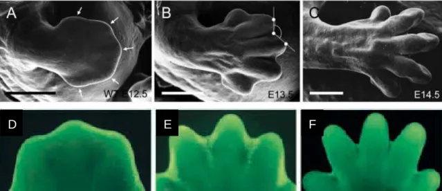
Figure 1.1: Apoptosis in the developing mouse forelimb ... 50
Figure 1.2: Insults associated with neurodegenerative disease and stroke that lead to neuron death ... 52
Figure 1.3: Apoptosis pathway induced in sympathetic neurons following NGF deprivation ... 54
Figure 1.4: Members of the Bcl-2 family of proteins ... 56
Figure 1.5: Formation and activation of the apoptosome complex ... 58
Figure 1.6: Basic overview of caspase domains ... 60
Figure 1.7: Domains of XIAP ... 62
Figure 1.8: Overview of the extrinsic pathway of apoptosis ... 64
Figure 1.9: Neurons become increasingly resistant to apoptosis with maturation ... 66
Figure 1.10: Comparison of a healthy adult brain to a highly atrophied brain of an Alzheimer’s disease patient ... 68
Figure 1.11: Localized regressive events occur during normal development ... 70
Figure 1.12: Massive pruning events sculpt the mature nervous system ... 72
Figure 1.13: Loss of key apoptotic proteins during development causes gross morphological and lethal defects in the brain ... 74
Figure 1.14: Distal portions of severed axons degenerate following axotomy during Wallerian degeneration ... 76
Figure 1.15: Schematic of developmental mushroom body neurite pruning in Drosophila ... 78
Figure 1.16: Current models of non-canonical and canonical functions of apoptotic molecules in neurons ... 80
xvi
Figure 2.2: Diagram of a microfluidic chamber and the fluidic isolation between soma and axon compartments ... 95 Figure 2.3: Sample of an ideal microfluidic chamber containing sympathetic
neurons after 5 days in culture ... 97 Figure 2.4: Campenot chambers permit the fluidic isolation of soma and axon ... 99 Figure 3.1: Casp6 activation in axons is selective to NGF deprivation ... 119
Figure 3.2: Casp6 is required for axon degeneration during axon pruning but not
apoptosis ... 121
Figure 3.3: Axon-selective degeneration is Apaf-1-independent but requires
Casp9 and Casp3 ... 123 Figure 3.4: Local deprivation triggers cyt c release in axons ... 125 Figure 3.5: The proteasome restricts caspase activation to axons during
axon-specific degeneration ... 127 Figure 3.6: XIAP protects the soma from caspase activation during axon-specific
degeneration ... 129 Figure 3.7: Mature neurons inhibit apoptosis during global deprivation but
degenerate axons during local deprivation ... 131 Figure 3.8: Working model of the pathways mediating apoptosis versus
axon pruning ... 133 Supplemental Figure 3.1: Casp6 activation in axons is selective to NGF
deprivation ... 135 Supplemental Figure 3.2: The extrinsic pathway is not involved in axon-selective
degeneration induced by local deprivation ... 137 Supplemental Figure 3.3: Time course of axon degeneration in proteasome-
inhibited and XIAP-deficient neurons during local deprivation ... 139 Figure 4.1: Transcription and axon-specific translation are required for axon
degeneration induced by local NGF deprivation ... 159 Figure 4.2: Axon-specific translation is required for c-Jun phosphorylation during
xvii
Figure 4.4: Levels of Bim mRNA increase in locally deprived axons ... 165 Figure 4.5: Bim -/- neurons degenerate their axons during local deprivation ... 167 Figure 4.6: ABT-737 treatment enhances axon degeneration induced by local NGF
deprivation ... 169 Figure 4.7: Overview of microRNA biogenesis ... 171
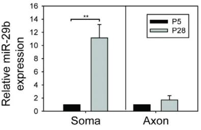
Figure 4.8: miR-29b levels drastically increase in the soma but not axons during
neuronal maturation ... 173
Figure A.1: Caspase-6 is required downstream of c-Jun phosphorylation during
xviii
LIST OF ABBREVIATIONS
AD: Alzheimer’s disease
ALS: Amyotrophic lateral sclerosis
Apaf-1: Apoptosis protease activating factor 1 APP: Amyloid precursor protein
ATP: Adenosine triphosphate
BACE1: β-site APP-cleaving enzyme 1 BH: Bcl-2 homology
BIR: Bacculoviral IAP repeat
CARD: Caspase activation and recruitment domain Casp: Caspase
C. elegans: Caenorhabditis elegans CNS: Central nervous system
dATP: Deoxyadenosine triphosphate DD: Death domain
DED: Death effector domain
DIABLO: Direct IAP binding protein with low pI DIV: Days in vitro
DNA: Deoxyribonucleic acid DR: Death receptor
xix ER: Endoplasmic reticulum
FADD: FAS-associated protein with Death Domain GAPDH: Glyceraldehyde phosphate deyhydrogenase HD: Huntington’s disease
HSP27/70/90: Heat shock proteins IAPs: Inhibitor of apoptosis proteins ICE: Interleukin-1β converting enzyme
JNK: c-Jun N-terminal kinase miR-29: microRNA-29 miRNA: microRNA
MLK: Mixed lineage kinase
MOM: Mitochondrial outer membrane
MOMP: Mitochondrial outer membrane permeabilization mRNA: Messenger RNA
NB-ARC: Nucleotide-binding adaptor shared by Apaf-1, R proteins and ced-4 NGF: Nerve growth factor
NOD: Nucleotide-binding oligomerization domain P: Post-natal day
PD: Parkinson’s disease
PIDD: p53-induced death domain protein PNS: Peripheral nervous system
qRT-PCR: quantitative RT-PCR
xx RING: Really interesting new gene domain RISC: RNA-induced silencing complex RNA: Ribonucleic acid
ROS: Reactive oxygen species Ser: Serine
SMAC: Second mitochondrial activator of caspases TRAIL: TNF-related apoptosis-inducing ligand TRAMP: Trf4/Air2/Mtr4p polyadenylation complex
1
CHAPTER ONE: INTRODUCTION
1. Overview of Apoptosis
“If you don't know how to die, don't worry; Nature will tell you what to do on the spot, fully and adequately. She will do this job perfectly for you; don't bother your head about it.”
A historical perspective:
Death is a fundamental and universal part of life; every living cell, vibrant organism, and breathing creature has an end. Cells can die in a variety of ways, yet the most
2 cancer to neurodegeneration.
Apoptosis is well appreciated as a genetically programmed and tightly regulated pathway required for proper development and tissue homeostasis (Danial and Korsmeyer 2004), yet this has not always been the case. Studies from Dr. Bob Horvitz’s lab during the mid-1980s laid the foundation for our understanding of the genetic and biochemical processes that control programmed cell death. The Horvitz lab mapped the fate of every single cell in the nematode C. elegans and found that exactly 131 cells die during normal development, always resulting in a worm with precisely 959 cells (Sulston and Horvitz 1977; Sulston, Schierenberg et al. 1983). A forward genetic screen then revealed two genes, ced-3 and ced-4, whose function is absolutely required for the death of those 131 cells (Ellis and Horvitz 1986; Yuan and Horvitz 1990). The Horvitz lab also identified ced-9, the first anti-apoptotic gene; mutations in ced-9 resulted in excessive and unnecessary cell death in the nematode (Hengartner, Ellis et al. 1992). These insightful discoveries about the genetic underpinnings of apoptosis earned Dr. Horvitz the Nobel Prize and pioneered the search for conserved cell death machinery beyond the nematode and into mammalian cells.
Apoptosis is essential for proper development:
Cell death occurs at astonishingly high levels during normal mammalian
3
Wood, Turmaine et al. 2000). Apoptosis is also essential for proper nervous system development for the following three known functions:
1.) to achieve balance between the number of innervating neurons and their target cells (Oppenheim 1991);
2.) to limit the number of proliferating neuronal precursors in germinal zones of the CNS and PNS (Voyvodic 1996; Kuan, Roth et al. 2000), therefore secondarily affecting the final number of neurons in the nervous system;
3.) to correct any errors, most notably by removing neurons that have migration deficits or misprojecting axon outgrowth (Kim and Sun 2011).
Apoptosis is required for adult homeostasis:
4
cells rather than risk passing genetic damage down to daughter cells (Green and Evan 2002). Notably, in contrast to cells that are replaced throughout life, post-mitotic cells with little or no proliferative capacity (such as neurons, cardiomyocytes and myotubes) must engage pathways that strictly inhibit apoptosis (Wright and Deshmukh 2006).
Apoptosis in disease:
Too little or too much apoptosis can cause developmental disease, cancer, autoimmunity, and neurodegeneration. The importance of developmental cell death is highlighted by the disrupted tissue formation observed in genetically altered mice that cannot execute apoptosis (Kuida, Haydar et al. 1998). In the immune system, apoptosis is critical for proper development of immune B and T cells to ensure the execution of any cells that recognize self-antigens (Marsden and Strasser 2003); failure to complete this process results in the survival of cells that recognize and respond to self-antigens (O'Reilly and Strasser 1999), leading to autoimmune disease. Lastly, the ability of cancer cells to evade apoptosis promotes their vigorous survival and uncontrolled growth (Vaux, Cory et al. 1988; Hanahan and Weinberg 2000).
In contrast to situations involving too little apoptosis, disease also occurs in cases of over-active apoptosis. One highly relevant example is neurodegeneration, which results when unwanted, excessive apoptosis occurs in the mature nervous system. While many diverse events contribute to the pathologies and neuron death observed in
5
6 1.2 Apoptosis in developing neurons
Nerve growth factor and the neurotrophic hypothesis:
Seminal studies by Viktor Hamburger and Rita Levi-Montalcini ultimately led to the discovery of the first neurotrophic factor (NGF, nerve growth factor)(Cohen,
Levi-Montalcini et al. 1954; Levi-Levi-Montalcini and Cohen 1956; Levi-Levi-Montalcini and Booker 1960; Aloe 2004) and the neurotrophic hypothesis, which provided insight into the basic
mechanism that initiates cell death in developing neurons. Simply stated, the neurotrophic hypothesis posits that an abundance of neurons are generated during development that must compete for limited amounts of survival-promoting growth factors secreted by target cells; the fates of developing neurons are ultimately regulated by their targets (Northcutt 1989; Yuan and Yankner 2000; Dekkers and Barde 2013; Dekkers, Nikoletopoulou et al. 2013). Neurons that reach and properly innervate their target will survive and incorporate into the developed nervous system, while those without adequate trophic factor support are eliminated by apoptosis. As many as 50% of neurons undergo apoptosis during this developmental period, elegantly matching the number of target cells to a precise
population of neurons.
Molecular modulators of neuronal apoptosis induced by NGF deprivation:
7
1979; Crowley, Spencer et al. 1994; Smeyne, Klein et al. 1994; Deshmukh and Johnson 1997; Putcha, Deshmukh et al. 2000).
In the presence of NGF, a neuron promotes its own survival through the tyrosine kinase receptor (known as TrkA), which signals through the PI3-kinase/Akt and
MEK/MAPK pathway (Kaplan and Miller 2000). One long-held and reasonable assumption posited that neurons died following NGF deprivation because NGF signaling promoted neuron survival. Simply stated, the loss of trophic factor support meant the loss of pro-survival signals and a passive death. We now know, however, that neurons initiate an active death process following NGF deprivation that requires signal transduction,
transcription and translation; strikingly, treating neurons with transcription or translation inhibitors during NGF deprivation protects them from death (Martin, Schmidt et al. 1988). Although new factors involved in neuronal death continue to be discovered, many of the essential events and modulators of the apoptosis pathway induced by NGF deprivation are well known (Figure 1.3). When an NGF-dependent neuron is deprived of trophic factor, the following initial changes occur: glucose uptake rapidly decreases, protein synthesis is globally reduced, levels of reactive oxygen species (ROS) spike, and the Rho GTPase Cdc42 becomes activated (Greenlund, Deckwerth et al. 1995; Bazenet, Mota et al. 1998). Precisely how each of these events is triggered remains unknown, yet their downstream effects are well characterized; following these events, a kinase cascade is initiated that involves the mixed lineage kinases (MLKs) and c-Jun N-terminal kinase (JNK), leading to the
phosphorylation of the transcription factor c-Jun (Mota, Reeder et al. 2001; Xu, Maroney et al. 2001). Phosphorylated c-Jun translocates from the cytosol to the nucleus where it
8
Puma, and Bmf (Estus et al., 1994; Ham et al., 1995; Imaizumi et al., 1997; Eilers et al., 1998; Xu et al., 2001; Mota et al., 2001; Harris and Johnson, 2001; Whitfield et al., 2001; Kristiansen et al., 2011). While phosphorylated c-Jun activity is absolutely required for NGF-deprivation-induced death in sympathetic neurons (Estus, Zaks et al. 1994; Ham, Babij et al. 1995; Eilers, Whitfield et al. 1998), its targets for transcriptional upregulation appear to act redundantly as the loss of any one BH3-only protein’s expression only modestly protects neurons from death (Putcha, Moulder et al. 2001; Imaizumi, Benito et al. 2004).
9 1.3 Intrinsic Apoptosis: The Key Players
“After all, to the well-organized mind, death is but the next great adventure.” –Albus Dumbledore in Harry Potter and the Sorcerer’s Stone by J.K. Rowling
Bcl-2 family of proteins: Key initiators of cell death:
The translocation of cytochrome c from the mitochondria into the cytosol is one major point of apoptosis regulation, and this step is primarily controlled by the Bcl-2 family of proteins. The discovery of ced-9 in C. elegans led to the critical identification of its
mammalian homolog, Bcl-2 (B-cell lymphoma 2) protein, as both proteins potently prevent apoptosis (Hockenbery, Nunez et al. 1990; Garcia, Martinou et al. 1992; Vaux, Weissman et al. 1992; Chipuk, Moldoveanu et al. 2010). Since its initial discovery in the 1990s, the Bcl-2 family of proteins has grown extensively and is divided into the following three distinct classes based on structure and function:
1.) the effector proteins that oligomerize and permeabilize the mitochondrial outer membrane (MOM), thereby inducing the release of intermembrane components (such as cytochrome c and SMAC/DIABLO) that lead to downstream caspase activation;
2.) the BH3-only proteins that sense cellular stress and become activated, either directly or indirectly;
10
More specifically, each Bcl-2 protein is classified by its pro- or anti-apoptotic role and its number of Bcl-2 homology (BH) domains (Figure 1.4). The first group of
pro-apoptotic members contains multi-domain proteins that have BH1, BH2 and BH3 domains; this group comprises two proteins, Bax and Bak. In healthy cells, Bax and Bak are normally expressed but remain inactive. During an apoptotic stimulus, however, both proteins can insert into the MOM and oligomerize to form a pore-like structure, resulting in
mitochondrial outer membrane permeabilization (called MOMP)(Chipuk, Bouchier-Hayes et al. 2006). MOMP permits the release of cytochrome c into the cytoplasm where, as described in the previous section, it associates with Apaf-1 and promotes caspase activation. Interestingly, in neurons, only Bax is capable of triggering MOMP as
Bax-deficient neurons are highly resistant to intrinsic apoptotic stimuli (Deckwerth, Elliott et al. 1996). Bak is present in neurons but exists in a uniquely spliced isoform called N-Bak that cannot directly permeabilize mitochondria (Sun, Yu et al. 2001; Uo, Kinoshita et al. 2005). Rather, the domain structure of N-Bak indicates this Bcl-2 protein may function like a BH3-only protein (described below) in neurons (Ham, Towers et al. 2005). While little is known about the function of N-Bak, the redundancy between Bax and Bak is clearly lost in
neurons, which may permit tighter apoptosis regulation and prevent the release of cytochrome c until absolutely necessary.
Members of the second group of pro-apoptotic Bcl-2 proteins contain only one BH3 domain and are aptly named the BH3-only proteins (Giam, Huang et al. 2008; Lomonosova and Chinnadurai 2008). This group included the following proteins: Bid, Bim, Bik, Bad, Bmf, Hrk/DP5, Puma, and Noxa. As the key responders to apoptotic stimuli, the BH3-only
11
phosphorylation, and altered localization. For example, in the presence of trophic factor, Bad is phosphorylated, bound to the protein 14-3-3, and safely held in the cytoplasm. Upon trophic factor deprivation, however, Bad becomes dephosphorylated and re-localizes to the mitochondria to inhibit the anti-apoptotic protein Bcl-ΧL (Zha, Harada et al. 1996;
Puthalakath and Strasser 2002). Inactive Bim, in turn, is normally secured to the microtubule-associated dynein motor complex but can be freed and translocate to the mitochondria to block anti-apoptotic Bcl-2 (Puthalakath, Huang et al. 1999). Additionally, full length Bid is cleaved into truncated Bid (tBid) in response to stimulation of the
extrinsic apoptotic pathway, resulting in Bax activation and cytochrome c release from the mitochondria (Esposti 2002). Transcriptional upregulation of Bim, DP5/HRK, Puma, and Noxa, respectively, has also been observed in various apoptotic situations (Dijkers,
Medema et al. 2000; Harris and Johnson 2001; Nakano and Vousden 2001; Puthalakath and Strasser 2002; Freeman, Burch et al. 2004). Activated members trigger apoptosis either indirectly by blocking anti-apoptotic Bcl-2 protein activity or by directly binding Bax and Bak. Only Bim, Bid, and Puma are classified as direct activators of apoptosis due to their ability to directly bind Bax and Bak (Ren, Tu et al. 2010). The rest of the BH3-only proteins are classified as sensitizers because of their ability to indirectly inhibit apoptosis by
blocking the anti-apoptotic Bcl-2 proteins (Letai, Bassik et al. 2002). With the activation of BH3-only proteins ultimately resulting in MOMP and cytochrome c release, these Bcl-2 family members are accepted as some of the most significant cell death initiators.
The third group within the Bcl-2 family consists of anti-apoptotic proteins that contain four BH domains (BH1-BH4). Members include Bcl-2, Bcl-ΧL, Bcl-w, and Mcl-1, all of
12
oligomerization and activation (Cory and Adams 2002). These anti-apoptotic Bcl-2 proteins must be relieved by BH3-only sensitizers or bypassed in order for apoptosis to proceed.
The apoptosome: Apaf-1, caspase-9, dATP and cytochrome c:
The effort to identify proteins capable of cleaving and activating caspase-3 resulted in the discovery of the mammalian homolog of ced-4, called apoptotic protease-activating factor (Apaf-1)(Liu, Kim et al. 1996; Zou, Henzel et al. 1997). Apaf-1 contains an N-terminal CARD domain, a nucleotide binding and oligomerization domain (NOD, or NB-ARC), and several repeats of the regulatory WD40 domain at its C-terminus (Riedl and Salvesen 2007). More complex than its nematode precursor, mammalian Apaf-1 contains a WD40 repeat motif that keeps Apaf-1 in an auto-inhibited conformation and prevents caspase-3 activation in the absence of the cytosolic Apaf-1 binding partner, cytochrome c (Hu, Ding et al. 1998); identified concomitantly with Apaf-1, cytochrome c is a well-studied, critical component of the mitochondrial respiratory chain (Liu, Kim et al. 1996). Securely sequestered away from Apaf-1 and destructive caspases in the mitochondrial
intermembrane of healthy cells, cytochrome c must translocate from the intermembrane space to the cytoplasm in order to induce Apaf-1 activation and downstream caspase activation during apoptosis (Wang, Zhou et al. 2001).
Apaf-1 normally exists in a monomeric, inactive state in healthy cells because the WD40 repeats mask the NB-ARC and CARD domains to form an auto-inhibited
conformation (Hu, Ding et al. 1998). Apaf-1 becomes active when cytochrome c binds to the WD40 domain, which alters Apaf-1’s conformation to expose its CARD and NB-ARC
13
CARD-CARD interactions) to form the caspase-activating scaffolding complex called the apoptosome (Liu, Kim et al. 1996; Hu, Benedict et al. 1998; Acehan, Jiang et al. 2002; Yu, Acehan et al. 2005). The main function of the apoptosome is to initiate enzymatic activity of caspase-9. With functional Apaf-1 in place at the central ‘hub of the wheel’, caspase-9 is recruited around Apaf-1 via its CARD domains and becomes active after dimerization on the apoptosome (Boatright, Renatus et al. 2003; Boatright and Salvesen 2003); active initiator caspase-9 then cleaves and activates effector caspase-3 to cause the ultimate destruction of the cell.
The powerful association between Apaf-1 and caspase-9 on the apoptosome enhances processed caspase-9 activity more than 1000-fold (Rodriguez and Lazebnik 1999), yet this model of activation (termed the “induced proximity model”) does not entirely explain caspase-9 activation. For example, constitutively dimeric caspase-9 is far less catalytically active than its apoptosome-associated counterparts (Chao, Shiozaki et al. 2005), and non-Apaf-1-mediated mechanisms of caspase-9 activation have been described (explored in more detail in the Discussion).
Unlike other initiator caspases, neither prodomain removal nor cleavage is required to achieve catalytic activation of caspase-9 (Riedl and Salvesen 2007). Whether the
14
‘molecular timer’ of apoptosis - the overall length of the timer is set by the intracellular concentration of procaspase-9, procaspase-9 autoprocessing starts the timer, and the rate of active caspase-9 dissociation from the complex (resulting in its inability to activate caspase-3) determines the speed at which the timer ‘ticks’ (Malladi, Challa-Malladi et al. 2009). Overall, this line of thinking maintains that, rather than merely activating caspase-9, the chief purpose of procaspase-9 autoprocessing is to initiate a molecular timer that regulates the duration of apoptosome activity (Bratton and Salvesen 2010). Although the precise events mediating caspase-9 activation continue to be explored, the requirement for the apoptosome in activating caspase-9 during apoptosis remains undeniably clear as the phenotypes of caspase-9-/- and Apaf-1-/- animals are very similar - the deletion of either apoptosome-associated protein potently inhibits neuron death during development (Kuida, Zheng et al. 1996; Cecconi, Alvarez et al. 1998; Hakem, Hakem et al. 1998).
Regulators of apoptosome function:
Our lab and others have shown that fully differentiated postmitotic cells lose their ability to undergo cytochrome c-mediated apoptosis, and this heightened resistance to apoptosis enables these cells to survive for the lifetime of the organism. For postmitotic cells such as neurons, myotubes, and cardiomyocytes, the decreased sensitivity to cytochrome c is partly due to the substantial transcriptional downregulation of Apaf-1 (Wright, Linhoff et al. 2004; Potts, Vaughn et al. 2005). Such low levels of Apaf-1 render the apoptosome incapable of activating caspases at adequate levels required to overcome the cell’s endogenous levels of caspases inhibitors, such as XIAP (X-linked inhibitor of
15
regulate apoptosome function as splice variants encoding fewer WD40 domains have reduced activity (Benedict, Hu et al. 2000).
Activity of cytochrome c can also be regulated at transcriptional, translational and redox levels. For example, cytochrome c mRNA and protein levels are induced in response to an apoptotic stimulus (Sanchez-Alcazar, Ault et al. 2000; Chandra, Liu et al. 2002). Post-translationally, addition or removal of a heme group can confer or remove cytochrome c’s pro-apoptotic facilities, respectively (Yang, Liu et al. 1997; Martin and Fearnhead 2002). Tri-methylation (at lysine 72) and the redox state of cytochrome c have also been shown to regulate this molecule’s ability to promote apoptosis (Pan, Voehringer et al. 1999; Hancock, Desikan et al. 2001; Suto, Sato et al. 2005; Vaughn and Deshmukh 2008). More specifically, oxidized cytochrome c is a more potent caspase activator than the reduced form. While healthy neurons maintain a highly reducing environment that restricts cytochrome c -mediated caspase activation (even after cytochrome c is released into the cytosol),
cytochrome c potently activates caspases when the environment becomes more oxidizing following insults such as NGF withdrawal (Vaughn and Deshmukh 2008). It is interesting to note that, unlike most cells, neurons have the capability to survive and even recover after the point of cytochrome c release. If caspase activation is prevented during NGF
withdrawal, sympathetic neurons can maintain their mitochondrial membrane potential for a short period after cytochrome c is released into the cytosol; this ‘safety window’ provides neurons with an opportunity to reverse apoptotic processes before mitochondrial membrane potential is lost (Martinou, Desagher et al. 1999; Deshmukh, Kuida et al. 2000).
16
Kornbluth 2006). First, cytochrome c can be negatively modulated by Heat Shock Protein 27 (HSP27), which binds and sequesters cytochrome c away from Apaf-1 (Bruey, Ducasse et al. 2000). In turn, Apaf-1 binding by Heat Shock Protein 90 (HSP90) inhibits apoptosome assembly (Pandey, Saleh et al. 2000), and Heat Shock Protein 70 (HSP70) has also been shown to bind Apaf-1 and block caspase-9 processing (Beere, Wolf et al. 2000; Saleh, Srinivasula et al. 2000; Beere 2004). Apaf-1 can be inhibited by Rsk-mediated
phosphorylation, which promotes the recruitment of 14-3-3 to effectively block
cytochrome c from binding to Apaf-1 (Kim, Parrish et al. 2012). Two proteins, Aven and ProT, can effectively block apoptosome assembly (Chau, Cheng et al. 2000; Jiang, Kim et al. 2003), and TUCAN, APIP, JNK, and various truncated forms of caspase-9 are all able to inhibit caspase-9 recruitment to and activation on the apoptosome (Srinivasula, Ahmad et al. 1999; Angelastro, Moon et al. 2001; Pathan, Marusawa et al. 2001; Cao, Xiao et al. 2004; Tran, Andreka et al. 2007). Even non-protein interactors (including nitric oxide donors, high potassium and calcium levels, and intracellular nucleotides) have been shown to negatively regulate Apaf-1 oligomerization by binding to cytochrome c or inhibiting
nucleotide exchange (Purring-Koch and McLendon 2000; Cain, Langlais et al. 2001; Bao and Shi 2006; Chandra, Bratton et al. 2006; Mei, Yong et al. 2010). Lastly, several kinases have been shown to modulate caspase-9 activity. Specifically, ERK2, DYRK1A, CDK1-cyclin B1
17
Clarke 2009). In all of these cases, however, the physiological importance of caspase-9 modification remains unclear.
When viewed in the context of the growing evidence for non-apoptotic caspase activation, the potential role of the apoptosome (as well as its many modulators) in this process is particularly exciting and puzzling. If the apoptosome is involved in non-apoptotic caspase activation, how does it function without killing the cell? Chapter Three of this dissertation examines the role of the apoptosome in one situation – developmental axon pruning – that involves caspase activation without death.
Caspase proteases execute cell death:
One of the principal families of apoptotic proteins was discovered during the search for the mammalian homolog to the ced-3 gene: the cysteine-dependent aspartate-directed proteases called caspases (Yuan, Shaham et al. 1993; Hengartner and Horvitz 1994). Over the past twenty years, our knowledge about mammalian caspases has been steadily
growing with regard to their physiological functions, distinct mechanisms of activation and regulation, and the diverse signal transduction pathways that trigger their activation and consequential involvement in disease pathogenesis.
18
Within each heterodimer the large and small subunits interdigitate to form a core that is unique to proteases. For some caspases (such as caspase-1 and -3), the maturation of caspase precursors depends on the p10 subunit from one zymogen forming a complex with the p20 subunit of a second zymogen. For others, however, maturation can occur via self-activation (caspase-6) or transself-activation with the aid of scaffolding proteins (caspase-9). Notably, caspase-9 is the only caspase reported to possess catalytic properties in the absence of any cleavage; mutating all active sites within caspase-9 does not block its downstream apoptotic effects, suggesting that caspases can mature in different fashions (Stennicke, Deveraux et al. 1999).
Active caspases cleave diverse intracellular polypeptides, such as major nuclear and cytoskeletal structures, to cause the characteristically deliberate and neat disassembly of the cell during apoptosis. Caspases recognize at least four contiguous amino acids in their designated substrates, P4-P3-P2-P1, and cleave after P1, which must always be an
aspartate residue. Of the thirteen identified mammalian caspases (eleven are in humans and ten are in mice), caspase-3 and caspase-9 are essential for developmental apoptosis; deletion of either caspase in C57/Black6 mouse background prevents the substantial apoptosis that occurs during nervous system development, resulting in hydrocephaly (enlarged brain) and embryonic lethality (Kuida, Haydar et al. 1998).
19
a. the death effector domain (DED), which is present in caspase-8 and -10 and mediates interactions with signaling adapter proteins such as FADD;
b. the caspase recruitment domain (CARD), found in caspase-1, -2, -4, and -9, promotes the ability of caspases to interact with each other as well as a wide range of other regulatory and adapter proteins.
Adapter proteins recruit specific caspases via interactions with the long prodomain. The following four specific adapter protein complexes that support caspase activation have been identified in mammalian cells:
a. the apoptosome, which mediates caspase-9 activation by the adaptor Apaf-1 bound to cytosolic cytochrome c in the intrinsic pathway;
b. the deathinducing signaling complex (DISC), which activates caspase8 and -10 by interactions with the adaptor FADD in the extrinsic pathway;
c. the inflammasome, which activates caspase-1 and -5 by interactions with the adaptors ASC (apoptosis-associated speck-like protein containing a CARD) or the NLR family during inflammatory responses;
20
Allocating separate caspase activation complexes to distinct insults allows cells to fine-tune and carefully control apoptosis, ensuring that specific caspases are activated appropriately (Hyman and Yuan 2012).
The effector caspases with short prodomains cleave various cellular substrates to perform the downstream execution steps of apoptosis, and these proteases are typically processed and activated by the upstream initiator caspases. To date, almost 400 distinct substrates have been reported for mammalian caspases (Luthi and Martin 2007).
It is important to note that not all caspases are created equal. Initiator caspases are much more specific and cleave only their own precursors and downstream caspases, whereas effector caspases are far more promiscuous during the demolition phases of apoptosis. Additionally, the upstream-downstream relationship between caspases is likely a transient and death-specific one; increasing evidence suggests important roles of
caspases in non-apoptotic processes (detailed in the Discussion), and understanding the non-apoptotic substrates of caspases continues to be an important ongoing area of study.
XIAP and the regulation of apoptosis:
21
Mammalian IAPs include the following: XIAP, cIAP1, cIAP2, ML-IAP, NAIP, Survivin, and Apollon/Bruce (Salvesen and Duckett 2002). All IAPs contain a specific motif called the baculovirus IAP repeat (BIR) domain, so called because this motif was originally isolated from baculovirus during a screen to identify regulators of host-cell viability during viral infection. Overexpression of most IAP family members can suppress apoptosis (Duckett, Nava et al. 1996; Liston, Fong et al. 2003), yet XIAP reigns as the most potent inhibitor of caspases (Salvesen and Duckett 2002; Scott, Denault et al. 2005; Eckelman and Salvesen 2006; Eckelman, Salvesen et al. 2006).
XIAP binds directly to cleaved caspases to block their proteolytic activity. XIAP contains three BIR domains, the third of which binds to cleaved and activated caspase-9 to prevent its interaction with other substrates (Figure 1.7)(Sun, Cai et al. 2000; Srinivasula, Hegde et al. 2001). The small linker region on the N-terminal side of the BIR2 domain forms a reversible, steric occlusion of substrates from caspase-3 and caspase-7 (Figure 1.7)(Chai, Shiozaki et al. 2001; Huang, Park et al. 2001; Riedl, Renatus et al. 2001). XIAP also contains a C-terminal RING finger domain that possesses E3 ubiquitin ligase activity (Yang, Fang et al. 2000). Despite evidence demonstrating XIAP’s ability to ubiquitinate several proteins in the apoptotic pathway (Silke, Ekert et al. 2001; Suzuki, Nakabayashi et al. 2001; MacFarlane, Merrison et al. 2002; Sun 2003; Morizane, Honda et al. 2005), how the RING finger regulates apoptosis in intact cells remains unclear.
Our lab has shown that XIAP is a primary safety brake on caspase activation induced by the release of mitochondrial cytochrome c. Even though XIAP-/- mice show no overt phenotype, XIAP-deficient neurons and cardiomyocytes are more susceptible to
22
Similarly, neurons in XIAP-deficient mice are more vulnerable to stroke injury in vivo (West, Stump et al. 2009). In contrast to mice, the deletion of the XIAP homolog DIAP1 in Drosophila causes massive apoptosis and fly lethality, and it was discovered that four proteins (Reaper, Grim, HID, and Sickle) bind and inhibit DIAP1 in the cytosol (Martin 2002). The search for mammalian XIAP binding partners uncovered two proteins,
SMAC/DIABLO and Omi/HtrA2, that localize to the mitochondria in healthy cells (Du, Fang et al. 2000; Verhagen, Ekert et al. 2000; Suzuki, Imai et al. 2001; Hegde, Srinivasula et al. 2002; Martin 2002; van Loo, van Gurp et al. 2002; Verhagen and Vaux 2002). Upon apoptotic stimulation, SMAC and HtrA2 are released into the cytosol and bind to XIAP at the same regions as caspase-9, -3, and -7 (Sun, Cai et al. 2000; Srinivasula, Hegde et al. 2001; Salvesen and Duckett 2002; Shi 2002; Verhagen, Silke et al. 2002). At sufficiently elevated ratios in the cytoplasm, these pro-apoptotic mitochondria-released proteins can exclude caspase-9 and disrupt caspase-3/7 binding with XIAP, resulting in freed active caspases and cell destruction. Curiously, endogenous SMAC does not inhibit XIAP in
neurons undergoing cytochrome c-mediated apoptosis (Vaughn and Deshmukh 2007); only ectopic expression of SMAC by microinjection allows cytochrome c to induce death in sympathetic neurons (Potts, Singh et al. 2003; Potts, Vaughn et al. 2005).
Importantly, XIAP activity in neurons can be regulated by SMAC-independent mechanisms. XIAP is degraded in neurons undergoing apoptosis in response to NGF-deprivation (Potts, Singh et al. 2003), although the mechanism that mediates this
23
activation maintains XIAP as an extremely effective apoptosis inhibitor. However, a high level of Apaf-1 (and hence apoptosome activity) causes maximal levels of caspase
activation that overpower XIAP’s ability to prevent apoptosis (Wright, Linhoff et al. 2004; Vaughn and Deshmukh 2007). Our lab has shown that differentiating neurons and
24 1.4 Extrinsic Pathway
The extrinsic cell death pathway is induced by ligand binding to death receptors (DRs) of the tumor necrosis factor receptor (TNFR) family that contain the death domain (DD). This receptor family includes the following members: CD95, TNF receptor (TNF-R1), DR3/APO-3/TRAMP, TRAIL receptor 1/DR4, TRAIL-2/DR5, and DR6 (Peter, Scaffidi et al. 1999). Following ligand binding (by ligands such as Fas and TRAIL), the death receptors undergo conformational changes that permit the recruitment of multiple proteins that form the death-inducing signaling complex (DISC). The ligand-bound death receptors then
recruit a DD-containing adapter protein called FADD (Fas-associated DD). FADD contains the critical death-effector domain (DED) that allows FADD to recruit initiator caspases-8 and -10. FADD-bound caspase-8/10 then undergoes autoproteolytic cleavage on the DISC and is released into the cytoplasm where it can cleave and activate effector caspase-3 to fully engage the apoptotic caspase cascade (Figure 1.8). In neurons, caspase-8 connects the intrinsic and extrinsic pathways by cleaving the BH3-only protein Bid into its
Bax-activating form, tBid (Ricci, Kim et al. 2007; Dekkers and Barde 2013). The extrinsic
pathway can be regulated by IAPs (Vaux and Silke 2005) and cFLIP, which inhibits caspase-8 and blocks formation of the DISC at the plasma membrane (Wang, Wang et al. 2005).
Dependence receptors:
25
simply due to the loss of positive survival signals. Extensive work over the past two
decades, however, supports a novel form of signal transduction that is mediated by unique receptors called dependence receptors. Dependence receptors support cell survival
signaling when bound to their respective ligands (just as seen with other transmembrane receptors), yet they can fully switch to activating cell death in the absence of ligand
(Bredesen, Mehlen et al. 2005; Tauszig-Delamasure, Yu et al. 2007). Dependence receptors create a highly tuned state of cellular dependence on their ligands. Fifteen dependence receptors had been identified as of 2010, although more likely exist that have yet to be discovered (Eilers, Whitfield et al. 1998).
A. DCC
Among the first dependence receptors to be characterized is the protein Deleted in Colorectal Carcinoma (DCC). DCC was discovered to promote apoptosis in the absence of its ligand, netrin, while inhibiting apoptosis when bound to netrin (Llambi, Causeret et al. 2001; Bredesen, Mehlen et al. 2005; Vanderhaeghen and Cheng 2010). Curiously, the death induced by netrin is believed to be dependent on caspase-3 and caspase-9 but independent of Apaf-1 or cytochrome c (Forcet, Ye et al. 2001). Although, as its name implies, DCC was first discovered due to its association with cancer, it also plays an important role in neurons. Netrin-DCC signaling is crucial for axon guidance during nervous system
26 B. Trks and p75NTR:
It was only recently shown that TrkA, the receptor for NGF in sympathetic neurons, is a dependence receptor: TrkA causes neuronal cell death by merely being expressed, and its death-inducing activity is blocked by the presence of NGF. Correspondingly, sympathetic neurons deficient for TrkA survive following NGF deprivation (Tauszig-Delamasure, Yu et al. 2007; Nikoletopoulou, Lickert et al. 2010). Closely related to TrkA, TrkC also acts as a dependence receptor. Exactly how TrkA and TrkC trigger cell death remains unclear. Proteolysis of the receptors themselves may be involved (Tauszig-Delamasure, Yu et al. 2007) or the proteolysis of the TrkA/C-associated receptor p75NTR could be responsible (Nikoletopoulou, Lickert et al. 2010). p75NTR colocalizes with TrkA and TrkC in lipid rafts in the plasma membrane, which is significant because the localization of certain pro-apoptotic proteins is known to be important for their death-inducing function. For example, during the extrinsic apoptotic pathway, activated death receptors must translocate to lipid rafts in the membrane in order to assemble the DISC, and cFLIP
prevents this translocation to mitigate cell death (Davis, Lotocki et al. 2007; Song, Tse et al. 2007). Likewise, DCC’s localization in lipid rafts is required for its death-inducing function in the absence of netrin (Furne, Corset et al. 2006). Notably, despite its overall structural similarity to TrkA and TrkC, TrkB does not function as a dependence receptor, most likely due to differences in both its membrane localization (TrkB does not colocalize with
27 1.5 Resistance of mature neurons to apoptosis
With the exception of neurons located in the olfactory bulb and dentate gyrus, neurons that survive the massive cull during nervous system development must
subsequently last the lifetime of the organism (which, for the average human, means over seven decades). As neurons are post-mitotic and have extremely limited potential for regeneration, these cells must strictly control the apoptotic pathway as they mature to prevent unwanted neuron loss (and risk neurodegeneration). Growing evidence supports the idea that mature neurons are far more resistant to apoptosis than young neurons (Figure 1.9). Although the exact changes that occur during neuronal maturation continue to be identified, several distinct mechanisms have been discovered that allow neurons to switch from a state of apoptotic susceptibility to one in which apoptosis is nearly completely shut off.
Mechanisms that restrict apoptosis in mature neurons:
Mature neurons engage several mechanisms that act at multiple points to restrict the apoptotic pathway, and this redundancy is necessary to safeguard neuronal survival in the event that one safety mechanism fails. The diverse ways in which a neuron becomes resistant to NGF deprivation-induced apoptosis are detailed below.
28
its ligand to the cell body where it promotes neuronal survival. When NGF is withdrawn from young NGF-dependent neurons, TrkA is dephosphorylated within one hour. However, in mature neurons deprived of NGF, TrkA remains phosphorylated for many hours and likely promotes the robust resistance of these cells against the loss of trophic support (Tsui-Pierchala and Ginty 1999). 2.) Failed translocation of Bax and cytochrome c release: Instead of translocating
from the cytoplasm to mitochondria to induce cytochrome c release following NGF deprivation (as seen in young neurons), Bax remains cytoplasmic in
mature neurons (Putcha, Deshmukh et al. 2000) and cytochrome c maintains its mitochondrial localization (Easton, Deckwerth et al. 1997).
3.) Transcriptional restriction of Apaf-1: Our lab has shown that mature neurons rearrange their chromatin to form a more condensed state at the Apaf-1 locus, resulting in a block on Apaf-1 expression (Wright, Smith et al. 2007). The addition of exogenous cytochrome c fails to induce apoptosis in both mature wildtype (essentially Apaf-1-deficient) and XIAP-deficient neurons, indicating that high levels of XIAP expression do not underlie mature neuron survival after this post-mitochondrial insult.
4.) MicroRNA 29b (miR-29b) inhibits multiple BH3-only mRNAs: Recent work from
our lab shows that while the vast majority of miRNAs are downregulated during neuronal maturation, the expression of miR-29 is markedly induced.
29
While the mechanisms utilized by maturing neurons to restrict apoptosis have been best characterized in sympathetic neurons, studies performed on other types of neurons indicate that they, too, restrict apoptosis with maturation. Cerebellar, cortical, and
photoreceptor neurons have been found to greatly downregulate Apaf-1 expression as they mature (Northcutt 1989; Kuan, Roth et al. 2000; Yuan and Yankner 2000; Aloe 2004). In contrast to sympathetic neurons, however, many neurons also downregulate caspase-3 expression (Northcutt 1989; Kuan, Roth et al. 2000; Yuan and Yankner 2000; Aloe 2004; Liu, Siesjo et al. 2004). Even the IAPs have been shown to increase their reach in maturing neurons - motor neurons downregulate the endogenous XIAP inhibitor, XAF, to allow XIAP to more potently inhibit caspases (Kim and Sun 2011).
Neuronal cell death in the mature nervous system:
Despite the protective mechanisms described above, neurons in the adult nervous system are still vulnerable to death, particularly in neurodegenerative situations (Orike, Middleton et al. 2001; Walsh, Orike et al. 2004). Why do the multiple brakes developed over the course of neuronal maturation become ineffective in neurodegenerative states? Are these mechanisms somehow reversed or disengaged during processes of degeneration and injury? As my research career was born from a deep personal interest in
neurodegeneration and a hope to positively impact those suffering from neurodegenerative disease, I have dedicated the following sections to providing a brief overview of apoptosis in several neurodegenerative situations.
30
amyotrophic lateral sclerosis, and stroke, among others. The extremely limited
regenerative capacity of the central nervous system makes neurodegenerative diseases particularly devastating – dead neurons cannot be replaced, resulting in worsening
symptoms and a declining quality of life as neurodegeneration progresses over time. Thus, understanding how to limit or prevent the damage caused by various destructive
mechanisms will enable the development of more effective and preventative treatments across the spectrum of neurodegeneration.
Neurodegenerative disorders are caused by multi-factorial stimuli that may include genetic and environmental factors, metabolic dysregulation, excitotoxicity,
neuroinflammation, cellular stressors such as oxidative stress and the overproduction of free radicals, disrupted calcium regulation, mitochondrial dysfunction, interrupted cell transport, sustained activation of microglia (the brain’s immune cells), and the
accumulation of misfolded or toxic proteins (Figure 1.2). Delineating the causes of
neuronal cell death in degeneration and injury is complicated by the fact that one stimulus can trigger multiple death pathways depending on its degree, duration, cell type, brain region, and the affected neuron’s bioenergetic state.
Several different pathways of neuronal cell death are implicated in
31
(Unal-Cevik, Kilinc et al. 2004). A brief overview of neuronal cell death across several of the most prevalent neurodegenerative diseases and stroke is described here.
The following sections on neurodegeneration and stroke were previously published in Cusack CL, Annis RP, Kole AJ, and Deshmukh M. Neuronal Death Mechanisms in
Development and Disease. In: Wu H, ed. Cell Death: Mechanism and Disease. New York, NY: Springer Science and Business Media; 2014: 167-188.
A. Alzheimer’s disease:
Alzheimer’s disease (AD)is the most common form of neurodegeneration
worldwide, and the vast majority of AD cases are sporadic. This disease is characterized by the loss of neurons and synapses in the cerebral cortex and several subcortical regions, resulting in massive brain atrophy of these areas (Figure 1.10). AD is most often diagnosed in people over 65 years of age, although the less prevalent early-onset form occurs much earlier. AD is progressive, meaning that the disease pathologies and symptoms become more severe over time. As neurodegeneration spreads throughout the brain, bodily functions are eventually lost, ultimately leading to death.
There is currently no known cure for AD, and understanding the causes and progression of AD is an area of intense investigation. The clinical hallmarks of AD include extracellular senile plaques, which are mostly composed of amyloid-β (Aβ) peptides, and neurofibrillary tangles composed of intracellular, hyperphosphorylated
32
dynamics, and these aggregates also induce inflammation; all of these processes can trigger death cascades.
The direct relationship between aggregate lesions, their toxic soluble monomers, and neuronal loss in AD is still unclear, but evidence of caspase activation in AD may provide a link. Initiating and executioner caspases are both upregulated in the brains of patients with AD (Matsui, Ramasamy et al. 2006), and caspases mediate the cleavage of multiple targets associated with AD pathology (LeBlanc, Liu et al. 1999; Guo, Albrecht et al. 2004; Albrecht, Bogdanovic et al. 2009; Halawani, Tessier et al. 2010; D'Amelio, Cavallucci et al. 2011; Hyman 2011). In particular, Caspase-6 has been shown to cleave both Aβ and tau, and caspase activation plays a role in the synaptic loss associated with Aβ toxicity (LeBlanc, Liu et al. 1999; Guo, Albrecht et al. 2004; Albrecht, Bogdanovic et al. 2009; D'Amelio, Cavallucci et al. 2011). There is now evidence that neurons can tolerate chronic low levels of caspase activation without dying in a mouse model of AD(de Calignon, Spires-Jones et al. 2010), and caspase-mediated cleavage of Aβ and tau produces toxic protein aggregates (Graham, Ehrnhoefer et al. 2011; Reddy 2011; Zhang, Thompson et al. 2011). These observations may help to explain how caspase activation – a normally acute, rapid event during apoptosis – can occur at sub-apoptotic levels for long periods of time, thus long-preceding but culminating in the progressive neurodegeneration of AD.
B. Parkinson’s disease:
33
shaking, rigidity, slowness, and unstable posture. More advanced stages of the disease involve dementia as well as cognitive, sleep-related, and behavioral issues. PD symptoms are considered to be the direct result of the progressive dysfunction and loss of
dopaminergic neurons, yet how and why these particular neurons are targeted for neurodegeneration remains unclear.
The pathology of PD is strongly linked to the progressive accumulation of alpha-synuclein into inclusions called Lewy bodies in neurons. The formation of Lewy bodies is a leading theory as the development and spread of these inclusions appears to correspond with disease progression and region-specific degeneration (Schulz-Schaeffer 2010); however, in some cases alpha-synuclein has been shown to be protective (Alves da Costa, Paitel et al. 2002; Li and Lee 2005). Alpha-synuclein can be secreted by neurons, partake in cell-to-cell transmission, induce neurotoxicity, make neurons more sensitive to apoptosis, and trigger inflammatory glial responses (Tanaka, Engelender et al. 2001; Schulz-Schaeffer 2010; Arduino, Esteves et al. 2011). Inflammation and dysfunction of the proteasomal and lysosomal systems also contribute to PD pathologies, and defects in mitochondrial quality control processes such as mitochondrial fission, fusion, and autophagy have also been linked to substantia nigral neuron degeneration (Obeso, Rodriguez-Oroz et al. 2010; Youle and van der Bliek 2012).
Caspase-dependent and independent death pathways contribute to
34
overexpression of Bcl-2 family members, and overexpression of XIAP, respectively, protect nigrostriatal neurons in certain PD mouse models. Additionally, inhibiting the opening of the mitochondrial permeability transition pore or overexpressing the E3 ubiquitin ligase Parkin to block cytochrome c release prevents apoptosis in dopaminergic neurons.
There is also evidence of paraptosis, a caspase-independent death pathway, in PD. Paraptosis depends on the activation of poly(ADP-ribose)polymerase (PARP-1), a DNA repair enzyme that, along with p53, is induced by genotoxic stress and DNA damage. PARP-1 activation leads to the translocation of apoptosis inducing factor (AIF) from the inner mitochondrial membrane to the nucleus where it participates in chromatin condensation and large-scale DNA fragmentation (Yu, Wang et al. 2002). Indeed, nuclear AIF is observed in postmortem PD patient brain tissue (Burguillos, Hajji et al. 2011). Additionally, PARP-1 inhibition blocks alpha-synuclein cytotoxicity as well as cell death in toxic (MPTP/MPP+) models of PD (Venderova and Park 2012). With multiple signaling pathways regulating several types of neuronal cell death in PD (and in other neurodegenerative diseases), basic research on neurodegeneration will provide much needed insight into the pathogenesis and regulation of cell death mechanisms in neurons.
C. Amyotrophic lateral sclerosis:
35
and mitochondrial damage and dysfunction are widely observed in patients with ALS and in mouse models of the disease (i.e. transgenic animals that overexpress the gene for human mutant superoxide dismutase (SOD1), mutant TAR DNA-binding protein 43 (TDP-43), or mutant fused in liposarcoma (FUS)). Disrupted axonal transport, impaired
mitochondrial fusion, decreased mitochondrial size and density, and defective
mitochondrial membrane potential are also present in degenerating motor neurons of ALS mouse models (Johri and Beal 2012).
Excitotoxicity also contributes to ALS pathology. Glutamate-mediated excitotoxicity caused by repetitive neuronal firing and/or elevation of intracellular calcium levels by calcium-permeable glutamate receptors is known to cause neuronal death. In normal physiological conditions, glutamate is the primary excitatory neurotransmitter in the nervous system. The release of glutamate into the synaptic cleft activates glutamate
receptors, which leads to the influx of calcium and sodium into the post-synaptic neuron to trigger its depolarization. The removal of synaptic glutamate by glutamate transporters is required to prevent repetitive firing, and the glial glutamate transporter EAAT2 is
responsible for approximately 90% of glutamate clearance in motor neurons (Rothstein, Dykes-Hoberg et al. 1996; Tanaka, Watase et al. 1997). Elevated levels of glutamate are found in 40% of sporadic ALS patients (Spreux-Varoquaux, Bensimon et al. 2002), and loss of EAAT2 protein causes significantly reduced glutamate transport in brain regions affected in ALS (Rothstein, Van Kammen et al. 1995). The abundance of data indicating
36
Despite the combined contributions of oxidative damage, axonal stress, and toxicity to motor neuron dysfunction in ALS, it is well established that Caspase-3 activation is the final event in the death cascade of these neurons (Cleveland and Rothstein 2001). Other evidence for the role of apoptosis in ALS includes the observation that administration of a pan-caspase inhibitor to ALS mice is neuroprotective and significantly extends survival, indicating a role for caspase-1 and caspase-3 in ALS (Li, Ona et al. 2000). As ALS mice age, there is a progressive transcriptional upregulation of Caspase-1 and Caspase-3 messenger RNA (mRNA)(Li, Ona et al. 2000; Vukosavic, Stefanis et al. 2000). These findings support that clinical relevance of mouse models of ALS as both Caspase-1 and Caspase-3 activation have been found in spinal cord samples from human ALS patients. In addition, other central markers of apoptosis – including Caspase-9 activation, the release of cytochrome c from mitochondria, and pro-apoptotic changes in the Bcl-2 family - have also been found in the spinal cords of ALS mice, and ALS mice that overexpress the anti-apoptotic Bcl-2 gene survive longer than other ALS mice (Sathasivam and Shaw 2005).
D. Huntington’s Disease:
Huntington’s Disease (HD) is a universally fatal autosomal dominant
neurodegenerative disease in which neostriatal medium spiny neurons and eventually certain regions of the cortex progressively waste away. Onset typically occurs during the mid-thirties and forties, and persons with HD usually die within 15 to 20 years. Symptoms include abnormal involuntary movements, cognitive decline, and psychiatric problems. The disease is caused by a genetic mutation encoding for the aberrant expansion of
37
mutant huntingtin protein (mHtt). Post-translational modifications of mHtt result in cleavage of the protein that leaves behind toxic, shorter fragments that form damaging protein aggregates instead of folding properly into functional proteins. These aggregates, called inclusion bodies, accumulate over time and ultimately interfere with proper
neuronal functions. There is also extensive evidence for deficient energy metabolism, mitochondrial dysfunction, and glutamate-mediated excitotoxicity in HD (Estrada Sanchez, Mejia-Toiber et al. 2008; Johri and Beal 2012). With regard to key apoptotic proteins, in a mouse model of HD, cytochrome c changes its localization from a mitochondrial to cytosolic pool as the animal ages and the disease progresses (Mangiarini, Sathasivam et al. 1996; Kiechle, Dedeoglu et al. 2002). Caspases, particularly caspase-6, have also been strongly implicated in HD (Graham, Deng et al. 2006; Majumder, Chattopadhyay et al. 2006; Leyva, Degiacomo et al. 2010). Caspase-mediated cleavage of Htt appears to be critical for
initiating striatal degeneration, and inhibiting such cleavage blocks Htt-mediated toxicity (Graham, Deng et al. 2006).
E. Stroke:
Stroke is an acute, rapid form of neurodegeneration that is caused by ischemia (lack of blood supply) or hemorrhage. Both the severity and duration of the insult induce
38
Cell death mechanisms induced by ischemic injury occur on a dynamic time-scale. At early time points, neurons in the necrotic core display many features of apoptosis, such as nuclear condensation and evidence of caspase-1 and caspase-8 activation (Benchoua, Guegan et al. 2001). As the penumbral region develops in later time points, the intrinsic apoptotic pathway is activated in the infarct core but appears to be aborted due to severe energy depletions and mitochondrial failure. Reperfusion of blood to the penumbral region permits energy-dependent caspase activation and complete apoptosis (Hyman and Yuan 2012). Bcl-2 protein family members play a significant role in neuronal death following ischemia as the overexpression of anti-apoptotic Bcl-2 protein (Martinou, Dubois-Dauphin et al. 1994) and the loss of Bid (Plesnila, Zinkel et al. 2002), a BH3-only protein,
respectively protect against ischemic injury by reducing mitochondrial damage. Caspases are also critical mediators of stroke-induced neuronal death as treatment with caspase inhibitors following stroke improve functional outcomes in ischemic mouse models (Akpan, Serrano-Saiz et al. 2011; Al-Jamal, Gherardini et al. 2011).
Neuronal death following stroke is largely due to the massive disruption of
39
2009; Yenari and Han 2012). Future therapies for stroke may therefore involve an
appropriate combination of treatments that prevent apoptosis, antagonize glutamate, and reduce oxidative stress.