Clearing Things Up: A Vertical Mucus Clearance Assay to
Investigate Rheological and Biochemical Parameters
Governing Mucociliary Clearance.
Jerome Carpenter
A dissertation submitted to the faculty of the University of North Carolina at Chapel Hill in partial fulfillment of the requirements for the degree of Doctor of Philosophy in
the Curriculum in Applied Sciences and Engineering.
Chapel Hill 2013
Approved by
Richard Superfine
Al Banes
Greg Forrest
Scott Randell
ABSTRACT
JEROME CARPENTER: Clearing Things Up: A Vertical Mucus
Clearance Assay to Investigate Rheological and Biochemical
Parameters Governing Mucociliary Clearance.
(Under the direction of Richard Superfine)
Mucociliary clearance (MCC) plays an essential role to protect the airways
from pathogens and pollutants. Impaired MCC leads to a host of diseases and
initiates a cascade of chronic infection, inflammation, and reduced lung function.
Understanding the mechanisms of clearance is a critical step in preventing and
ultimately treating disease. The fundamental mechanisms guiding clearance are still
poorly understood. Existing models have focused on fluid rheology, but haven’t
reached a consensus on which parameters govern the system. In this work I build an
in vitro model capable of evaluating rheological and biochemical parameters and
their effect on clearance.
My model integrates the air-liquid interface (ALI) bronchial epithelial cell
culture model with microfluidics to create a mucus clearance assay (MCA). Growing
well-ciliated cells inside of a fluidics channel allows me to use external flow to
investigate cilia-ASL interactions. I use external flow to build and validate
a model of cilia hydrodynamics. Then I demonstrate external flow can be used as a
Next I integrate the MCA with a tilting microscope to explore the effects of gravity on
mucociliary clearance. Gravity has been largely ignored in mucus clearance
experiments despite vertical transport in vivo and therapies exploiting gravitational
drainage. I observe fluids transporting horizontally, but failing to transport when the
cilia driven flow is opposed by gravity. This establishes gravitational effects as a
necessary requirement for evaluating the parameters that govern MCC. Next I tested
a 4.5% 1MDa PEG solution that matched the apparent viscosity of mucus and
observed that unlike mucus, the PEG solution didn’t transport vertically. This
experiment demonstrates that viscosity alone is not a predictor of vertical transport.
Finally I suggest that the inability to transport vertically is a result of the system
dehydrating and posit that mucus’ ability to transport vertically is the result of
Acknowledgements
I would like to thank my advisor, Rich Superfine. Thanks for your guidance,
your patience, but above all else teaching me how to be a scientist. I don’t know how
many times the light bulb has come on for me hours later, days, or even months
later when I finally realized where you were steering me. Beyond teaching, thank
you for creating such a great environment for science and the exchange of ideas.
Your passion and enthusiasm for science is contagious. Also I think I probably still
owe you a few bags of peanut M&Ms.
Sean thanks for the guidance and support that you’ve given me. It doesn’t
seem that long ago that I was a TA for your electronics lab. I’m pretty sure that I still
use the fire hose approach to communicate. You’ve played a major role in my
growth as a scientist. I learned so much in device meeting and while I still have a
ways to go, I at least cringe when I look at the first few iterations of a plot or figure.
Thanks to others in NSRG. Tim thanks for you bio and microscopy expertise.
Mike thanks for the guidance. Russ thanks for the amazing tools that enabled my
project. Cassandra thank you for all of the POs, the emergency gas cylinders and
supplies so that my cells would live another day. David thanks for being my mucus
supplier.
To everyone at the UNC CF Tissue Procurement and Cell Culture Core, but
with a tricky cell type, but also being there whenever there was a contamination
scare or other cell related crisis.
Everyone else in the Physics department, again too many to name, but
thanks Sallie, Jean, Bev and everyone else. Thanks to the machine shop for not only
getting me through the shop class with all my digits intact, but helping to build and
consult on several key pieces of equipment in my project. Thanks to the BME lab for
access to the 3D printer and laser cutter: equipment which without I couldn’t have
built my system. Thanks to everyone in CHANL for all of the help with microscopy
and fabrication.
To all of my fellow graduate students past and present thank you for making
my time here amazing. Whether in Phillips Hall or the Chapman fishbowl it’s been a
great honor to work with so many wonderful people whose talent is only exceeded
by their character. I hesitate in trying to name everyone (I have the dubious
distinction of overlapping with the most students in the group), but I’m going to give it
a shot. Thanks Adam H., Adam S., Ben, Briana, Kwan, Lamar, Nathan, Kris,
Jeremy, Suzy, Rob, Luke, Nadira, Kellie, Ricky, Jay, Sreeja, Michael, Cory and
Vinay. (I roughly tried alphabetical order, but that quickly fell apart.) I apologize for
any omissions and could easily write a couple of pages thanking all of you
specifically, but I’ll save that for the academy speech, also I think I hear the
orchestra beginning to play me off stage. Suzy thanks for helping to push me over
the finish line. Also I couldn’t have done it with your help along with Evan, John,
Finally I would like to thank my parents. Mom and Dad thanks for, well
everything. Thanks for nurturing curiosity, giving me support, and working so hard to
give me opportunities. I don’t say it enough and I’m not sure if it’d be possible to say
Table of Contents
List of Figures ... xiii
Abbreviations ... xvi
Chapter 1 Introduction ... 1
1.1 Motivation ... 1
1.2 Thesis Statement and contributions ... 2
1.3 Outline for the document ... 4
Chapter 2 Mechanisms of Mucociliary Clearance ... 6
2.1 Lung physiology and airway defense ... 6
2.2 Epithelium ... 9
2.2.1 Cells of the epithelium ... 9
2.2.2 Epithelial Cross-Section ... 14
2.3 Cilia ... 16
2.3.1 Cilia Primer ... 17
2.3.2 Cilia Organization and Entrainment ... 19
2.3.3 Beyond Cilia: Cilium Surface and the PCL ... 24
2.4 Airway Surface Liquid ... 25
2.4.1 Mucins and Mucus ... 26
2.4.2 Properties of the Mucus Gel ... 33
2.4.3 Mucociliary Transport and Viscoelasticity ... 38
2.4.4 Fluid Regulation ... 40
2.4.6 Other Forces acting on the ASL ... 42
2.5 Model Systems ... 43
2.5.1 Animal MCC studies ... 44
2.5.2 In vitro MCC studies ... 45
Chapter 3 Building the Mucus Clearance Assay ... 50
3.1 Design Considerations ... 50
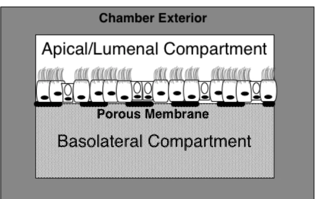
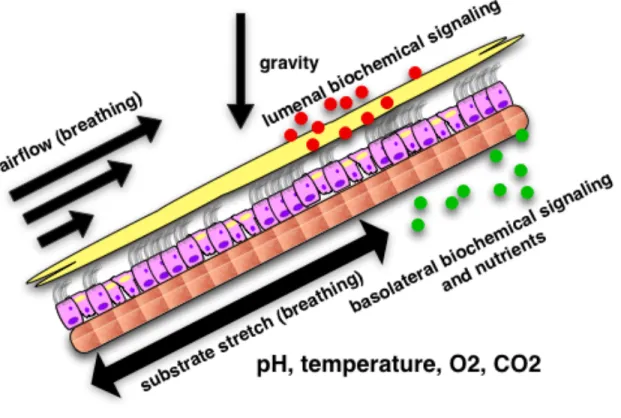
3.1.1 Designing the chamber: Physiological Considerations ... 52
3.1.2 Cell perfusion rate to determine basolateral volume ... 59
3.1.3 Experimental Requirements ... 60
3.1.3.1 Controlling the Airway Surface Liquid ... 61
3.1.3.2 Optimizing optics: integrating the MCA with a microscope ... 62
3.2 Channel Fabrication and Assembly ... 65
3.2.1 Mold Fabrication ... 66
3.2.2 Channel Fabrication ... 67
3.3 Ciliated, transporting cultures in the Mucus Clearance Assay ... 70
3.3.1 Cells span the MCA chamber and can be patterned ... 70
3.3.2 MCAs differentiate and produce regions of long range transport ... 72
3.4 Conclusion ... 75
3.5 Methods ... 75
3.5.1 Channel Sterilization ... 75
3.5.2 Growing cells in the Clearance Assay ... 76
3.5.3 Cell viability and confluence ... 77
3.5.4 Measuring Ciliation ... 77
3.5.5 Measuring ASL flow with PIV ... 80
4.1 Fluid Mechanics Primer ... 83
4.1.1 Navier-Stokes ... 83
4.1.2 Poiseuille flow ... 87
4.1.3 Couette Flow ... 88
4.1.4 Couette + Poiseuille Flow ... 89
4.2 Initial Channel Fluid Characterization (no external flow) ... 89
4.2.1 Lateral Heterogeneity of particle velocity due to variations in cilia diminish as a function of height ... 90
4.2.3 Channels take on a Couette-Poiseuille flow profile in the absence of external flow ... 92
4.3 External Flow ... 95
4.3.1 Syringe pump driven Poiseuille profile. ... 96
4.3.2 Verifying negligible CBF change with applied shear stresses ... 97
4.3.3 Couette + Poiseuille revisited with an external fluid flow ... 99
4.3.4 Future flow experiments ... 101
4.4 Adhesion Assay ... 102
4.4.1 Forces on the bead ... 103
4.4.2 Releasing the bead ... 108
4.4.3 WGA beads demonstrate stronger adhesion than PEG beads on an untreated epithelial surface ... 110
4.4.4 WGA beads demonstrate similar adhesion as PEG beads on an epithelium incubated with free WGA ... 112
4.4.5 Future Direction for the Adhesion Assay ... 115
4.5 Conclusions ... 117
4.6 Experimental Methods ... 117
4.6.1 Preparing channels for flow experiments ... 117
4.6.2 Performing experiments with an external fluid flow ... 119
4.6.4 Modifying the chamber for adhesion assay experiments ... 120
4.6.5 Functionalizing beads with Wheat Germ Agglutinin and PEG ... 121
4.6.6 Performing the Adhesion Assay experiments ... 122
Chapter 5 Clearing against gravity ... 125
5.1 Gravity and Clearance ... 126
5.2 Tilting the clearance assays ... 127
5.2.1 Submerged Zone ... 129
5.2.2 Meniscal Zone ... 130
5.2.3 Air-Liquid Interface ... 131
5.3 Adding Drainage to the model ... 132
5.4 Vertical Air-Liquid Interface (VALI) transport ... 138
5.4.1 Mucus Transports at VALI while Buffer does not ... 139
5.4.2 Vertical Hurricanes ... 142
5.5.1 A True Clearance Assay ... 145
5.5.2 Developing a Methodology ... 145
5.5.3 Choosing Simulants ... 147
5.5.4 DTT treated mucus: A direct assault on rheology ... 147
5.5.5 Matching mucus’ apparent viscosity with PEG ... 155
5.6 Conclusions ... 163
5.7 Future Directions ... 165
5.7.1 Clearance isn’t the same as transport ... 165
5.7.2 DTT Revisited ... 166
5.7.3 Mucin covered particles ... 167
5.8 Experimental Methods ... 167
5.8.2 Clearance Assay Experiment Protocol ... 168
List of Figures
Figure 2.1 Lung branching structure and the characteristic epithelium ... 7
Figure 2.2 Epithelial profile with various cell types ... 14
Figure 2.3 SEM image showing a side view of cilia ... 17
Figure 2.4 Sialyl Lewis X capping mucins ... 28
Figure 2.5 Structure and sizes of Membrane bound mucins ... 30
Figure 2.6 Organization and structure of the gel forming mucins ... 32
Figure 2.7 Distribution of different mucin types in the ASL. ... 33
Figure 3.1 Cross section of the bilayer design ... 51
Figure 3.2 Diagram showing the cellular microenvironmen ... 53
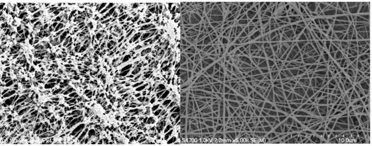
Figure 3.3 Left: SEM image of scaffolds ... 56
Figure 3.4 H&E stains of HBE cells grown on custom scaffolds. ... 57
Figure 3.5 Left: Elastin-Collagen Scaffold ... 57
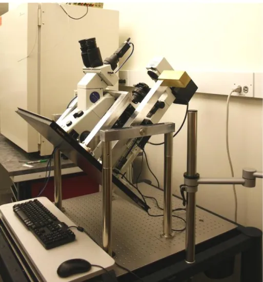
Figure 3.6 Tilting microscope "Ixion".. ... 64
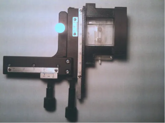
Figure 3.7 Custom built stage ... 65
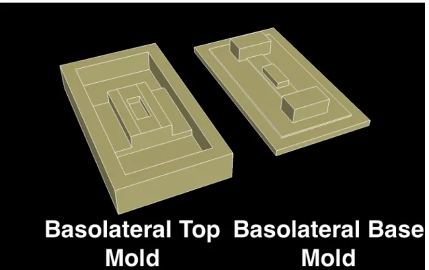
Figure 3.8 MCA mold design ... 66
Figure 3.9 MCA molds ... 67
Figure 3.10 Apical chamber fabrication ... 68
Figure 3.11 Basolater chamber fabrication and final assembly ... 69
Figure 3. 12 Mucus Clearance Assay Device. ... 70
Figure 3.13 Live/dead assay of an entire channel. ... 71
Figure 3.14 Live/dead assay of HBE cells forming a Y junction. ... 72
Figure 3.16 Processing used to measure
Cilia Beat Frequency (CBF). ... 80
Figure 4.1 Correlative microscopy showing lateral heterogeneity ... 92
Figure 4.2 Flow profile of 2um beads in a ciliated fluidics channel ... 95
Figure 4.3 Poiseuille flow profiles at several volumetric flow rates ... 97
Figure 4.4 Cilia beat frequency vs. flow ... 99
Figure 4.5 Flow profiles of opposed cilia driven fliuid flow ... 100
Figure 4.6 Free body diagram of an adhered bead undergoing fluid flow ... 104
Figure 4.7: Diagram showing the mechanism of bead rolling ... 110
Figure 4.8 Adhesion of PEG beads and WGA beads on HBE16 cells ... 111
Figure 4.9 HBE 16 cell surface decorated with FITC-WGAs ... 114
Figure 4.10 Adhesion of beads to an untreated surface ... 115
Figure 4.11 Adhesion of beads to a WGA treated surface ... 116
Figure 4.12 Bubble trap diagram ... 121
Figure 4.13 Bead functionalization protocol. ... 122
Figure 4.14 WGA functionalized microbeads (16um) on HBE 16 cells layer. ... 123
Figure 5. 1 The custom built microscope IXION. ... 128
Figure 5. 2 Different fluid regimes in a partially filled channel ... 129
Figure 5. 3 The velocity profile for vertical liquid films ... 135
Figure 5. 4 Draining buffer and mucus velocity profiles ... 136
Figure 5. 5 The velocity profile for a 1um film of buffer on a vertical surface. ... 137
Figure 5. 7 Buffer transport vs tilt angle ... 141
Figure 5. 8 Particle transport for buffer and mucus at a 60° tilt ... 142
Figure 5. 9 Mucus hurricane streaklines ... 143
Figure 5. 10 Mucus hurricane vs tilt angle ... 144
Figure 5.11 DTT reduction ... 148
Figure 5.12 Effective viscosity of normal and DTT treated mucus ... 149
Figure 5.13 Mucus and DTT Mucus streaklines ... 151
Figure 5.14 Mucus/DTT Mucus velocity field ... 152
Figure 5.15 Buffer/Mucus velocity vs distance from meniscus ... 153
Figure 5.16 Loss and Storage modulus of 4.5% 1MD PEG ... 156
Figure 5.17 Apparent viscosity of 4.5% 1MDalton PEG ... 157
Figure 5.18 Flow profile of a vertical 10um film of 4.5% 1MDalton PEG ... 158
Abbreviations
ABS Acrylonitrile Butadiene Styrene
ALI Air-liquid Interface
ASL Airway Surface Liquid
ATI Alveolar Type I
ATII Alveolar Type II
ATP Adenosine Triphosphate
CBF Cilia Beat Frequency
CCSP Clara Cell Secretory Protein
CF Cystic Fibrosis
CFTR Cystic Fibrosis Transmembrane Conductance Regulator
CISMM Computer Integrated Systems for Microscopy and
Manipulation
Con A Concanavalin A
COPD Chronic Obstructive Pulmonary Disease
DMEM Dulbecco's Modified Eagle Medium
DMT Derjaguin-Muller-Toporov
DTT Dithiothreitol
ECM Extracellular Matrix
EDAC 1-Ethyl-3-[3-dimethylaminopropyl]carbodiimide
hydrochloride
EGF Epithelial Growth Factor
ENaC Epithelial Sodium Channel
HBE Human Bronchial Epithlial
HBE16 Human Bronchial Epithelial 16
IgA Immunoglobulin A
MCA Mucus Clearance Assay
MCC Mucociliary Clearance
MIP Maximum/Minimum Intensity Projection
NA Numerical Aperture
PA Pseudomonas Aeruginosa
PBS Phosphate Buffer Saline
PCD Primary Cilia Dyskinesia
PCL Periciliary Liquid
PCP Planar Cellular Polarity
PDMS Polydimethylsiloxane
PEG Polyethylene Glycol
PIV Particle Image Velocimetry
PTFE Polytetrafluoroethylene
PU Polyurethane
PVC Polyvinyl Chloride
RA Retinoic Acid
SEM Scanning Electron Microscope
TRIS tris(hydroxymethyl)aminomethane
UV Ultraviolet
Vangl2 Van gogh like 2
VNTR Variable Number Tandem Repeats
VWD Von Willebrand Factor Type D
Chapter 1 Introduction
1.1 Motivation
Breathing is important. It shouldn’t come as a surprise that diseases that
affect breathing take a huge toll on human health and life. Respiratory infections and
chronic obstructive pulmonary disease (COPD) are the 3rd and 4th leading killers
worldwide: combined they are responsible for over 7 million deaths a year (Mathers
et al., 2008) How do these diseases sicken and kill? Ultimately it comes down to the
ability of the lung to clear pathogens and toxins. Impaired clearance obstructs the
airways, but long term it triggers a cascade that changes the fabric of the lung itself.
Impaired clearance leads to infection, inflammation and ultimately remodeling of the
lung. Treating and ultimately curing these diseases relies on restoring effective
clearance.
How do we fix clearance? The first step is to understand the mechanisms of
clearance. At its essence mucociliary clearance seems a rather simple concept.
Cells secrete an assortment of molecules in an effort to impede or ensnare potential
invaders and irritants. Ciliated cells transport the resulting detritus out of the airway.
The simplicity of the system quickly disappears when trying to understand all of the
parameters involved in clearance. “Ciliated cell transport” is a complicated process
that pushes our understanding of fluid dynamics, rheology, biochemistry, and cell
protein level. As a result, work in answering these questions has spawned clearance
treatments ranging from using body posture to enhance clearance down to drugs
that correct misshapen proteins. Furthering our understanding of mucociliary
clearance will advance the development of treatments for these awful pulmonary
diseases.
1.2 Thesis Statement and contributions
This thesis argues that in vivo mucociliary clearance is a product of both the
biophysical and biochemical properties of the airway surface liquid. Specifically,
mucociliary clearance relies on the viscoelastic properties of mucus and biochemical
interactions between mucus and cilia to successfully transport mucus against
gravity. One of the key contributions of this work is the development of a mucus
clearance assay (MCA). The MCA integrates cell culture techniques with a
microfluidics platform to provide the first in vitro system capable of
1) Probing adhesion interactions between the epithelium and ligands
found in the airway surface liquid (ASL), and
2) Challenging mucociliary clearance with external fluid flows (including
breathing regimens), and
3) Evaluating the ability of mucus simulants (rheological and
Furthermore as a clearance assay the system can be used to explore clearance of
diseased states and the efficacy of pharmaceuticals in treating those states.
The primary goal of this work and primary use of the MCA is to elucidate the
mechanism of clearance. The mechanisms of mucociliary clearance are still very
much an open question. An important unsolved aspect of mucociliary clearance is
the interaction between the cilium and the overlying mucus gel. Is the cilium merely
an actuator pushing the gel forward, or is there a biochemical connection that
provides a transient adhesion between the cilium and the gel? Prior in vitro systems
have always had the airway surface liquid normal to gravity, this orientation forces
the liquid to self-level over the epithelial surface. The MCA makes it possible to
orient the culture at varying degrees, including parallel to gravity. The vertical
surface orientation removes the artifact of self-leveling as the liquid height is
determined by the surface energy between the luminal surface and liquid along with
cilia driven transport of the liquid. By using fluids with varying rheological and
biochemical profiles, it makes it possible to probe for and to isolate the properties
required for vertical mucociliary clearance. Experiments from the system have
demonstrated that fluids transporting horizontally aren’t necessarily capable of
transporting against gravity. Also the system has demonstrated that viscosity as a
rheological parameter isn’t sufficient in predicting clearance. Finally the system can
be modified to test for adhesion interactions between ASL species and the
epithelium. Results from this system make it possible to form a clearer model of the
1.3 Outline for the document
Chapter 2 starts with a brief overview of the mucociliary system. The chapter
starts by examining the different elements involved with mucociliary clearance MCC:
the epithelium, cilia, mucus and assorted miscellaneous factors. The goal is to
familiarize the reader with the mechanisms of MCC while building a “requirements
list” for a clearance model; this requirements list is important in evaluating existing
model systems and the model system described in this work. The chapter then
explores current model systems. A literature survey describes notable model
systems that have been used to advance the knowledge of lung clearance and
discusses findings as well as shortcomings and remaining questions.
Chapter 3 focuses on the model system, the MCA, which I developed during
the course of my dissertation work. The chapter starts by compiling the design
requirements from chapter 2 and adding specifications relating to cell culture and
intended experiments. These requirements and specs are then weighed and used to
make design decisions. Next the chapter details the fabrication process, sterilization
procedure and associated cell culture. Finally, the chapter closes by looking at data
demonstrating that not only can cells be grown in the system, but also they are well
differentiated, well-ciliated and demonstrate long range ciliary entrainment. Example
measurements of CBF, %ciliation, and flow are shown as well as discussion of the
techniques.
Chapter 4 starts building a simple hydrodynamic model. Initially the goal of
the model is to demonstrate that the beating cilia can be coarse-grained into a shear
agreement with canonical solutions to the Navier-Stokes equations. Ultimately this
model will resurface in chapter 5 to demonstrate that hydrodynamics alone aren’t
enough to describe transport (or lack thereof) in the tilted conformation. I then use
the fluidics aspect of the channel as a force probe to measure the adhesion between
functionalized beads and the epithelial surface. I demonstrate that the clearance
assay can also be used as an adhesion assay to identify and quantify adhesion of
moieties attached to the beads and the epithelial surface.
Chapter 5 extends the model developed in chapter 4 and includes
gravitational drainage. Using this model I determine that if the boundary condition
holds, then buffer, mucus, and pathological mucus would all be able to transport up
a vertical surface. I test this with buffer and mucus and discover that while buffer
transports horizontally, it failed to transport vertically. Next I develop the system into
a mucus clearance assay; I developed a methodology to test which properties
(biochemical or rheological) enable transport against gravity. Finally I test viscosity
with a Polyethylene Glycol solution and determine that viscosity alone doesn’t
enable transport. I’m able to conclude that the boundary condition developed in
chapter 4 changes when the system is in a vertical conformation; gravitational
drainage changes the interaction between the cilia and the ASL. I suggest that
mucus is able to transport vertically in spite of drainage because of biochemical
Chapter 2 Mechanisms of Mucociliary
Clearance
In this chapter I present an overview of mucociliary clearance. The first part of
the chapter introduces different aspects of the mucociliary clearance system. The
goal of these sections is to familiarize the reader with clearance and to discuss key
parameters that potentially govern clearance. Current literature and models suggest
that the major guiding parameters are cilia behavior (beat frequency and distribution)
as well as the ASL’s properties (rheology, hydration). In working towards my
hypothesis that there is a biochemical interaction that plays a role in clearance, I
also present potential binding interactions between cilia and the ASL that could
potentially play a role in ASL transport. The end of the chapter is a survey of existing
animal and in vitro model systems. The goal of this section is to understand the
successes of current model systems, but also their limitations. Finally I close the
chapter by reviewing the key parameters and current model systems to generate
requirements for the model system that I design and build in chapter 3.
2.1 Lung physiology and airway defense
The lungs handle gas exchange for the entire body; the cellular respiration of
trillions of cells depends on the ability of the lungs to take in oxygen and dump
requires that the lungs have a large surface area available for gas exchange, a
surface area that ranges from 24-69m2(Hasleton, 1972).
The lungs achieve a large surface area through a bifurcating tree structure
(Fig. 2.1). Air enters (and exits) the trachea where it splits into two main bronchi that
deliver air to each lung. These bronchi further split into another generation of bronchi
that feed the different lobes of the lung. This branching continues on for a total of
approximately 23 generations (Weibel & Gomez, 1962), at which point the path ends
at alveolar sacs. In general these generations can be classified into two zones. The
conducting zone consists of the approximately the first 14 generations and is
responsible for filtering and conditioning inhaled air. Beyond lies the acinar zone.
The acinar zone makes up about 90% of the lung volume (Weibel & Gomez, 1962)
and is responsible for gas exchange in the lung.
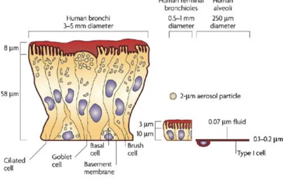
Figure 2.1 Lung branching structure and the characteristic epithelium at each level. Taken from Scotton
The large surface area, however, acts as a double-edged sword. Every
additional square foot available for gas exchange also represents an area that has to
be fortified against potential pathogens. Furthermore this fortification has to protect
the airway, while having minimal impact on breathing. In meeting this challenge of a
large, yet unobtrusive defense system, the airway employs several different lines of
defense. The epithelium itself represents a line of defense: the epithelial cells form a
barrier and can control what crosses the epithelial barrier. In the conducting airways,
mucociliary clearance acts as a large moving filter: pathogens are trapped in a
mucus gel and then transported via cilia out of the airway. Additionally the mucus
gel contains agents such as secretory IgA, lactoferrin, lysozyme, and defensins that
have anti-microbial properties (Goldman et al., 1997; Wanner et al., 1996). Finally
macrophages act as a final line of defense: they actively seek and destroy any
pathogens that are able to penetrate the airway tree.
Amongst these mechanisms, mucus clearance seems to be the primary
defense mechanism(Knowles & Boucher, 2002). The work in this dissertation will
focus exclusively on the mucociliary apparatus. As a consequence the model system
developed for this work will attempt to recreate the environment of the conducting
airways. Specifically my model will attempt to match the geometry and structure of
the bronchioles. While a more thorough discussion of scale and choice will appear in
chapter 3, I should note an important physiological point regarding this choice.
Typically the trachea and large bronchi have a large number of sub-mucosal glands
In order to build a mucociliary clearance model, it’s necessary to have a
strong background of the different components. The next few sections will detail the
different components of the mucociliary apparatus: the epithelium, cilia, and the
airway surface liquid (ASL).
2.2 Epithelium
In discussing MCC in the airways, understanding the pulmonary epithelium is
of vital importance. Mucociliary clearance is an epithelial process. The epithelium’s
importance lies in both the generation of cilia and the airway surface liquid, but also
the regulation of the two. To get a better sense of the epithelium and its involvement
in the MCC process, I will first talk about the most common cells found in the airway
epithelium and the roles as they relate to mucociliary clearance and model systems.
Next I’ll discuss the epithelial structure; how the cells assemble and how that
assembly shapes the epithelial surface. Finally I’ll discuss epithelial secretions as
well as how the epithelium regulates fluids.
2.2.1 Cells of the epithelium
The epithelium is filled with a variety of cells that serve a variety of purposes. The
cells of the airway epithelium include
• Serous cells • Clara cells • Mucous cells • Brush cells
Additionally in the acinar zone there are
• Alveolar Type I pneumocytes • Alveolar Type II pneumocytes
While there are several other types of cells in the airway, I’ll limit discussion to
those above, as they are the cells found superficially and/or directly contribute to the
epithelial environment. The cells mentioned above can be categorized as secretory
cells, brush and ciliated cells, and alveolar cells. The secretory cells produce
several key components of the ASL and thus play a major role in the physical and
chemical properties of the liquid. The ciliated cells play a vital role in transport, but
their contribution to the epithelial surface structures along with brush cells may also
play a role in mucus dynamics. The in vitro model on which I base my clearance
assay produces well-differentiated cells including secretory and ciliated cells
(Fulcher et al., 2005). Furthermore the secreted products of these cells are highly
similar to induced sputum samples. The major differences being saliva, plasma
exudate, immune cells, and surfactant produced by alveolar cells (Kesimer et al.,
2008). My model will not include alveolar cells, but future iterations may incorporate
them to study their effects on clearance and host defense.
2.2.1.1 Secretory Cells
Serous cells
In a 1990 Annual Review of Physiology paper Basbaum et al. compare the
anti-microbial products. Serous cells are the origin of most of the non-mucin protein
found in the ASL (Basbaum, Jany, & Finkbeiner, 1990). Secretory products of these
cells include lysozyme, lactoferrin, peroxidases, secretory IgA, as well as protease
inhibitors, albumin, and high molecular weight glycoproteins. In humans, serous cells
are found only in submucosal glands, and occupy 60% of the gland volume
(Takizawa & Thurlbeck, 1971). In vitro, serous cells have also been grown by
isolating submucosal glands (Finkbeiner, Zlock, Mehdi, & Widdicombe, 2009). The
presence of lactoferrin and lysozyme suggests that serous cells are present in
cultures generated from general tracheal/bronchial isolations. Interestingly the
proportion of serous cells to mucous cells seems to decrease as retinoic acid (RA),
an additive used to promote mucociliary-differentiation, concentration is increased in
media (Gray, Guzman, Davis, Abdullah, & Nettesheim, 1996; Mammoto, Montoya-‐
Zavala, Hsin, & Ingber, 2010).
Clara cells
Clara cells are found in the terminal bronchioles of the airway where they
perform diverse tasks (Jeffery & Li, 1997). One function is that of a precursor cell;
Clara cells have the ability to differentiate into both mucous and ciliated cells (Ayers
& Jeffery, 1988). Clara cells also have a diverse array of secretory products. Clara
cell secretory protein (CCSP) has been shown to act as an anti-inflammatory by
regulating macrophage behavior (Snyder et al., 2010). Cytochrome P450 helps
metabolize toxins (Devereux et al., 1981). Clara cells also produce surfactant protein
D, a C-type lectin (Crouch, Parghi, Kuan, & Persson, 1992).
Mucous cells, also known as goblet cells are found in both the surface
epithelium and in sub-mucosal glands. In the trachea there are ~6,500 mucous cells
per mm2(Ellefsen & Tos, 1972), but this number decreases further down the airway
tree (Wanner et al., 1996). Mucous cells are often referred to as goblet cells because
of their characteristic shape. Histology sections show that they resemble a goblet:
the nucleus is towards the bottom of the cell while a large secretory granule
dominates the top half of the cell (Rhodin, 1966). Goblet cells are the major source
of the gel forming mucins that form the mucus layer, which I’ll discuss later in this
chapter. Functionally the cells can store large mucin reserves and secrete those
reserves into the ASL. The stored mucus capacity for the lung has been estimated at
4ml (Reid, 1965) and it takes between 2-4 hours for a goblet cell to synthesize and
secrete Muc5AC(Sheehan et al., 2004).
2.2.1.2 Brush and Ciliated Cells
Brush Cells
Brush cells are found throughout the airway and feature a tuft of microvilli on their
surface. Their function isn’t well understood but the following roles have been
speculated (Reid et al., 2005).
1. Absorptive
2. Chemoreceptors
3. Immune surveillance
4. Detoxification
Ciliated Cells are terminally differentiated cells, each of which is covered with
an average of 200 hair like projections called cilia(Rhodin, 1966). Cilia drive the
mucociliary system; understanding their movement, organization, and interaction
with mucus is a critical step in understanding mucociliary clearance and an area in
which I will go into greater detail in subsequent sections. In addition to cilia, these
cells also have microvilli and glycocalyx on their apical surface(Rhodin, 1966). The
number of ciliated cells varies with the location in the airway tree. In the human
trachea ciliated cells outnumber mucous cells by a ratio of 5:1, but the numbers
decrease in more distal generations (Wanner et al., 1996).
2.2.1.3 Alveolar cells
Alveolar Type I
Alveolar Type I (ATI) pneumocytes are found in the respiratory zone of the
lung and are the main constituents of the alveoli; they make up 90% of the alveolar
surface (Haies, Gil, & Weibel, 1981). Structurally ATI cells are ideal for diffusion; they
are extremely thin with a large surface area. Each cell has a surface area of
5100um^2 (Crapo, Barry, Gehr, Bachofen, & Weibel, 1982) and an average thickness
of only 200nm (Weibel, Gehr, Haies, & GIL, 1976)
Alveolar Type II
Alveolar Type II (ATII) pneumocytes make up 10% of the alveolar epithelial
surface (Haies et al., 1981). They produce surfactant proteins A, B, C,D lysozyme,
and lysosomal enzymes. (Voorhout, Weaver, Haagsman, Geuze, & Van Golde, 1993)
2.2.2 Epithelial Cross-Section
As shown in figure 2.1, the airway epithelium changes dramatically depending
on the location in the lung. Figure 2.2 further illustrates the degree to which the
epithelial profile changes. The thickness of the epithelium transitions from a ~60um
pseudostratified columnar profile in the proximal lung to a 100nm single cell layer in
the alveoli (Patton & Byron, 2007). In addition to the thickness, the airway structure
and distribution of cell types changes. The percentage of ciliated and mucous cells
decrease distally as do the number of sub-mucosal glands (Whimster, 1986), which
are mainly absent after the 10th generation (Wanner et al., 1996).
Figure 2.2 Epithelial profile with various cell types. The epithelial profile transitions from a 60um
thickness down to a 100 nanometer thickness in the distal airways. Taken from Patton 2007
The epithelium serves as a barrier itself; cells of the epithelium are bound
lumen from the rest of the body. This seal allows the epithelial cells to function as
gatekeepers. This regulation extends down to the ion level; ions can only cross the
epithelium through ion channels on the plasma membrane of epithelial cells. Using
this mechanism the epithelium is able to regulate the ASL volume via osmotic
pressure (J. Widdicombe, Bastacky, & Wu, 1997), a mechanism that I’ll revisit in
section 2.4.4.
The epithelial surface is covered by a variety of surface structures, which
range in size from nanometers to microns. Understanding these structures and their
biochemistry is important for eliciting any potential interactions with mucins.
2.2.2.1 Glycocalyx
Closest to the cell membrane is the glycocalyx. The glycocalyx is a collection
of various polysaccharides extending 500-1500nm from the cell surface (Knowles &
Boucher, 2002) and it covers all of the various cell types. The glycocalyx has many
roles including cell adhesion, protection from pathogens, and signaling. A review by
Martins et al. covers more of the specifics of the pulmonary glycocalyx (Martins &
Bairos, 2002). The glycocalyx’s ubiquity and large surface area make it a prime
candidate for potential biochemical adhesion between cilia and the ASL.
Looking for these adhesions requires knowledge of the specific saccharides
present in the bronchial epithelial glycocalyx. The bronchial epithelial glycocalyx is
rich in sialic acid residues, MUC1 (discussed in section 2.4.1.1), and localized
keratan sulfate. In vitro cultures express the same moieties as in vivo, but the
Understanding the availability of these moieties will be important in developing the
adhesion assay in chapter 4.
2.2.2.2 Microvilli
Microvilli are small ~1 micron tall protrusions coming from the cell surface,
particularly brush cells and ciliated cells. Microvilli are themselves covered by the
cell membrane and glycocalyx (Alexander, Ritchie, Maloney, & Hunter, 1975).
2.3 Cilia
Figure 2.3 SEM image showing a side view of cilia. Cilia are about 6-7um long .
Cilia are small hair-like organelles expressed on the luminal side of the airway
epithelium (fig 2.3). Through regular beating, cilia work in concert to transport mucus
up the pulmonary tree. The mechanism of this interaction, however, is still very much
an open question. Specifically this thesis investigates the nature of the interaction
mucus layer, or is there a biochemical interaction between cilia and mucus?
Exploring their role as actuators requires knowledge of cilia size, coverage, and cilia
dynamics: the dynamics of individual cilia as well as their organization. Additionally
in creating the cilia model discussed in chapters 4 and 5, it’s important to know what
external factors could change cilia behavior. Finally in exploring potential
biochemical interactions an awareness of the cilia surface is necessary.
I’ll start with a brief background on cilia structure and function. Next I’ll review
a couple of papers that discuss a particularly important aspect of cilia dynamics, cilia
entrainment. Cilia entrainment, the alignment of cilia across the epithelial surface, is
required for mucociliary clearance. It’s of particular interest to my project because it
is found in vivo, but the mechanisms of reproducing it in vitro are just starting to be
understood. I’ll conclude this section by discussing the cilia surface and its local
environment.
2.3.1 Cilia Primer
The epithelial surface of the proximal conducting zone is dominated by cilia:
as stated earlier ciliated cells outnumber mucous cells 5:1 in the trachea (Wanner et
al., 1996). Each ciliated cell is home to approximately 100-200 cilia located on the
apical surface. Each individual cilium has a length of about 6 microns and a diameter
of 300 nanometers(Rhodin, 1966).
In order to propel mucus each cilium oscillates at a range of 10-20Hz, this
large range being a product of normal variation, environmental conditions as well as
location in the airway tree (Luk & Dulfano, 1983) (Rautiainen et al., 1992)(Rutland,
components; an effective stroke and a recovery stroke. The effective stroke is in the
“forward” direction and occupies about 40% of the stroke cycle. During this phase
the cilium stretches the farthest away from the cell, presumably to interact with the
mucus gel. The recovery stroke occupies the remaining 60% of the cilia beat and
travels in the “backwards” direction. During the recovery stroke the cilium travels
closer to the surface of the cell, which presumably minimizes the interaction with the
overlying gel (Sanderson, 1981)(Marino, 1982).
Several studies have examined external factors that influence cilia beat
frequency. Clary-Meinesz et al. showed that CBF had a temperature dependence in
nasal and tracheal brushings. They observed a 50% decrease in CBF when
temperature was decreased from 20C to 9C (Clary-‐Meinesz, Cosson, & Huitorel,
1992). Additionally, several groups have also looked at mechanosensitivity of CBF.
Lansley et al. showed CBF rapidly increased from an increase in intracellular
calcium, an increase triggered by mechanical stimulation. Rabbit tracheal cilia at
37C increased their beat frequency from 17.2 to 26.7Hz with a corresponding rise
intracellular calcium concentration of 100-650nM (Lansley & Sanderson, 1999).
Winters et al. observed mouse tracheal CBF increase in response to an applied
shear-stress(Winters, Davis, & Boucher, 2007). Button et al. observed an increase in
CBF as a result of a cyclically applied trans-epithelial pressure(Button, Picher, &
Boucher, 2007a).
Equally important to CBF is cilia coordination. In general the cilia of the lung
are entrained in such a way that their effective strokes are all pointed in the
demonstrate temporal coordination; the beats of neighboring cilia are offset so that
they produce a metachronal wave(Gheber & Priel, 1989). The origin of this
coordination/entrainment is a heavily studied area, and rightly so. It represents a
major element still poorly understood in in vitro cell cultures.
2.3.2 Cilia Organization and Entrainment
Cilia are coordinated at the cell level and on the tissue level. This coordination
is an essential element of mucociliary clearance and highly desirable in a clearance
assay. Starting at the cellular level, cilia are all “pointed” in the same direction. After
a certain level of maturation, cilia are locked into a certain direction. Each cilium has
an “anchor” in the cell called the basal foot, which locks into a certain direction as
the cilium grows and matures (Lemullois, Klotz, & Sandoz, 1987). In healthy cells
roughly all of the cilia are oriented such that their effective strokes are in the same
direction. This coordination goes beyond the cellular level; in healthy in vivo systems
neighboring ciliated cells also have their cilia pointing in the same direction. In fact
this coordination spans the tissue, and is referred to as tissue polarization. Tissue
polarization is essential to functional mucociliary clearance, it guarantees that all of
the cilia have their effective strokes oriented to clear mucus out of the airway as
opposed to deeper into the lung. Outside of the airway, the signaling that
coordinates tissue polarization has been a hot field of study. Several groups are
looking at the Planar Cellular Polarity (PCP) pathway and the proteins that regulate it
in several organisms across several different types of tissue. Several studies have
specifically tried to understand the role that the PCP signaling pathways and
In a 2007 Nature paper, Mitchell et al. investigated the developmental
mechanisms that polarize cilia on the skin of Xenopus embryos. Initially they looked
at the basal body orientation in several cells in vivo. Basal feet were roughly aligned
prior to cell differentiation. This alignment was present at the cell level as well as
across several cells (clusters). The degree of alignment improved as the system
matured. The cilia produced a strong directed flow, which Mitchell called the
“Refinement Phase”. The initial alignment is created by a “Patterning Phase”. Next
they looked at in vitro cells taken before and after the patterning phase. Explants
taken prior to the patterning phase didn’t align the same way and were “chaotic”,
explants taken after the patterning demonstrated alignment similar to in vivo results.
Next Mitchell et al. investigated the relationship between cilia function and
polarization. They injected morpholinos to block expression of gene products
required for cilia motility: essentially they gave the embryos the equivalent of Primary
Cilia Dyskinesia (PCD), a genetic disease that disrupts normal cilia motility. The
embryos still showed the rough alignment, but never experienced the refinement
step. Again they looked at explants that were taken before the patterning stage.
They exposed these to an external flow and saw an improvement in alignment, but
the alignment wasn’t as good as the wild type. They also used a 1Hz oscillating flow
and saw no alignment. Finally they exposed mutated PCD-like explants to flow and
didn’t see alignment. Their major conclusions were
1. Most of the alignment takes place before differentiation/ciliation
3. In the absence of a patterning phase, external flow (with a shear-stress of
0.5 dynes/cm2, the same shear-stress applied to the ASL surface from
breathing (Tarran et al., 2005)) will still lead to alignment, but not the same
degree as seen in vivo.
4. Motile cilia are required for flow to affect alignment.
Guirao’s 2010 Nature Cell Biology paper investigated cilia orientation in
mouse brain ventricles (ependymal cells). They argue that this model, as well as
other mammalian systems, requires a mechanism other than the 2-step
patterning/refinement mechanism observed in Xenopus. Initially they looked at cilia
coordination in in vivo models as a function of time. The immature cells had
randomly aligned basal feet, which is opposite to the patterning that Mitchell et al.
observed in Xenopus.
In their in vitro system, 60% of the ciliated cells formed in clusters
surrounded by non-ciliated cells. They first concluded that isolated cells can align
their own cilia, and that cell clusters align as well. They also cite their earlier
theoretical work(Guirao & Joanny, 2007) for the mechanism by which cilia are
aligned by an external force. If a cilium is not beating in the same orientation as the
flow, then the flow will apply a torque to the cilium. If the basal body is free to rotate,
the cilium will align itself with the flow. Next, they exposed in vitro cultures to an
external flow via a rotating plate. The plate was situated atop the media above the
cells. They rotated the plate so that the cell layer experienced a shear stress of
aligned with the external flow, and that cultures exposed after this time period didn’t
realign. Cultures not exposed to flow showed “typical” cluster behavior. They then
investigated the effect of PCP signaling, specifically the PCP protein Van Gogh-like
2 (Vangl2). In immature cultures they found Vangl localized at the apicocaudal
borders of cells. More mature cultures showed asymmetric localization and that the
protein localized along cilia from the tip to the base. They looked at Vangl2 mutants,
which produced structurally normal cilia. In these mutants, theVangl2 protein
aggregated in the cytoplasm instead of localizing at the membrane As a result, the
cilia generated flow didn’t affect each other at the cell or cluster level. They
concluded that Vangl2 is required for cilia to be influenced by hydrodynamic forces.
They explored this in vivo and found the same result. They then looked at a mutant
that didn’t have cilia. They found the asymmetric localization of Vangl2 even without
cilia present; however, the basal bodies were still misaligned in mature cultures. So
Vangl2 alone isn’t enough for orientation. They conclude the following
1. There doesn’t appear to be a patterning phase that aligns cilia in
mouse ependymal cells and possibly mammalian cells in general.
2. Cilia will tend to align themselves in clusters.
3. Cilia can be aligned via an external force, but only during a small
window
4. Not only are motile cilia required for flow-driven alignment, but also
5. Despite the lack of “patterning”, Vangl2 still localizes
asymmetrically.
6. In mutants that prevent ciliogenesis, the basal bodies still misalign
despite the Vangl2 localization.
Further studies have shown that both actin and microtubules play a large role in
establishing PCP protein asymmetry across cells and orienting a basal body network
(Vladar, Bayly, Sangoram, Scott, & Axelrod, 2012; Werner et al., 2011).
There are still many unanswered questions relating to cilia alignment
particularly to lung epithelial cilia entrainment. What are the origins of entrainment in
vivo? At what stage of cilia growth can cilia be entrained? Do airway cilia undergo a
patterning step? Can airway cilia be entrained with an external force? The last
question is of particular interest in designing the clearance system. By having a
mechanism to deliver external flow, the mucus clearance assay could potentially be
used to test this result in human bronchial epithelial (HBE) cell cultures.
In addition to spatial coordination, cilia are also coordinated temporally
(Wanner et al., 1996). Neighboring cilia have an offset in their strokes so that there is
a time lag. This time lag produces metachronal waves; a frequent example often
cited is “the wave” that a crowd performs at a football stadium. The net effect of this
is that there is a longer wavelength, slower frequency wave propagating along the
ASL boundary. As I’ll discuss later, mucus’ viscoelasticity leads to a time-dependent
mechanical response. For this reason it’s important to be aware of the metachronal
2.3.3 Beyond Cilia: Cilium Surface and the PCL
Understanding the cilium surface is important in identifying potential
biochemical interactions with mucus in the ASL. The cilia membrane is similar to the
cell membrane and it has recently been shown that MUC16 and MUC4 are present
on the cilia membrane (Button et al., 2012). In attempting to identify a biochemical
interaction, there are several examples of the cilia membrane having adhesive
functions. Chlamydomonas uses adhesive cilia for motility (Bloodgood, 1990) and
mating (Mesland & Ende, 1979). Cilia adhesion is used by mammalian oviduct cilia to
help transport oocyte-cumulus complexes (Talbot, Shur, & Myles, 2003). I discuss
cilia-mucin adhesion in greater detail in section 2.4.5. More examples of the
adhesive functions of cilia and a more complete picture of the cilia membrane can be
found in a 2008 Pazour chapter of Current Topics in Developmental Biology (Pazour,
2008).
The periciliary layer/periciliary environment (PCL) is the intermediate region
that separates the epithelial surface from the overlying gel phase of the ASL. The
PCL varies in height over different cell types: it ranges from 7um over ciliated cells to
3um over other non-ciliated cell types(Tarran et al., 2001). It’s been hypothesized
that the membrane-bound mucins and other glycolipids on the cell surface create a
framework for the movement of water into this region(Randell, 2006). Button et al.
further developed this into a “Gel-on-brush” model that proposed that membrane
spanning mucins and mucopolysaccharides attached to cilia, microvilli and the
epithelial surface sterically block large particles (large than 20-40nm) from
transport exists in the PCL, but that this transport is highly dependent on the
overlying mucus layer. PCL transport is reduced by more than 80% without an
overlying mucus layer(Matsui, Randell, Peretti, Davis, & Boucher, 1998b).
2.4 Airway Surface Liquid
The airway surface liquid (ASL) performs several important functions in
maintaining pulmonary health and sterility. The first function is that of a barrier and
trap: pathogens and toxicants are impeded and ensnared by the mucus gel.
Throughout the pulmonary airway, the epithelial surface is normally covered by a
liquid layer ranging from 5-20 um in thickness (J. Widdicombe et al., 1997). Secondly,
the ASL has a variety of antimicrobial agents that can disable potential invaders.
Finally the ASL acts a cilia-driven transport vehicle to remove detritus from the
airway(J. H. Widdicombe, 2002). This dissertation is chiefly concerned with the last
point: understanding how the cilia interact with the mucus to remove it from the
system. Knowledge of the other two functions, however, is still important in that their
actions may have an effect on mucus transport and could potentially hint at
biochemical interactions.
The ASL features a diverse collection of species that provide the
aforementioned functions. A 1996 review of mucociliary clearance lists
glycoproteins, proteoglycans, lipids, secretory IgA immunoglobulins, lysozyme,
peroxidase, lactoferrin, DNA, actin as the constituents of the ASL (Wanner et al.,
1996). A 2002 review further lists several additional polypeptides and defensins that
that act as antimicrobials in the ASL (Ganz, 2002). Finally, it’s important to remember
Boucher, Stutts, Bromberg, & Gatzy, 1981). Furthermore, these electrolytes have been
implicated in defensin activation (J. Smith, Travis, & Greenberg, 1996), and ASL
volume regulation/hydration (Matsui, Grubb, Tarran, Randell, Gatzy, Davis, et al.,
1998a)(Mall, Grubb, Harkema, & O'Neal, 2004). In exploring potential biochemical
interactions between the ASL and the cilia, it is important to realize that any of these
species could act as potential intermediaries or catalyzing agents. In an effort to
keep the problem somewhat tractable, I’ll focus on the high molecular weight
glycoproteins, the mucins, present in the ASL.
2.4.1 Mucins and Mucus
Mucins are high molecular weight glycoproteins that form the structure of the
mucus gel (Verdugo, 1991). Structurally mucins have a central protein core that has
at least one, and often times many, domains that are heavily glycosylated with
oligosaccharide side chains. The heavily glycosylated regions, mucin-like domains,
feature an abundance of serine and threonine, which serve as attachment points for
O-glycans These O-glycans attach to the serine/threonine through an
N-acetylgalactosamine sugar. The rest of the O-glycan is composed of varying
combinations of N-acetlygalactosamine, N-acetlyglucosamine, galactose, fucose,
sialic acid, and sulfate (Thornton, 2004). A more thorough review of the O-glycans
and their assembly can be found in Essentials of Glycobiology (Varki, 1999).
The O-glycans in the mucin-like domain confer a couple of important
properties to the mucins and by extension the mucus gel. Firstly the O-glycans play
a major part in the conformation of the mucin. The high-concentration of
nature of the carbohydrates heavily influence the shape of the mucin. The
carbohydrates sterically prevent secondary and tertiary structures, which results in a
linear extended conformation as compared to other proteins of the same contour
length (Jentoft, 1990). Another important function of the O-glycans is their
biochemical functionality. The oligosaccharides are biochemically similar to the
glycocalyx, and thus they can intercept microorganisms that target the glycocalyx
(Wanner et al., 1996) (Carnoy et al., 1994)(Lamblin et al., 1992; 1991). It’s also
important to note that there are oligosaccharide moieties present in mucins that are
associated with known biochemical adhesions. Sialyl Lewis X (fig 2.4), for example,
is present in mucins (Allahverdian, Wojcik, & Dorscheid, 2006), but also plays a major
role in leukocyte tethering to the endothelium (Lawrence & Springer, 1991; McEver,
2005). Due to these interactions and the variation present in the O-glycans, they
Figure 2.4 PNAd and PSGL-1, mucins expressed on endothelial cells and leukocytes respectively, are
capped by Sialyl Lewis X. L-selectin binds to the sialic acid and fucose ends of Sialyl Lewis X. This bond
plays a major role in leukocyte adhesion and rolling. (Taken from McEver 2005)
Structurally the mucin-like domains have a variable number of tandem
repeats (VNTR) and thus these domains can vary in size between individuals
(Hattrup & Gendler, 2008). The organization of these mucin-like domains into mucin
monomers can be separated into two different families: membrane bound (tethered)
2.4.1.1 Membrane Bound Mucins
Membrane bound mucins are named so because they are characteristically
found near the cell surface and are tethered to the cell membrane. Structurally they
are composed of two subunits. The larger of the subunits is dominated by a VNTR
domain and located outside of the cell. The smaller subunit consists of a small
extracellular region, a transmembrane domain, and a cytoplasmic tail. In the airway,
the primary membrane bound mucins are MUC1, MUC4, and MUC16 (Fig 2.5). The
structure of these mucins prevents them from polymerizing, so they are found in
monomeric form. It should be noted that despite their name, they can also be found
in the mucus layer and represent about 10% of the ASL mucins. A much deeper
more detailed description of membrane bound mucins can be found in Hattrup’s
2008 review Structure and Function of the Cell Surface (Tethered) Mucins (Hattrup
Figure 2.5 Structure and sizes of Membrane bound mucins. Hattrup 2008
2.4.1.2 Gel Forming Mucins
Unlike membrane bound mucins, gel-forming mucins linearly polymerize into
polydisperse chains ranging in length from 0.5-10um. This polymerization is enabled
by structural differences in the mucin monomers. In gel-forming mucin monomers,
the mucin-like domain is sandwiched between protein domains rich in cysteine
residues (Fig 2.6). The monomers polymerize into linear chains through disulfide
linkages between the cysteine rich, protein domains. The mucous gel is then formed
by a combination of entanglement and reversible Ca+2 dependent crosslinks
forming mucins are MUC5AC and MUC5B, where together they make up 90% of the
mucin raft (Fig 2.7)(Hattrup & Gendler, 2008). These two species also have an
interesting difference in their origins. MUC5AC is produced primarily by goblet cells
on the surface of the epithelium, while MUC5B is typically produced in sub-mucosal
glands. Thornton speculated that their differing origins and secretory control
mechanisms could allow a mechanism for controlling the properties of the mucus gel
by fine-tuning the ratio of MUC5AC to MUC5B (Thornton, 2004).
In creating a mucus clearance assay, it is important to have a solid
understanding of differences between their presence and properties in vivo versus in
vitro. In a 2009 paper, Kesimer et al. compared the apical secretions from ALI
cultured HBE cells versus Induced Sputum. They found that overall the compositions
were similar, but there were some important distinctions. MUC5B and MUC5AC
appear at similar percentages 38% to 30% respectively in induced sputum vs. a 45%
to 21% difference in cell culture respectively. Percentages were determined by
looking at the non-glycosylated regions of the protein, so in essence it is the number
of mucin monomers of a species expressed over all of the mucin monomers
detected (Kesimer et al., 2009). Another interesting difference exists between the
types of MUC5B expressed in cell cultures. In vivo MUC5B is produced in two
different glycoforms: one is high charge and the other is low charge. MUC5B from
cell culture, however, seems to be more homogeneous with only one glycoform
appearing (MUC5AC is homogeneous in both cases) (Thornton, 2004). I would like
to reemphasize that this section on the structure and components of the mucins
Polymeric Mucins in Airway Mucus (Thornton et al., 2008)for more information about
gel-forming mucins and their structure and Kesimer’s Tracheobronchial air-liquid
interface cell culture: a model for innate mucosal defense of the upper airways?
(Kesimer et al., 2009)for an in depth comparison of in vivo versus. in vitro
constituents of the ASL.
Figure 2.6 Organization and structure of the gel forming mucins found in the airway. (Thornton 2008)
Figure 2.7 Distribution and origin of different mucin types in the airway surface liquid. Muc5B and
Muc5AC make up 90% of the mucins on the epithelial surface, the remaining 10% are cleaved membrane
bound mucins (from Hattrup 2008)
2.4.2 Properties of the Mucus Gel
The gel forming mucins account for the majority of the “structure” of mucus,
and as a result they heavily influence the mechanical properties of the mucus gel. As
discussed previously, the individual mucins are relatively stiff molecules that are
crosslinked near their ends. The crosslinked mucus network behaves as a
viscoelastic liquid. An understanding of mucus viscoelasticity is necessary to study
how the mucus gel reacts to cilia, gravity, and ultimately clears out of the
enough of a background to understand some basics of mucus behavior, but more
importantly to understand the theories and experiments presented in sections 2.4.3
through 2.5.
2.4.2.1 Viscoelasticity
Viscoelastic materials exhibit viscous and elastic properties in response to an
applied force. In modeling the elastic behavior, we fall back on Hooke’s law and the
concept of an ideal spring. If we displace the spring by distance x, we will have a
restoring force that opposes that displacement by a spring constant k.
𝐹= −𝑘𝑥 (2.1)
Which can also be expressed in terms of an applied stress (force per area) σ, an
extensional strain ϵ and the elastic modulus E.
𝜎= 𝐸𝜖 (2.2)
This equation is for extensional stress, a stress applied normal to the incident
surface. If instead we apply a stress along a surface, we refer to it as a shear stress
σ, the resulting displacement a shear strain 𝛾, and the ratio of proportionality is the
shear modulus.
𝜎=𝐺𝛾 (2.3)
In dealing with viscoelastic fluid, our forces will be in the form of shear stresses, so
we will primarily use equation 2.3. A quick point in regards to elasticity;
fundamentally, elasticity is a function of bonds. Whether we are talking about a
elastic relationship only holds true as long as the bonds are still intact. When a yield
stress, a stress strong enough to irreversibly break the bonds is applied, the material
no longer exhibits elastic behavior.
If elastic behavior is the mechanism by which a system stores energy from an
applied stress, viscous behavior is the mechanism by which the system dissipates
energy from an applied stress. Conceptually it helps to think of this dissipative loss
as a friction between strata of a fluid. If a shear-stress is applied to the top stratum,
that stratum will move at a certain velocity. Subsequent strata will also move, but the
frictional force reduces the velocity. This can be expressed as
𝜎= 𝜂𝛾 (2.4)
The applied shear-stress is proportional to the strain rate 𝛾 by the viscosity 𝜂.
This equation follows our intuition; if we apply the same amount of shear to materials
with different viscosities, the more viscous material will strain at a lower rate.
In modeling material responses, simple mechanical schematics are useful in
understanding the dynamics. Elasticity is represented by a spring, while viscosity is
modeled as a dashpot. Viscoelastic materials are thus modeled as a combination of
springs and dashpots. The Kelvin-Voigt model consists of a spring and dashpot in
parallel and can be represented mathematically as the sum of the elastic and
viscous responses
𝜎= 𝐺𝛾+ 𝜂𝛾 (2.5)
The Kelvin-Voigt model is often used for viscoelastic solids; the spring
any displacement. The Maxwell model consists of a spring and dashpot in series
and can be represented mathematically by
𝛾 = 𝜎
𝐺+ 𝜎
𝜂 (2.6)
This model is often used for viscoelastic fluids, and further components can be
added to model more complicated fluid behavior.
We can extend these models to look at oscillating stresses or strains. G* is
the complex modulus and can be represented as
𝐺∗(𝜔) = 𝜎!
𝛾! (2.7)
Where 𝜎! and 𝛾! are the maximum shear and stress for a given frequency 𝜔. Along
with G*, the phase lag between the stress and strain, δ, is useful in characterizing
the mechanical response. It’s also useful to express the complex shear modulus as
a sum of the real and imaginary components. Expressing in this manner gives
𝐺∗ 𝜔 = 𝐺! 𝜔 +𝑖𝐺"(𝑤) (2.8)
Where G’ is referred to as the storage modulus and G” is referred to as the loss
modulus. These quantities, along with the tangent of δ will be very useful in
describing the rheological properties of mucus.
2.4.2.1 Mucus rheology parameters
Now that I’ve defined a few basic concepts about viscoelasticity, I’ll briefly
discuss some properties of mucus viscoelasticity. In a 1998 paper, King lists the
following properties of mucus (King, 1998)