Sara Hanna
A dissertation submitted to the faculty of the University of North Carolina at Chapel Hill in partial fulfillment of the requirements for the degree of Doctor of
Philosophy in the Curriculum of Genetics and Molecular Biology.
Chapel Hill 2013
Approved by William Y. Kim, M.D Victoria L. Bautch, PhD. Charles M. Perou, PhD.
ABSTRACT
SARA HANNA: HIF1α and HIF2α independently activate SRC to promote melanoma invasion and metastasis
(Under the direction of Dr. William Kim)
Melanoma incidence is increasing at an epidemic rate of 3% per year. When caught early, the disease is easily curable with surgical removal of the primary tumor. However, melanoma is notable for its propensity to metastasize, the leading cause of death in cancer patients. Here we study the role of the hypoxia-inducible factors in the malignant progression of melanoma, both in vivo, in a genetically engineered mouse model of metastatic melanoma, and in vitro, in human melanoma cell lines. The hypoxia-inducible factor (HIF) family of
These results establish the importance of HIFs in melanoma progression and demonstrate that HIF1α and HIF2α activate independent transcriptional
ACKNOWLEDGEMENTS
I want to thank the numerous people who have supported me during graduate school. Through good times and bad, I have been surrounded by people who have always believed in me and my ability to achieve to my goals. I wish to first thank my family, my mom, dad, brothers, and sisters-in-law, who have always been there to pick me up and to push me forward. Their
unconditional love and support has gotten me through multiple tough times and make me laugh even when things are tough. I wish to thank Guillermo for supporting me and encouraging me during the whole dissertation process. Through the late nights and emotional highs and lows, he has been a rock and has truly helped me.
During my graduate career, I have been fortunate to work with so many wonderful, fun, intelligent, and hard-working people. I would like to thank Billy, my mentor, for taking me on as his first graduate student and for giving me incredible support and guidance throughout the past seven years. I would also like to thank Ned for giving me a place to work in the beginning and for
members of the Sharpless lab, past and current, who have helped me throughout these past years. Thanks to the members of the Kim lab for all their help and for putting up with me for the past years. You guys really made me look forward to coming into lab and I appreciate all your help and guidance through the hard times, and especially the fun and laughter through the good times. I want to especially thank Bhavani and Sean for helping me with this project.
LIST OF TABLES ... viii
LIST OF FIGURES ...ix
Chapter I. Melanoma: The Disease and Its Genetics... 1
A. Melanoma Development ... 2
B. Histiologic Types of Melanoma ... 4
C. Melanoma Staging... 6
D. Melanoma Treatment... 10
E. Melanoma Epidemiology and Risk Factors... 11
F. Inherited Predisposition to Melanoma... 13
G. Genetics of Melanoma... 14
H. Genetically Engineered Mouse Models of Melanoma ... 26
I. Melanoma and Metastasis ... 30
J. Summary ... 33
II. Hypoxia-Inducible Factors ... 35
A. Structure of Hypoxia-Inducible Factors... 35
B. Regulation of HIF ... 37
G. HIF and Melanoma ... 52
III. HIF1α and HIF2α activate SRC promoting melanoma invasion and metastasis... 56
A. Overview ... 56
B. Introduction ... 57
C. Results... 60
D. Discussion ... 75
E. Materials and Methods... 81
F. Supplemental Methods ... 89
G. Figures... 93
H. Supplemental Figures... 101
IV. Conclusions and Future Directions ... 111
A. HIF Expression and Metastasis ... 113
B. HIF and Metastasis to Regional Lymph Nodes... 116
C. HIF Expression Increased SRC Activation ... 119
D. Clinical Applicatoins of Findings ... 123
E. Summary... 126
Table
1. Clinical Subtypes of Melanoma... 5
2. Melanoma TNM Classification ... 8
3. AJCC Stage Groupings for Melanoma... 9
4. Common Mutations in Melanoma ... 15
5. Genetically Engineered Mouse Models of Melanoma... 28
6. HIF Activation in Human Cancer... 42
Figure
1. The Initiation and Progression of Melanoma ... 3
2. RAS Pathway... 17
3. CDKN2A Locus and Pathway ... 23
4. PTEN Pathway... 25
5. Domain Structure of HIF ... 36
6. Post-translational Regulation of HIF ... 38
7. Targets of HIF1α and HIF2α... 40
8. Targeting the HIF Pathway in RCC... 46
9. Steps to Metastasis... 47
10. HIF Activation in Melanoma Pathways ... 54
11. Inactivation of HIF1α or HIF2α does not affect initiation, survival, or growth of Pten; Braf melanomas, but abrogates lymph node metastasis... 93
12. Hypoxia enhances melanoma cell invasion and invadopodia formation... 94
13. Knock-down of HIF1α or HIF2α reduces the hypoxia- inducible invasion and invadopodia formation of melanoma cell lines... 95
14. HIF1α and HIF2α upregulate PDGFRα and FAK and are necessary for hypoxia-induced SFK activation... 96
15. Expression of stabilized HIF1α and HIF2α are suffcient to enhance normoxic melanoma cell line invasion... 97
18. Knock-down of Hif1α and Hif2α reduces the hypoxia-induced invasion
of cell lines derived from Braf-activated, Pten-deficient melanomas ... 100
19. HIF Regulates a Transcripional Program to Increase Invasion in Melanoma ... 112
Supplemental Figures 1. Inactivation of Hif1α and Hif2α does not affect tumor free survival ... 101
2. A subset of mleanoma cell lines express HIF1α in normoxia ... 102
3. Hypoxia promotes melanoma cell line motility in a HIF1α and HIF2α dependant manner... 103
4. Hypoxia promotes melanoma cell line invasiveness in a HIF1α and HIF2α dependent manner... 104
5. HIF1α and HIF2α directly regulate PDGFRα and FAK transcription ... 105
6. HIF1α and HIF2α stabilization at normoxia increases invadopodia formation ... 106
7. Stabilized HIF1α and HIF2α decrease the stiffness of A375 SM cells ... 107
8. Inhibition of SRC activity decreases HIF=induced invasion... 108
LIST OF ABREVIATIONS
α-MSH α-Melanocyte Stimulating Hormone
AJCC American Joint Committee on Cancer
ALM Acral Lentiginous Melanoma
ARNT Aryl Hydrocarbon Receptor Nuclear Translocator
ATP Adenosine Triphosphate
bHLH Basic Helix-Loop-Helix
cAMP Cyclin Adnenosine Monophosphate
ccRCC Clear Cell Renal Cell Carcinoma
CDKN2A Cyclin-Dependent Kinase Inhibitor 2A
CDK4 Cyclin-Dependent Kinase 4
COX-2 Cyclooxygenase-2
CODDD Carboxy-terminal Oxygen-Dependent Domain
CPD Cyclobutane Pyrimidine Dimers
Cre Cyclization Recombination
CSC Cancer Stem Cells
CTAD Carboxy-terminal Activation Domain
CTC Circulating Tumor Cells
DCT Dopachrom Tautomerase
DNA Deoxyribonucleic Acid
ECM Extracellular Matrix
EMT Epithelial to Mesenchymal Transition
FAK Focal Adhesion Kinase
FAMMM Familial Atypical Multiple Mole Melanoma
FIH Factor Inhibiting HIF
FLT-3 FMS-like Tyrosine Kinase 3
FTI Farnesyltransferase Inhibitor
GAG Glycosaminogylcans
GAP GTPase Activating Factors
GDP Guanosine Diphosphate
GEF Guanine Nucleotide Exchange Factors
GEMM Genetically Engineered Mouse Model
GLUT1 Glucose Transporter 1
GTP Guanosine Triphosphate
HGF Hepatocyte Growth Factor
HIF Hypoxia-Inducible Factor
HNSCC Head and Neck Squamous Cell Cancer
HRE Hypoxia Response Element
HSP90 Heat Shock Protein 90
IFNα Interferon-α
IFP Interstitial Fluid Pressure
IHC Immunohistochemistry
IL-2 Interleukin-2
IL-8 Interleukin-8
LEC Lymphatic Endothelial Cell
LM Lentigo Maligna
LOX Lysyl Oxidase
lox P Locus of X-over P1
MAPK Mitogen-Activated Protein Kinase
MCR1 Melanocortin Receptor I
MET Mesenchymal to Epithelial Transition
MMP Matrix Metalloproteinase
MITF Microphthalmia Transcription Factor
NGF Nerve Growth Factor
NM Nodular Melanoma
NODDD Amino-terminal Oxygen-Dependent Domain
NSCLC Non-small-cell Lung Cancer
NTAD Amino-terminal Activation Domain
OCT-4 Octamer-Binding Transcription Factor 4
ODD Oxygen-Dependent Domain
OIS Oncogene-Induced Senescence
PAS PER AHR ARNT and SIM
PDGF Platelet Derived Growth Factor
PDGFβ Platelet Derived Growth Factor β
PER Period Circadian Protein
PHD Prolyl Hydroxylase
PIP2 Phosphatidylinositol-(3,4)-P2
PIP3 Phosphatidylinositol-(3,4,5)-P3
PLGF Placental Growth Factor
PTEN Phosphate and Tensin Homologue
RAS Ras Sarcomas
RB Retinoblastoma
RCC Renal Cell Carcinoma
RGP Radial Growth Phase
RTK Receptor Tyrosine Kinase
SCF Stem Cell Factor
SIM Single-Minded Protein
siRNA Small interfering RNA
SSM Superficial Spreading Melanoma
STAT Signal Transducers and Activators of Transcription
SV40 Simian Vacuolating Virus 40
TGFα Transforming Growth Factor α
TNM Tumor, Regional Lymph Node, Metastasis
TRP1 Tyrosinase Related Protein 1
Tyr Tyrosinase
UV Ultraviolet
UVB Ultraviolet-B
VEGF Vascular Endothelial Growth Factor
VEGFR3 Vascular Endothelial Growth Factor Receptor 3
VGP Vertical Growth Phase
VHL von-Hippel Lindau
WHO World Health Organization
Melanoma is a cancer of melanocytes, melanin producing cells located at
the epidermal / dermal junction of the skin. These cells produce melanin, the
pigment found in skin, eyes, and hair. Exposure to ultraviolet (UV) light from the
sun increases melanogenesis, or the production of melanin, and this increase in
melanin protects the skin from damage by absorbing the UV light (Agar & Young,
2005). The number of people diagnosed with melanoma has been increasing
epidemically at almost 3% per year in the past thirty years (Krapcho, Neyman,
Aminou, & Howlader, 2009). With advancements in screening and early
detection, as well as increased public knowledge of the disease, melanoma has
become the sixth most common form of cancer in the United States. When
detected early, melanoma can usually be cured with surgical removal of the
primary tumor and limited to no adjuvant therapy. However, if diagnosed at a
later stage, and particularly with metastatic disease, up until recently there have
been very few treatment options. Recent scientific discoveries uncovering the
genetic drivers of melanoma have led to improvements in the treatment of
A. Melanoma Development
Melanocytes are derived from neural crest cells. These are transient,
multipotent cells that migrate during embryogenesis to give rise to multiple cell
types – including smooth muscle, peripheral and enteric neurons, glia, and
melanocytes. Once situated in the epidermis, the proliferation of melanocytes
remains the under tight control of keratinocytes through direct epidermal-specific
cell-cell contact though E-cadherin. Melanocytes only proliferate after stimulation
by paracrine factors, such as hepatocyte growth factor (HGF), platelet-derived
growth factor (PDGF), and endothelin-1 (ET-1), secreted by keratinocytes (Haass
& Herlyn, 2005). It is through the acquisition of multiple mutations within the
DNA (deoxyribonucleic acid) of the melanocyte that the cell escapes this tight
regulation and becomes dysplastic and, ultimately, cancerous.
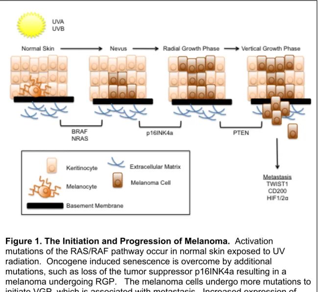
The transformation of a melanocyte into melanoma is believed to occur
through the acquisition of multiple mutations, usually through UV-induced DNA
damage (Figure 1). Once such pathway hypothesized is that an initial oncogenic
mutation occurs within a member of the RAS (Ras sarcoma) pathway. The most
common RAS pathway mutations in melanoma are BRAFV600E (occurring in
~50-66% of all melanomas (Davies et al., 2002; Maldonado et al., 2003)), NRASQ61R
(occurring in ~13-25% of all melanomas (Ball et al., 1994; Curtin et al., 2005; van
't Veer et al., 1989)), and HRASG12V (occurring in 1-7% of all melanomas (Forbes
et al., 2008; Jafari et al., 1995; Nogueira et al., 2010)). In normal human
melanocytes, ectopic introduction of BRAFV600E or NRASQ61R in vitro leads to
2010). This oncogene-induced senescence (OIS) is a major barrier to oncogenic
transformation. Benign nevi, which are considered to be non-proliferative
melanocytic lesions, represent one of the best in vivo examples of OIS.
BRAFV600E mutations are found in 60% of benign nevi and are thought to result in
the initial increase in proliferation of melanocytic cells followed by induction of cell
cycle arrest and senescence (Pollock et al., 2003).
Figure 1. The Initiation and Progression of Melanoma. Activation mutations of the RAS/RAF pathway occur in normal skin exposed to UV radiation. Oncogene induced senescence is overcome by additional mutations, such as loss of the tumor suppressor p16INK4a resulting in a melanoma undergoing RGP. The melanoma cells undergo more mutations to initiate VGP, which is associated with metastasis. Increased expression of
genes associated with metastasis, such as TWIST1, CD200, and HIF1α, and
In order to develop fully into a melanoma, the cells must acquire additional
alterations or mutations that allow them to overcome OIS. Loss of p16INK4a or
activation of the MAPK, PI3K/AKT, and Notch signaling pathways have been
shown to be important for OIS escape and melanocyte transformation (Slipicevic
& Herlyn, 2012). With this transformation, the nevus becomes dysplastic and
begins to superficially spread along the epidermal basal layer but it remains in
situ and lacks the capacity to invade the dermis and metastasize. This is
generally known as the radial growth phase (RGP). These cells then acquire
more mutations that allow them to invade into the dermis in what is known as the
vertical growth phase (VGP). From this stage, the cells are more able to spread
throughout the body, metastasizing to lymph nodes as well as distant organs,
such as the lung, liver, brain, and bone.
B. Histiologic Types of Melanoma
There are four basic categories of melanoma: superficial spreading
melanoma (SSM), lentigo maligna (LM), acral lentiginous melanoma (ALM), and
nodular melanoma (NM) (Table 1). Superficial spreading melanoma is the most
common, accounting for about 70% of all diagnosed cases. This type of
melanoma, as its name suggests, grows along the top layer of the skin before
penetrating more deeply. While SSM can be found anywhere on the body, it is
most commonly found on sun exposed areas of the skin such as the trunk in
men, the legs of women, and on the upper back of both. Lentigo maligna,
malignant cells but does not show invasive growth. It typically progresses very
slowly and can remain non-invasive for years. Once LM begins to grow
vertically, and invades down into the dermis, it is called lentigo maligna
melanoma, though this only occurs in about 2-5% of patients. Acral lentiginous
melanoma, while accounting for only 5% of all diagnosed cases of melanoma, is
the most common form of melanoma in Asians and African-Americans. It is
commonly found on the palms, soles, under nails, and in oral mucosa. Unlike
other forms of melanoma, ALM does not appear to be linked to UV exposure.
Finally, nodular melanoma is the second most common form of melanoma,
accounting for about 15% of all diagnosed cases. It is the most aggressive form
of melanoma and tends to grow more rapidly in thickness (vertically) than in
diameter (radially). NM generally do arise from a pre-existing mole and appear in
a spot where a lesion did previously not exist (James, Berger, & Elston, 2011).
Melanomas can also be classified based on site of occurrence, level of Table 1: Clinical Subtypes of Melanoma
TYPE OF MELANOMA
% OF ALL MELANOMAS
COMMON LOCATION
Superficial Spreading 70 Any sun-exposed area, most
commonly the back and lower legs among women
Lentigo Maligna 10 Any sun-exposed area
Acral Lentiginous 5 Soles, palms, under nails,
mucous membranes
melanomas that develop on skin with limited sun exposure. Patients presenting
with BRAF mutations often have many nevi. Melanomas without mutations in
BRAF or RAS have frequent copy number increases and overexpression of
cyclin D1 or CDK4 (cyclin dependent kinase 4), genes that are downstream of
BRAF. This could be important in treating patients as those with amplification of
these genes may not respond well to BRAF inhibitors as the drug is targeting too
far upstream of the mutated pathway. KIT mutations can generally be found in
melanomas occurring on chronically exposed skin, such as the lower dorsal arms
or the head and neck and these patients tend to have relatively few nevi (Curtin
et al., 2005). The discovery of specific genetic mutations occurring within
melanomas located in certain areas of the body will enable screening patients
more quickly for treatment with therapies that can best target their specific tumor.
C. Melanoma Staging
Melanoma is most lethal form of skin cancer. While melanoma accounts
for less than 5% of skin cancer cases, due to its propensity to metastasize, it is
responsible for 75% of all skin cancer related deaths. In 2009 the American Joint
Committee on Cancer (AJCC) set forth an official guideline for the clinical staging
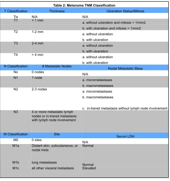
of melanoma. The staging system is based on the TNM system where T
describes the size of the original or primary tumor and whether it has invaded
into nearby tissue, N describes the regional lymph nodes that are involved, and
Melanoma stages are also based on defining characteristics of the primary
melanoma and its metastases: tumor thickness, known as Breslow’s thickness;
the presence or absence of tumor ulceration; the number of metastatic lymph
nodes; mitosis within the primary tumor; whether metastasis to the lymph node is
microscopic or macroscopic; the site of distant metastases; and the level of
lactate dehydrogenase (LDH) in the serum (Balch et al., 2009). Melanomas are
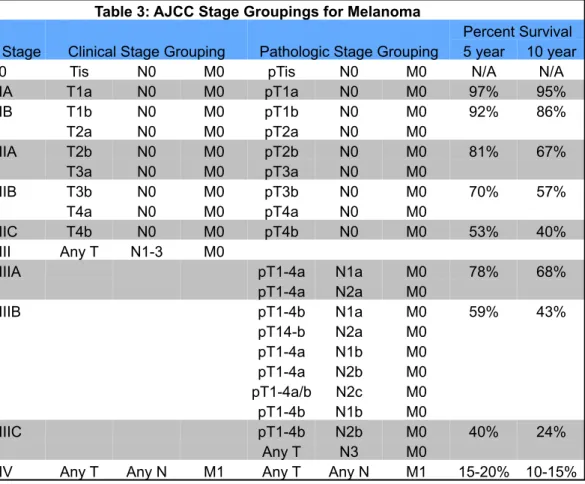
staged from 0-IV, with stage 0 having no evidence of a primary tumor to stage IV,
which is any primary tumor presenting with lymph node and distant metastases
Table 2: Melanoma TNM Classification
T Classification Thickness Ulceration Status/Mitosis
Tis N/A N/A
T1 < 1 mm
a. without ucleration and mitosis < 1/mm2 b. with ulceration and mitosis > 1/mm2
T2 1-2 mm
a. without ulceration b. with ulceration
T3 2-4 mm
a. without ulceration b. with ulceration
T4 > 4 mm
a. without ulceration b. with ulceration N Classification # Metastatic Nodes
Nodal Metastatic Mass
No 0 nodes
N/A
N1 1 node
a. micrometastases b. macrometastases
N2 2-3 nodes
a. micrometastases b. macrometastases
c. in-transit metastasis without lymph node involvement N3 4 or more metastatic lymph
nodes or in-transit metastasis with lymph node involvement
M Classification Site
Serum LDH
M0 0 sites
N/A M1a Distant skin, subcutaneous, or
nodal mets
Normal
M1b lung metastases
Thanks to increased public awareness of the disease and increased
dermatological screening, 85% of melanoma patients present at stage I or II,
15% percent of patients have lymph node metastasis, stage III, and less than 2%
of patients have distant metastasis, stage IV. The five-year survival rate of
melanoma diagnosed at stage I, where the melanoma in less than 2.00 mm thick
and there is no lymph node or distant metastasis, is 98%. However, if the
melanoma is diagnosed at stage IV, where the cancer has spread to parts of the
body remote from the primary tumor, either by discontinuous metastasis to
distant organs, tissues, or via the lymphatic system to distant lymph nodes, the
five-year survival rate drops to just 15% (J. Jiang, Tang, & Liang, 2011; Krapcho
Table 3: AJCC Stage Groupings for Melanoma
Stage Clinical Stage Grouping Pathologic Stage Grouping
Percent Survival 5 year 10 year 0 Tis N0 M0 pTis N0 M0 N/A N/A IA T1a N0 M0 pT1a N0 M0 97% 95% IB T1b N0 M0 pT1b N0 M0 92% 86%
T2a N0 M0 pT2a N0 M0
IIA T2b N0 M0 pT2b N0 M0 81% 67% T3a N0 M0 pT3a N0 M0
IIB T3b N0 M0 pT3b N0 M0 70% 57% T4a N0 M0 pT4a N0 M0
IIC T4b N0 M0 pT4b N0 M0 53% 40% III Any T N1-3 M0
IIIA pT1-4a N1a M0 78% 68%
pT1-4a N2a M0
IIIB pT1-4b N1a M0 59% 43%
pT14-b N2a M0 pT1-4a N1b M0 pT1-4a N2b M0 pT1-4a/b N2c M0 pT1-4b N1b M0
IIIC pT1-4b N2b M0 40% 24%
Any T N3 M0
D. Melanoma Treatment
As mentioned above, stage I and II melanomas are treated by wide
excision, with the amount of normal skin removed depending on the thickness of
the melanoma. The patient may undergo a sentinel lymph node biopsy as well.
If the lymph node does contain cancerous cells, a lymph node dissection will also
be done. For more aggressive stage II and stage III patients, the above
treatments as well as the adjuvant immunotherapy therapy of interferon-α (IFNα)
or interleukin-2 (IL-2) is generally administered. Immunotherapy is given to
melanoma patients in the hopes of activating their body’s immune system so it
will destroy any cancer cells in the body. Some stage III patients may also
benefit from radiation or chemotherapy. Stage IV patients generally have both
tumor and lymph node excisions, as well as removal of distant metastases that
can be reached surgically. Metastases that cause symptoms but cannot be
reached surgically may also be treated with radiation, immunotherapy, targeted
therapy, or chemotherapy. Since it is often very hard to cure metastatic
melanoma, many stage III and stage IV patients will enter a clinical trial of new
chemotherapy drugs, new specific-target drugs, or combinations of different
types of treatments. The discovery of specific genetic mutations that factor in
both the initiation and progression of the disease have allowed for the creation of
targeted therapy that may be more specific and beneficial for the individual
E. Melanoma Epidemiology and Risk Factors
Melanoma is typically seen in white male patients over the age of 40.
However, in patients under the age of 40, melanoma is more common in women.
In men, melanomas are commonly found on the upper back or on the head and
neck. However, in women, melanomas generally develop on the lower legs (W.
Y. Kim et al., 2009; Siegel, Naishadham, & Jemal, 2012). While melanomas are
10 times more likely to occur in Caucasians than Africa-Americans,
Asian-Americans, or Hispanics, patients with darker skin are more likely to present with
advanced stage melanoma and it is frequently fatal for these populations (Cress
& Holly, 1997; S.-H. Kim, Turnbull, & Guimond, 2011). In dark-skinned people,
the most common form of melanoma is acral melanomas commonly appear
under the fingernails or toenails, on the palms on the hands, or on the soles of
the hands. While these areas of the body are the most common places for
melanomas to develop, they can appear anywhere on the skin and even in the
eye (uveal melanoma) or the bowel.
Melanoma occurs through the unchecked proliferation of melanocytes.
This can be through inheriting genetic mutations in genes involved with
melanocyte proliferation and maintenance, such as mutations in MC1R or
CDKN2A, or through exposure to environmental factors that cause DNA
mutations, such as activating oncogenes like BRAF or NRAS. The most
common cause of melanoma is UV damage from the sun (Erler et al., 2006;
Situm, Buljan, Bulić, & Simić, 2007) and there are numerous risk factors that can
produces less melanin to protect oneself from damaging UV radiation. People
with blond or red hair, blue or green eyes, and who freckle or sunburn easily are
more likely to develop melanoma than someone with a darker complexion.
Those who have a history of sunburn, where they have had one or more
blistering sunburns as a child, have an increased risk of melanoma as an adult.
Those who live closer to the equator or a higher elevation, where the sun’s rays
are more direct, experience a higher level of UV radiation. People with jobs who
work outdoors are exposed to more UV radiation than those who work indoors
and have an increased risk of melanoma. Finally, people who naturally have
more than 50 ordinary moles on their body have an increased risk of melanoma
(Egeblad & Werb, 2002; Volkovova, Bilanicova, Bartonova, Letašiová, &
Dusinska, 2012).
While ultraviolet radiation direct from the sun is the most common cause
of melanoma, ultraviolet radiation exposure from indoor tanning beds has been
linked to an increase in melanoma susceptibility. The International Agency for
Research on Cancer, an affiliate of the World Health Organization (WHO),
includes ultraviolet tanning devices in its Group 1, a list of the most dangerous
cancer-causing substances. Group 1 also includes agents such as plutonium,
cigarettes, and solar UV radiation (Choi, Jang, Min, & Song, 2011; Ghissassi et
al., 2009; Shyu, Hsu, Wang, Wang, & Lin, 2007; Zhou et al., 2011). Just one
indoor tanning session increases users’ risk of developing melanoma by 20
another two percent (Ameln, Muschter, Heimesaat, Breier, & Ben B Wielockx,
2012; Boniol, Autier, Boyle, & Gandini, 2012).
F. Inherited Predisposition to Melanoma
There are also genetic mutations that increase an individuals’ chance of
having the disease. Some rare genes have a relative high risk of causing
melanoma, thereby having high penetrance, while some more common genes
have relatively low risk, or low penetrance. One such common gene is the
G-protein-coupled receptor MC1R (melanocortin 1 receptor). Investigations into the
genetics of mouse coat color have shown that this gene regulates both the
tanning response of the skin and hair and skin color in humans (Beaumont et al.,
2011; Sullivan & Graham, 2007). MC1R variants that are associated with having
red hair and pale skin lead to inefficient cyclic AMP (cylic adenosine
monophospate) signaling stimulation upon UV exposure. This leads to an
impairment of melanin production in the skin and to a reduction in
photoprotection (Beaumont et al., 2005; Chambers et al., 1995). Another
commonly mutated gene in familial melanoma is CDKN2A (cyclin-dependent
kinase inhibitor 2A). About 5-12% of all melanoma are estimated to be
hereditary and about 40% of these are associated with CDKN2A mutations
(Goldstein et al., 2006; Sullivan & Graham, 2007). This gene, located on
chromosome 9p21.3, encodes for two genes, p14ARF and p16INK4a. Loss of
p14ARF results in decreased p53 activation and decreased cell apoptosis while
proliferation. Damaged cells lacking the ability to undergo apoptosis or stop cell
cycle progression are likely to proliferate unchecked and form melanomas.
Mutations that cause the skin condition xeroderma pigmentosum (XP) also
seriously predispose one to melanoma. Scattered throughout the genome, these
mutations reduce a cells ability to repair DNA. The development of melanoma
within families carrying inherited mutations in CDKN2A or XP is highly penetrant.
Finally, a gain-of-function mutation in the melanocyte lineage-specific
microphthalmia transcription factor (MITF) has recently been found to predispose
individuals to melanoma. The variant allele was also associated with increased
nevi and non-blue eye color (Koop et al., 1995; Yokoyama, Woods, Boyle, &
Aoude, 2011).
Both environmental and genetic factors contribute to an individuals’ risk of
developing melanoma. By studying these factors, scientists and clinicians are
able to better understand the disease and are able to offer better treatment
options to patients presenting with malignant melanoma.
G. Genetics of Melanoma
An individuals’ cancer is made up of unique mutations and characteristics
that have generated the malignant phenotype of cancer. As scientists and
physicians learn more and more about what genetic events contribute to
melanoma, they are able to offer better treatments and therapies for the
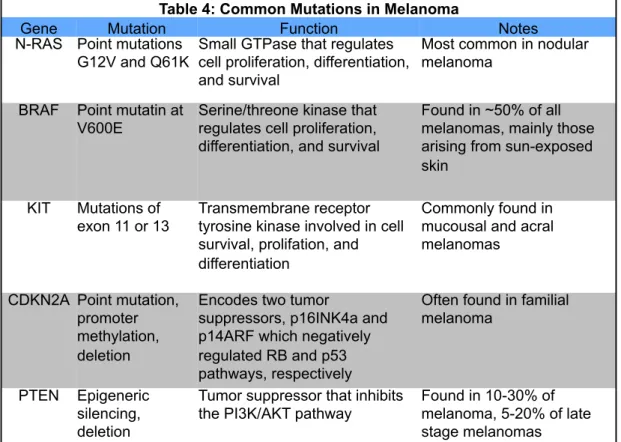
activating mutations of the RAS/RAF pathway and c-KIT and loss of tumor
suppressor genes PTEN or CDKN2A (Table 4).
RAS
The Ras sarcoma (Ras) oncoproteins are small GTPases that serve as
signaling nodes activated in response to numerous extracellular stimuli.
Activated Ras interacts with multiple, catalytically distinct downstream effectors
to regulate cytoplasmic signaling networks in order to control gene expression
and the regulation of cell proliferation, differentiation, and cell survival (Evans,
Schrlau, Chalian, Zhang, & Koch, 2006; Wennerberg, Rossman, & Der, 2005).
There are three Ras genes: HRAS, KRAS, and NRAS in humans. HRAS and
KRAS were initially discovered from two cancer-causing viruses in rats, the
Table 4: Common Mutations in Melanoma
Gene Mutation Function Notes
N-RAS Point mutations G12V and Q61K
Small GTPase that regulates cell proliferation, differentiation, and survival
Most common in nodular melanoma
BRAF Point mutatin at V600E
Serine/threone kinase that regulates cell proliferation, differentiation, and survival
Found in ~50% of all melanomas, mainly those arising from sun-exposed skin
KIT Mutations of
exon 11 or 13
Transmembrane receptor tyrosine kinase involved in cell survival, prolifation, and differentiation
Commonly found in mucousal and acral melanomas
CDKN2A Point mutation, promoter methylation, deletion
Encodes two tumor
suppressors, p16INK4a and p14ARF which negatively regulated RB and p53 pathways, respectively
Often found in familial melanoma
PTEN Epigeneric silencing, deletion
Tumor suppressor that inhibits the PI3K/AKT pathway
Harvey sarcoma virus and the Kirsten sarcoma virus (Bedogni et al., 2005;
Malumbres & Barbacid, 2003), while NRAS was initially discovered in human
neuroblastoma cancer cells (Dische, Bennett, Orchard, Stratford, & Wardman,
1989; Hall, Marshall, Spurr, & Weiss, 1983; Lartigau et al., 1997).
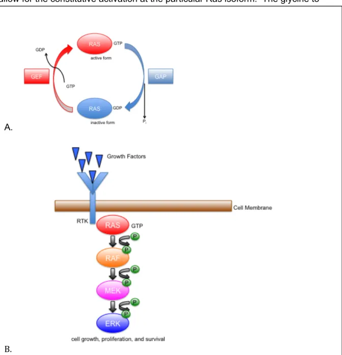
As a G-coupled protein, Ras has an “on” and “off” state. While in the “off”
state, it is bound to the nucleotide guanosine diphosphate (GDP), while when the
Ras protein is in the “on” state it is bound to the nucleotide guanosine
triphosphate (GTP). The exchange of GDP for GTP causes a conformational
change that switches it to its activated form. The process of exchanging the
bound phosphate group is mediated by guanine nucleotide exchange factors
(GEFs), which facilitate Ras activation, and GTPase activating proteins (GAPs),
which facilitate Ras inactivation (Vetter & Wittinghofer, 2001).
Activated Ras binds to and promotes the translocation of the Raf
serine/threonine kinase to the plasma membrane where additional
phosphorylation events promote full Raf kinase activity. Raf phosphorylates and
activates the MEK1/2 dual specificity protein kinase, which in turn phosphorylates
and activates the ERK1/2 mitogen-activated protein kinase (MAPK). Activated
ERK translocates to the nucleus where it phosphorylates Ets-family transcription
factors, which in turn activate Ets-responsive promoters (Figure 2).
Ras is commonly mutated in 20-30% of all human cancers (Bos, 1989).
KRAS mutations are most frequently detected in colorectal tumors, lung
carcinomas (particularly non-small-cell lung cancer [NSCLC]), and in pancreatic
neck; and NRAS mutations are common in haematopoietic malignancies (Pylayeva-Gupta, Grabocka, & Bar-Sagi, 2011). The most common mutations
allow for the constitutive activation at the particular Ras isoform. The glycine to
A.
B.
valine mutation at residue 12 (G12V) renders the GTPase domain of Ras
insensitive to inactivation by GAP and thus stuck in its “on” state. The glutamine
to lysine mutation at residue 61 (Q61K) impairs GTP hydrolysis, again sticking
Ras in its “on” state (Scheffzek, Ahmadian, Kabsch, & Wiesmüller, 1997).
The most commonly mutated RAS isoform in melanoma is NRAS.
Approximately 15-20% of melanoma cases harbor point mutations, generally
occurring at codons 12 or 61. Activating NRAS mutations are generally
correlated with nodular lesions, sun exposure, higher melanoma thickness, and a
higher mitotic rate (Ellerhorst et al., 2011). However, NRAS mutations are rarely
found in dysplastic nevi, the most common precursor to melanoma (Albino et al.,
1989). HRAS mutations are found in 30% of Spitz nevi, these are benign
melanocytic lesions that occur most frequently in the first 2 decades of life
(Bastian, LeBoit, & Pinkel, 2000). Transgenic studies in mice have shown that
activated HRAS mutations in melanocytes can lead to aberrant proliferation and
transformation, especially in cooperation with inactivating mutations in tumor
suppressors such as Cdkn2a or Trp53 (Powell et al., 1995).
Pharmacological inhibition of NRAS is challenging because its GTPase
activity has so far precluded the successful design of specific small-molecule
antagonists. Small interfering RNA (siRNA)-mediated depletion of NRAS in
melanoma cell lines inhibits proliferation and makes cells much more sensitive to
chemotherapy (Eskandarpour et al., 2005). Farnesyltransferase inhibitors (FTIs)
block farnesylation, a key post-translational modification necessary for RAS
FTI, Tipifarnib, or R115777, was evaluated in a single-agent, single-arm Phase II
trial in patients with metastatic melanoma, but the lack of response among the
first 14 patients led to the early closure of the trial (Gajewski et al., 2012). Since
it is so difficult to directly target NRAS activation, more studies are being done to
investigate the inhibition of RAS effector pathways.
RAF
Directly downstream of Ras, Raf is commonly mutated in multiple cancers,
including malignant melanoma, colorectal cancer, thyroid, and ovarian cancers.
There are three isoforms of Raf, A-Raf, B-Raf, and C-Raf. While mutations in
either A-Raf or C-Raf are rare in cancer, the valine to glutamic acid mutation of
B-Raf at residue 600 is the most common Raf-activating mutation, accounting for
~90% of the B-Raf mutations that are seen in human cancers (Wellbrock,
Karasarides, & Marais, 2004). This mutation is thought to disrupt the interaction
between the glycine-rich loop and the activation segment of the B-Raf kinase
domain, freeing the activation segment and allowing the kinase to fold into the
active confirmation.
BRAF is most highly expressed in neuronal tissues and melanocytes as
well as in testis and haematopoietic cells and at this time MEK is its only known
substrate. Activating mutations in BRAF have been identified in approximately
50% of all melanomas, with the vast majority found in melanomas that arise from
intermittently sun-exposed skin. The high frequency of BRAF mutation in
to UVB radiation, α-MSH (α-melanocyte stimulating-hormone) and
proopiomelanocortin-derived peptides bind to MCR1, which upregulate
cyclic-AMP. This increase in cAMP leads to increased proliferation and melanogenesis
(Halaban, 2000) through the upregulation of BRAF and subsequently ERK
(Buscà et al., 2000). That a principle melanocyte-specific signaling pathway
controlling proliferation and differentiation operates through the activation of
BRAF suggests a possible explanation for the high frequency of BRAF mutations
in melanoma relative to other cancer types (Davies et al., 2002).
There have been positive clinical results from phase I, II, and III trials that
show that vemurafenib (PLX4032), a potent and selective BRAFV600E inhibitor,
caused complete or partial tumor regression in 80% of patients carrying
BRAFV600E tumors as well as an increased progression-free survival rate
(Chapman et al., 2011; Flaherty et al., 2010). Unfortunately, after an initial period
of response to vemurafenib, most patients relapse and develop resistance to the
drug (Flaherty et al., 2010). Identification of the mechanisms leading to BRAF
resistance have led to new clinical trials combining inhibitors of BRAF with MEK
and AKT inhibitors in patients with metastatic melanoma as well as other tumor
types harboring the BRAFV600E mutation (Curti & Urba, 2012). Murine models of
vemurafenib-resistant tumors have even shown that some tumors acquire a
dependency on the drug, where removal of vemurafenib treatment results in
tumor regression (Thakur et al., 2013). Increased understanding of how
melanomas become resistant to this treatment will enable better options for
KIT
Although BRAF mutation melanomas represent the largest genetic
subtype of melanoma, c-Kit mutations are more common in mucosal and acral
melanomas and they are also present in melanomas that originate in chronic
sun-damaged skin (Beadling et al., 2008). KIT encodes a type III
transmembrane receptor tyrosine kinase. Binding of KIT’s ligand, stem cell factor
(SCF), to the extracellular domain causes the dimerization of the KIT receptor
resulting in the autophosphorylation of its intracellular tyrosine kinase domains.
Once activated, the tyrosine kinase domains initiate signaling in a variety of
downstream pathways including MAPK/MEK, phosphatidylinositol 3-OH kinase
(PI3K)/AKT, JAK/signal transducers and activators of transcription (STAT)
pathways.
Many of the KIT mutations found in melanomas affect exon 11, which
codes for the juxtamembrane domain. This membrane is involved in
autoinhibition of the receptor when it is not bound with its ligand, when it is
mutated, this inhibitory function is prevented and ligand independent receptor
dimerization and activation is enhanced (Curtin, Busam, Pinkel, & Bastian, 2006).
Phase II clinical trails have found that patients with mutations in either exon 11 or
exon 13 of KIT respond well to treatment with imatinib (Carvajal et al., 2011).
Currently, patients with mucosal or acral melanomas harboring these activating
KIT mutations are encouraged to enroll in Phase II or Phase III clinical trials
CDKN2A
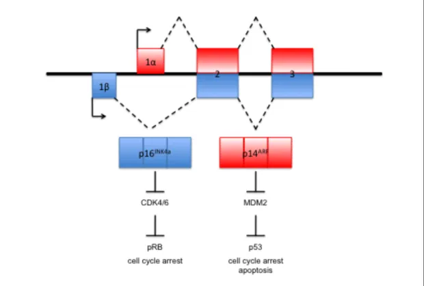
The CDKN2A locus encodes for two distinct tumor-suppressing proteins,
p16INK4A and p14ARF. p16INK4A (inhibitor of cyclin-dependent kinase) inhibits
CDK4/6-cylin D-mediated phosphorylation and inactivation of retinoblastoma
(RB). In the absences of p16INK4A inhibition, CDK4/6-cylcin D phosphorylates RB,
resulting in the release of E2F. E2F then activates genes that are necessary for
a cell to transition from G1 to S phase (Serrano, Hannon, & Beach, 1993).
p14ARF (alternative reading frame) inhibits MDM2-mediated ubiquitylation and
subsequent degradation of p53 (Kamijo et al., 1998; Pomerantz et al., 1998; Stott
et al., 1998; Y. Zhang, Xiong, & Yarbrough, 1998), thereby inhibiting activation of
the p53 apoptotic pathway (Figure 3). The two products p16INK4A and p14ARF
negatively regulate RB and p53 pathways respectively, and their loss
predisposes to the development of melanoma.
CDKN2A mutations are commonly found in individuals with familial melanoma and melanoma-prone families (Hussussian, Struewing, & Goldstein,
1994; Kamb et al., 1994). These families are diagnosed with familial atypical
multiple mole melanoma (FAMMM), an autosomal dominant disease
characterized by multiple, melanocytic nevi, usually more than 50, and a family
history of melanoma. Interestingly, BRAFV600E induces p16INK4A expression and
senescence in primary human melanocytes in vitro (Gray-Schopfer et al., 2006;
Michaloglou et al., 2005). p16INK4A upregulation contributes to the induction of
senescence in nevi and p16INK4A defects are found in dysplastic, but not benign,
BRAF-induced melanocyte transformation. However, with further mutations that
lead to the silencing of p16INK4A, the OIS is overcome and the melanoma
progression continues.
In the situation of CDKN2A loss, drugs that inhibit CDK4/6 have been
suggested as potential therapeutics. Multiple in vitro studies combining CDK4/6
inhibitors with MEK inhibitors or BRAF inhibitors have shown increased
melanoma cell apoptosis (J. Li, Xu, Yang, Li, & Dong, 2010). However, a Phase
II clinical trial of flavopiridol, a pan CDK inhibitor, showed no significant clinical
Phase I and II trials are being done looking novel and more specific CDK4/6
inhibitors in combination with other chemotherapies or specific targeting drugs.
PTEN
Loss of the lipid phosphataste PTEN (phosphate and tensin homologue) is
detected in 10-30% of cutaneous melanomas and is also seen in 5-20% of late
stage melanomas (Wu, Goel, & Haluska, 2003). PTEN dephosphorylates the 3’
position of phosphatidylinositol-(3,4)-P2 (PIP2) and
phosphatidylinositorl-(3,4,5)-P3 (PIphosphatidylinositorl-(3,4,5)-P3). This directly antagonizes the activity of PI3K, which phosphorylates
PIP2 and PIP3 resulting in their activation of AKT, a serine/threonine kinase that
normally exists in the cytoplasm in an inactive conformation. Upon activation of
PI3K, AKT is recruited to the cell membrane and phosphorylated at 2 critical
residues (Thr308 and Ser473) and activates its downstream targets, including
mTOR (Figure 4). AKT activity has been shown to regulate many cellular
processes, including metabolism, growth/size, survival, motility, invasion, and
angiogenesis (D DD Sarbassov, Ali, & Sabatini, 2005; Restuccia & Hemmings,
2010; Yecies & Manning, 2011). Therefore, PTEN loss in melanoma results in
PTEN gene mutations and deletions are mutually exclusive with activating
NRAS mutations in melanoma. In contrast, many melanomas with loss of PTEN
have concurrent BRAF activating mutations (Tsao, Goel, Wu, Yang, & Haluska,
2004). In melanoma, NRAS and BRAF mutations are mutually exclusive.
mutually activating so the pathways cooperate to stimulate proliferation. Indeed,
genetically engineered mouse models (GEMMs) have shown that loss of PTEN
is required for the transformation of melanocytic lesions to invasive melanomas
in the setting of activated BRAFV600E (Dankort, Curley, Cartlidge, & Nelson,
2009).
There have been numerous scientific and clinical studies on the use of
PI3K-AKT and its pathway inhibitors in melanoma. An example is the use of the
mTORC1 inhibitor Rapamycin. Use of this drug inhibits signaling downstream of
mTORC1, but also results in feedback activation of PI3K, resulting in
hyperactivation of AKT(Tabernero et al., 2008). This compensatory activation of
AKT may be the reason behind the lack of clinical activity of single-agent
treatment with rapamycin in several cancers, including melanoma (Margolin et
al., 2005). Future studies and clinical trials using PI3K/AKT pathway inhibitors
alone and in tandem with other therapeutics will be necessary to better
understand how to treat metastatic melanoma patients with PTEN loss.
H. Genetically Engineered Mouse Models of Melanoma
Genetically engineered mouse models of melanoma have been used to
phenocopy the human disease and allow us to better understand the initiation
and progression of melanoma in vivo. By manipulating the expression of
different genes, such as the ones mentioned above, believed to be involved in
also how that gene affects the entire system of the mouse, from tumor
microenvironment to immune system.
The earliest mouse models were used in the early 1900’s when
melanocytic tumors arose spontaneously in inbred mouse strains. These
spontaneous melanomas, including the well-known B16 cell line, were
transplanted to congenic mice and could also be cultured, studied, and
manipulated in vitro (Damsky & Bosenberg, 2010). As technology improved,
methods of manipulating gene expression, such as the CRE-lox P system, have
been developed to allow scientists to control when and where the genes are
expressed or deleted. These systems of in vivo genetic manipulation have
allowed great insight into the initiation and progression of melanoma.
The most popular system to generate conditional mouse models is the
CRE-Lox P system (Agar & Young, 2005; Argos et al., 1986; Davies et al., 2002;
Haass & Herlyn, 2005; Krapcho et al., 2009; Maldonado et al., 2003; Sauer &
Henderson, 1988; Sternberg, Sauer, Hoess, & Abremski, 1986). This system
allows for site-directed recombination of target sequences to regulate or
manipulate gene expression. In this system, Cre (cyclization recombination) is a
site-specific DNA recombinase that recognizes a specific 34-bp site, known as
lox P (locus of X-over P1) sites. When two lox P sites flank a region of DNA, the
Cre enzyme will excise all DNA sequences located between the two sites.
Mouse models generally make use of this technology by placing lox P sites on
either side of an exon to create a null allele or on either side of a stop codon to
In order to generate mouse models of melanoma, scientists have taken
advantage of the melanocyte-specific expression of the tyrosinase family of
genes: tyrosinase (Tyr); tyrosinase related protein 1 (TRP1); and dopachrom
tautomerase (DCT) to drive expression of their genes of interests only in those
cells (Ball et al., 1994; Bansal & Nikiforov, 2010; Forbes et al., 2008; Jafari et al.,
1995; Mintz & Klein-Szanto, 1992; Slipicevic & Herlyn, 2012). By combining
melanocyte specific expression with the Cre-lox P system, many models of
melanoma have been developed to increase our understanding of the disease
and the genes involved in its initiation and progression, some of which are
described below (Table 5).
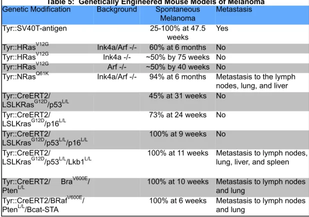
* References cited in text Table 5: Genetically Engineered Mouse Models of Melanoma
Genetic Modification Background Spontaneous Melanoma
Metastasis Tyr::SV40T-antigen 25-100% at 47.5
weeks
Yes Tyr::HRasV12G Ink4a/Arf -/- 60% at 6 months No Tyr::HRasV12G Ink4a -/- ~50% by 75 weeks No Tyr::HRasV12G Arf -/- ~50% by 40 weeks No
Tyr::NRasQ61K Ink4a/Arf -/- 94% at 6 months Metastasis to the lymph nodes, lung, and liver Tyr::CreERT2/
LSLKRasG12D/p53L/L
45% at 31 weeks No Tyr::CreERT2/
LSLKrasG12D/p16L/L
73% at 24 weeks No Tyr::CreERT2/
LSLKrasG12D/p53L/L/p16L/L
100% at 9 weeks No Tyr::CreERT2/
LSLKrasG12D/p53L/L/Lkb1L/L
100% at 11 weeks Metastasis to lymph nodes, lung, liver, and spleen Tyr::CreERT2/ BraV600E/
PtenL/L
100% at 10 weeks Metastasis to lymph nodes and lung
Tyr::CreERT2/BRafV600E/ PtenL/L/Bcat-STA
One of the first models of melanoma had melanocyte-specific expression
of the SV40 (Simian Vacuolating Virus 40) large T antigen that disrupts both the
Rb and p53 pathways (Mintz & Klein-Szanto, 1992). These mice developed
spontaneous and UV-induced melanomas that metastasized. In this model, the
majority of the melanomas were ocular melanomas as opposed to the more
commonly seen cutaneous melanomas.
Multiple models were generated to examine the role of CDKN2A in
melanoma. Models utilized either complete loss of p16Ink4a and p19Arf, or loss of
either tumor suppressor, combined with Tyr::HRasV12G, a constitutively
activated HRas transgene on a melanocyte specific promoter. Mice with loss of
both p16Ink4a and p19Arf developed cutaneous melanomas that were highly
vascularized, locally invasive, amelanotic, and nodular (Chin, Pomerantz, Polsky,
& Jacobson, 1997). Mice that had loss of either p16Ink4a or p19Arf developed
spontaneous melanomas at 75 and 40 weeks respectively. Mice with
homozygous loss of p16Ink4a demonstrated loss of p19Arf expression and mice
with homozygous p19Arf loss demonstrated loss of p16Ink4a expression. These
results suggest the p16Ink4a and p19Arf work together to hinder melanoma
progression in vivo (Sharpless, Kannan, Xu, Bosenberg, & Chin, 2003). Finally,
loss of p16Ink4a and p14Arf was combined with melanocyte-specific activated NRas
(Tyr::NRasQ61K). As mentioned previously, NRas is the most common isoform
of Ras mutated in human melanoma, while HRas and KRas are rarely mutated.
These tumors were also able to metastasize to the lymph nodes, lung, and liver
(Ackermann et al., 2005).
The development of the conditional TyrCreERT2 allele allowed for the
possibility of mouse melanoma models with controlled spatial expression of a
gene of interest (Balch et al., 2009). This allele is under the control of a
tamoxifen-inducible system, so that only when tamoxifen is administered to the
mouse will the Cre-lox P recombination occur in melanocytes. Conditional
activation of KRasG12D with somatic loss of both p16Ink4a and p53 has been
shown to cooperate in melanoma formation (Monahan et al., 2010) and adding
Lkb1 loss to this model generates metastatic disease. (Liu et al., 2012) In
another model, the conditional activation of BrafV600E with somatic loss of Pten
induces metastatic melanoma (Dankort et al., 2009) and adding stabilized β
-catenin to this model increases distant metastases (Damsky et al., 2011).
Genetic mouse models of melanoma have generated much knowledge as
to the genes involved in the initiation and progression of the disease. These
models help scientists to understand how these genetic events not only affect the
melanoma, but also the tumor microenvironment and immune system.
I. Melanoma and Metastasis
While the majority of patients are diagnosed with stage I or II melanoma,
17% of patients are diagnosed with stage III or IV melanoma, meaning that they
have metastases to lymph nodes (stage III) or distant organs (stage IV).
with the disease (Balch, Soong, et al., 2001b). The one-year survival rates in
melanoma patients with clinically apparent metastasis to one, two or three
different visceral sites is 36%, 13%, and 1%, respectively (Damsky, Rosenbaum,
& Bosenberg, 2010). Even “new” melanomas may metastasize as metastasis
can occur early in melanoma progression, even from a thin primary tumor
(Bedrosian et al., 2000). Though treatment options have improved for patients
with metastatic disease, it is an important area of research to find new targets for
treatment as patients often relapse and those that are thought to be cured of the
disease can sometimes discover new metastases many years after the “curative”
treatment (Balch, Soong, et al., 2001b).
Once melanoma cells switch from RGP to VGP the tumor has obtained
invasive potential and is more likely to metastasize. These malignant cells grow
deeper into the dermis and have access to lymphatic or blood vasculature that
can aid in the dissemination of the cells. Melanoma cells also undergo genetic
changes that can aid in its progression to metastasis, such as the decrease in
E-cadherin expression. Decreasing E-E-cadherin frees the cells from linkages with
adjacent keratinocytes and allows the cell to become more motile (Haass &
Herlyn, 2005).
Primary melanomas generally first metastasize to regional lymph nodes
(Meier et al., 2002). It is known that melanoma cells can secrete VEGF-C
(Saharinen, Tammela, Karkkainen, & Alitalo, 2004), which induces
lymphangiogenesis, and high levels of VEGF-C are related to deep lymph node
increased levels of matrix metalloproteinases (MMPs) that help to degrade the
extracellular matrix (ECM) surrounding the tumors thereby allowing the
melanoma cells to move more freely (Seftor et al., 2001). By creating new lymph
vessels, the now-motile malignant cells are able to intravasate into the lymphatic
system and spread to regional lymph nodes. Patients diagnosed with melanoma
exceeding 1mm in depth generally receive sentinel lymph node dissection in
order to prohibit the spread of the disease.
The most common sites of distant metastasis are skin, lung, brain, liver,
bone, and intestine (Damsky et al., 2010). How a cancer cell is able to
extravasate and seed sites of distant metastasis is an increasingly popular field
of research. Multiples studies looking at protein expression in human melanoma
samples by immunohistochemistry (IHC) have found that the expression of
specific genes correlates with melanoma metastasis to specific sites. Melanoma
cells express chemokine receptors, including CXCR4, which could possibly aid in
the honing of the cells to sites of distant metastasis. CXCR4 expression has also
been linked to poor prognosis and increases distant metastasis in patients with
malignant melanoma (Scala et al., 2005). The expression of different integrins
can also predict sites of metastasis. Melanomas that express integrin αvβ3 are
more likely to develop lung metastases (Hieken, Ronan, Farolan, Shilkaitis, &
Gupta, 1999), while melanomas expressing αvβ1 develop lymph node metastasis
(Hieken, Ronan, Farolan, Shilkaitis, & Gupta, 1996). Finally, expression of the
p75 NGF (nerve growth factor) receptor in melanoma correlates with brain
suggest the melanoma cells in the process of metastasis can develop multiple
genetic changes that confer advantages in seeding new sites of distant
metastasis.
Melanoma is known to be more aggressive than other cancers. The most
common mutation in melanoma is activation of the Ras/Raf pathway. Introducing
Ras into normal melanocytes results in melanomas that are significantly more
metastatic than introducing Ras into normal fibroblasts or epithelial cells (P. B. P.
Gupta et al., 2005). These findings show that the intrinsic features of
melanocytes, or at least their response to RAS transformation, may be
responsible for their quick metastasis. This may be a reflection of the fact that
melanocytes are derived from neural crest cells. These cells are characterized
by their expression of motility-associated genes, such as Slug, ERBB3, CD44,
and Nodal (Siegel et al., 2012), that can not only mediate the neural crest cell
motility, but also tumor migration. Understanding the mechanisms that allow
melanoma to spread to both lymph nodes and other sites of distant metastasis
will allow for the discovery of therapies and screens to better treat patients with
advanced disease.
J. Summary
Here we have discussed the development and progression of melanoma.
There are many factors that contribute to an individuals’ risk of the disease, from
environmental risk factors, like exposure to UV radiation, to the hereditary
familial melanoma patients. Melanomas may also contain many different genetic
mutations that drive their development. Activation mutations in oncogenes, such
as NRAS, BRAF, and c-KIT, and loss of tumor suppressor genes, such as
p16INK4A, p14ARF, or PTEN, are seen in multiple types of melanomas. By
studying how these mutations and genes interplay in in vitro melanoma cell lines
and in vivo melanoma mouse models, scientists and clinicians will be able to discover therapies and therapeutics that will ultimately give the individual
The availability of oxygen in the cellular environment has many effects on
multiple cellular functions. Oxygen is necessary for cellular respiration, the
metabolic reaction within cells that converts biochemical energy from nutrients
into ATP (adenosine triphosphate) which the cell then uses to perform a myriad
of functions, including: biosynthesis of amino acids, fatty acids, and other natural
products; locomotion; and the transport of molecules across cell membranes. If
the amount of oxygen in the cellular environment is limited, then the cells must
switch to an anaerobic pathway of respiration and must also regulate multiple
pathways in order to maximize the use of its resources. The genes involved in
the ability of a cell to “sense” the availability of oxygen in the environment have
been of interest to both basic scientists as well as translational medicine.
A. Structure of Hypoxia-Inducible Factors
The hypoxia-inducible factor (HIF) is a heterodimeric transcription factor
composed of a labile α-subunit and a stable β-subunit that are members of the
PAS family (PER, ARNT, and SIM family). There are three alpha subunits in the
human genome, HIF1α, HIF2α, and HIF3α, and three HIFβ genes, also known
as ARNT (aryl hydrocarbon receptor nuclear translocator) (Semenza, 2001). The
elements (HRE) that are associated with a broad range of transcriptional targets
(Semenza et al., 1996).
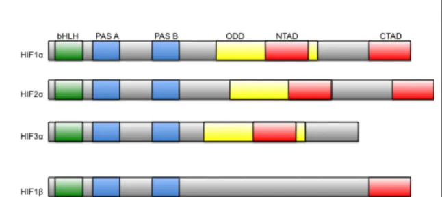
Both HIFα and HIFβ have basic helix-loop-helix (bHLH) domain at their
N-terminus for dimerization and DNA binding. Following the bHLH domain are two
PAS domains, PER (period circadian protein), Arnt, and Sim (single-minded
protein) PAS-A and PAS-B. PAS domains frequently mediate protein-protein
interactions and are often regulated by the binding of their partner to their
hydrophobic core. One or both of the HIFα PAS domains have been functionally
Figure 5. Domain structure of HIF. Both HIFα and HIFβ have basic
helix-loop-helix (bHLH) domain at their N-terminus for dimerization and DNA binding. Following the bHLH domain are two PAS domains. PAS domains frequently mediate protein-protein interactions and are often regulated by the
binding of their partner to their hydprophobic core. The HIFα-subunits contain
two oxygen-dependent degradation domains located in the central region of the protein: the amino-terminal oxidation-dependent degradation domain (NODDD) and the carboxy-terminal oxygen-dependent degradation domain
(CODDD),. There are also two transactivation domains on the 1 and 2α
-subunits, the amino-terminal activation domain (NTAD) and a
implicated in nuclear localization, HIF stabilization via heat shock protein 90
(HSP90) association, and binding with cofactors for target gene specificity, but
the most important function is their heterodimerization of the α-subunit with
ARNT (J. J. Yang et al., 2005). The HIFα-subunits contain two
oxygen-dependent degradation domains located in the central region of the protein: the
amino-terminal oxidation-dependent degradation domain (NODDD) and the
carboxy-terminal oxygen-dependent degradation domain (CODDD),. There are
also two transactivation domains on the α-subunits, the amino-terminal activation
domain (NTAD) and a carboxy-terminal activation domain (CTAD) that
associates with the CH-1 (cysteine/histidine rich) domain on HIF’s transcriptional
co-activator p300 (Figure 5) (Schofield & Ratcliffe, 2004).
B. Regulation of Hypoxia-Inducible Factors
HIF is a major regulator of the cellular response to the availability of
oxygen in the cellular environment, but it also regulates many of the genes
involved in the adaptation of cancer cells to the hypoxic microenvironment
created by a tumor as it outgrows its blood supply. When oxygen is freely
available in the cellular environment, HIFα is hydroxylated at specific proline
residues, Pro402 and Pro564, by an oxygen-dependent prolyl hydroxylase
(PHD). There are three PHD isoforms, PHD1, PHD2, and PHD3. PHD 2 has
been shown to generally target HIF1α while PHD1 and PHD3 generally target
HIF2α (Fong & Takeda, 2008). The hydroxylation of the α-subunit allows the
(VHL) tumor suppressor protein, pVHL. pVHL polyubiquinates HIFα within its
oxygen-dependent domain (ODD), thereby targeting HIFα for proteosomal
degradation (Ivan et al., 2001; Jaakkola et al., 2001). In times of hypoxia, where
there is little or no oxygen available in the cellular environment, or in the setting
is
Figure 6. Post-translational Regulation of HIF. When oxygen is freely
available in the cellular environment, HIFα is hydorxylated at specific proline
residues, Pro402 and Pro564, by an oxygen-depenent prolyl hydroxylase
(PHD). The hydroxylation of the α-subunit allows the alpha subunit to be
recognized by its E3 ubiquitin ligase, the von-Hippel Lindau (VHL) tumor suppressor protein, pVHL. pVHL polyubiquinates HIFα within its
oxygen-dependent domain (ODD), thereby targeting HIFα for proteosomal
degradation. Both HIF1α and HIF2α are also regulated by the hydroxylation
of an asparaginyl residue, thereby disrupting association with p300 in normoxic conditions. In times of hypoxia, where there is little or no oxygen
available in the cellular environment, or in the setting of pVHL loss, HIFα is no
longer degraded and can translocate into the nucleus, bind with its β-subunit
of pVHL loss, HIFα is no longer degraded and can translocate into the nucleus,
bind with its β-subunit and regulate its target genes (Figure 6). Over 60 direct
HIF target genes have been identified, including many implicated in the
pathogenesis of melanoma: VEGF, PDGFβ, IL-8, alphaVbeta3 integrin, and
N-Cadherin (Semenza, 2003).
Both HIF1α and HIF2α are also regulated by the hydroxylation of a
specific asparaginyl residue located at the N-terminus, Asn803 and Asn851
respectively (Lando, Peet, Whelan, Gorman, & Whitelaw, 2002). This
hydroxylation is catalyzed by factor inhibiting HIF (FIH) and asparginyl
hydroxylation causes the disruption of the association of the α-subunit with the
CH-1 domain of the p300 co-activator (Figure 6) (Mahon, Hirota, & Semenza,
2001).
C. Function of HIF
The most commonly studied of the alpha subunits are HIF1α and HIF2α.
There is little known about HIF3α, though it lacks the c-terminal transactivation
domain (CTAD) found on HIF1α and HIF2α and thus cannot bind with p300.
HIF3α has multiple splice variants that can interact with HIF1α, HIF2α, and HIFβ.
It may act as a negative regulator of hypoxia-inducible gene expression as it
competes for HIFβ binding with HIF1α and HIF2α (Heikkilä, Pasanen, Kivirikko, &
Myllyharju, 2011). While HIF1α and HIF2α share many of the same target
genes, they are not wholly redundant in function. HIF1α -/- mice have lethality at
defects (Iyer et al., 1998). HIF2α -/- mice have lethality between embryonic days
9.5 and 13.5 with divergent phenotypes depending on the background of the
mice – including lung maturation defects and catecholamine deficiency
(Compernolle et al., 2002; Tian, Hammer, Matsumoto, Russell, & McKnight,
1998). HIF1α is expressed ubiquitously while HIF2α expression is tissue
specific, occurring in endothelial cells, type II pneumocytes, cardiomyocytes,
fibroblasts of the kidney, interstitial cells of the pancreas and duodenum, and in
hepatocytes (Ema et al., 1997; Flamme, Fröhlich, Reutern, & Kappel, 1997; Tian,
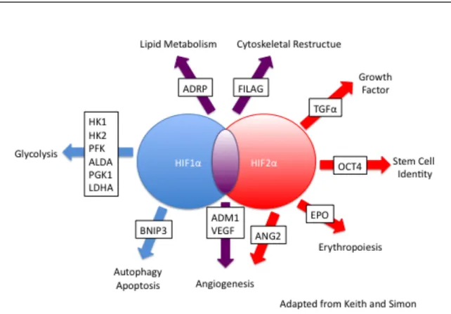
Figure 7. Targets of HIF1α and HIF2α. HIF1α and HIF2α produce
overlapping yet distinct gene expression profiles, with HIF1α uniquely
targeting the genes involved in glycolysis and HIF2α uniquely targeting genes
McKnight, & Russell, 1997). Finally, HIF1α and HIF2α produce overlapping yet
distinct gene expression profiles, with HIF1α uniquely targeting the genes
involved in glycolysis and HIF2α uniquely targeting genes such as Oct4,
CyclinD1, TWIST1, TGFα, and EPO (Figure 7) (Keith, Johnson, & Simon, 2012).
D. HIF In Cancer
The majority of human cancers and their metastases have been shown to
have increased levels of HIF1α or HIF2α (or both) protein (relative to surrounding
normal tissue) (Zhong, De Marzo, Laughner, Lim, & Hilton, 1999) (Table 4).
Clinical data has shown an association between HIF1α and / or HIF2α protein
levels with increased human mortality in many cancers. In addition to this
correlative data from the clinic, there is a large body of experimental data
showing that HIFα loss of function results in decreased tumor growth,
vascularization, and metastasis, whereas HIFα gain of function has the opposite
Adapted from Giaccia (2008) and Semenza (2010)
When HIF is activated, it regulates numerous genes that might actually
help the cancerous cells adjust to the harsh microenvironment of the tumor. HIF
activates genes involved in angiogenesis, such as VEGF (vascular endothelial
growth factor), PDGFβ (platelet derived growth factor β), IL-8 (interleukin-8), and
OCT-4 (octamer-binding transcription factor 4), allowing for the increased
delivery of oxygen and nutrients to the quickly growing tumor (Hickey & Simon,
2006). HIF also activates genes that can breakdown the extracellular matrix,
such as MMPs (matrix metalloproteinase). This allows the tumor to grow and
metastasize. HIF regulates genes that are involved in the epithelial to
mesenchymal transition (EMT), such as N-Cadherin and TWIST, thought to be Table 6: HIF Activation in Human Cancer
Tumor Type HIF1! HIF2! HIF! and Poor Prognosis
Astrocytoma + ND HIF1!
Bladder + + HIF1!
Breast + + HIF1!
Colorectal + + HIF1! and HIF2!
Cervical + ND HIF1!
Endometrial + ND HIF1!
Esophageal, SCC + ND HIF1!
Gastric + ND HIF1!
Glial + + HIF1!
Head and Neck + + HIF1! and HIF2!
Hepatocellular + + HIF2!
Laryngeal + ND HIF1!
Lung NSCLC + + HIF2!
Melanoma + + HIF2!
Nasopharyngeal + + HIF1!
Osteosarcoma + + HIF1!
Ovarian + + HIF1! and HIF2!
Pancreatic + + HIF1!
Prostate + + HIF2!