EPIGENETIC REGULATION OF DNA REPLICATION IN DROSOPHILA MELANOGASTER
Robin Leigh Armstrong
A dissertation submitted to the faculty at the University of North Carolina at Chapel Hill in partial fulfillment of the requirements for the degree of Doctor of Philosophy in the
Curriculum in Genetics and Molecular Biology.
Chapel Hill 2020
ii © 2020
iii ABSTRACT
Robin Leigh Armstrong: Epigenetic regulation of DNA replication (Under the direction of Robert J. Duronio)
iv
v To Lily Rose.
vi
ACKNOWLEDGEMENTS
I could never have successfully completed this journey without the support of many incredible individuals. I would first like to thank Bob, for without his mentorship and guidance, I would not be half of the scientist that I am today. Being one of Bob’s graduate students is truly special—we get to experience the amazing combination of an unbelievably talented scientific mentor and an incredibly supportive advocate. I’m forever grateful that Bob both trusted me and encouraged me to be independent in almost everything that I did in his lab. The skills that I have gained from him will only continue to propel my career
forward. Finally, I couldn’t be more thankful to Bob for accepting and supporting who I am as a scientist and as a person. The Duronio lab became my second family over the past five years.
I would also like to thank the members of my dissertation committee for their support and scientific feedback during my graduate career. Matt, you taught me to thoroughly think through every experiment before I performed it and continued to challenge me throughout my graduate career with the difficult and often overlooked questions: “Why did you choose to do that experiment? Why does that particular result matter?”. Dave, the scientific input and technical insight I received from you was paramount to the completion of my PhD. I
vii
instilled in me to be a strong woman in science. Our conversations at CSH about managing scientific success in a healthy balance with life truly shaped the way I view my career and my career goals. I cannot describe the extent to which you have served as my role model during graduate school, and, for that, I am so incredibly thankful. Dan, when I came to UNC, I knew I wanted my chair to know the ins and outs of my personality and work ethic. I did not expect to gain a mentor and colleague who I now view as a second PI. Thank you for all the small, but incredibly significant, things you did and said to me to ensure that I made it through my PhD better than how I started. Also, a huge thank you to Piper. You got me through one of the toughest weeks of my life without even knowing it.
To the members of the Blue Pod, past and present, thank you for being the ultimate support system. Spencer and Chris, thank you for bringing an insane amount of intelligence and kindness to the workplace. I am incredibly appreciative of the interactions we shared over the past five years. Amanda, from day one you have been the glue that has held the pod together; but personally, you have been the glue that has held me together. You are one of the most amazing people I have ever met, and your love and support for others is unmatched. Every day, your friendship has pulled me through this wild ride, and I’m so grateful to have you in my life. Also, thank you from the bottom of my heart for being a Patriots fanatic. Taylor, working with you was one of the greatest professional experiences I could ever hope for—when I fell behind, you picked me up, and when you needed a hand, you encouraged me to take the lead. I treasure our friendship, and I am looking forward to seeing all the professional and personal successes you attain. Also, thank you for Phantom, who has
viii
strongest and hardest working person I know, and I will always be in awe of the beautiful person you are. You taught me to chase my dreams and surpass my limits. I cannot wait to see the amazing feats we can, and will, accomplish together.
To my friends outside of lab, you made this graduate school experience an absolute blast. Kelsey and Ian, thank you for the nights spent screaming at the Tarheels at Hickory Tavern. We made it through the past six years together, and I am so proud of you both for landing your dream jobs. Adele, you are always there right when I need you most. Every dinner and coffee date made being a graduate student exponentially better, and I truly cherish our friendship.
To the current members of the Duronio lab. I do not have the words to thank you for your support and your friendship. Our lab has become a truly unique family, and it has been the ultimate privilege to work with each of you. Chris, Ashlesha/Sharon, Jim, and Aaron, thank you for accepting me for my authentic self—you all are truly amazing friends. Mary, you took me under your wing, welcomed me into your home, and made me feel like a part of your family. There are no words that encompass what you mean to me. I love you and Tim dearly and look forward to the mischief we will certainly get ourselves into in the years to come.
ix
x
TABLE OF CONTENTS
LIST OF FIGURES ... xii
LIST OF TABLES ... xv
LIST OF ABBREVIATIONS ... xvi
CHAPTER 1 – INTRODUCTION ... 1
DNA replication of metazoan genomes ... 1
Regulation of DNA replication timing ... 3
Replication timing: Genome instability and human disease ... 9
CHAPTER 2- CHROMATIN CONFORMATION AND TRANSCRIPTIONAL ACTIVITY ARE PERMISSIVE REGULATORS OF DNA REPLICATION INITIATION IN DROSOPHILA ... 12
Introduction ... 12
Materials and Methods ... 14
Results ... 24
Discussion ... 76
Acknowledgements ... 82
CHAPTER 3- H3K9 PROMOTES UNDER-REPLICATION OF PERICENTROMERIC HETEROCHROMATIN IN DROSOPHILA SALIVARY GLAND POLYTENE CHROMOSOMES ... 83
Introduction ... 83
Materials and Methods ... 88
Results ... 90
Discussion ... 106
Acknowledgements ... 109
xi
Introduction ... 110
Materials and Methods ... 112
Results ... 116
Discussion ... 166
Acknowledgements ... 168
CHAPTER 5- EXPLORING DISTINCT ROLES FOR H3K9 AND RIF1 IN PERICENTRIC HETEROCHROMATIN DNA REPLICATION ... 170
Introduction ... 170
Materials and Methods ... 172
Results ... 173
Discussion ... 183
CHAPTER 6- DISCUSSION AND FUTURE DIRECTIONS ... 186
Map origins of replication in mutant backgrounds ... 188
Identify how Rif1 and H3K9 regulate RT in pericentric heterochromatin ... 190
Identify whether transposon expression is sufficient to induce replication initiation ... 192
Investigate the relationship between RT and genome instability in mutant backgrounds 194 APPENDIX 1 ... 196
APPENDIX 2 ... 199
xii
LIST OF FIGURES
Figure 2.1. Measuring genome-wide replication timing in vivo. ...25
Figure 2.2. Generation of wild type replication timing profiles. ...27
Figure 2.3. Replication timing in Drosophila wing discs correlates with features of active and repressive chromatin. ...29
Figure 2.4. Replication timing profiling in Drosophila tissue is highly reproducible. ...31
Figure 2.5. Wild-type 3rd instar imaginal wing discs and cell culture replication timing profiles are highly correlated. ...34
Figure 2.6. Analysis of replication timing in H3K9R mutants. ...38
Figure 2.7. Replication timing profile for H3K9R mutants and control. ...40
Figure 2.8. Characterization of altered replication timing in H3K9R mutants. ...42
Figure 2.9. Open chromatin is permissive to advancement but not delay of replication timing. ...47
Figure 2.10. Disrupting heterochromatin does not always result in altered replication. ...49
Figure 2.11. Altered transposon expression occurs at advanced replication domains in H3K9R mutants. ...54
Figure 2.12. Regions of advanced replication in H3K9R mutants exhibit altered transposon expression. ...56
Figure 2.13. Transposon density and H3K9me2/me3 status are distinguishing features of regions with advanced replication. ...59
Figure 2.14. H4K16 promotes hyper-expression of the Drosophila male X chromosome. ...64
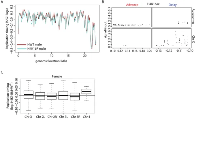
Figure 2.15. Replication timing profile for H4K16R females and control. ...68
Figure 2.16. Replication timing profile for H4K16R males and control. ...70
Figure 2.17. H4K16R mutation reduces gene expression and delays replication of the male X...72
xiii
Figure 2.19. Domains of altered replication in H3K9R mutants do not overlap
those identified after HP1a knockdown. ...80
Figure 3.1. H3K9 promotes endoreplication of the Drosophila salivary gland. ...94
Figure 3.2. DNA copy number in pericentric heterochromatin is elevated in H3K9R mutants. ...98
Figure 3.3. DNA copy number in pericentric heterochromatin is elevated in H3K9R mutants ...100
Figure 3.4. Under-replication of pericentric heterochromatin is H3K9-dependent. ...104
Figure 4.1. Cell lineage is a major driver of DNA replication timing in Drosophila. ...118
Figure 4.2. Characterization of RT in wildtype wing discs and follicle cells. ...120
Figure 4.3. Replicate correlations of RNA-seq data. ...126
Figure 4.4. Transcriptional change does not drive differential RT between lineages. ...128
Figure 4.5. Cell type-specific transcription does not drive changes in RT. ...130
Figure 4.6. S phase strategy does not affect DNA replication timing within the follicle cells of the adult ovary...134
Figure 4.7. Characterization of RT between wildtype mitotically cycling and endocycling follicle cells. ...135
Figure 4.8. Characterization of RT in Rif1- wing imaginal discs...138
Figure 4.9. Characterization of RT in Rif1-mitotically cycling follicle cells. ...140
Figure 4.10. Rif1 regulates RT in a lineage-specific manner ...142
Figure 4.11. Rif1 promotes late replication of pericentric heterochromatin across lineages. ...146
Figure 4.12. Characterization of RT in Rif1-/+mitotic follicle cells. ...148
Figure 4.13. Characterization of RT in Rif1-endocycling cycling follicle cells. ...150
xiv
Figure 4.15. Under-replication does not contribute to RT differences between
mitotically cycling and endocycling follicle cells. ...156
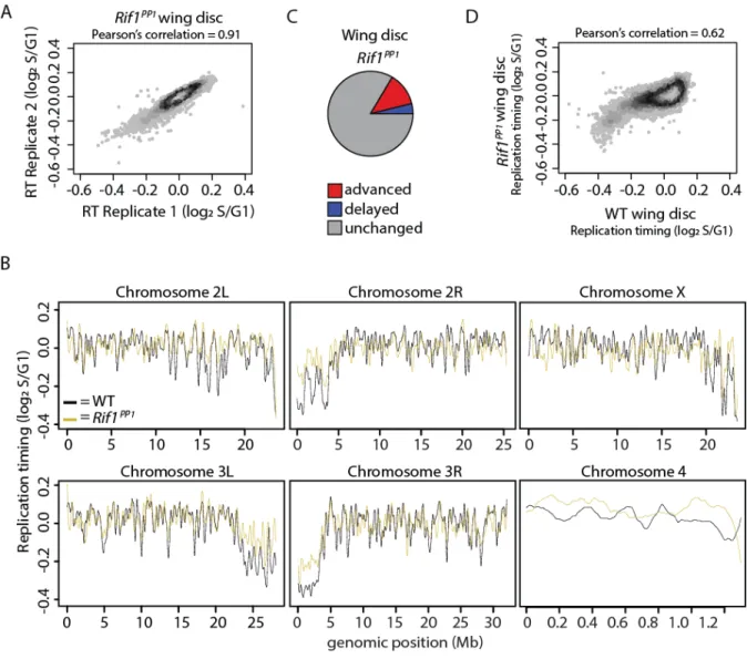
Figure 4.16. Characterization of RT in Rif1PP1wing discs. ...161
Figure 4.17. Characterization of RT in Rif1PP1mitotic follicle cells. ...162
Figure 4.18. Rif1’s PP1 binding motif is essential for Rif1-mediated RT control. ...164
Figure 5.1. Rif1 and H3K9 promote S phase progression of the second mitotic wave. ...175
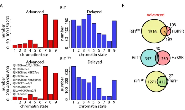
Figure 5.2. Rif1 and H3K9 regulate RT of unique heterochromatic domains. ...177
Figure 5.3. Chromatin accessibility profiling in Rif1- mutants. ...180
Figure 5.4. A modest relationship exists between chromatin accessibility and RT in Rif1- mutants. ...182
Figure A1. H3K9, H4K16, and H3K56 promote genome stability. ...198
xv
LIST OF TABLES
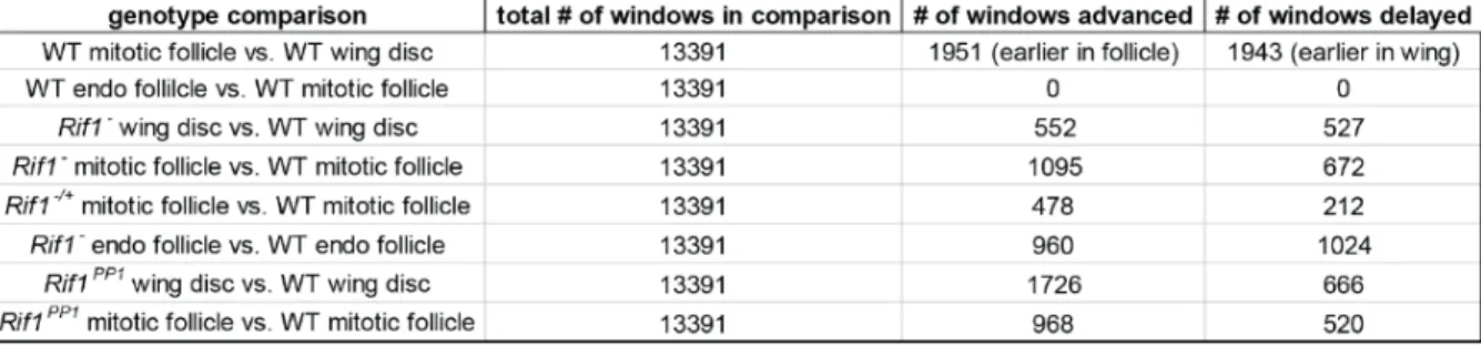
Table 4.1. Quantification of differential RT at 100kb windows using a 10kb slide
xvi
LIST OF ABBREVIATIONS
ac Acetyl
ARS Autonomously replicating sequence BAC Bacterial Artificial Chromosome
bp Base pair
BSA Bovine serum albumin
CDK Cyclin-dependent kinase
CDS Coding sequence
Cdt1 Cdc10-dependent transcript 1 gene Cdc6 Cell division cycle 6 gene
ChIP Chromatin immunoprecipitation
Chr Chromosome
CMG The replicative helicase containing Cdc45, MCM2-7. And GINS CTR Constant timing region
CUT&RUN Cleavage under targets and release using nuclease DAPI 4’,6-Diamidino-2-phenylindole
DDK Dbf4-dependent kinase
DNA Deoxyribonucleic acid
EdU 5-Ethynyl-2´-deoxyuridine
FACS Fluorescence activated cell sorting FISH Fluorescence in situ hybridization
FAIRE Formaldehyde assisted isolation of regulatory elements
xvii GFP Green fluorescent protein gRNA Guide ribonucleic acid H2Av Histone 2a variant v
H3 Histone 3
H4 Histone 4
H3.3K9R Lysine to arginine mutation at the ninth lysine of histone H3.3
H3.3WT Wild type histone H3.3
H3K9 Ninth lysine of histone H3
H3K9R Lysine to arginine mutation at the ninth lysine of histone H3 H4K16R Lysine to arginine mutation at the sixteenth lysine of histone H4 HEPES 4-(2-hydroxyethyl)-1-piperazineethanesulfonic acid
His4r Histone H4 replacement
HisC Df(2L)HisCED1429
HP1 Heterochromatin Protein 1
HWT Histone wild-type
K Lysine
kb Kilobase pair
Mb Megabase pair
MCM Mini-chromosome maintenance
me Methyl
MNase Micrococcal nuclease
xviii NEB Nuclear extraction buffer ORC Origin recognition complex PBS Phosphate buffered saline
PCNA Proliferating cell nuclear antigen PCR Polymerase chain reaction PTM Post-translational modification RFP Red fluorescent protein
RNA Ribonucleic acid
RPKM Reads per kilobase of transcript per million mapped reads
RPM Reads per million
RT Replication timing
SMW Second mitotic wave
SuUR Suppressor of under-replication SSC Saline sodium citrate
TAD Topologically associated domain TPM Transcripts per million
TTR Timing transition region twi Promoter for the twist gene YFP Yellow fluorescent protein UAS Upstream activating sequence
1
CHAPTER 1 – INTRODUCTION
DNA replication of metazoan genomes
In an average human lifetime, 1016 cell divisions occur within the body (Milo et al.
2010). During each cell division, the genetic information encoded from one parent cell is passed to two daughter cells. Genome integrity is maintained during cell division through the coordination of many tightly controlled mechanisms that ensure complete and accurate genome duplication while preventing deleterious mutation and chromosome mis-segregation. If these mechanisms go awry, severe developmental outcomes, such as the onset of disease, can occur. Therefore, understanding how DNA replication is regulated in space and time is critical to understanding the fundamental aspects of cell division and disease.
2
Stillman 1992; Liang et al. 1995; Coleman et al. 1996; Rowles et al. 1996; Nishitani et al. 2000). The first step in origin licensing involves ORC binding to chromatin (Duzdevich et al. 2015). Next, Cdc6 binds to ORC, which is necessary to recruit a single Cdt1-bound MCM2-7 hexamer (Duzdevich et al. 2015). The origin of replication is considered licensed when a second Cdt1-bound MCM2-7 hexamer is loaded adjacent to the first to form a head-to-head double hexamer (Evrin et al. 2009; Remus et al. 2009).
3
Regulation of DNA replication timing
Initiation of bidirectional DNA synthesis from origins of replication is staggered in space and time during S phase. This results in spatially separated regions of actively
replicating DNA; this asynchrony of replication is termed the DNA replication timing (RT) program (Taylor 1958; Taylor 1960; Woodfine et al. 2004; Ryba et al. 2010; Pope et al. 2014). Although asynchronous origin firing is evolutionarily conserved among eukaryotes, the biological function and control mechanisms of RT programs are not completely
understood. Importantly, altered RT of cancer-related genes correlates with changes in gene expression and contributes to malignant states (De and Michor 2011; Koren et al. 2012; Black et al. 2013; Fritz et al. 2013; Sima and Gilbert 2014; Polak et al. 2015; Rivera-Mulia and Gilbert 2016). Furthermore, genome-wide RT changes observed in cancer cells have been postulated to occur early during disease progression and may be sufficient to predict common disease-associated translocations (Koren et al. 2012; Donley and Thayer 2013; Rivera-Mulia et al. 2017; Du et al. 2019). We still do not understand whether RT change is a cause or consequence of disease progression, necessitating further understanding of the mechanisms regulating RT programs in both normal and disease contexts.
Local chromatin structure
4
function and density along DNA, establishing chromatin states that are either “open” (euchromatin, enriched in H3/H4 acetylation (ac) and H3K4 methylation (me)) or “closed” (heterochromatin, enriched in H3K9me2/3, H4K20me3, and H3K27me3). The chromatin landscape influences the binding of trans-acting factors primarily through either direct binding of factors to specific histone PTMs or through recruitment of factors to open, accessible chromatin. In metazoans, chromatin is thought to influence both recruitment of abundant trans-acting factors (ORC, Cdc6, Cdt1 and MCM2-7) that license origins in G1 and accessibility of DNA to limiting replication initiation factors (Sld2, Sld3, Dbp11, and Dbf4) that activate origins of replication in S phase (Mantiero et al. 2011; Collart et al. 2013; Das et al. 2015; Miotto et al. 2016). This is in contrast to S. cerevisiae, where origins of replication are sequence defined such that ORC binds to a conserved motif termed the autonomously replicating sequence (ARS) (Stinchcomb et al. 1979). As all other eukaryotes studied to date lack sequence-defined origins of replication, the exact mechanisms through which chromatin dictates the genome-wide landscape of licensed origins remain unclear.
ORC preferentially binds to G-rich, accessible chromatin, resulting in more licensed origins within “open” euchromatic regions of the genome relative to “closed”
heterochromatic regions (Delgado et al. 1998; MacAlpine et al. 2010; Cayrou et al. 2011). Furthermore, current models suggest that increased chromatin accessibility promotes the loading of more MCM complexes per ORC, further increasing the density of origins in euchromatic regions relative to heterochromatic regions (Das et al. 2015). Upon entry into S phase, replication initiation is thought to follow a stochastic model based largely on
5
probability than heterochromatic origins due to an increased density of licensed origins in accessible regions of the genome relative to inaccessible regions. Differential origin
activation within euchromatic and heterochromatic regions of the genome contributes to the relatively earlier RT of euchromatin relative to heterochromatin (Mantiero et al. 2011; Collart et al. 2013; Das et al. 2015). However, correlations between early RT and
euchromatin and late RT and heterochromatin are not absolute, suggesting additional modes of regulation beyond chromatin structure.
Transcriptional activity
Transcriptionally active regions of the genome tend to replicate earlier during S phase whereas transcriptionally repressive regions tend to replicate later during S phase (Goldman et al. 1984; Lubelsky et al. 2014). Because transcriptional activity is also strongly correlated with chromatin structure, it remains unclear whether the relationship between transcriptional activity and RT is a consequence of chromatin structure or if transcriptional activity directly influences RT. Despite strong correlations between active transcription and early RT, extremely high levels of transcription have been shown to inhibit replication initiation (Martin et al. 2011). Furthermore, transcriptional activity can displace the MCM complex, changing the origin location from the initial site of ORC binding (Gros et al. 2015; Powell et al. 2015). Although transcriptional activity can directly influence origins of replication, increasing evidence suggests that RT and transcriptional activity are regulated by a common chromatin environment (Lubelsky et al. 2014).
6
promoters is thought to be driven by the open chromatin conformation of promoters—active transcription maintains accessibility at promoters, thus promoting origin licensing in G1 or activation in S phase (MacAlpine et al. 2010; Dellino et al. 2013; Miotto et al. 2016). Dellino et al. mapped ~13,000 ORC1 binding sites and found that almost all mapped origins were associated with transcription start sites of either coding or noncoding RNAs (Dellino et al. 2013). Similarly, Miotto et al. mapped ORC2 binding genome-wide and found similar binding profiles to the independently derived ORC1 data (Miotto et al. 2016). Importantly, the ORC enrichment at promoters is most likely a consequence of the open chromatin environment as there is only a modest genome-wide correlation between ORC binding and transcriptional activity (Miotto et al. 2016).
Three-dimensional genome architecture
While both local chromatin structure and transcriptional activity influence genome-wide RT from yeast to humans, emerging evidence points to three-dimensional genome architecture as a previously unappreciated, key contributor to RT control (Rivera-Mulia and Gilbert 2016). The genome is organized within subnuclear compartments such that early replicating regions tend to be located at the nuclear interior (active compartment “A”), and late replicating regions tend to be located at the nuclear or nucleolar periphery (inactive compartment “B”) (Visser et al. 1998; Lieberman-Aiden et al. 2009). Within each
7
and TADs and cytologically-defined replication foci likely represent the same structures (Xiang et al. 2018). However, TAD boundaries are not absolutely required for maintenance of RT as TAD boundary disruption, either through deletion of DNA sequences at TAD boundaries or depletion of the protein components required for establishing interactions between adjacent TADs, has no effect on RT (Oldach and Nieduszynski 2019; Sima et al. 2019). Interestingly, while TAD boundaries are not required for RT maintenance, emerging evidence suggests that sequence elements within TADs (early-replication control elements, ERCEs) drive interactions between TADs required for maintenance of early RT (Sima et al. 2019). While further study is necessary to provide mechanistic insight into the relationship between three-dimensional genome architecture and RT, our understanding of RT control mechanisms is beginning to parse out correlative versus causal relationships.
Cell lineage
During animal development, cells undergo progressive changes in genome structure and function in order to generate more differentiated cell types. Transcriptional programs differ between cell types, and cell type-specific transcriptomes are reflected by genome-wide changes in both three-dimensional arrangement of DNA within the nucleus and local
chromatin structure. Interestingly, replication domain boundaries and TAD boundaries are stable structural units during cellular differentiation where the RT and subnuclear
8
TTR is positioned in compartment A in cells where RT has advanced. In fact, Heinz et al. demonstrated that manipulating the nuclear position of pericentric heterochromatin was sufficient to advance its RT in mammalian cells (Heinz et al. 2018).
Cellular differentiation provides a unique system to track the dynamics of RT, transcription, chromatin accessibility, and three-dimensional genome architecture in developmental time. Studying RT in this fashion has revealed multiple instances where genome architecture and RT are mechanistically separable. Studies in the early Drosophila embryo have demonstrated that the onset of late replication precedes the establishment of constitutive heterochromatin suggesting that the hallmarks of heterochromatin (H3K9me2/3 and HP1a enrichment) are not required for late RT (Yuan and O'Farrell 2016). Furthermore, establishment of RT programs was shown to anticipate transcriptional programs in the early zebrafish embryo (Siefert et al. 2017) while transcriptional change often preceded RT change during differentiation of mammalian cells (Rivera-Mulia et al. 2015). From studies
conducted in mammalian cells, we now know that correlations between RT, transcription, chromatin, and three-dimensional genome architecture become stronger as cells differentiate. Interestingly, strong correlations were shown to be restricted to genes located in CTRs (Rivera-Mulia et al. 2015), and these correlations are much weaker in TTRs (Besnard et al. 2012; Takebayashi et al. 2012; Dileep et al. 2015). Collectively, these data raise the
9
Replication timing: Genome instability and human disease
Normal RT programs contribute to genome instability
Many biological processes contribute to genomic instability, including the normal RT program. RT has been proposed to contribute to the non-random genome-wide distribution of mutations, where early replicating regions are more susceptible to trans chromosomal
rearrangements and late replicating regions are more prone to cis translocations and to point mutations (Watanabe et al. 2002; Stamatoyannopoulos et al. 2009; Cui et al. 2012; Sima and Gilbert 2014; Supek and Lehner 2015; Du et al. 2019). Furthermore, late replicating regions and origin-depleted regions that are passively replicated by an adjacent origin (TTRs),
experience a greater overall mutational burden than early replicating regions (Watanabe et al. 2002; Hiratani et al. 2008; Watanabe and Maekawa 2010; De and Michor 2011). Many common structural mutations, fragile sites, hotspots for copy number alterations, and genomic rearrangement sites in cancer are found in TTRs, possibly due to the complicated nature of replicating these origin-depleted regions (Watanabe et al. 2002; Donley and Thayer 2013; Rhind and Gilbert 2013). It has been postulated that minimizing the mutational burden in early replicating regions of the genome helps to prevent mutation of ubiquitously
expressed “housekeeping” genes. Consequently, tissue-specific genes located in TTRs and late replicating gene-poor regions of the genome experience the bulk of the mutational burden.
RT alterations in cancer
10
Ozgyin et al. 2019; Takahashi et al. 2019). The robustness of RT programs is further highlighted by the fact that almost every attempt to disrupt RT, including genetic perturbation of key RT control factors, results in little to no effect on genome-wide RT (Yokochi et al. 2009; Pope et al. 2014; Foti et al. 2016; Armstrong et al. 2018; Oldach and Nieduszynski 2019; Sima et al. 2019). Despite the robust nature of RT, RT programs are commonly altered in cancer, and altered RT programs have been proposed to be an early epigenetic event in disease progression (Ryba et al. 2012; Koren et al. 2014; Rivera-Mulia et al. 2017). Importantly, the proportion of, and the specific loci within, the genome that display altered RT are cancer type-specific with, for example, LNCaP prostate cancer cells
displaying altered RT at 5.7% of the genome and acute lymphoblastic leukemia patient cells displaying altered RT at 9-18% of the genome (Ryba et al. 2012; Du et al. 2019). Because, in some instances, RT can differentiate disease and normal tissue in ways conventional
transcriptomic analysis cannot, RT profiling has been proposed as a potential diagnostic tool for cancers with unique RT signatures (Rivera-Mulia et al. 2017).
11
12
CHAPTER 2- CHROMATIN CONFORMATION AND TRANSCRIPTIONAL ACTIVITY ARE PERMISSIVE REGULATORS OF DNA REPLICATION
INITIATION IN DROSOPHILA1
Introduction
Animal cells duplicate large, complex genomes by initiating replication at distinct locations within the genome at different times during S phase. An evolutionarily conserved feature of this regulatory paradigm is a temporal order of DNA replication initiation events that results in characteristically early and late replicating regions of the genome (Rhind and Gilbert 2013). Such “replication timing” (RT) programs appear at early stages of animal development and ensure genome integrity during cell proliferation (Shermoen et al. 2010; Mantiero et al. 2011; Collart et al. 2013; Hamperl and Cimprich 2016; Yuan and O'Farrell 2016; Almeida et al. 2018). Importantly, RT is associated with mutational burden and SNP density, as spontaneous mutations occur less frequently in early compared to late replicating regions of the genome (Stamatoyannopoulos et al. 2009; Donley and Thayer 2013).
Furthermore, perturbed RT is thought to be an early epigenetic event that predisposes cancer and disease-associated genome rearrangement (Ryba et al. 2012; Donley and Thayer 2013). Notwithstanding their importance, mechanisms that control where and when DNA
replication initiates within an animal genome remain poorly understood.
13
In contrast to replication initiation in single-celled eukaryotes such as budding yeast, replication of animal genomes does not initiate at well-defined sequence motifs (Bell and Stillman 1992; MacAlpine et al. 2010; Miotto et al. 2016). Rather, two levels of genome organization have emerged as putative regulators of replication initiation: three dimensional arrangement of DNA within the nucleus and local chromatin structure, characterized in part by differential DNA accessibility (i.e. differential nucleosome occupancy) (Hiratani et al. 2008; Pope et al. 2014; Heinz et al. 2018). Current models posit that these features of genome organization regulate replication by influencing trans-acting factor recruitment to sites of replication initiation (i.e. origins) (Mantiero et al. 2011; Collart et al. 2013; Pope et al. 2014; Das et al. 2015; Miotto et al. 2016; Rivera-Mulia and Gilbert 2016). In all metazoan organisms examined to date, transcriptionally active, accessible euchromatin generally replicates early during S phase, whereas transcriptionally repressive, inaccessible
heterochromatin generally replicates late (Bell et al. 2010; Eaton et al. 2011; Lubelsky et al. 2014). Despite strong genome-wide correlations between replication and chromatin structure in animal cells, efforts to determine a causal relationship between the two have been
hampered by imprecise methods for manipulating chromatin structure in vivo. We therefore developed an approach for altering the distribution of accessible chromatin throughout the genome and determined if and how these changes in chromatin structure affect genome replication.
14
substrates that may participate in DNA replication (Glozak et al. 2005; Huang and Berger 2008). Therefore, to reduce potential pleiotropic effects of mutating histone-modifying enzymes, we employed a strategy in Drosophila to more precisely manipulate chromatin structure by mutating the histone genes themselves, an approach that is not currently feasible in other animal models. This strategy involves deleting the endogenous wild type histone genes and replacing them with transgenic copies encoding a single amino acid substitution that prevents PTMs of a particular histone residue (Günesdogan et al. 2010; McKay et al. 2015). Here, we determine how two different histone mutations that affect chromatin organization and transcription in heterochromatin (H3K9R) and euchromatin (H4K16R), respectively, affect DNA replication initiation throughout the genome.
Materials and Methods Complete genotypes
“12xHWT” (Histone Wild Type) refers to a control Bac-based transgene containing 12 copies of the 5kb histone wild type repeat unit containing all five replication dependent histone genes (McKay et al. 2015). “12xH3K9R” and “12xH4K16R” are identical transgenes except with a Lys to Arg substitution mutation at the 9th residue of histone H3 and 16th residue of
histone H4, respectively.
HWT: yw; ΔHisC, twi-Gal4/ΔHisC,UAS-2xEYFP; 12xHWT/+ (McKay et al. 2015) H3K9R: yw; ΔHisC, twi-Gal4/ΔHisC,UAS-2xEYFP; 12xH3K9R/+ (Penke et al. 2016) H4K16R: yw; ΔHisC, twi-Gal4/ΔHisC,UAS-2xEYFP; 12xH4K16R/+
15
H3.3K9R; H3K9R: yw, H3.3BK9R; H3.3A2x1, ΔHisC, twi-Gal4/Df(2L)BSC110,Δ HisC,UAS-2xEYFP; 12xH3K9R/+ (Penke et al. 2018)
Zygotic, replication-dependent HWT: yw; ΔHisC, twi-Gal4/ΔHisC,UAS-2xEYFP; 12xHWT/+
Zygotic replication-dependent H4K16R: yw; ΔHisC, twi-Gal4/ΔHisC,UAS-2xEYFP; 12xH4K16R/+
Maternal/zygotic, replication-dependent HWT: yw; ΔHisC, twi-Gal4/Δ HisC,UAS-2xEYFP; 12xHWT/+ (from mothers of genotype ΔHisC, UAS-2xeYFP; 12xHWT) Maternal/zygotic, replication-dependent H4K16R: yw; ΔHisC, twi-Gal4/Δ HisC,UAS-2xEYFP; 12xH4K16R/+ (from mothers of genotype ΔHisC, UAS-2xEYFP; 12xH4K16R) Zygotic, replication-dependent and replication-independent HWT: yw; ΔHisC, twi-Gal4/ΔHisC,UAS-2xEYFP; 12xHWT, His415-4/His4r15-4
Zygotic replication-dependent and replication-independent H4K16R: yw; ΔHisC, twi-Gal4/ΔHisC,UAS-2xEYFP; 12xH4K16R, His4r15-4/His4r15-4
Generation of H3K9R and H4K16R mutant genotypes
All fly stocks were maintained on standard corn medium and crossing schemes to generate replication-dependent histone genotypes were performed as in (Penke et al. 2016). For first instar larval brain EdU experiments, the following crosses were performed: H3.3WT; H3WT) yw; H3.3A2x1, ΔHisC, twi-Gal4/CyO females were crossed to yw; Df(2L)BSC110,
ΔHisC,UAS-2xEYFP/CyO; 12xHWT males and for H3.3K9R; H3K9R) yw, H3.3K9R; H3.3A2x1, ΔHisC, twi-Gal4/CyO females were crossed to H3.3K9R; Df(2L)BSC110,
16
generate zygotic, replication-dependent HWT and H4K16R mutants, ΔHisC, twi-Gal4/CyO mothers were crossed to ΔHisC, UAS-2xEYFP/CyO; 12xHWT/12xHWT or ΔHisC, UAS -2xEYFP/CyO; 12xH4K16R/12xH4K16R fathers, respectively. To generate flies where both the maternal and zygotic contribution of histones were HWT or H4K16R mutant, ΔHisC,
UAS-2xeYFP; 12xHWT or ΔHisC, UAS-2xEYFP; 12xH4K16R mothers, respectively, were crossed to ΔHisC, twiGal4/CyO fathers. To generate zygotic, replication-dependent and replication-independent HWT and H4K16R mutants, ΔHisC, twi-Gal4/CyO; His4r15-4 mothers were crossed to ΔHisC, UAS-2xEYFP/CyO; 12xHWT, His4r15-4 or ΔHisC, UAS-2xEYFP/CyO; 12xH4K16R, His4r15-4 fathers, respectively. For each H4K16R viability experiment, groups of fifty GFP+ first instar larvae of each genotype were separated from their wild type siblings into vials of standard corn medium and allowed to complete development.
CRISPR-Cas9 Mutagenesis of His4r
Two different gRNA oligos targeting the 5’UTR (target sequence:
17 Culture conditions for embryo sorting
A Union Biometrica BioSorter for large particle flow cytometry equipped with a 488-nm solid state laser and accompanying FlowPilot software was used for identification and high throughput isolation of GFP-positive ΔHisC, UAS-2xEYFP/ΔHisC, twi-GAL4 mutant embryos from their GFP-negative siblings. For this purpose, three hundred to four hundred
ΔHisC, twi-Gal4/CyO females and 100 ΔHisC, UAS-2xEYFP/CyO; 12xHWT/12xHWT, 100
ΔHisC,UAS-2xEYFP/CyO; 12xH3K9R/12xH3K9R, or 100 ΔHisC,UAS-2xEYFP/CyO; 12xH4K16R/12xH4K16R males were placed in a large embryo collection cage (fits 100mm petri dishes) at 25°C and allowed to lay eggs on apple juice agar plates. Overnight collections were dechorionated in 100% bleach for two minutes and collected in embryo wash buffer (0.7% NaCl, 0.07% Triton X-100) prior to embryo sorting. Aliquots of one hundred GFP-positive embryos were transferred to vials containing standard corn medium and cultured at 25°C to obtain third instar larvae.
Sample preparation for FACS and sequencing
Third instar wing imaginal discs were dissected over a period of four hours and stored in Grace’s insect medium (supplemented with L-Glutamine, 3.33g/L Lactalbmin
Hydrolysate, and 3.33g/L Yeastolate) on ice prior to nuclear isolation. Nuclear isolation was performed similarly to (Ma and Weake 2014) with the following adjustments. In brief, a 2mL dounce homogenizer was pretreated with nuclear extraction buffer (NEB; 10mM HEPES-KOH, pH 7.5; 2.5 mM MgCl2; 10mM KCl) supplemented with 5% BSA and placed on ice.
18
CellTrics 30 μm filter and isolated nuclei were stained with 1.5μg/mL DAPI prior to FACS. Nuclei were sorted into G1, S, and G2 populations based on DNA content as measured by DAPI intensity on a FACSAria II or III (using NEB-0.1% Tween sheath). Gates were chosen conservatively to prevent contamination of either fraction with nuclei from a neighboring fraction. Analyses of cell cycle indices were performed on DAPI profiles generated from FACS using the Dean-Jett Fox model included with the FlowJo software (Dean and Jett 1974; Fox 1980). Isolated populations of nuclei were pelleted, flash frozen, and stored at -80°C prior to DNA isolation and library preparation. Genomic DNA libraries were prepared with the Rubicon ThruPLEX DNA-seq kit and sequencing was performed on an Illumina HiSeq 2500 in the UNC-Chapel Hill High Throughput Sequencing Facility.
Sequence data analysis
Analyses were performed using R (Team 2017). Replication Timing Profiles
19
were then averaged. As an additional control, another RT value was generated using half the G2 value, which should be equivalent to the G1 value. RT profiles were generated by
plotting the RT value at each window versus the genomic location. RT profiles normalized to G1 or G2 copy number controls were very similar; we therefore used G1 for all subsequent analyses. LOESS regression lines using loess.model were created to smooth RT profiles (span=0.02 for chromosome arms, span=0.05 for Chromosome 4). Although the genomic location where RT regression lines changed direction was similar across all genotypes and replicates, we note that the range of RT values for HWT female samples was slightly smaller than the other two genotypes. HWT female samples exhibited a higher percentage of cells in S phase, which decreased the clarity of the G1/S boundary when performing FACS. We speculate that a small increase in the number of late G1 cells in S phase populations limited the dynamic range of HWT female RT values. We therefore used quantile normalization through the preprocess Core R package to equalize the dynamic range of RT values for each female genotype (Bolstad 2016). We note that regions altered in H3K9R mutants compared to HWT were similar without quantile normalization. The limma statistical package was used to identify windows with significantly altered RT values between HWT and H3K9R female, HWT and H4K16R female, and HWT and H4K16R male samples (lmFit, adjusted p value Benjamini and Hochberg, p<0.01) (Newville et al. 2014). The adjusted p value corrects for multiple testing. An additional significance parameter of an absolute log2 fold-change greater
20
chromatin states were obtained from (Kharchenko et al. 2011) and converted to dm6 coordinates using the UCSC liftOver tool (Karolchik et al. 2004).
Wild-type Replication Timing Characterization
To calculate replication domain sizes, we identified the genomic coordinates halfway between each peak and valley of an RT profile and determined the distance from one halfway point to the next. We used modENCODE ChIP-seq data from whole 3rd instar larvae to
calculate histone PTM enrichment at 100kb windows across the genome
(
ftp://data.modencode.org/D.melanogaster/Histone-Modification/ChIP-seq/raw-seqfile_fastq/). Accession numbers for each data set are as follows: H3K36me3 (GSE47248),
H3K4me1 (GSE47282), H3K4me2 (GSE47261), H3K4me3 (GSE49491), H3K79me1 (GSE49492), H3K27ac (GSE49488), H3K79me2 (GSE49493), H3K79me3 (GSE49494), H4K20me1 (GSE47254), H2Bubi (GSE49487), H3K36me1 (GSE47249), H3K23ac (GSE47257), H3K9ac (GSE48510), H3K9me1 (GSE47289), H3K9me2 (GSE47260), H3K9me3 (GSE47258), H3K9acS10P (GSE47288), H2Av (GSE47259), and H4K16ac (GSE49497) (Roy et al. 2010). For each histone PTM, raw reads for two ChIP replicates and two input replicates were aligned to the genome using Bowtie 2 (v2.3.2) (Langmead et al. 2009). BEDTools coverage (v2.25.0) was used to count the number of reads mapping to each 100kb window, and the resulting counts were normalized to read depth (Quinlan and Hall 2010). Histone PTM enrichment for each replicate was calculated by dividing the ChIP normalized read counts by the input for each replicate; the resulting values were then
21
RNA-seq data from 3rd instar imaginal wing discs (from GSE85374) (Penke et al. 2016) was
used to calculate transcript density or transcript activity at 100kb windows. The imaginal wing disc transcriptome was assembled using Cufflinks (v2.2.1) (Trapnell et al. 2012) with the following parameters: library-type fr-firsttrand, masked rRNA, and provided dm6 transcriptome obtained from Flybase release 6.04. Subsequently, the number of transcripts overlapping each 100kb window was determined. To calculate transcript activity of a window, the normalized read per million of each transcript overlapping a window was summed.
FAIRE, HP1a, and RNA Analyses
For H3K9R experiments, RNA reads from three HWT and three H3K9R replicates were aligned using TopHat default parameters (v2.1.1) (Trapnell et al. 2012), and a
transcriptome was generated using Cufflinks (v2.2.1, see above for parameters). Previously, we showed that the H3K9R mutation causes widespread de-repression of transposons (Penke et al. 2016). Therefore, in the current analysis, we combined the Cufflinks generated
transcriptome with transposons annotated by RepeatMasker (Smit et al. 2013-2015). For H4K16R experiments, 30 wing imaginal discs per replicate were homogenized in Trizol and flash frozen in liquid nitrogen. RNA was chloroform extracted and isopropanol precipitated before column purification on Qiagen RNeasy purification with DNase digestion.
22
from three HWT female, three HWT male, three H4K16R female, and three H4K16R male replicates were aligned using TopHat default parameters (v2.1.1) (Trapnell et al. 2012), and a transcriptome was generated using Cufflinks (v2.2.1, see above for parameters). For both H3K9R and H4K16R experiments, raw counts of RNA reads at each transcript were used as input for edgeR statistical analysis (p value <0.01) (Robinson et al. 2010; McCarthy et al. 2012). We then identified transcripts within or that overlapped each 10kb window and selected the transcript with the lowest p value.
To determine RT values at 10kb windows, we used the previously calculated log2
fold-change and p values from 100kb windows. We used RT values from 100kb windows as this size closely matches average replication domain size (~100-200kb), but similar results were obtained using RT values determined from 10kb windows (Fig S1). For each 10kb window, we calculated the median fold change and median p value of all overlapping 100kb windows. 10kb windows were identified as having significantly altered RT between H4K16R or H3K9R and HWT if p<0.05 (adjusted for multiple testing) and the absolute log2
fold-change was at least 0.1. To focus our analysis on more mappable regions of the genome, we analyzed 10kb windows on the major chromosome scaffolds (Chr 2L, Chr 2R, Chr 3L, Chr 3R, Chr 4, and Chr X) that, for H3K9R experiments, had an average FAIRE and HP1a counts per million (CPM) value of greater than zero. Comparisons of RT, FAIRE, HP1a, and RNA signal between H3K9R and HWT samples were performed with all reads or “uniquely” mapping reads (MAPQ>10) with similar results.
23
identified in Cufflinks were included in this edgeR analysis to facilitate modeling of variability. Data was visualized using the Integrative Genomics Viewer.
Immunofluorescence
Third instar wandering larvae were dissected and the carcasses inverted to expose attached imaginal discs and incubated for 60’ in 0.1mg/mL EdU. Tissues were then fixed in 3.7% paraformaldehyde in PBS for 25 min. EdU incorporation was detected using the Click-It EdU Alexa Fluor 488 Imaging Kit (ThermoFisher Scientific). Carcasses were washed for 10 min in PBS-Tx (3% Triton X-100), then treated with 200ug/mL RNaseA in PBS-Tx for 2h and washed for 1 h in PBS-Tx. Individual imaginal discs were then separated from the carcass and groups of discs were successively incubated for 20 min in each of four pre-hybridization solutions: 1) 80% PBS-Tx, 20% pHM (50% formamide, 4xSSC, 100mM NaH2PO4 pH 7.0, 0.1% Tween 20), 2) 50% PBS-Tx, 50%pHM, 3) 20%PBS-Tx, 80% pHM,
4) 100%pHM. Denatured 359bp probe (Joyce et al. 2012) was hybridized with wing discs overnight at 37°C at 450rpm in an Eppendorf tube. Discs were successively incubated in four post-hybridization solutions for 20 min at 37°C at 800rpm: 1) 50% formamide, 2XSSC, 2) 40% formamide, 2XSSC, 3) 30% formamide, 70% PSS-Tw (1x PBS, 0.1% Tween20), 4) 20% formamide, 80% PBS-Tw and three post-hybridization solutions at 25°C: 1) 10% formamide, 90% PBS-Tw, 2) PBS-Tw, 3) PBS-Tx. DNA was stained with DAPI, and the discs were mounted in ProLong Gold antifade reagent and imaged on a Leica confocal microscope.
24 Results Profiling replication timing in a Drosophila tissue
To probe the relationship between chromatin structure and replication in an intact animal, we adapted a genome-wide measure of RT for use in Drosophila wing imaginal discs, a relatively simple epithelium of proliferating diploid cells (Koren et al. 2014; Sasaki et al. 2017; Siefert et al. 2017). Our method is based on the premise that in S phase cells early replicating DNA sequences are over-represented relative to late replicating ones, due to a higher probability of replication initiation (Rhind et al. 2010; Mantiero et al. 2011; Collart et al. 2013; Das et al. 2015). Consequently, RT data are a proxy for the propensity of replication initiation in a particular region of the genome. We performed whole-genome sequencing on DNA isolated from populations of G1 and S phase nuclei collected from wing discs by fluorescence-activated cell sorting (FACS) (Fig 2.1A). Replication profiles were generated by determining the log2 transformed S/G1 read count at 100kb intervals using a 10kb slide
25
Figure 2.1. Measuring genome-wide replication timing in vivo.
A) Experimental paradigm: (1) Nuclei were FACS sorted into G1 (yellow), S (red) and G2 (blue) populations based on DNA content. (2) Sequenced DNA was mapped to the dm6 genome. More reads map to early than late replicating sequences. (3) Log2 S/G1 ratio
generates RT profiles. Normalizing to G1 or G2 phase controls gave similar results. B) LOESS regression line showing average yw (“yellow,white” control genetic background used for all fly lines)RT values (log2 S/G1) in 100kb windows with 10kb slide across Chr 2
and 3. Chromosome schematics show approximate locations of constitutive pericentric heterochromatin (green) and largely euchromatic arms (blue) (Riddle et al. 2011; Hoskins et al. 2015). C) Heatscatter plot of yw log2 S/G1 (RT) versus gene density at all 10kb windows
across the genome with LOESS regression line (black). D) Heatmap of relative modENCODE histone PTM enrichment in bins of equally sized RT quintiles (early, early/mid, mid, mid/late, and late) generated using RT values (log2 S/G1) within 100kb
windows. modENCODE data is from third instar larvae (Celniker et al. 2009) (see
27
Figure 2.2. Generation of wild type replication timing profiles.
A) To determine the most appropriate window size for analyzing replication timing, we considered windows of 1kb (top left), 10kb (top right), 10kb with 1kb slide (bottom left), and 100kb with 10kb slide (bottom right). Chr 3R coordinates 10000000-15000000 are shown. A heatscatter of raw log2 S/G1 values (grey) and LOESS regression line (red) are included. All
29
Figure 2.3. Replication timing in Drosophila wing discs correlates with features of active and repressive chromatin.
A) Representative 5Mb region on Chromosome 3R of S/G1 (log2) replication timing values
within 100kb windows with a 10kb slide. RT values are an average of replicate yw samples. LOESS regression line is indicated in red. B) LOESS regression line showing average yw S/G1 (log2) replication timing values at 100kb windows using a 10kb slide across
Chromosome X and 4 scaffolds. Approximate locations of constitutive heterochromatin (green) and largely euchromatic regions (blue) are indicated (Riddle et al. 2011; Hoskins et al. 2015). C) Histogram of yw replication domain sizes. D) Heatscatter plot of yw S/G1 (log2)
replication timing values and RNA expression levels within all 10kb windows across the genome with LOESS regression line in black. E) Heatmap of relative H3K9me2 and
H3K9me3 enrichment in bins of equally sized RT quintiles (early, early/mid, mid, mid/late, and late) generated using S/G1 (log2) RT values within 100kb windows and normalized
modENCODE H3K9me2/me3 data from third instar larvae. Color indicates average
31
Figure 2.4. Replication timing profiling in Drosophila tissue is highly reproducible. Quantile normalized S/G1 (log2) replication timing values for each replicate for the indicated
33
Our wing disc replication profiles are similar to those previously generated from Drosophila cell lines and most closely correlate with RT data obtained from a cell line derived from the same developmental stage as wing discs (Fig 2.5) (Lubelsky et al. 2014). Replication domain sizes ranged from 20kb-570kb (Fig 2.3C), closely matching previous measurements (MacAlpine et al. 2004). Consistent with previous studies in zebrafish embryos and in fly and mammalian cultured cells (Bell et al. 2010; Eaton et al. 2011;
34
Figure 2.5. Wild-type 3rd instar imaginal wing discs and cell culture replication timing profiles are highly correlated.
A) Heatscatter plot of S/G1 (log2) replication timing value at 100kb windows from yw
imaginal wing discs and previously generated timing profiles from three Drosophila cell culture lines (Kc, S2, and Bg3) (Lubelsky et al. 2014). Top row shows correlations between each of the three cell culture lines, and bottom row shows the correlations between yw wing discs and the three cell culture lines. Windows with earlier timing values in S2 cells
36
Replication timing is largely unchanged in H3K9R mutants
To determine how chromatin structure influences replication, we first tested if modification of H3K9 determines the difference in RT between heterochromatin and euchromatin. Defining features of heterochromatin are the presence of methylated H3K9 (H3K9me) and Heterochromatin Protein 1a (HP1a). HP1a binds H3K9me and facilitates heterochromatin formation through multimerization of HP1a molecules and recruitment of other factors (Canzio et al. 2011; Larson et al. 2017; Strom et al. 2017). Previously, we showed that H3K9R mutants are depleted of H3K9me and HP1a within pericentric heterochromatin (Penke et al. 2016). In addition, we found that loci within the pericentric heterochromatin of H3K9R mutants are nucleosome depleted relative to controls, as measured by increased FAIRE-seq signal (Formaldehyde-Assisted Isolation of Regulatory Elements) (Penke et al. 2016).
If increasing chromatin accessibility directly resulted in earlier replication initiation, we would expect large-scale advancement of RT at nucleosome-depleted H3K9R
pericentromeres. We assigned RT values to 100kb windows tiled 10kb across the genome and used stringent significance thresholds (p<0.01 (adjusted for multiple testing), absolute log2 fold change>0.1; limma) to identify differential RT between H3K9R and control.
37
38
Figure 2.6. Analysis of replication timing in H3K9R mutants.
A) Log2 S/G1 RT values at 100kb windows with 10kb slide for 12x HWT (histone wild type;
yellow) and 12x H3K9R (purple) plotted across Chr 3R. See Fig S5 for other chromosomes. B) ~5 Mb region of the pericentromeric heterochromatin of Chr 3R. Red vertical bars designate significant RT changes between H3K9R and HWT (p<0.01, p value adjusted for multiple testing; absolute log2 fold change>0.1; limma). C) H3.3WT H3WT and H3.3K9R H3K9R
(see supplementary materials for full genotype) first instar brains pulse labeled for 1hr with EdU (yellow) and stained for DNA (blue; DAPI). White arrowheads designate late patterned EdU incorporation. D) Percentage of EdU+ cells with early or late EdU incorporation
40
Figure 2.7. Replication timing profile for H3K9R mutants and control.
LOESS regression line applied to S/G1 (log2) averaged replicates from HWT (yellow) and
42
Figure 2.8. Characterization of altered replication timing in H3K9R mutants.
A) HWT and H3K9R eye imaginal discs labeled for 60’ with EdU (yellow), stained for 359-bp FISH probe (X Chromosome pericentromere; magenta), and counterstained with DAPI (blue). White arrow indicates colocalization of EdU late replication focus and 359-bp FISH probe. Shown is a single slice from a Z projection (top). Box plot of the percentage of colocalization between the 359-bp FISH focus and late-patterned EdU focus in HWT and H3K9R eye imaginal discs (bottom; * = P < 0.05, Student’s T-test). B) Histogram of the number of domain sizes with advanced (red), delayed (blue), or all replication timing change (grey). C) Correlation analysis of the absolute H3K9R/HWT log2 RT fold change versus the
average enrichment of H3K9me2 (top) or H3K9me3 (bottom) signal at 10kb windows with significantly advanced (left) or delayed (right) replication. ChIP-seq enrichment was
determined from modENCODE datasets from wild-type whole 3rd instar larvae. Experiments
44
Advanced replication occurs at newly accessible chromatin in H3K9R mutants Despite largely unchanged RT in H3K9R mutants, 3% of the genome nevertheless exhibited altered RT (~2% advanced and ~1% delayed). Importantly, these changes do not result from pre-existing copy number differences between the G1 genomes of H3K9R and control (Fig 2.2). We used these changes to investigate the relationship between chromatin structure and replication initiation (Fig 2.8B). We found that the majority (82.1%) of earlier replicating 100kb windows in H3K9R mutants are located in pericentric heterochromatin (Fig 2.6B) or on the small 4th Chromosome (Fig 2.7), which is primarily heterochromatic (Haynes
et al. 2007). Importantly, these changes are unlikely to be caused by changes in the
expression of genes encoding replication factors or other protein-coding genes, as the H3K9R mutation does not significantly affect their expression (File 2.3; (Penke et al. 2016)). By contrast, 76.2% of later replicating 100kb windows are located along euchromatic chromosome arms (Fig 2.6A; Fig 2.7).
To compare our RT data to other genome features like histone PTMs, we assigned a RT value to non-overlapping 10kb windows across the entire genome (see supplementary materials). Notably, 10kb windows with advanced replication in H3K9R mutants are enriched for H3K9me2/me3 in a wild type genome and not for other histone PTMs such as H3K27me3, a marker of facultative heterochromatin (Fig 2.6F,G; Fig 2.8C). This
45
We hypothesized that if chromatin structure directly influences replication, then RT changes should occur at newly accessible chromatin in H3K9R mutants. To compare
chromatin accessibility and RT in H3K9R mutants, we compared FAIRE-seq and RT values at 10kb windows across the genome (Materials and Methods) (Penke et al. 2016). While most pericentric regions included in the current genome assembly are more accessible in H3K9R mutants compared to control (Penke et al. 2016), we found that the vast majority (92.9%) of windows with increased FAIRE signal do not display altered RT (Fig 2.9A). Thus, despite established correlations between accessible chromatin and early replication, increasing chromatin accessibility by H3K9R mutation does not invariably result in earlier replication.
Importantly, this conclusion does not mean that high chromatin accessibility makes no contribution to early replication. Indeed, nearly all windows (230/243) that exhibit significantly advanced replication in H3K9R mutants also have increased FAIRE signal (Fig 2.9A-D; Fig 2.10A,E). This result suggests that a more accessible chromatin environment may be necessary for earlier replication in H3K9R mutants. In contrast, most windows with delayed RT exhibit no change in FAIRE signal, suggesting that delayed replication occurs independently from chromatin accessibility changes in H3K9R mutants (Fig 2.9A-D; Fig 2.10A,F).
46
H3K9R mutants do not have altered RT. These results indicate that HP1a loss does not invariably result in advanced replication in H3K9R mutants, although it may be necessary. Overall our observations are surprising in that the hallmarks of heterochromatin – high levels of H3K9me and HP1a within a relatively inaccessible chromatin environment – are not
47
Figure 2.9. Open chromatin is permissive to advancement but not delay of replication timing.
A) Heatscatter plot of the H3K9R/HWT ratio of RT values (log2 S/G1) versus the
H3K9R/HWT ratio of FAIRE signal at all 10kb windows across the major chromosome scaffolds. 10kb windows with significantly advanced (red) or delayed (blue) RT are indicated. Darker color indicates higher density of windows. B) Cumulative count of advanced (red) or delayed (blue) 10kb windows ordered by increasing FAIRE signal in H3K9R compared to HWT. C) Heatscatter plot of the H3K9R/HWT ratio of HP1a ChIP signal versus the H3K9R/HWT ratio of FAIRE signal at all 10kb windows across the major
chromosome scaffolds. D) Venn-diagram of all 10kb windows with significantly altered FAIRE or HP1a signal in H3K9R compared to HWT (p<0.01; edgeR). For all panels, significantly different RT was determined as p<0.05, log2 fold change>0.1 using limma.
49
Figure 2.10. Disrupting heterochromatin does not always result in altered replication. A) Venn-diagram of 10kb windows with significantly altered FAIRE signal and significantly advanced or delayed replication in H3K9R mutants compared to control. B) Heatscatter plot of the H3K9R/HWT ratio of normalized replication timing values (S/G1 (log2)) plotted versus
51
Elevated transposon expression accompanies advanced replication in H3K9R mutants We next considered the transcriptional activity of domains of altered replication. We compared our newly generated RT profiles with our previously generated wing disc
transcriptome profiles from H3K9R and control (Penke et al. 2016). We focused on transcripts (genes or transposons) most likely to drive RT changes by identifying the transcript that was most significantly different within each 10kb window between H3K9R and control (i.e. the transcript with the lowest p-value in differential expression analysis; edgeR). We then compared the fold change of this transcript to the RT value assigned to the same 10kb window. We found that only a small fraction (6.8%) of the 3,371 10kb windows containing a transcript with a significant expression change also exhibited a RT change (Fig 2.11A,B; Fig 2.12A). This observation indicates that, despite strong correlations between active transcription and early replication (MacAlpine et al. 2004; Liu et al. 2012; Lubelsky et al. 2014; Rivera-Mulia and Gilbert 2016), transcriptional activity and RT are separable. Conversely, we found that the majority (76.5 %) of windows with advanced replication in H3K9R mutants exhibited a change in gene expression (Fig 2.11A,B; Fig 2.12B). Because most (97.3%) changes were increases in expression, we speculate that transcription might promote early replication initiation in pericentric heterochromatin. Similar results were obtained by using the average expression change of all transcripts that overlap each window with advanced RT, rather than the transcript with the most significant change in expression across the window (Fig 2.12G).
52
changes within advanced replication domains (Fig 2.12C). Therefore, we also identified transposons belonging to families that were differentially expressed between H3K9R and control (Materials and Methods; Fig 2.11C; Fig 2.12C-F). All 243 windows of advanced replication in H3K9R mutants contain either a transposon belonging to a family that was differentially expressed in H3K9R compared to control (96.4%) or that neighbored a window containing multiple differentially expressed transcripts (Fig 2.11C,D). Although we cannot determine whether individual transposons within all 243 advanced windows changed expression, these data suggest that altered transcription may promote advancement of replication in H3K9R mutants.
Along with transposon enrichment (Fig 2.13A,B), advanced replication domains in H3K9R mutants are normally enriched for H3K9me2/me3 (Fig 2.13C) and exhibited a lower GC content (Fig 2.13D) compared to domains of increased chromatin accessibility or
increased RNA expression with unaltered replication (FAIRE only and RNA only,
respectively). Although transposon density distinguished advanced domains, the majority of domains with altered transposon expression have no change in RT (Fig 2.11C; Fig 2.12D). Therefore, we surmise that altered transposon expression is necessary, but additional events must occur within accessible chromatin to advance replication.
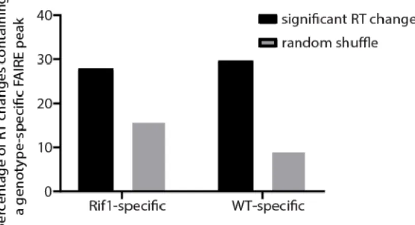
Our data thus far indicate that increased chromatin accessibility and gene expression act upstream of advanced replication within pericentric heterochromatin in H3K9R mutants (Fig 2.11D). To further understand the relationship between transcription and DNA
53
54
Figure 2.11. Altered transposon expression occurs at advanced replication domains in H3K9R mutants.
A) Heatscatter plot of the H3K9R/HWT ratio of RT values (log2 S/G1) plotted versus the
H3K9R/HWT ratio of RNA-seq signal at all 10kb windows across major chromosome scaffolds. RNA-seq differences were determined based on the transcript with the lowest p-value across the 10kb window. 10kb windows with significantly advanced (red) and delayed (blue) RT are indicated (p<0.05, log2 fold change>0.1; limma). B) Histogram of the number
56
Figure 2.12. Regions of advanced replication in H3K9R mutants exhibit altered transposon expression.
A) Histogram of the number of differentially expressed transcripts in 10kb windows of delayed replication (blue; left). Venn-diagram comparing the number of windows with differentially expressed transcripts and number of windows with delayed replication (right). B) Venn-diagram comparing the number of advanced windows in H3K9R mutants compared to control containing a differentially expressed transposon and/or a differentially expressed transcript. C) Genome browser shot of a 10kb window with significantly advanced
replication in H3K9R mutants but no detectable accompanying change in RNA expression via edgeR analysis. FAIRE-seq, HP1a ChIP-seq, or RNA-seq signal are shown for H3K9R (purple) and HWT (yellow) samples. Note the low mappability of this region due to high transposon density. Red transposons indicate individual transposons belonging to a family that is differentially expressed in H3K9R mutants. Browser shot provides a representative example of transcriptional changes that are likely occurring but cannot be directly examined due to low mappability. D) Venn-diagram comparing the number of windows with a
differentially expressed transposon to the number of windows with advanced replication (see also Figure 4C). Because high transposon density (Figure 2.12A,B) and low sequence
57
Histograms in the top left panel show the number of transposons belonging to a family that is differentially expressed in H3K9R mutants compared to control at 57 10kb windows that exhibited advanced replication in H3K9R mutants but no initially detected transcriptional change (see also Venn diagram in Figure 4B). Bottom left panel shows number of
59
Figure 2.13. Transposon density and H3K9me2/me3 status are distinguishing features of regions with advanced replication.
61
H4K16 is necessary for hyper-expression of the Drosophila male X Chromosome The Drosophila dosage compensation mechanism is mediated by the Male-Specific Lethal (MSL) complex, which specifically localizes to and promotes higher gene expression from the male X. The MSL complex includes MOF, a histone acetyltransferase that
acetylates lysine 16 of histone H4, resulting in higher levels of H4K16ac on male X
Chromosomes relative to autosomes or the female X (Hilfiker et al. 1997; Smith et al. 2000; Gelbart et al. 2009). Furthermore, hyper-acetylation of H4K16 correlates with increased chromatin accessibility of the male X (Bell et al. 2010). These data suggest that H4K16ac is required for dosage compensation in flies. In accordance with these findings, MOF mutations cause a male-specific lethal phenotype; however, MOF performs both H4K16-dependent and -independent functions (Hilfiker et al. 1997; Buscaino et al. 2003; Sykes et al. 2006). A requirement for H4K16 in dosage compensation, therefore, has not been directly tested.
62
contrast, females of all these H4K16R genotypes are viable, indicating that H4K16 modification is not generally required for organismal viability.
We next performed gender-specific total RNA-seq from replication dependent H4K16R and control wing discs, generated transcriptomes (Cufflinks), and identified
differentially expressed transcripts between H4K16R males and females and their respective controls (Trapnell et al., 2012). We observed 1789 differentially expressed transcripts (608 increased and 1181 decreased) in H4K16R males relative to control males and 105
differentially expressed transcripts in H4K16R females relative to control females (39 increased and 66 decreased) indicating that the H4K16R effect on gene expression is greater in males than in females (p<0.05, edgeR; Fig 2.14B). Of the 1181 genes with decreased expression in H4K16R males, 72% are located on the X. In addition, the majority (92%) of the down-regulated, X-linked genes in H4K16R males have a log2 fold change less than 1,
which would be expected for a disruption in X Chromosome dosage compensation. In contrast, only 3.6% of genes with increased expression in H4K16R males are on the X.
63
64
Figure 2.14. H4K16 promotes hyper-expression of the Drosophila male X chromosome. A) Table of observed HWT and H4K16R adult females and males where first instar larvae of each genotype were isolated from their wild-type siblings and mono-cultured in aliquots of 50 larvae per vial (see supplementary materials for crosses and complete genotypes: Rows 1 and 2) zygotic, replication-dependent HWT and H4K16R; Rows 3 and 4) maternal/zygotic, replication-dependent HWT and H4K16R; and Rows 5 and 6) zygotic, replication-dependent and replication-independent HWT and H4K16R (left; Chi squared comparisons performed against the male to female ratio of zygotic, HWT, p<0.01;). Percentage of viable male (grey) and female (black) adults for H4K16R and HWT (right). B) Heatscatter plot of the