MOLECULAR DISSECTION OF BREAST LUMINAL TRANSCRIPTION NETWORKS
Francesca Bargiacchi
A dissertation submitted to the faculty at the University of North Carolina at Chapel Hill in partial fulfillment of the requirements for the degree of doctor of philosophy in the department of
Pharmacology in the school of Medicine.
Chapel Hill 2015
ABSTRACT
Francesca Bargiacchi: Molecular Dissection of Breast Luminal Transcription Networks (Under the direction of H. Shelton Earp)
During mammary development, cellular differentiation and lineage commitment to various epithelial and mesenchymal cell types are driven by hormonal and paracrine signaling mechanisms. Understanding mechanisms that govern differentiation into distinct cell populations is critically important for a complete understanding of development and breast tumorigenesis. Previous studies have shown that retroviral transduction of fibroblasts with four transcription factors can initiate the conversion of a somatic cell into an embryonic stem cell-like state with capabilities of differentiating into cell types of all three germ layers. The goal of the proposed study was to determine whether mammary specific transcription factors (TFs) could directly induce transdifferentiation to an ER+/luminal cell phenotype in mouse embryonic fibroblasts via an iPS-type approach. After screening 9 candidate TFs for their abilities to induce various epithelial-specific and breast-specific attributes, we focused subsequent efforts on ESR1,
TABLE OF CONTENTS
LIST OF TABLES ... vii
LIST OF FIGURES ... viii
CHAPTER 1: MOLECULAR DISSECTION OF BREAST LUMINAL TRANSCRIPTION NETWORKS ... 1
Introduction ... 1
1.1 Overview of Mammary Development ... 3
1.1.1 General Process of Development ... 3
1.1.2 Mammary Development in the Embryo... 4
1.1.3 Mammary Development During Puberty ... 6
1.1.4 Pregnancy and Lactation ... 8
1.1.5 Involution ... 9
1.2 Current Mouse Models of Mammary Development ... 11
1.2.1 Genes Required for Mammary Function ... 11
1.2.2 Comparing Mammary Development to the Five Intrinsic Subtypes of Breast Cancer ... 23
1.3 Reprogramming/Transdifferentiation: What Has History Taught Us? ... 27
1.4 Summary ... 33
CHAPTER 2: SPECIFIC COMBINATIONS OF TRANSCRIPTION FACTORS INDUCE TRANSDIFFERENTIATION TO A MAMMARY LUMINAL EPITHELIAL CELL PHENOTYPE ... 41
Introduction ... 41
2.1 Materials and Methods ... 44
2.1.1 Plasmids ... 44
2.1.2 Virus Production ... 45
2.1.5 RNA Isolation and cDNA Synthesis ... 47
2.1.6 Quantitative PCR ... 47
2.1.7 Western Blotting ... 48
2.1.8 Proliferation Assays/IC50 Curve Analysis... 48
2.1.9 Flow Cytometry ... 48
2.1.10 Immunofluorescence with and without Matrigel ... 49
2.1.11 Gene Expression Microarrays ... 50
2.1.12 Informatics Analysis ... 50
2.2 Results ... 51
2.2.1 Transcription Factors Enriched in Luminal Tumor/Cell Lines ... 51
2.2.2 Stable Cell Lines Expressing Candidate Transcription Factors ... 52
2.2.3 ESR1 Expression in BABES Induces Cellular Senescence ... 53
2.2.4 MEFS Transduced with EFP (M–EFP) Exhibit Epithelial Phenotype ... 54
2.2.5 M-EFP Cells Exhibit Expression of Breast Epithelial Markers ... 56
2.2.6 Genomic Analysis of M-EFP Suggests Shifts towards Mammary Development ... 57
2.2.7 M–EFP Exhibit Estrogen-Dependent Growth ... 59
2.2.8 M-EFP Cell Line Is Responsive to Milk Producing Stimulus In Vitro ... 60
2.3 Discussion ... 61
CHAPTER 3: ROLE OF GATA3 IN BREAST DEVELOPMENT AND LUMINAL TUMORS ... 83
Introduction ... 83
3.1 Methods ... 86
3.1.1 GATA3 Expression and Reporter Plasmid Construction ... 86
3.1.2 Cell Culture and Transfection ... 87
3.1.3 Luciferase Assay ... 88
3.1.4 Structural Analysis ... 88
3.2 Results and Discussion ... 88
LIST OF TABLES
LIST OF FIGURES
Figure 1.1. Embryonic Mammary Gland Development ... 36
Figure 1.2. Mammary Ductal Network from a 20 Week Old Mouse ... 37
Figure 1.3. The IL4/13-STAT6-GATA-3 Pathway in Mammary Gland Development ... 38
Figure 1.4. Differentiation Hierarchy for Mouse Mammary Development ... 39
Figure 1.5. Human Breast Cancer/Normal Cell Line Alignment along a Differentiation Axis ... 40
Figure 2.1. TF Characterization in the BABE Cell Line ... 71
Figure 2.2. Cell Cycle Gene Expression Analysis of Single Transductants in the BABE Cell Line ... 72
Figure 2.3. Initial Transduction Experiments in the MEFS ... 73
Figure 2.4. Epithelial Characterization of M–EFP Cell Line ... 74
Figure 2.5. Interrogation of Breast-Specific Phenotypic Markers in the M–EFP Cell Line ... 75
Figure 2.6. Breast-Specific TFT Enrichment and Differentiation Analysis ... 76
Figure 2.7. ESR1 Responsiveness and KI67 Expression ... 77
Figure 2.8. 96 hr Treatment with Tamoxifen in Phenol-Red Free Media ... 78
Figure 2.9. M–EFP Exhibit Breast Functional Characteristics ... 79
Figure 2.10. STAT5A Activation in Prolactin Treated M–EFP Cells ... 80
Figure 2.11. TF Regulation in the BABE Cell Line ... 81
Figure 2.12. EpCAM/CD49f FACS Analysis in the Mouse Model ... 82
Figure 3.1. GATA3 Protein Domains and Functions ... 96
Figure 3.2. GATA3 Open Reading Frame and Lentiviral Plasmid Development ... 97
Figure 3.3. Genomic Location of FOXA1-Reporter Sequence ... 98
Figure 3.4. Cloning Result of FOXA1 Genomic DNA Sequence and FOXA1-luc Promoter Double Digest ... 99
Figure 3.5. Normalized FOXA1 – Reporter Activity in 293T Cells ... 100
Figure 3.6. C–Terminus GATA3 Mutations in TCGA Data Set ... 101
CHAPTER 1: MOLECULAR DISSECTION OF BREAST LUMINAL
TRANSCRIPTION NETWORKS
Introduction
The molecular characterization of human breast cancers and normal mammary
developmental processes has revealed commonalities that have proven to be useful in clinical cancer diagnostics and in understanding normal and neoplastic breast biology. During mammary development, cellular differentiation and lineage commitment are driven by distinct growth cycles under the control of local epithelial and mesenchymal paracrine signaling mechanisms. Multipotent stem cells give rise to various luminal and myoepithelial cell types organized in lobular and ductal architectures that ultimately comprise normal breast tissue. Likewise, our lab and others have established that human breast cancers consist of multiple “intrinsic” subtypes, several of which are defined by their similarity to normal mammary cell counterparts on the basis of gene expression patterns.
These tumor subtypes differ in their responses to therapy and survival outcomes. Importantly, luminal A tumors are the most common and diagnostically are ER+ and PR+. Luminal A tumors tend to grow slowly, and patients with these tumors have an overall better prognosis than those with the other subtypes; yet these tumors do not respond well to
yet to be successfully propagated in vitro and to be captured as an immortal cell line (with MCF7 being a luminal B line). Thus, creating new cell lines that capture the pertinent aspects of the luminal A cell-type biology would have tremendous value for a better understanding of these tumors and for enabling new drug development.
To address the goal of creating a ER+/luminal A cell line, we propose an approach similar to the one taken to develop induced pluripotent stem cells (i.e., iPS cells). In this
approach, we propose using two different lineage cell lines and transducing them with candidate lineage determining genes, in an attempt to transdifferentiate this basal-like cell line into a luminal A–like cell line. The nine candidate transcription factors chosen are all highly expressed in ER+/luminal A cells and are thought to be luminal lineage-specific factors. Our preliminary data suggested that ectopic overexpression of these transcription factors in HMECs have both unique and overlapping contributions toward inducing the expression of genes responsible for luminal differentiation. One of these transcription factors, GATA3, is expounded upon in Chapter 3. The process that results in the development of a luminal-like cell line requires a thorough understanding of mammary development in both mice and humans. In addition, knowledge of which mouse mammary models are utilized for the study of development is key, particularly with regard to gene knockout models, which result in either no mammary system or an abrogated one. Concluding the introduction with a discussion regarding
reprogramming/transdifferentiation emphasizes not only the technical approach of creating the cell line but also the biological process necessary to achieve the change in the cell line lineage. The aim of this introduction is to give a relevant and meaningful understanding of the
1.1 Overview of Mammary Development
1.1.1 General Process of Development
principally after birth. Typically under the control of hormones, development occurs through distinctive stages throughout the life span of a mammal, starting with the embryo, continuing with puberty, and concluding with reproduction. At each developmental stage, there are cues specific to that developmental stage that initiate changes in both the mammary cells and the surrounding stroma. Functionally speaking, the mammary gland’s role is to provide ample nutrition for offspring. Comprised of milk protein and fat, milk also provides immune factors such as antibodies that offer some protection to offspring against disease (Rogier et al., 2014). The mammary gland halts development after birth, until puberty, with the growth and expansion of milk ducts. It matures via lobuloalveolar development during pregnancy. How these intricate signaling processes come together at different stages of mammary development will be discussed further, with a primary focus on mouse mammary development. There are several reasons for this: First, the ability to genetically engineer mice makes them an attractive model to study. Second, there are developmental similarities between humans and mice, although it should be noted that there are some subtle structural and hormonal differences between the two (Watson and Khaled, 2008).
1.1.2 Mammary Development in the Embryo
Studies have shown that the mesenchyme from the mammary region is responsible for the mammary bud’s epithelial development. Initial signals for epithelial development come from the mesenchyme and include transcription factors’ estrogen and androgen receptors, Lef-1, Msx1, and the growth factors BMP4 and FGF7. These factors are induced in a paracrine fashion by the developing bud and are silenced at later stages (Heuberger et al., 1982). As mentioned earlier, mammary gland development was derived from apocrine glands from the skin. Hence, some of these transcription factors are not unique to the development of the mammary gland. Once the bud is formed, Lef-1 and Msx1/2 are turned off, leading to a lack of other ectodermal appendages such as teeth and hair (Hennighausen and Robinson, 2001). With the formation of the bud comes the elongation of the primary spout, driven largely by the mammary bud’s expression of parathyroid hormone related protein (PTHrP). PTHrP signals to the adjacent mesenchyme (which expresses PTHrP receptors) to give rise to the nipple. By Day E18.5, several small, tree-like glands have developed and are dependent on factors supplied by the fat pad. Signaling from PTHrP continues, facilitating the activation of downstream marker BMP4.
From a mouse model perspective, PTHrP knockout mice exhibit greatly reduced ductal branching morphogenesis. Finally, PTHrP and BMP4 induce Msx2 to regulate the suppression of hair follicle formation (Watson and Khaled, 2008). The development of the mammary gland elicits signals from the nascent mesenchyme, which is the primary driver of epithelial bud development, while working synergistically with the epithelial sprout to further differentiate into other ectoderm lineages by rapid changes in transcription factor expression. Interestingly,
1.1.3 Mammary Development During Puberty
During this time, the development of the mammary gland relies heavily on hormones, leading to the creation of a complex ductal network. This complex network is a result of
branching morphogenesis, which is regulated by the various components of the mammary gland. This includes cellular players such as the stromal compartment-fibroblasts, adipocytes, and immune cells, as well as the mammary epithelial cells themselves. Hormones and growth factors act in both a paracrine and autocrine fashion to control the growth of the ductal network,
assisting the invasion into the fat pad. At the tip of these ducts are terminal end buds (TEBs), which comprise of two layers of cells. The outer layer consists of cap cells, which are highly proliferative, hormone receptor-negative cells involved in the remodeling and extension of the bud. In contrast, the body cells, which line the inner portion of the TEB and are responsible for lumen development, are less proliferative than the cap cells, and are responsive to
hormonal/lactogenic stimulus. Please see Figure 1.2. In addition, ductal elongation will result in bifurcation of the existing ducts to create branches. In the mouse, this process will be complete by 10–12 weeks of age. At that point, TEBs have invaded the fat pad to the full extent and growth of the branches has ceased (Watson and Khaled, 2008). One of the key regulators of ductal morphogenesis is estrogen. Estrogen has two receptors, ESR1 (ERα) and ESR2 (ERβ). ERα has been shown to be critical for mouse mammary development, as ERα-null mice fail to develop TEBs and the ducts that do form do not invade the fat pad (Mallepell et al., 2006).
Along with the process of morphogenesis comes lumen formation. Lumen formation is a process that occurs early in the development of mammals and continues through organ
cells within the TEB become polarized, recognizing via contact with the basement membrane just prior to lumen formation. Once polarity is established, cells that do not become polarized undergo apoptosis (Debnath et al., 2002). Some key proteins that regulate polarity and lumen formation include ROBO1 and SLIT2. Knockout mouse models with either ROBO1 or SLIT2 deficiency not only exhibit defects in TEB structure but also ductal abnormalities that result in separation between the luminal epithelial and myoepithelial cell layers (Macias et al., 2011). These data highlight the complex signals needed for lumen development.
There are several transcription factors that drive this stage of mammary development. As mentioned before, estrogen signaling is critical for branching morphogenesis and becomes an invaluable signal for alveologenesis during pregnancy. ERα-null mice not only have reduced levels of prolactin, which augments milk production, but also have insufficient progesterone synthesis, which is necessary for ductal development (Hennighausen and Robinson, 2001). Progesterone mediates its effects on the development of the mammary gland through two main isoforms, PR-A and PR-B. Mice lacking both isoforms become anovulatory and develop a severely growth-retarded mammary gland. Progesterone signaling controls cell proliferation through the activation of the Wnt pathway, promoting alveolar formation (Brisken and Duss, 2007). Although estrogen mediates much of the branch morphogenesis, its regulation of progesterone receptors (PRs) results in the elaborate extension of the branches, known as side branching, in the virgin mouse (Fernandez-Valdivia et al., 2009; Ismail et al., 2003). The role of estrogen and progesterone signaling will be further discussed in Section 3.
activation of TGF-β was thought to correlate with hormonal, differentiation status, as well as proliferation/apoptosis (Silberstein and Daniel, 1987). Normally TGF-β is expressed throughout all phases of mammary development. TGF-β1 is regulated by estrogen and progesterone, and the downstream effectors of TGF-β1 regulate morphogenesis in a variety of ways, including
apoptosis, matrix remodeling, and loss of proliferation (Moses and Barcellos-Hoff, 2011). Although TGF-β signaling can induce apoptosis during morphogenesis, within the mammary gland it can exert its effects on the stroma via the production of extracellular matrix (ECM) (Moses and Barcellos-Hoff, 2011). This production of ECM may be an attempt to control the growth of mammary epithelial cells during development (Gomm et al., 1991).
1.1.4 Pregnancy and Lactation
Stat6, whose downstream effector molecule is GATA3. GATA3 is necessary for Th2
development due to its role in chromatin remodeling and facilitates the expression of IL-4, IL-13, and IL-5 cytokines (Watson and Khaled, 2008). Thus, the regulation of these cytokines promotes Th2 while suppressing Th1 differentiation. Mouse knockout models of IL-4/IL-13 (responsible for Th2 regulation) resulted in reduced proliferation and milk protein production, as well as delayed alveolar development during gestation, a similar phenotype to the Stat6 knockout mouse model (Khaled et al., 2007). These data also suggested that under lactogenic stimulation,
expression of Th1 cytokines was suppressed and that the mechanisms that mediate Th2 development also determine the fate of the mammary luminal epithelial lineage. The IL-4/IL-13/Stat6/GATA3 pathway in mammary development is illustrated in Figure 1.3. GATA3, a potent regulator of alveolar differentiation, is discussed in further detail in Chapter 3. In addition to Stat6 signaling, Stat5, a potent downstream signal from prolactin receptor (PRLR), is critical for the differentiation of mammary alveoli during pregnancy (Liu et al., 1997). Although the mechanisms are still unclear, it appears that Stat5a and Stat6 work in parallel to induce IL-4/IL-13 expression.
1.1.5 Involution
As with pregnancy and lactation, the immune-regulated genes are involved in mouse mammary involution, a process of tissue remodeling and cell death. There are two phases of involution: the first occurs within hours of weaning and entails the accumulation of milk within the alveolar lumen and subsequent decrease of lactogenic hormones, while the second phase is a signal initiated by systemic hormonal regulation. This results in a remodeling of the ECM and the basement membrane of the ductal-lobular architecture due to the activation of matrix
al., 2004). All of this occurs within 2 weeks post weaning, making involution a quick and efficient process in the mouse. Examining Phase 1 in more detail, we determined that this phase is responsible for the apoptotic events within the alveolar lumen and is initiated by Stat3
signaling. Stat3 is activated by the leukemia initiation factor (LIF). Downstream targets of Stat3, IGFPB-5 and C/EBPδ have been shown to be instrumental in the apoptotic response. Knockout models of these two genes have suggested a diminished ability to undergo involution
(Thangaraju et al., 2005; Tonner et al., 2002). Cells with activated Stat3 signaling begin to shed, filling the lumen and expressing proapoptotic markers such as Caspase-3, Bax, and decreased levels of Bcl-x. Other downstream targets of Stat3 ensure that transition to the second phase of involution ensues.
Approximately 48 hours after Phase 1, Phase 2 begins, primarily by remodeling the lobulo-ductal architecture. This is achieved by the stromal secretion of matrix metalloproteases, which not only degrade the lobulo-ductal supportive matrix but also initiate further apoptosis through the detachment of the ECM to the epithelial cells. Adipocytes migrate to the newly remodeled matrix and differentiate via MMP3 and plasminogen (Watson, 2006). Ultimately, NF-κB signaling recruits immune cells, primarily macrophages, to phagocytose and remove the cells and debris, a process that involution mediates.
The four main stages of mouse mammary development have unique roles and complex signaling mechanisms regulated by hormones, growth factors, and lactogenic signals.
influence the risk of neoplastic events during child-bearing years. These different stages of development also complement the mouse mammary differentiation hierarchy, as the earlier stages correspond to a less differentiated (more stem cell-like) phenotype, while lactation is more in line with a luminal differentiated phenotype. It is essential in understanding the developmental phases especially when trying to recapitulate a mammary epithelial cell line de novo. This will be touched upon in Chapter 2.
1.2 Current Mouse Models of Mammary Development
1.2.1 Genes Required for Mammary Function
Mammary gland development and function are tightly controlled processes that are heavily regulated by hormones and growth factors both produced by the epithelial and/or stromal compartments, as well as by more distant locations such as the ovaries and/or pituitary gland. Understanding the critical mediators of development and function involve multiple cell types, particularly within the lobulo-ductal network. The focus will be on the myoepithelial and more differentiated basal/luminal epithelial cells. There are several considerations when determining which mouse model to utilize for the scientific question at hand. First, in which phase of
development does the scientific interest rest? For example, as mentioned in Section 1, hormonal dependence and necessity become apparent during puberty, when such signals are required for ductal elongation and morphogenesis. An ERα-knockout model would thus be a poor choice for studying embryonic development of the mammary gland.
system will delete GATA3 when the MMTV promoter is turned on, presumably in breast tissue. This method not only assures expression in the tissue types being studied but also prevents the effects of the knockout on the overall development of the embryo. More recently, there has been interest in comparing normal breast tissue to breast tissue in tumor development. Here, one may want to focus on certain cell types, whether myoepithelial in origin or the cell source that gave rise to a luminal tumor. Inducible Cre knock-in models for these questions may include the use of a KRT14, KRT8, or KRT18 promoter, depending on the mammary cell type desired
(Keymeulen et al., 2011).
Some of the key regulators of mammary development and their phenotypic characteristics when knocked out are shown in Table 1.1. The essential gene knockout developmental models either result in a breast phenotype that is severely abrogated or
completely defective in morphology and/or function. It is not surprising to see that loss of ERα results in not only branching morphogenesis loss but also a loss in overall reproductive function. Interestingly, knocking out ERβ in place of ERα results in the normal development of the
mammary gland, which suggests that both isoforms have unique downstream targets and that functional ERβ cannot compensate for the loss of ERα (Couse and Korach, 1999). Loss of both reproductive and lactation function is also observed with progesterone receptor and prolactin knockout models, suggesting that genes involved with specific developmental cues across multiple tissue types may have a profound effect on those systems when lost. Reduced functions are even seen in a PRLR-heterozygous knockout mouse model (Ormandy et al., 1997).
arrest mammary development at the embryonic bud stage, however only when both are knocked out is bud development arrested. This suggests that both genes may have redundant functions when either one is knocked out (Cowin and Wysolmerski, 2010).
During each stage of development, signaling cues give rise to particular cellular types, be it multilineage, as seen in embryonic development, or within a specific lineage, such as epithelial differentiation, as seen in pregnancy. During the embryonic stage, the formation of a rudimentary gland includes initial bud formation, which gives rise to the mammary epithelial cells. These epithelial cells are poorly differentiated and are not hormone dependent at this stage of
development. Exhibiting a “basal” phenotype, embryonic cells at this phase express Keratin 14 and have multipotent capabilities. Keratin 14 positive progenitor cells arise from the embryonic epidermis at embryonic Day 17 (E17). Studies have shown that at birth, mouse mammary glands consist of a small network of rudimentary ducts comprising of myoepithelial and luminal cells. The myoepithelial cells are positive for Keratins 5 and 14 and smooth muscle actin (SMA). These are considered markers for the basal phenotype seen in both mammary development and cancer and will be further discussed later in this chapter.
during puberty and/or pregnancy, such cells lose their self-renewal and multipotent functions. Studies have provided evidence for the abilities of both bi- and unipotent progenitor cells that could contribute to the expansion of the mammary gland during puberty and pregnancy (Keymeulen et al., 2011). Interestingly, others have shown that while prospective alveolar progenitors fail to form outgrowths in vivo, when such cells are coinjected with Matrigel into mammary fat pads, they in fact can create small, branched mammary structures (Jeselsohn et al., 2010; Vaillant et al., 2011).
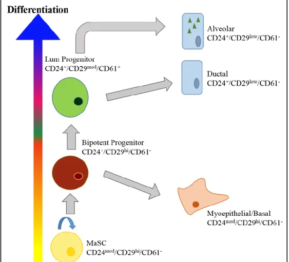
There are several markers used to determine cellular plasticity and differentiation in the mouse. The four main markers are CD24, CD29 (ITGβ1), CD61 (ITGβ3), and CD49f (ITGα6). Initial studies have shown that CD24 is an excellent marker in delineating between epithelial and stromal cells (Sell, 2013). Within the mammary epithelial lineage, CD24 expression also
delineates between the basal (CD24mod/+) and luminal (CD24hi) subpopulations (Sleeman et al.,
2006). Interestingly, the same study also recognized that sorted mouse mammary epithelial cells with high CD24 expression had limited repopulation capacity, suggesting that the CD24low cell fraction was responsible for the ability to repopulate a mammary gland. CD29hi and CD49fhi have been shown to represent cells with repopulation capacity (Wang, 2006).
While mammary stem cells (MaSC) exhibit the CD24mod/+/CD29hi/Cd49fhi phenotype,
commonly used marker of the two (Asselin-Labat et al., 2007; Sleeman et al., 2006). The mature luminal population (CD24+/CD29low/CD49flow/-/CD61-) will have loss of expression of the
integrins CD29, CD49f, and CD61 and have higher expression of CD24 than the
MaSC/progenitor cell types (Lim et al., 2010). There will also be few progenitor cells within this population (Lim et al., 2010). As a result, luminal cells are considered to be unipotent with no self-renewal capabilities (see Figure 1.4).
Studies suggest that EpCAM is superior to CD24 as a marker for mouse mammary luminal cells. EpCAM is used in place of CD24 in human mammary studies due to its specificity for luminal/luminal progenitor populations, as CD24 expression is found in basal progenitor/ MaSC and even some stromal cells (Oakes et al., 2014; Smalley, 2015; Visvader, 2009).
Depending on the experimental question, either marker is considered acceptable for the isolation of progenitor/luminal mammary cells; however, if the desire is to study the basal/myoepithelial component of the mammary gland, then EpCAM is a better marker for separating those cells from the luminal component (Smalley, 2015).
Rios and colleagues (2014) determined that bipotent MaSCs could collectively be called multipotent MaSCs due to the fact that these cells contributed to the development of alveolar branches during pregnancy. Notably, these cells were traced over a couple of pregnancy and involution cycles, suggesting that they were involved in ductal remodeling (Rios et al., 2014; Visvader et al., 2014). These bipotent MaSCs may also give rise to lineage-specific progenitor cells (basal, luminal). Ultimately, these cells have been found to maintain mammary gland homeostasis and development, postnatally, instead of the rare multipotent stem cell that had been shown to give rise to a completely functional mammary gland (Mark et al., 2006; Visvader et al., 2014). It is worth mentioning while discussing the subtle nuances between the multi-/bipotent progenitor cells and their functions that the characteristics and functions of the differentiated luminal cell have been better characterized. Transplantation studies with mammary stem cells typically have basal-like characteristics, perhaps in part due to their basal apical location in the TEBs and their role of giving rise to a functional ductal network (Smith and Boulanger, 2003).
As mentioned earlier, lineage-committed progenitor cells have features that contain either basal or luminal characteristics, although some lineage-tracing studies suggest a bipotent
progenitor may contain features that are passed onto and maintained in its differentiated
counterparts, as has been seen using Keratin 5–labeled cells (Rios et al., 2014). Keratin 8–labeled stem cells have been shown to give rise to a luminal progenitor cell that can give rise to both luminal and milk-producing cells. In addition, these labeled cells were found to be self-renewing, after as many as 3 pregnancy/involution cycles, indicating that these cells are not being replaced by a mammary stem cell (Keymeulen et al., 2011).
widely thought that cells expressing ESR1 within the luminal component are rarely activated (Clarke et al., 1997); however, others have discovered rare ESR1-positive cycling cells (Booth and Smith, 2006). This suggests that not all ESR1-positive cells are quiescent; they may represent a minor fraction of the luminal progenitor population. Luminal progenitor (LP) cells expressing ESR1 tend to be CD24+/Sca1+/c-kit+. This is in contrast to LP cells without ESR1, whose expression of lineage markers’ CD24+/Sca1-/c-kit+ only varies by the loss of Sca1
(Sleeman et al., 2007).
Both LP populations exhibit bipotent capabilities; however, in the mouse at least, ESR1-expressing LPs appear to have a survival advantage over ESR1-negative LPs in the presence of oestrogens. The basal level of oestrogens in the mammary gland is generally low and thus shifts the LP population from ESR1-negative to ESR1-expressing luminal cells. The ESR1-positive LP cells are likely to be ductal-restricted progenitor due to their expression of ESR1 and FOXA1. ESR1 and FOXA1 are known to be involved in ductal, not alveolar, development, which may explain why ESR1-negative LP cells have been shown to be alveolar-restricted progenitor cells Shehata et al., 2012). These cells are long living and prepare the gland for pregnancy and involution, giving rise to functional differentiated luminal cells (Keymeulen et al., 2011).
Differentiated luminal cells will have high CD24 expression, low to moderate CD29 expression, and low to no CD61 expression (Sleeman et al., 2006). The functions of
CD24+/CD29low/mod/CD61- luminal epithelial cells are well defined, as they exhibit two main responsibilities: First, ductal luminal cells are responsible for the lining of the duct, which assists in draining the lobuloalveolar structures during lactation. Second, during pregnancy, signaling cues (more on this in the next section) initiate the production of milk in an otherwise quiescent cell, which explains why such differentiated cells are difficult to culture in vitro (Clarke et al., 1997; Smalley et al., 1998). Consequently, human studies with reduction mammoplasty tissue grown in vitrotend to lose some of the key luminal subpopulations, leaving behind a largely proliferating mammary epithelial cell with basal-like features (Gordon et al., 2000). More recently, new lineage markers used for the separation of subpopulations have been discovered within the stem cell hierarchy. While CD24/CD29/CD61 have been instrumental in defining populations with repopulation capacity, metabolic activity, and milk-producing function, new markers have been shown to help further define the subpopulations, expanding our
understanding of the layers involved in plasticity, self-renewal, and function (see Table 1.2). The prepubertal and pubertal/gestational development phases are largely defined by their dependence on hormones. Unlike the prepubertal phase, hormone dependence occurs after puberty and results in ductal elongation and side branching, and in pregnancy, inducing alveologenesis and milk production. Mouse experiments involving the surgical ablation of hormone-secreting organs from mature female mice have resulted in lack of mammary gland development. The organs involved in mammary development, mainly the pituitary gland and the ovaries, are responsible for the secretion of the hormones GH, PRL, 17β-estradiol, and
Subsequent studies have demonstrated that when the pituitary and/or ovaries were removed, treatment of mice with 17β-estradiol, progesterone, growth hormone, cortisol, and prolactin could recapitulate the development of the gland (Brisken and O’Malley, 2010). The steroid 17β-estradiol is one of the most potent and prevalent of the estrogens. Other members of the estrogen family include, estrone and estriol, are also present in the mammary gland. The effects of these ligands are mediated through two estrogen receptors, ESR1 and ESR2, and when bound to a ligand, ESR1 undergoes a conformational change, resulting in its interaction with other transcription factors such as AP-1, SP-1, STAT3, or NF-κB (Tanos et al., 2012). ESR1 also recruits coactivators and/or corepressors to help promote these transcriptional changes (Moggs and Orphanides, 2001). Due to the action of 17β-estradiol, this genomic mechanism of ESR1 signaling induces expression of amphiregulin (AREG) which is then acted on by ADAM17, a protease that cleaves AREG to its secreted form (Sternlicht and Sunnarborg, 2008). Via paracrine mechanisms, AREG subsequently binds to the epidermal growth factor receptor (EGFR) on fibroblasts, further inducing the expression of various growth factors, including several members of the fibroblast growth factor (FGF) family of ligands (Coleman-Krnacik and Rosen, 1994). Within the luminal cell, ESR1 signaling is also highly influenced by coregulators, which include coactivators and corepressors. This is also the case for progesterone receptor (PGR) signaling, which will be discussed next.
2003; Leo and Chen, 2000). While ESR1 is necessary for mammary development in both mice and humans, ESR1 levels in luminal progenitor cells are lower in mice than in humans (Asselin-Labat et al., 2007; Asselin-(Asselin-Labat et al., 2010; Lim et al., 2009), although the reasons for this are not well understood. This may explain, in part, why there are no real ESR1-positive mammary models.
It is also difficult to study ESR1 signaling in mice, as both the stroma and epithelium express ESR1. This is in contrast to the human mammary gland, where ESR1 is uniquely expressed in the epithelium. As a result, studies surrounding the role of ESR1 signaling in development have been focused on human normal and breast cancer cell lines. Two of the critical mediators of ESR1 signaling are FOXA1 and GATA3. Both are considered pioneer factors: FOXA1 has been shown to mediate ESR1 binding to a subset of gene promoters whose genes are involved in proliferation, such as Cyclin D1 (CCND1) (Eeckhoute et al., 2006, 2007).
regulation (PIP) (Wilson et al., 2006). Interestingly, several of these genes are also
overexpressed in human luminal A/B tumors, which highlights the remarkable similarities between normal and tumorigenic genomic profiles.
In addition to mammary development, estrogens act on the pituitary gland by stimulating prolactin synthesis and secretion. In turn, prolactin controls the synthesis of progesterone and induces ESR1 expression in various tissues (Bachelot and Binart, 2007; Frasor and Gibori, 2003). In order to better understand the direct versus indirect mechanisms behind hormonal regulation of the mammary gland, hormone receptor-deficient mouse strains have been developed. For the cognate hormone receptors ESR1, ESR2, and PGR, and prolactin receptor (PRLR), mice without any of these receptors are in fact viable but have severe reproductive abnormalities, including sterility (Bole-Feysot et al., 1998; Hamilton et al., 2014; Ismail et al., 2003). In the mouse mammary gland, ESR1 is expressed in both the epithelium and the stroma. Epithelial ESR1 expression is required for ductal elongation and for subsequent side branching in alveologenesis. Studies addressing the necessary role of ESR1 in the stroma were performed by grafting ESR1-/- fat pads along with wild-type ESR1 mammary epithelium into the muscle wall of mice. The presence of stromal ESR1 was not required for mammary gland development (Mallepell et al., 2006).
during the luteal phase of the reproductive cycle, whose role in the mammary gland is the promotion of ductal side branching and alveolar development (Graham and Clarke, 1997). PGR receptor signaling is thought to be mediated in the mammary gland via a series of signaling events. First, progesterone activates PGR-negative cells in the TEBs, undergoing proliferation to promote side branching and alveolar development, while the adjacent PGR-positive cells in the TEB remain quiescent. Downstream targets of PGR have been implicated as effector genes for proliferation and side branching morphogenesis (Obr and Edwards, 2012). These genes include Wnt4, amphiregulin, and RANKL, among others. During pregnancy, progesterone suppresses differentiation by inhibiting the expression of tight junction proteins until parturition in order to assume the proliferative role of alveolar expansion up until lactation. During lactation,
progesterone levels will decline, allowing for milk production and secretion (Obr and Edwards, 2012). Currently, the molecular mechanisms regulating tight junctions between luminal cells, which progesterone mediates, are unknown.
The hormonal regulation of milk production falls under the regulation of two key
lactogenic hormones: prolactin (PRL) and glucocorticoids. As mentioned earlier, PRL induction is regulated by ESR1. Prolactin receptor (PRLR) signaling activates STAT5A via
phosphorylation by JAK-2. In addition to ESR1, glucocorticoids may also potentiate PRL signaling by recruiting glucocorticoid receptors (GR) near STAT5 binding sites. Other
coactivators of milk protein production, such as C/EBPβ will form a complex with STAT5A and GR to help further recruit other proteins involved in chromatin remodeling, allowing for
(Brisken and Rajaram, 2006). In the presence of progesterone, a STAT5A-PGR complex can inhibit activation of downstream genes, importantly β-casein (Buser et al., 2013), highlighting the timely importance of progesterone stimulation during pregnancy and lactation.
1.2.2 Comparing Mammary Development to the Five Intrinsic Subtypes of Breast Cancer
Gene expression microarray analysis of human breast cancers has revealed five intrinsic molecular subtypes: luminal A, luminal B, basal-like, HER2-enriched, and claudin-low (Perou et al., 2000; Prat et al., 2010). Each of these subtypes has unique histological features and different responses to therapy and clinical outcomes. The term “intrinsic” subtype was used to
characterize these tumors in part due to their developmental characteristics (Perou et al., 2000). Concerted efforts have been underway for several years now to understand where along the breast developmental hierarchy cellular transformation occurs. One of the more prevailing thoughts is that a normally quiescent stem cell undergoes a “genomic” hit, or series of hits, causing oncogenic transformation. These stem cells are found at different stages of development; however they may likely give rise to a less differentiated tumor type, such as a basal or claudin-low tumor.
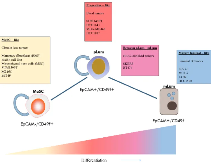
Alternatively, an oncogenic hit can occur within a well-defined, mature luminal cell, resulting in a HER2-enriched or luminal tumor type (Visvader, 2009; Visvader and Stingl, 2014). As an attempt to exploit the relationship between mammary development and the intrinsic
progenitor, and luminal fractions, respectively. The subpopulations were introduced into NOD-SCID mice to determine their mammary regenerative capacity.
The EpCAM-/CD49f+, MaSC population was determined to have such capacity (Lim et al., 2009). Furthermore, each of the sorted fractions were characterized by various basal and luminal markers. The EpCAM-/CD49f+ MaSC group exhibited expression of KRT14, p63, and Vimentin—all basal markers—while the progenitor group, EpCAM+/CD49f+, expressed KRTs 5/6, 8/18, 19, GATA3, and MUC1. As expected, the MaSC group lacked hormone receptor expression. The mature luminal group, EpCAM+/CD49-, stained positive for KRTs 8/18, 19, GATA3, and MUC1, although at higher frequencies than the progenitor. Hormone receptor detection of ESR1 and PGR was highest in the EpCAM+/CD49f- group, indicating this is the mature luminal population.
In terms of mammary development, membrane markers EpCAM and CD49f also
segregate the mouse mammary hierarchy of development similarly, which is important to know, as the genomic classification of human breast EpCAM and CD49f subpopulations discovered in the Lim et al.(2009) studywill be used to extrapolate the differentiation status of various mouse models in Chapter 2 (Shehata et al., 2012). There are other aspects of development in both species that appear to be conserved. For example, hormone expression is lacking in the EpCAM
-/CD49f+ MaSC subpopulation. The MaSC subpopulation expresses KRTs 5/6, p63, and EGFR while there is no expression of ERBB2/HER2, all of which are characteristics of the basal-like tumors (Bertucci et al., 2012; Kreike et al., 2007). However, the MaSC genomic signature was most enriched in both normal mammary and claudin-low tumors. This is likely due to the low proliferative profile and mesenchymal-associated genes found in both tissue types
The progenitor subpopulation contains both basal and luminal features; yet this group was found to have the greatest enrichment in the basal-like tumors, including the expression of ESR1 in a small subset of cells (Asselin-Labat et al., 2010; Nielsen, 2004). This may explain why approximately 10%–15% of basal-like tumors are ESR1 positive (Nielsen, 2004). It has been established that BRCA1 mutant breast tumors in both mouse and human are basal-like, which makes for a good model in understanding the origin of basal-like tumors (Akslen et al., 2003; Liu, 2008; Turner et al., 2006). The study of the origin of BRCA1 tumors may be best studied in human subjects with germline BRCA1 mutations. Lim et al. (2009) compared the EpCAM and CD49f subpopulations in both BRCA1 wild-type and mutant normal breast samples from reduction mammoplasties. Curiously, the BRCA1-mutant normal progenitor fraction expanded significantly more than the matched BRCA1 wild-type group. Moreover, the growth properties of the mutant fraction were significantly greater than that of the wild-type group in vitro, confirming the consequences of BRCA1 loss in regulating cell cycle checkpoints (Lim et al., 2009).
Finally, the mature luminal subpopulation, EpCAM+/CD49f-, described in the Lim et al. (2009) study as ESR1, PGR, and KRTs 8/18 positive, are also found to be highly enriched in luminal A, luminal B, and HER2-enriched tumor types. To further characterize the functional aspects of this population, there was lack of growth in a 3-D Matrigel assay, a typical growth pattern seen in differentiated, quiescent cells, in the absence of lactogenic hormone stimulus (Gordon et al., 2000).
resource for understanding the molecular mechanisms behind diseases and have given us an ability to effectively study the effectiveness and potency of drugs. While there are no luminal A models of breast cancer, MCF-7, a luminal B cell line, has allowed the elucidation of ESR1 signaling through study of the effects of tamoxifen, an ESR1 antagonist. A reason for the lack of luminal A cell lines is that they are slow-growing tumors and are extremely difficult to propagate in culture. Basal cell lines such as SUM149 and HCC1143 have demonstrated expression of dual populations of MaSC (EpCAM-) and progenitor (EpCAM+) subpopulations.
These results have highlighted others’ work confirming that basal-like tumors may, in fact, exhibit both MaSC and progenitor phenotypes and growth characteristics (Liu, 2008; Prat et al., 2013). More importantly, these data have helped adopt the theory as to why basal tumors initially respond well to chemotherapy and then relapse, as such subpopulations are slower cycling and resistant to chemotherapy (Bertucci et al., 2012; Fillmore and Kuperwasser, 2008). The claudin-low tumors and normal mammary cell lines were classified in the Prat et al. (2013) study as representing the MaSC subpopulation, as these cell types are EpCAM-, express
mesenchymal genes, and are slower cycling than their progenitor/basal EpCAM+ counterparts (Prat et al., 2013). These data and others confirm that the current repertoire of human breast normal and cancer cell lines recapitulate all of the hierarchical states seen in breast tumors, with the exception of luminal A tumors (Kenny et al., 2007; Neve et al., 2006; Prat et al., 2013; Prat et al., 2010). Figure 1.5 illustrates a summary of commonly used normal and cancerous cell lines and their respective locations along the developmental axis of differentiation.
human, stressing the importance of reliably using mouse models in interpreting normal and oncogenic events in humans. The knowledge regarding where tumors are derived from yields valuable insight into how developmental cues can influence the characteristics of a tumor, including its response to therapy and metastatic potential. In addition, these studies have shown that the cell lines characterized closely resemble the tumors/normal tissue they are derived from, reinforcing their vital roles as reliable models for research. A critical issue that remains,
however, is the lack of a cell line that represents the most commonly diagnosed breast cancer subtype, luminal A. Laboratories, including our own, are trying to utilize innovative ways to develop a working luminal A cell line for in vitro use. Some of these technical advances include the reprogramming of cells from one type to another, and such techniques have demonstrated success in a limited number of tissue types.
1.3 Reprogramming/Transdifferentiation: What Has History Taught Us?
Cellular reprogramming, by definition, is the conversion from a somatic cell, such as a fibroblast, to a pluripotent stem cell (iPSC) by a number of mechanisms addressed in this section. The world went wild in 1996 when a laboratory at the Roslin Institute teamed up with a small biotech firm to create the first cloned animal, Dolly the Sheep. Ian Wilmut and colleagues transferred the nuclei of cultured epithelial cells into enucleated oocytes to create Dolly, who, ironically, lived a short 6 years (Edwards, 2010). The key to mammalian cloning success,
The discovery and isolation of mouse embryonic stem cells (mESCs) in the early 1980s, followed by the establishment of human embryonic stem cells (hESCs) in the 1990s by James Thomson at the University of Wisconsin, were incredible breakthroughs not only for
understanding the concept of pluripotency but also as a tool to manipulate cells into different lineages. Expanding on the nuclear transfer methods mentioned earlier, scientists discovered that transferring mESCs into a blastocyst, implanting it in vivo,and subsequently breeding can create mice entirely from the ESC DNA fingerprint (Graf, 2011). Basic biological tools used in the lab have been fine-tuned and designed to assist with reprogramming to pluripotency. The use of growth factors was found to be essential for many specialized cell types.
Cytokines, in particular LIF, were found to be essential for ESC growth and maintenance (Williams et al., 1988). RNA tumor viruses were later found to be an indispensable tool for the insertion of genes into a host genome at high efficiencies (Yu et al., 2007). Retroviral gene transduction was instrumental in the first positive iPS studies as a way to introduce and
overexpress ectopic genes essential for reprogramming (Graf, 2011; Stadtfeld and Hochedlinger, 2010; Takahashi and Yamanaka, 2006). Finally, it is worth mentioning that the creation of transgenic knock-in/out mouse models utilized in performing lineage-tracing experiments. The creation of a mouse line with a reporter construct inserted into Fbx15, a locus specific for ESC, was important for the discovery of iPSC reprogramming. Takahashi and colleagues used this line to select for Fbx15-positive cells, thus enriching for the propagation of iPS coloniesin vitro (Takahashi and Yamanaka, 2006).
in vitro, although the conversion is not always a complete one and has extremely low efficiency (< 1% of cells). Confirmation of the function of candidate iPS colonies was determined in two ways. First, colonies were transplanted into NOD-SCID mice and gave rise to all three lineages. Secondly, transfer of GFP-labeled iPS cells into a blastocyst resulted in embryos positive for GFP. After the pups were born, GFP-labeled cells gave rise to all three lineages, confirming that the induction of the four transcription factors in a somatic cell can create cells with pluripotent capabilities (Takahashi and Yamanaka, 2006).
Meanwhile, at the University of Wisconsin, James Thomson and colleagues created an OCT4 knock-in hESC cell line to screen for combinations of genes that could reprogram the cell line. Candidate colonies with transcription factors OCT4, SOX2, NANOG, and LIN28 that grew in geneticin (a selection marker on OCT4 promoter plasmid) were transplanted into NOD-SCID mice to demonstrate teratoma formation with all three lineages. Yu et al. (2007) also discovered that the iPS cells were demethylated at the OCT4 locus compared to controls, an epigenetic feature of embryonic stem cells. Both labs utilized similar mechanisms to demonstrate the
feasibility of creating a functional iPS cell. The subtle differences in transcription factor selection were largely due to the species differences (mouse vs. human).
While their studies set off a wave of new research directions, limitations to
silenced than retroviruses; however, their constitutive activation can result in difficulties inducing differentiation of the iPS cell (Brambrink et al., 2008). A way to circumvent this issue is the creation of an inducible system, where induction/silencing of transcription factors can be controlled by the use of doxycycline. Moreover, reprogramming has been accomplished more recently without the use of viral vectors. Both human and mouse iPSCs have been derived by the delivery of recombinant proteins and whole-cell extracts isolated from ESCs, although the efficiency of this process is extremely low (Cho et al., 2010; Zhou et al., 2009). In order to overcome reprogramming inefficiencies, recent efforts have included the use of chemical compounds as another nonintegrating approach to iPSC development. While these agents, including histone deacetylase inhibitor valproic acid, increased reprogramming efficiencies, they have not been successful at replacing reprogramming factors (Desponts and Ding, 2010).
Unlike the single iPS reprogramming experiment that occurred in 2006, the discovery of converting one cell type into another occurred in increments that started from the directed differentiation of fibroblasts into muscle cells by Myod, followed by the demonstration that committed and fully differentiated cells can be switched within the hematopoietic system and then finding that cell types from different germ layers could be interconverted (Davis et al., 1987; Graf and Frampton, 1995; Huang et al., 2011). Many of these conversions were purposely sought out for therapeutic reasons: for example, b-islet cell production to alleviate
Some of the key transdifferentiation studies mentioned earlier relied on mouse embryonic fibroblasts (MEFs) as the host cell line. These cells are considered to be genomically stable due to nonconforming chromatin configurations and DNA modifications, among other attributes that can make working with MEFs both advantageous and troublesome (Vierbuchen and Wernig, 2012). In 2011, Huang and colleagues published data on the successful conversion of mouse fibroblasts into functional hepatocytes. One consideration when starting with a primary cell line is its proliferative limitations. Alternatively, the generation of immortalized mouse fibroblasts has become an invaluable tool in studying transdifferentiation, although it depends greatly on the method of immortalization, as some can create genetic instabilities and become tumorigenic (Muhammad and Mohammad, 2013; Oh et al., 2007). MEFs derived from cdkn2a-null mice tend to be used due to their genetic stability despite continuous cycles of proliferation (Ieda et al., 2010). Both the primary tail-tipped fibroblasts (TTF) and the cdkn2a-/- MEFs used in the Huang
study were transdifferentiated with three defined transcription factors important for liver function (FOXA2, HNF1α, and GATA4). Conversion into induced hepatocytes (iHEP) took 2–3 weeks, and while there was no selection based on a tagged reporter as seen in the iPS studies,
“candidate” colonies were selected based on morphology and expression of key epithelial markers. Production of key liver enzymes was confirmed, and ultimately the test for true functionality came when the iHEP cells were injected into Fah-/- mice (a mouse strain with severe liver disease that ultimately causes liver failure and death). The iHEP cells were
express and/or produce proteins at the same level as their tissue counterparts, despite the fact they function similarly in vivo(Vierbuchen et al., 2010; Virmani et al., 2001). Finally, the conversion of cdkn2a-/- MEFs does not seem to promote tumor growth, suggesting that silencing a key component of the cellular senescence pathway does not increase risk of transformation. Transdifferentiation studies have been successfully executed across several tissue types. Similar to iPS, conversion events are rare and colony selection is critical, as conversion includes several intermediate stages comprising heterogeneous cells. Other considerations going forward include understanding the role of chromatin modifications and how they affect the rate or completion of cellular conversion. Besides the established technical limitations, it is crucial to understand why conversion so rare. Is there an underlying biological process that can explain this? Moreover, the creation of an induced cell line representing reproductive tissue and utilizing viral transduction methods has yet to have been achieved.
development, a stark contrast to the 3–4 week time frame for iPS conversion (Takahashi and Yamanaka, 2006; Taylor et al., 2006).
Mammary tissue, similar to prostate tissue, develops slowly relative to other organs. It is reliant on hormonal signaling cues from distant organs, as well as local mesenchyme (Cooke et al., 2010; Tanos et al., 2012). The origin of the mammary epithelial cell is poorly understood. The complicating factors include the time for reaching maturity, which in humans is not until puberty. It is therefore possible that well-controlled epigenetic mechanisms maintain the quiescence of the gland until puberty/pregnancy is reached. The ability of such mechanisms to successfully keep cells in check for years in humans may explain the difficulties in creating a mature luminal breast cell line. The next few chapters will discuss attempts to create a luminal breast line, as well as the study of a key pioneer factor that may be crucial in reversing the epigenetic regulation of breast stem cells, GATA3.
1.4 Summary
Table 1.1. Current Mammary Mouse Models and their Phenotypes
Table 1.2. Mammary Specific Lineage Markers Commonly Used in Mice Fibroblast
s
MaSC Basal
Progenitor
Luminal Progenitor
Luminal
EpCAM
-
-
+
++
+++
CD49f
-
+++
++
++
-/+
ALDH-1
++
++
-
-/+
-
SCA-1
++
-
-
-/+
+
c-KIT
++
-
+
++
-
CD14
-
-
-/+
++
-
Figure 1.1. Embryonic Mammary Gland Development
Adapted from Christine J. Watson and Walid T. Khaled, “Mammary Development in the
Figure 1.2. Mammary Ductal Network from a 20 Week Old Mouse
Figure 1.3. The IL4/13-STAT6-GATA-3 Pathway in Mammary Gland Development
Adapted from Christine J. Watson and Walid T. Khaled, “Mammary Development in the
CHAPTER 2: SPECIFIC COMBINATIONS OF TRANSCRIPTION FACTORS
INDUCE TRANSDIFFERENTIATION TO A MAMMARY LUMINAL
EPITHELIAL CELL PHENOTYPE1
Introduction
Luminal A tumors, one of 5 intrinsic subtypes originally defined by Perou et al., account for roughly 40% of all breast tumors diagnosed (Prat et al., 2010). Patients with luminal A tumors have a favorable prognosis compared to the other intrinsic subtypes, with a 10-year relapse free survival rate of 70% (Koboldt et al., 2012). However, the 30% relapse rates indicate that additional knowledge and better treatment are still needed. Typically, treatment consists primarily of surgical resection and estrogen-modulating therapy. Radiation and chemotherapy are also considered depending on location, lymph node involvement, tumor size, and/or OncoTypeDX intermediate/high recurrence score. While these patients experience superior overall and disease-free survival compared to patients with luminal B, basal, HER2 positive, claudin-low tumors, they do have a risk for relapse. Studies that directly evaluated the response to neo-adjuvant chemotherapy across the subtypes have shown that luminal A tumors achieved a pathological complete response (pCR) of 7%, versus a pCR of 43%, 36%, and 17% for basal, HER2-enriched, and luminal B tumors, respectively (Keller et al., 2010). These and other studies have highlighted the significant differences in chemotherapeutic sensitivities between luminal (predominantly ER+) and basal (predominantly ER-) tumors. There have been several attempts
1 Francesca G. Bargiacchi, Joel Parker, Charles M. Perou, and H. Shelton Earp, “Specific
to explain this difference. Luminal A tumors are clinically classified as ER+, HER2-, and ki67-low, suggesting that slow cycling may cause these tumors to be relatively insensitive to
chemotherapy (Inic et al., 2014). Because these patient tumors are slowly proliferating, in vitro propagation has been largely unsuccessful. In addition, there are no luminal A mouse models due to a lack of hormone dependency in murine mammary development.
Unlike the pivotal iPS reprogramming experiments reported 8 years ago, the discovery of approaches to directly convert one differentiated cell type into another has occurred
incrementally, starting with the directed differentiation of fibroblasts into muscle cells by Myod (Davis et al., 1987). Later came the demonstration that committed and fully differentiated cells can be switched within the hematopoietic system and more recently the finding that cell types from different germ layers can be interconverted. For example, laboratories have recently published the successful transdifferentiation of fibroblasts into functional neurons and liver cells (Vierbuchen et al., 2010; Huang et al., 2011). While these studies showed that the phenotype of the new transdifferentiated cells might not be entirely identical to that of their “normal”
counterparts, the fact that they acquire critical cell-type specific functions in vivo raises hopes that defined transcription factor perturbations can be harnessed to generate useful cells for both research and clinical purposes.
and c-Myc (OSKM)—into MCF-7 cells, a human luminal breast cancer cell line (Corominas-Faja et al., 2013). While the MCF-7 cells did not adopt an iPS functional phenotype, they exhibited some stem cell characteristics, such as ES organoid formation in Matrigel and
expression of stem cell markers ALDH and CD44 (Corominas-Faja et al., 2013). Similar studies have introduced TGF-β and/or downstream SMAD family TFs to induce cancer “stemness” from normal mammary epithelia (Piek et al., 1999). However, there are few, if any, studies attempting to transdifferentiate breast epithelial cells either from more stem-like cell types or from cells representing other lineages. It is likely that fully differentiated breast cells may represent a quiescent state or even be committed to senescence or cell death under some circumstances. This could create a serious roadblock to creating model systems. Other considerations for
transdifferentiation strategies include host cell and/or genes used for direct reprogramming. Yamanaka’s iPS experiments were successfully executed using somatic fibroblasts (Takahashi and Yamanaka, 2006). However, the transcription factors used (OSKM) are also involved in epigenetic events that influence chromatin modifications and alterations in the methylation of DNA, which contribute to the formation of iPS cells (Patel and Yang, 2010). Hence, methylation is a concern when starting with a lineage-committed cell line, and the selection of transcription factors with chromatin modification abilities and/or methyltransferase inhibitors may represent strategies to circumvent this.
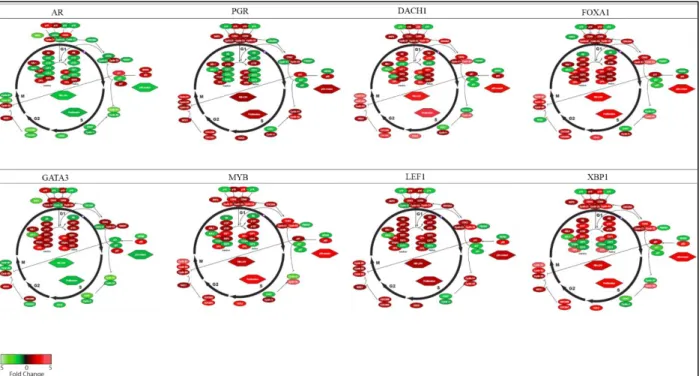
In the present study, the selection of transcription factors for breast luminal cell
factors with known relevance to breast biology: AR, DACH1, ESR1, FOXA1, GATA3, LEF1, MYB, PGR, and XBP1.
Estrogen receptor alpha (ESR1) and progesterone receptor (PGR) are used clinically to define hormone-positive breast cancer (Parker et al., 2009). FOXA1, a pioneering factor, has been shown to been an indispensable partner for ESR1 and PGR signaling (Hurtado et al., 2011). FOXA1 can effectively bind chromatin domains during development and enable gene activity. The FOXA1-DNA binding domain structurally resembles linker histones and can bind
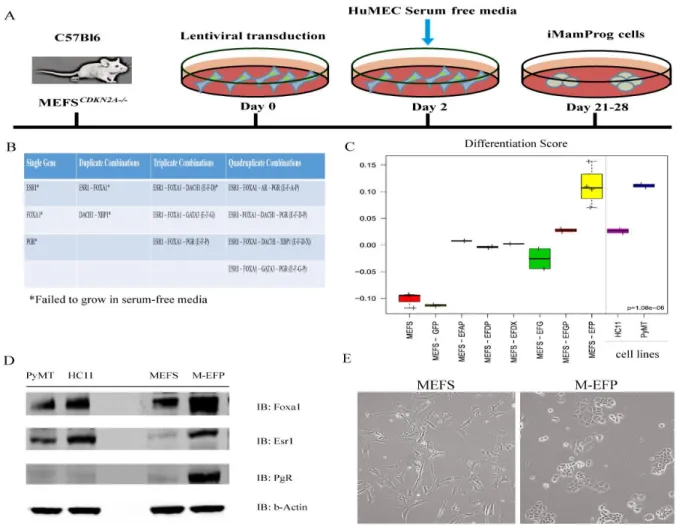
nucleosomes, thus influencing the accessibility of certain transcription factors to their binding sites on particular promoters (Gong et al., 2014). Utilizing two different cell types, first BABES, an hTERT-immortalized human mammary epithelial cell line, and later mouse embryonic
fibroblasts (MEFS), immortalized via the knockout of cdkn2a, we set out to determine if the creation of a luminal-like breast cell line would be feasible in either cell line. These two lines represent different lineages and begin with different methylation patterns (Bloushtain-Qimron et al., 2008; Doi et al., 2009). Experiments with the human BABE cell line led to a hypo
proliferating and senescent population. Subsequently, we discovered that 3 of the 9 genes tested—ESR1, FOXA1, and PGR—can transdifferentiate MEFS into functional epithelial cells with breast luminal and basal characteristics. These findings shed light on the necessary
molecular mechanisms for normal breast development and those that perhaps underlie the various intrinsic subtypes of breast cancers.
2.1 Materials and Methods
2.1.1 Plasmids
pLenti-PGK-blast-DEST, pLenti-PGK-neo-DEST, and pLenti-PGK-puro-DEST were used as the mammalian expression vectors (http://www.addgene.org). GFP-blast and the pLENTI-PGK-DEST backbones were used as positive/negative transduction controls. Final lentiviral
expression plasmids were created using Invitrogen’s recommended protocol for the Gateway system. Plasmids were then sequenced to verify ORF insertion and sequence.
2.1.2 Virus Production
293FT cells (Invitrogen, Life Technologies) were cultured in high-glucose DMEM (Gibco, Life Technologies) with 10% fetal bovine serum (FBS), 500 μg/ml Geneticin®, 1 mM MEM Sodium Pyruvate, 0.1 mM MEM Non-Essential Amino Acids (NEAA), 6 mM
L-glutamine, and 1% Pen-Strep up until the day before transfection. Then they were switched to the same media without Geneticin. On the day of transfection, in one tube, 3ug of either one of four pLENTI-PGK backbone plasmids was added to 9ug (9ul) Virapower (Invitrogen, Life Technologies) in 1.5 mls of Optimem media (Gibco, Life Technologies). This was performed for each of the 9 genes, the GFP, and the negative control plasmids (backbones only). In a separate tube, 36ul of Lipofectamine2000 (Invitrogen, Life Technologies) was added to 1.5 mls Optimem media. Both tubes sat for 5 min at room temp and then were mixed together and incubated at room temp for an additional 20 min. Each 10 cm dish of 293FT cells received 5 mls of DMEM growth media plus 3 mls of the plasmid-lipofectamine2000 mixture. Cells were incubated at 37
°C for 24 hours. Then the media were switched out for 10 mls fresh DMEM growth media for
2.1.3 Cell Culture
The BABE cell line (hTERT-immortalized human mammary epithelial cells from the Counter lab, Durham, NC) was maintained at 37 °C in HuMEC medium supplemented with bovine pituitary extract, supplements, and 1% Pen-Strep (Troester, 2004). Embryonic fibroblasts from cdkn2a-/- C57BL/6 mice were maintained at 37 °C in RPMI medium (Gibco, Life
Technologies) with 10% FBS and 1% Pen-Strep (kindly donated to us by the Sharpless Lab, UNC-Chapel Hill). For the 4-day proliferation studies, cells were treated with either a range of tamoxifen (Sigma) or estradiol (Sigma). For the prolactin (Sigma) and combination 17β-estradiol and progesterone 4-day treatments, 100ng/ml, 10ng/ml, and 100nM of each were used, respectively.
2.1.4 Generation of Stable Cell Lines
BABE cells were plated into 6-well plates at a seeding density of 100,000 per well. Lentiviral infections were performed with 500 ul single virus plus 8 ug/ml polybrene in 2 mls HuMEC media with supplements (no antibiotics), as described above. For the combination viruses (1 ml of pooled virus) was added to 1 ml HuMEC media for a final volume of 2 mls. Cells were incubated at 37 °C for 24 hours. The media were changed to HuMEC with supplements for another 24 hours. Finally, selection media were added (HuMEC plus
Cells were then serum starved and placed in HuMEC media plus supplements. Both cell lines were tested and confirmed for gene expression by quantitative PCR over several time points (data not shown).
2.1.5 RNA Isolation and cDNA Synthesis
Cells were grown to about 80% confluency, trypsinized, and washed twice with PBS (Gibco, Life Technologies) before isolating the total RNA with the Qiagen RNeasy kit (Qiagen). The RNA was quantified using the Nanodrop (ThermoFisher), and 1ug of RNA was made into cDNA with the iScript kit (BioRad).
2.1.6 Quantitative PCR
For each of the following human gene targets (AR, DACH1, ESR1, FOXA1, GATA3, LEF1, MYB, PR, and XBP1), forward and reverse primer mixes were purchased from Origene. The primer sequences for murine genes KRT14, CDH1, PRLR, Vimentin, COL1A2 are listed here in order: KRT14-F: GAAGAACCGCAAGGATGCTGAG,
KRT14-R:TGCAGCTCGATCTCCAGGTTCT. CDH1-F: GCTGTTGTGCTCAAGCCTTCAC, CDH1-R: CGGAAAGTGGAATCCTTGCAG. PRLR-F: GCATGATGACCTGCATCTTTCC, PRLR-R: CAAGGCACTCAGCAGTTCTTCT. VIM-F: AGCAGTGAGGTCAGGCTTGGAA, VIM-R: AGCAGTGAGGTCAGGCTTGGAA. COL1A2-F: TTCTGTGGGTCCTGCTGGGAAA,
COL1A2-R: TTGTCACCTCGGATGCCTTGAG. TATA Box Binding Protein (TBP) was used
as the endogenous control. TBP-F: CTACCGTGAATCTTGGCTGTAAAC, TBP-R: AATCAACGCAGTTGTCCGTGGC. Taqman SYBR Green Master Mix (ABI,
TBP. Then relative fold changes were calculated using either parental BABE or MEF as the control. Samples were run in triplicate.
2.1.7 Western Blotting
Cells were lysed in RIPA buffer containing 50 mM TRIS HCL, pH 8, 150 nM NaCl, 1 mM EDTA, 1% NP-40, 0.25% deoxycholate, and 30ug of protein were separated on
SDS-PAGE. Primary antibodies were used at a dilution of 1:1000, and secondary antibodies were used for two-color fluorescent detection at a dilution of 1:5000 for rabbit (680) and 1:10,000 for mouse (800). Analysis was conducted with the LI-COR Odyssey system (LI-COR Biosciences). 2.1.8 Proliferation Assays/IC50 Curve Analysis
BABE cells and their respective single transductants were seeded at a density of 5 x 10³ per well in a 96-well plate for 4 d. MEF and M-EFP cells were seeded at a density of 3 x 10³ per well in a 96-well plate for 4 d. The MEF experiments included a treatment of either β-estradiol 10 uM–1 pM) or tamoxifen (10 uM–1 nM) in a dose response fashion. Next, 20 ul of
CellTiter96® Aqueous One Solution cell proliferation assay solution (Promega) was added to each well and incubated at 37 °C for 2 hours. Then its absorbance was measured at 490 nm on a spectrophotometer (Molecular Probes). Four to 6 replicates for each cell line/condition were measured. This was repeated twice. We calculated the IC50 by calculating the nonlinear regression, sigmoidal dose-response, 3-parameter curve fit in GraphPad (GraphPad Software, Inc.). At least 5 replicates were used for each dose. Each experiment was repeated 2–3 times. The data shown include mean and SD values.
2.1.9 Flow Cytometry