Myogenic transcription factors regulate pro-metastatic miR-182
Rebecca D. Dodda,*, Mohit Sachdevaa,*, Jeffrey K. Mitob, William C. Ewardc, Brian E.
Brigmanc, Yan Maa, Leslie Doddd, Youngbaek Kime, Dina Levf, and David G. Kirscha,b,†
aDepartment of Radiation Oncology, Duke University Medical Center, Durham, NC
bDepartment Pharmacology and Cancer Biology, Duke University Medical Center, Durham, NC
cDepartment of Orthopaedic Surgery, Duke University Medical Center, Durham, NC
dDepartment of Pathology, University of North Carolina, Chapel Hill, North Carolina, 27599, USA
eDepartment of Clinical Pathology, College of Veterinary Medicine, Seoul National University,
Seoul, South Korea
fThe Sarcoma Research Center at The University of Texas MD Anderson Cancer Center,
Houston, Texas, USA
Abstract
Approximately thirty percent of patients with soft-tissue sarcoma die from pulmonary metastases. The mechanisms that drive sarcoma metastasis are not well understood. Recently, we identified miR-182 as a driver of sarcoma metastasis in a primary mouse model of soft-tissue sarcoma. We also observed elevated miR-182 in a subset of primary human sarcomas that metastasized to the lungs. Here, we show that myogenic differentiation factors regulate miR-182 levels to contribute to metastasis in mouse models. We find that MyoD directly binds the miR-182 promoter to increase miR-182 expression. Furthermore, mechanistic studies revealed that Pax7 can promote sarcoma metastasis in vivo through MyoD-dependent regulation of pro-metastatic miR-182. Taken together, these results suggest that sarcoma metastasis can be partially controlled through Pax7/ MyoD-dependent activation of miR-182 and provide insight into the role that myogenic transcription factors play in sarcoma progression.
Introduction
Approximately ninety percent of all cancer deaths are due to metastatic disease (1). Unlike primary tumors, which are often controlled locally with surgery and radiation therapy, systemic chemotherapy often fails to eradicate metastases. Many of the somatic mutations necessary for metastasis of epithelial tumors are present within distinct subclones of the primary tumor which ultimately give rise to distant metastases (2, 3). In some cases, the risk
Users may view, print, copy, and download text and data-mine the content in such documents, for the purposes of academic research, subject always to the full Conditions of use:http://www.nature.com/authors/editorial_policies/license.html#terms
†Corresponding author: David G. Kirsch, Duke University Medical Center, Box 91006, Durham, NC 27708, Phone: 919-681-8605,
HHS Public Access
Author manuscript
Oncogene
. Author manuscript; available in PMC 2016 May 18.Published in final edited form as:
Oncogene. 2016 April 7; 35(14): 1868–1875. doi:10.1038/onc.2015.252.
A
uthor Man
uscr
ipt
A
uthor Man
uscr
ipt
A
uthor Man
uscr
ipt
A
uthor Man
uscr
of metastasis correlates with changes in the genomic sequence (4), copy number (5), or gene expression (6–10) of the primary tumor. Despite the clinical importance of metastasis, the molecular events that confer these metastatic properties are not well-understood. Due to the multiple steps involved and the interaction with the stromal environment, it is challenging to distinguish which genes are directly responsible for contributing to metastatic growth (known as “drivers”) and which genes are elevated as a result of the metastatic process (known as “bystanders”). Thus, a current challenge in the field is to identify drivers of metastasis, which could serve as biomarkers in primary tumors to risk-stratify patients for systemic therapy and serve as potential anti-metastatic targets.
Soft-tissue sarcomas (STS) are malignant tumors of the connective tissue, including muscle, fibrous tissue, fat, blood vessels and nerves. These mesenchymal tumors metastasize to the lungs in over thirty percent of patients, resulting in a median patient survival of 15 months (11). Due to a lack of molecular markers that accurately classify sarcomas for their potential to metastasize, at our institution adult patients with most types of soft-tissue sarcoma do not routinely receive adjuvant chemotherapy because of its significant side effects and limited response rate (approximately 15–20%) (12). Efforts to understand the metastatic process in sarcoma are hindered by the heterogeneity and rarity of sarcoma samples; indeed, over 50 distinct sarcoma subtypes are known. One of the most common types of soft-tissue sarcoma diagnosed in adults is Undifferentiated Pleomorphic Sarcoma (UPS, previously known as Malignant Fibrous Histiocytoma, MFH). UPS tumors are characterized by complex karyotypes reflecting genomic instability and often harbor inactivating mutations in the p53 tumor-suppressor pathway. Moreover, mutations in oncogenes are also required for
sarcomagenesis, and mutations associated with Ras pathway signaling have been identified in both human and mouse UPS (13). Despite efforts to define gene signatures for distinct sarcoma subtypes, little is known concerning the molecular events contributing to sarcoma metastasis.
We recently used genetically engineered mice to delete or overexpress miR-182 in primary mouse sarcomas. We demonstrated that miR-182 drives sarcoma metastasis through regulation of several proteases that contribute to local invasion, thereby increasing
extravasation into the blood and ultimately lung metastases (14). Others have recently shown that miR-182 levels increase ~500 fold during muscle stem cell activation (15). Myogenic development in the adult begins in muscle stem cells, called satellite cells, which express the Pax family paired domain transcription factors Pax7 and/or Pax3. Activation of quiescent satellite cells leads to the expression of the MyoD family myogenic regulatory factors (MRFs) Myf5 and MyoD, which drive the expression of genes in myoblasts. Differentiation of myoblasts into terminal muscle fibers is regulated by Mrf4 and myogenin. Thus, a cascade of myogenic transcription factors are responsible for muscle differentiation (16) and could play a role in controlling miR-182 levels. In a series of 88 high grade soft-tissue sarcomas originally diagnosed as UPS, tumors with myogenic differentiation (n=26) showed a higher metastatic rate than tumors without myogenic differentiation (n=62, p=0.006) (17). Based on these data in human UPS, the identification of miR-182 as a driver of sarcoma metastasis (14), and the regulation of miR-182 during muscle development (15), we hypothesized that myogenic trascription factors might regulate miR-182 to drive metastasis in UPS.
A
uthor Man
uscr
ipt
A
uthor Man
uscr
ipt
A
uthor Man
uscr
ipt
A
uthor Man
uscr
Results
Myogenic transcription factors are enriched in a subset of metastatic mouse sarcomas
To investigate if transcription factors that regulate myogenic differentiation correlate with metastasis in primary mouse sarcomas, we used the Cre-LoxP system to generate primary soft-tissue sarcomas in mice (18). Sarcomas develop after intramuscular delivery of an adenovirus that expresses Cre (Ad-Cre) into compound mutant mice with conditional mutations in both oncogenic Kras (LSL-KrasG12D) and mutant p53 (p53flox/flox), which we refer to as KP mice. Cre-mediated recombination simultaneously (1) inactivates p53 function by deleting exons 2 through 10 and (2) activates oncogenic Kras by removing an upstream floxed transcription/translation STOP cassette (termed “loxP-STOP-loxP”, or LSL cassette). These KP mice form high-grade spindle cell sarcomas at the site of injection that resemble undifferentiated pleomorphic sarcoma (UPS) both histologically and by geneset enrichment analysis (18, 19). Although the available genomic data suggest that human UPS may often arise in the absence of Kras and p53 mutations, in the related soft tissue sarcoma rhabdomyosarcoma, where more comprehensive genomic data are available, mutations of Ras and p53 commonly occur (20, 21).
This model exhibits many hallmarks of human sarcoma pathogenesis, including the
development of lung metastases in ~40% of the mice (22, 23). However, the rapid growth of the primary tumors necessitates sacrifice of the animal prior to determining the full
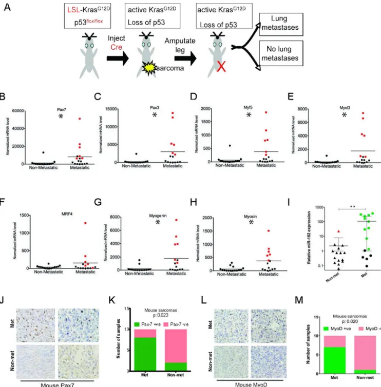
metastatic potential of the tumor. To mimic the development of pulmonary metastases that may occur following surgical resection in patients with no adjuvant therapy, we amputated the tumors to generate a cohort of metastatically-annotated sarcomas according to the schematic described in Figure 1A. After the sarcoma-bearing limb was amputated, mice were monitored for up to 6 months for the development of pulmonary metastases. The presence or absence of lung metastases was confirmed by histopathological examination of hematoxylin and eosin stained sections of lung by a sarcoma pathologist (L.D). With this approach, primary sarcomas were unambiguously divided into two phenotypes: metastatic (i.e. developed pulmonary metastasis) and non-metastatic (i.e. did not yield any discernible lung metastases).
We performed real-time PCR for multiple myogenic transcription factors in a cohort of 30 primary mouse sarcomas, of which 15 subsequently developed metastases. We examined satellite cell markers (Pax7 and Pax3), all four members of the myogenic regulator factor family (Myf5, MyoD, Mrf4 and myogenin) and a marker of terminal muscle differentiation, myosin (Figure 1B–H). We found that expression of Pax7, Pax3, Myf5, MyoD, myogenin and myosin were enriched in a subset of metastatic primary soft-tissue sarcomas from KP mice (Figure 1B–G). Although expression of myogenic regulator factor 4 (Mrf4) was elevated in metastatic tumors, this was not statistically significant (Figure 1H).
Immunohistochemical analysis for Pax7 and MyoD confirmed that protein levels for these myogenic factors were expressed at higher levels in metastatic primary tumors (n=10) than non-metastatic primary tumors (n=10, Figure 1J–M). Next, we examined the miR-182 levels in these tumors. We found a striking correlation between expression of myogenic factors and miR-182 (Figure 1I). Tumors with high miR-182 expression (shown in green Figure 1I) are
A
uthor Man
uscr
ipt
A
uthor Man
uscr
ipt
A
uthor Man
uscr
ipt
A
uthor Man
uscr
also illustrated in red in Figure 1 B–H. These findings suggested that miR-182 levels may be partially regulated by a myogenic differentiation program in primary sarcomas. We also examined the expression of miR-182, Pax7, and MyoD in lung metastases. Interestingly, the expression of miR-182 by RT-PCR was similar in the lung metastasis compared to the matched primary tumor, but Pax7/MyoD-positive primary tumors (n=5) did not maintain the expression of these factors (Supplemental Figure 1). This suggests that after colonization in the lung, there is no selective pressure to maintain expression of myogenic differentiation.
MyoD directly binds to and controls miR-182 expression
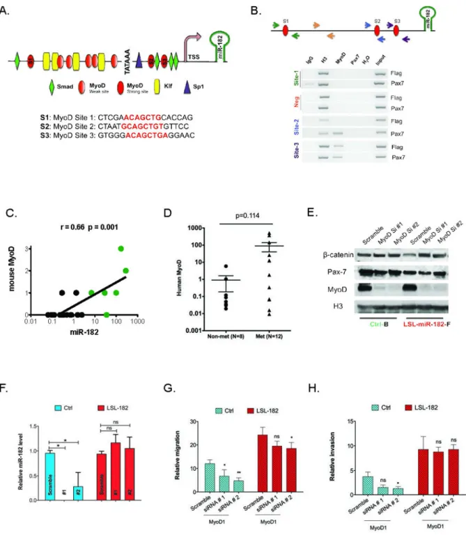
To determine if any of the myogenic transcription factors directly regulated miR-182 levels, we performed an in silico analysis (Genomatix) of the miR-182 promoter (14). This analysis identified several potential binding sites for the myogenic factor MyoD (Figure 2A). Chromatin immunoprecipitation (ChIP) demonstrated that MyoD, but not another myogenic transcription factor, Pax7, can directly bind the miR-182 promoter at two distinct sites (Figure 2B). We used a luciferase reporter assay to determine that MyoD sites 2 and 3 are most important for expression from the miR-182 promoter in C2C12 cells (Supplemental Figure 2). To determine if MyoD-specific control of miR-182 levels could contribute to metastasis, we examined an independent cohort of KP mouse sarcomas (n=18) and found a high correlation between miR-182 levels and MyoD protein expression. (Figure 2C). To determine if MyoD expression also correlates with metastasis in human sarcomas, we examined a cohort of human UPS tumors. Twenty fresh-frozen UPS samples were obtained from patients with known metastatic outcome that did not receive prior therapy (n=12 metastatic, n=8 non metastatic). Using real-time PCR, we determined that MyoD mRNA was enriched in a subset of metastatic tumors (n=5/12), while MyoD was expressed at much lower levels in non-metastatic samples (Figure 2D), although this difference was not statistically-significant. Importantly, the presence of MyoD in only 40% of the metastatic tumors supports the view of metastasis as a complex biological process that utilizes a diverse array of biological mechanisms, and suggests that alternative or complementary pathways may drive metastasis for the other tumors.
In cells derived from primary mouse sarcomas from KP mice (Ctrl), siRNA-mediated knockdown of MyoD decreased miR-182 transcript levels (Figure 2E–F and Supplemental Figure 3E). Little effect on miR-182 levels was seen following MyoD knockdown in sarcoma cells with constitutive expression of miR-182 from the ROSA26 promoter (LSL-miR-182, Figure 2E–F). To determine if the MyoD-dependent regulation of miR-182 could impact metastatic phenotypes, we examined the ability of MyoD expression to influence the in vitro migration and invasion of sarcoma cells with endogenous or constitutive expression of miR-182 (Figure 2G–H). While knockdown of MyoD blunted migration/invasion in control sarcoma cells, overexpression of miR-182 was sufficient to maintain invasion across the transwell membrane. Taken together, these results suggest that MyoD-mediated
regulation of miR-182 can influence metastatic properties in sarcoma.
Pax7 contributes to sarcoma metastasis
To further define the myogenic proteins that may influence pro-metastatic miR-182
expression, we sought to identify upstream factors that regulate MyoD expression in tumors.
A
uthor Man
uscr
ipt
A
uthor Man
uscr
ipt
A
uthor Man
uscr
ipt
A
uthor Man
uscr
In a subset of primary metastatic tumors, we found elevation of Pax7 and Pax3 (Figure 1B– C), two factors that are regulators of MyoD expression during embryonic and postnatal myogenesis (24). While both Pax7 and Pax3 are genetically upstream of MyoD, a review of the literature made Pax7 a more attractive candidate for several reasons. First, Pax7 is universally expressed in satellite cells, while Pax3 expression is limited to specific muscle groups (25). Second, Pax7 is known to directly bind to and activate the MyoD promoter and displays stronger transcriptional activation properties than Pax3 in C2C12 cells (26, 27). Third, expression of dominant-negative Pax7 abolishes MyoD expression in satellite cells in culture (28). Taken together, this makes Pax7 an attractive candidate for controlling MyoD levels in metastatic sarcoma.
We identified a statistically significant correlation between levels of miR-182 and Pax7 protein in the mouse tumor cohort (Figure 3A). Moreover, in the cohort of human UPS tumors, we determined that Pax7 mRNA was enriched in a subset of metastatic tumors (n=6/12, Figure 3B). Thus, in this cohort of human UPS tumors, elevated Pax7 expression was exclusive to metastatic tumors and correlated with miR-182 expression, making it a promising candidate to promote sarcoma metastasis. Of note, there was a trend for elevated Pax7 mRNA expression to correlate with high miR-182 expression in the primary tumors, but this correlation did not reach statistical significance (p=0.08) in this relatively small sample size (Figure 3C).
To determine if Pax7 expression could influence metastatic phenotypes in our mouse model similar to MyoD, we overexpressed Pax7 in KP sarcoma cell lines, which induced miR-182 expression (Figure 3D). As a control, we overexpressed Pax7 in sarcoma cells derived from miR-182flox/flox; LSL-KrasG12D; p53flox/flox mice (14) that lack expression of miR-182 (KO cells). While Pax7 expression increased miR-182 levels in wild-type sarcoma cells, it was unable to induce miR-182 expression in the knockout cell lines (Figure 3D). Of note, Pax7 increased both migration and invasion properties in wild-type cells, but was unable to influence in vitro metastatic phenotypes in miR-182 knockout (Figure 3E–F) or knockdown cells (Supplemental Figure 3A–C). In tandem, we knocked down Pax7 in sarcoma cells derived from mice with conditional overexpression of miR-182 (14). In these cells, Pax7 siRNA inhibited migration and invasion of the control cell line, but Pax7 knockdown showed minimal effect in cells with constitutive expression of miR-182 (Figure 3G–I). Taken together, these results suggest that Pax7-driven expression of miR-182 is sufficient to drive metastatic properties in vitro.
Pax7 and MyoD regulate pro-metastatic miR-182 to increase sarcoma metastasis
To further define the relationship between Pax7, MyoD, and miR182 in the metastatic sarcoma cohort, we determined that Pax7 and MyoD protein expression exhibited a strong degree of correlation in the mouse tumor cohort (Figure 4A). To determine if Pax7 acts through MyoD to regulate miR-182, we used siRNA to knockdown MyoD in sarcoma cells that overxpressed Pax7 (Figure 4B and Supplemental Figure 3D). While overexpression of Pax7 alone was capable of inducing miR-182 expression (Figure 4C, purple bars), siRNA knockdown of MyoD prevented Pax7 from elevating miR-182 levels (Figure 4C, yellow
A
uthor Man
uscr
ipt
A
uthor Man
uscr
ipt
A
uthor Man
uscr
ipt
A
uthor Man
uscr
bars). Taken together, these results suggest that Pax7-mediated regulation of miR-182 occurs through MyoD.
We next tested the role of Pax7 in modulating miR-182 levels, MyoD expression, and metastatic phenotypes through siRNA knockdown of Pax7 in two KP sarcoma cell lines. In both cell lines, knockdown of Pax7 with two distinct duplexes resulted in decreased miR-182 levels (Figure 2D). Knockdown of Pax7 also impaired migration and invasion of these cells in a transwell assay (Figure 2E–F). Of note, knockdown of Pax7 resulted in a corresponding decrease in MyoD protein levels, while MyoD knockdown did not alter Pax7 expression (Figures 4D, 2E).
Pax7 can regulate sarcoma metastasis in vivo
To further define the role of Pax7 in miR-182-mediated sarcoma metastasis, we generated stable mouse sarcoma cell lines that express either Flag-tagged Pax7 (29) or a Flag-alone control vector. We used two cell lines derived from primary sarcomas resulting from Ad-Cre injection into mice that were LSL-KrasG12D; p53flox/flox (KP cells) or LSL-KrasG12D; Ink4a/Arfflox/flox (KI cells). High levels of Pax7 were detected by qPCR and immunoblot in cells expressing Flag-tagged Pax7 (Supplemental Figure 4A). Overexpression of Pax7 did not alter the growth kinetics of the cells (Supplemental Figure 4B–D). Both Pax7-expressing cell lines showed elevated levels of metastatic phenotypes in vitro, including increased invasion/migration in both transwell and wound healing assays (Supplemental Figure 4E– H). To directly address the contribution of Pax7 to metastatic lung colonization in vivo, we injected sarcoma cells expressing either Flag-alone or Flag-tagged Pax7 orthotopically into immunocompetent mice. Following amputation of syngeneic orthotopic tumors, mice were sacrificed 6 months later, and the area of their lungs containing metastatic colonies was calculated from hematoxylin and eosin (H&E) stained slides. Lungs from mice receiving the Pax7-expressing cells showed increased metastatic lung area (Supplemental Figure 4I–J). Taken together, these data show that Pax7 expression can induce metastatic phenotypes in sarcoma cells in vivo.
miR-182 is required for Pax7-mediated metastasis in vivo
To further explore the Pax7/miR182 relationship in metastasis, we performed an orthotopic allograft amputation experiment using sarcoma cells that expressed Pax7 or Flag-alone with or without an anti-miR-182 sponge (n=9–10 mice per cell line; Figure 5A). The efficacy of the miR-182 sponge was assessed by the expression of the miR-182 target Rsu-1 (Figure 5A) (14). Following injection of cells into the lower left flank of nude mice, tumors were amputated at 200–300 mm3. Mice were sacrificed 7 weeks later or when they showed signs of pulmonary distress, and the lungs were examined for the presence of metastases. Expression of Pax7 greatly decreased the survival of the mice, while increasing the number and percent of metastatic lung growths (Figure 5B–E). Reduction of miR-182 levels by the anti-182 sponge decreased the Pax7-dependent metastatic phenotype by increasing the survival of mice and decreasing the area and number of lung metastases. These results suggest a model where Pax7-dependent metastasis requires miR-182 for sarcoma metastasis in vivo (Figure 5F).
A
uthor Man
uscr
ipt
A
uthor Man
uscr
ipt
A
uthor Man
uscr
ipt
A
uthor Man
uscr
Discussion
Currently, over 30% of adult sarcoma patients develop metastatic disease. We previously demonstrated that miR-182 can contribute to metastasis in a mouse model of soft-tissue sarcoma(14). However, the events controlling miR-182 expression in these tumors remained unexplored. In this current study, we describe a novel mechanism where myogenic
differentiation factors regulate miR-182 expression and contribute to sarcoma metastasis in vivo. Using a mouse model of soft-tissue sarcoma, we observed expression of several myogenic transcription factors, including Pax7 and MyoD, in primary metastatic mouse tumors. Expression of Pax7 and MyoD correlated with miR-182 levels in both mouse and human sarcoma samples. Chromatin immunoprecipitation and luciferase assays confirmed that MyoD regulates miR-182 expression through direct binding to the miR-182 promoter. In vitro analyses showed MyoD directly controls miR-182 expression and metastatic phenotypes, while Pax7 requires MyoD for the regulation of miR-182 levels. Using orthotopic allografts of cells engineered with an anti-miR-182 sponge, we determined that Pax7-expressing cells require miR-182 to drive sarcoma metastasis in vivo. Taken together, these data suggests that one pathway to activate miR-182 in metastatic sarcoma is through a Pax7/MyoD-dependent mechanism. As the miR-182 promoter contains multiple binding sites for other transcription factors, we postulate that miR-182 activation can occur through multiple mechanisms, some of which are independent of myogenic factors (Sachdeva et al, unpublished results).
Myogenic transcription factors play a critical role in muscle cell development during embryogenesis. For example, precursor cells originating in the somites require Pax3 for migration to the sites of muscle wall formation. We speculate that sarcoma cells could be hijacking this developmental program to activate migratory phenotypes required for metastasis. Just as many epithelial cancers utilize the epithelial-to-mesenchymal transition (EMT) to express genes that normally control cell motility during embryonic development, we postulate that myogenic transcription factors may also provide a metastatic advantage at the primary tumor site through increased migration.
This work expands on the observation that expression of myogenic markers in human undifferentiated pleomorphic sarcomas correlates with poor patient outcome(17). Our work suggests a mechanism for the role of myogenic transcription factors in promoting metastasis in UPS. Recent work from other groups suggests this theme can be extended to other sarcoma subtypes. In a particularly aggressive subtype of rhabdomyosarcoma, a recurrent somatic mutation in MyoD blocks wild-type function (28). Instead of activating canonical MyoD targets involved in differentiation, the mutated MyoD binds MYC consensus sequences. This switches downstream transcriptional profiles from muscle differentiation genes to proliferative targets. In combination with our data in UPS, these findings identify an important role for myogenic transcription factors, such as MyoD, in promoting aggressive behavior in sarcomas and suggest further investigations of these proteins in other sarcoma subtypes.
This work reveals one mechanism by which myogenic transcription factors regulate miR-182 to induce pro-metastatic phenotypes in murine systems in vitro and in vivo.
A
uthor Man
uscr
ipt
A
uthor Man
uscr
ipt
A
uthor Man
uscr
ipt
A
uthor Man
uscr
Therefore, further evaluating the role of Pax7, MyoD, and miR-182 in human sarcomas has the potential to provide information related to metastatic risk. If the analysis of this pathway in large, retrospective sarcoma patient cohorts supports this hypothesis, then prospective studies could be undertaken to define the prognostic value of this pathway for patients with UPS. Thus, our results provide support for future studies investigating the potential
prognostic value and biological role of myogenic transcription proteins in human soft-tissue sarcoma.
Experimental Procedures
Mouse Sarcoma Model
All mouse work was performed in accordance with Duke University Institutional Animal Care and Use Committee approved protocols. Tumors were generated by injection of Adenovirus–expressing Cre recombinase (Ad-Cre; University of Iowa, Vector core) into the lower left leg of p53flox/flox; LSL-KRasG12D compound mutant mice as previously described (18). Following amputation, mice were monitored for up to 6 months for development of lung metastases.
Quantitative RT-PCR
RNA was isolated from tumors and cells in TRIZOL, and cDNA synthesis was performed with iScript (BioRad). Quantitative RT-PCR was performed on an iQ5 instrument (BioRad) using the delta-delta Ct method. Primer sequences can be found in Table S1.
Human samples
UPS samples were obtained from MD Anderson under a protocol approved by the Duke University and MD Anderson Institutional Review Boards. Frozen tumor samples were processed using TRIZOL, and cDNA synthesis was performed with iScript (BioRad). Quantitative RT-PCR was performed on an iQ5 instrument (BioRad) using the delta-delta Ct method.
Histology and Immunostaining
Antibodies for immunohistochemistry included: Pax7 (a gift from Chen-Min Fan, Carnegie Institute), Ki67 (BD Pharmagen), and MyoD (Santa Cruz). All immunostaining was perfomed with citrate-based antigen retrieval, and M.O.M. Kits (Vector Labs) were used for Pax7 staining.
In vitro studies
The mouse sarcoma cell lines KP and KI were derived from primary sarcomas that developed after intra-muscular Ad-Cre injection into a p53 flox/flox; LSL-KrasG12D or Ink4a/Arf flox/flox; LSL-KrasG12D mouse, respectively. Mouse sarcoma cells for miR-182 experiments were described previously (14). Cells were cultured in DMEM + 10% FBS. For colony formation studies, cells were seeded at 500/well in a 6-well dish, and the number of existing colonies was determined by crystal violet staining after 2 weeks. Each experiment was conducted in triplicate, and the experiment was repeated three times. Data shown are
A
uthor Man
uscr
ipt
A
uthor Man
uscr
ipt
A
uthor Man
uscr
ipt
A
uthor Man
uscr
representative of one independent experiment. For cell doubling assays, 1,000 cells were seeded overnight and the number of cell doublings was counted daily for four days. Migration and invasion assays were completed using the Cultrex Cell Invasion Assay, per manufacturer’s instructions. Scratch assays were performed on confluent monolayers, with images obtained on an Olympus CKX41 microscope and distances quantified in ImagePro. Data shown represent the average of three independent experiments. For siRNA studies, sarcoma cell lines were transfected with siRNAs against Pax7 or MyoD with Lipofectamine (Invitrogen) following the manufacturer’s protocol. The cells were harvested after 36 hrs and 48 hrs post transfection for RNA and protein analysis, respectively.
In vivo allografts
129/SvJae or athymic female nude mice were injected intramuscularly with 10,000 cells in the lower left flank. Mice were randomly picked for which cell line to receive. Following 7– 14 days of growth, tumors were amputated and mice were followed for 6 months. Mice were sacrificed when they presented with symptoms of lung metastases, such as labored breathing and hunched posture. Investigators were blind to experimental groups when assessing animal health. Mice that died prior to lung harvest were excluded from analysis. Following
sacrifice, lungs were inflated with formalin prior to harvest and H&E stained sections were quantified on Image Pro to calculate the percent of lung volume containing metastases. Three sections of lung per mouse were examined.
Western blot analysis
Cells were washed once with cold PBS (Sigma) and lysed for 10 minutes on ice with RIPA buffer (Sigma). Protein concentration was performed with BCA Protein Concentration Assay (Thermo Scientific). MiniProtean TGX gels (BioRad) were transferred to PVDF by wet transfer. Antibodies include Histone H3 (Cell Signaling) and others described in these methods.
Chromatin Immunoprecipitation (ChIP) Assay
KP cells stably expressing Flag and Pax-7 were seeded at 50% confluence overnight. Nuclear proteins bound to the genomic DNA were cross-linked directly in the cell culture medium in 1% formaldehyde at room temperature. Chromatin was digested with
Micrococcal Nuclease for 20 mins at 37° C and incubated with respective antibodies overnight. Protein G agarose beads captured the antibody-chromatin complex. DNA was separated from beads and eluted using ChiP elution buffer (Cell signaling technology), purified with spin columns and quantified using specific PCR primers (see Table S1).
Luciferase Assay
Cells were transfected with different miR-182 promoter constructs with or without MyoD plasmid (lentiviral system from System Biosciences). Cells were harvested 36 h after transfection. Luciferase assays were performed as previously described (14). Plasmids with different miR-182 promoters were cloned into a luciferase vector (Promega). miR-182 promoter sequences were PCR amplified with high fidelity Taq polymerase enzyme (Invitrogen) using mouse genomic DNA as a template. Primer sequences are provided in
A
uthor Man
uscr
ipt
A
uthor Man
uscr
ipt
A
uthor Man
uscr
ipt
A
uthor Man
uscr
Table S1. The amplified fragment was cloned into a Pgl3-basic vector at Kpn1 and Xho1 sites (Cho-Cho Cloning Kit). To mutate the putative MyoD binding sites in the miR-182 promoter, we adopted a two-step PCR ligation method with two sets of overlapping primers (Table S1) that span the new binding site with mutations. The two amplified PCR fragments were used as a template for the second PCR using primers Mir-182-pro-5.4 -kpn1 and Mir-182-pro-3.1-rev-xho1 and cloned using a similar strategy as described above.
Supplementary Material
Refer to Web version on PubMed Central for supplementary material.
ACKNOWLEDGEMENTS
We thank Ron Dephino (MD Anderson) for providing the Ink4a/Arf flox mice, Tyler Jacks (MIT) for the LSL-Kras mice, and Anton Berns (Netherlands Cancer Institute) for the p53 flox mice. We thank Michael Rudnicki for the Pax7-FLAG tagged vectors. We thank Laura Jeffords, Rafaela Rodrigues, and Loretta Woodleif for assistance with mouse work. RDD is supported by postdoctoral fellowships from the American Cancer Society/Canary Foundation and the Children’s Tumor Foundation. MS is supported by a postdoctoral fellowship from the QuadW/AACR. This work is supported by RO1 CA 138265 (DGK) and a SARC SPORE pilot project 5U54 CA 168512-02 (DKG and RDD).
References
1. Nguyen DX, Massague J. Genetic determinants of cancer metastasis. Nat Rev Genet. 2007; 8(5): 341–352. [PubMed: 17440531]
2. Yachida S, Jones S, Bozic I, Antal T, Leary R, Fu B, et al. Distant metastasis occurs late during the genetic evolution of pancreatic cancer. Nature. 2010; 467(7319):1114–1117. [PubMed: 20981102] 3. Heitzer E, Ulz P, Belic J, Gutschi S, Quehenberger F, Fischereder K, et al. Tumor-associated copy
number changes in the circulation of patients with prostate cancer identified through whole-genome sequencing. Genome Med. 2013; 5(4):30. [PubMed: 23561577]
4. Liu W, Laitinen S, Khan S, Vihinen M, Kowalski J, Yu G, et al. Copy number analysis indicates monoclonal origin of lethal metastatic prostate cancer. Nature medicine. 2009; 15(5):559–565. 5. Kimmelman AC, Hezel AF, Aguirre AJ, Zheng H, Paik JH, Ying H, et al. Genomic alterations link
Rho family of GTPases to the highly invasive phenotype of pancreas cancer. Proceedings of the National Academy of Sciences of the United States of America. 2008; 105(49):19372–19377. [PubMed: 19050074]
6. Ramaswamy S, Ross KN, Lander ES, Golub TR. A molecular signature of metastasis in primary solid tumors. Nature genetics. 2003; 33(1):49–54. [PubMed: 12469122]
7. van 't Veer LJ, Dai H, van de Vijver MJ, He YD, Hart AA, Mao M, et al. Gene expression profiling predicts clinical outcome of breast cancer. Nature. 2002; 415(6871):530–536. [PubMed: 11823860] 8. van de Vijver MJHY, van't Veer LJ, Dai H, Hart AA, Voskuil DW, Schreiber GJ, Peterse JL, Roberts C, Marton MJ, Parrish M, Atsma D, Witteveen A, Glas A, Delahaye L, van der Velde T, Bartelink H, Rodenhuis S, Rutgers ET, Friend SH, Bernards R. A gene-expression signature as a predictor of survival in breast cancer. N Engl J Med. 2002; 347(25):1999–2009. [PubMed: 12490681]
9. Salazar R, Roepman P, Capella G, Moreno V, Simon I, Dreezen C, et al. Gene expression signature to improve prognosis prediction of stage II and III colorectal cancer. J Clin Oncol. 29(1):17–24. [PubMed: 21098318]
10. Gibbons DL, Lin W, Creighton CJ, Zheng S, Berel D, Yang Y, et al. Expression signatures of metastatic capacity in a genetic mouse model of lung adenocarcinoma. PloS one. 2009; 4(4):e5401. [PubMed: 19404390]
11. Borden EC, Baker LH, Bell RS, Bramwell V, Demetri GD, Eisenberg BL, et al. Soft tissue sarcomas of adults: state of the translational science. Clin Cancer Res. 2003; 9(6):1941–1956. [PubMed: 12796356]
A
uthor Man
uscr
ipt
A
uthor Man
uscr
ipt
A
uthor Man
uscr
ipt
A
uthor Man
uscr
12. Canter RJ, Qin LX, Downey RJ, Brennan MF, Singer S, Maki RG. Perioperative chemotherapy in patients undergoing pulmonary resection for metastatic soft-tissue sarcoma of the extremity : a retrospective analysis. Cancer. 2007; 110(9):2050–2060. [PubMed: 17828771]
13. Dodd RD, Mito J, Kirsch DG. Animal models of soft-tissue sarcoma. Disease Models and Mechanisms. 2010; 3(9–10):557–566. [PubMed: 20713645]
14. Sachdeva M, Mito J, Lee C, Zhang M, Li Z, Dodd R, Cason D, Luo L, Ma Y, Vanmater D, Gladdy R, Lev D, Cardona D, Kirsch D. MicroRNA-182 drives metastasis of primary sarcomas by targeting multiple genes. Journal of Clincal Investigations. 2014; 124(10):4305–4319. 15. Cheung TH, Quach NL, Charville GW, Liu L, Park L, Edalati A, et al. Maintenance of muscle
stem-cell quiescence by microRNA-489. Nature. 2012; 482(7386):524–528. [PubMed: 22358842] 16. Buckingham M, Relaix F. The role of Pax genes in the development of tissues and organs: Pax3
and Pax7 regulate muscle progenitor cell functions. Annu Rev Cell Dev Biol. 2007; 23:645–673. [PubMed: 17506689]
17. Fletcher CDGP, Rydholm A, Willen H, Akerman M. Clinicopathologic Re-Evaluation of 100 Malignant Fibrous Histiocytomas: Prognostic Relevance of Subclassification. J Clin Oncol. 2001; 19(12):3045–3050. [PubMed: 11408500]
18. Kirsch DG, Dinulescu DM, Miller JB, Grimm J, Santiago PM, Young NP, et al. A spatially and temporally restricted mouse model of soft tissue sarcoma. Nature medicine. 2007; 13(8):992–997. 19. Mito JK, Riedel RF, Dodd L, Lahat G, Lazar AJ, Dodd RD, et al. Cross species genomic analysis identifies a mouse model as undifferentiated pleomorphic sarcoma/malignant fibrous histiocytoma. PloS one. 2009; 4(11):e8075. [PubMed: 19956606]
20. Rubin BP, Nishijo K, Chen HI, Yi X, Schuetze DP, Pal R, et al. Evidence for an unanticipated relationship between undifferentiated pleomorphic sarcoma and embryonal rhabdomyosarcoma. Cancer Cell. 2011; 19(2):177–191. [PubMed: 21316601]
21. Blum JM, Ano L, Li Z, Van Mater D, Bennett BD, Sachdeva M, Lagutina I, Zhang M, Mito JK, Dodd LG, Cardona DM, Dodd RD, Williams N, Ma Y, Lepper C, Linardic CM, Mukherjee S, Grosveld GC, Fan CM, Kirsch DG. Distinct and Overlapping Sarcoma Subtypes Initiated from Muscle Stem and Progenitor Cells. Cell Reports. 2013
22. Mito JK, Min HD, Ma Y, Carter JE, Brigman BE, Dodd L, et al. Oncogene-dependent control of miRNA biogenesis and metastatic progression in a model of undifferentiated pleomorphic sarcoma. J Pathol. 2013; 229(1):132–140. [PubMed: 22951975]
23. Eisinger-Mathason TS, Zhang M, Qiu Q, Skuli N, Nakazawa MS, Karakasheva T, et al. Hypoxia-dependent modification of collagen networks promotes sarcoma metastasis. Cancer Discov. 2013; 3(10):1190–1205. [PubMed: 23906982]
24. Buckingham M, Rigby PW. Gene regulatory networks and transcriptional mechanisms that control myogenesis. Dev Cell. 2014; 28(3):225–238. [PubMed: 24525185]
25. Kassar-Duchossoy L, Giacone E, Gayraud-Morel B, Jory A, Gomes D, Tajbakhsh S. Pax3/Pax7 mark a novel population of primitive myogenic cells during development. Genes Dev. 2005; 19(12):1426–1431. [PubMed: 15964993]
26. Hu P, Geles KG, Paik JH, DePinho RA, Tjian R. Codependent activators direct myoblast-specific MyoD transcription. Dev Cell. 2008; 15(4):534–546. [PubMed: 18854138]
27. Soleimani VD, Punch VG, Kawabe Y, Jones AE, Palidwor GA, Porter CJ, et al. Transcriptional dominance of Pax7 in adult myogenesis is due to high-affinity recognition of homeodomain motifs. Dev Cell. 2012; 22(6):1208–1220. [PubMed: 22609161]
28. Relaix F, Montarras D, Zaffran S, Gayraud-Morel B, Rocancourt D, Tajbakhsh S, et al. Pax3 and Pax7 have distinct and overlapping functions in adult muscle progenitor cells. J Cell Biol. 2006; 172(1):91–102. [PubMed: 16380438]
29. McKinnell IW, Ishibashi J, Le Grand F, Punch VG, Addicks GC, Greenblatt JF, et al. Pax7 activates myogenic genes by recruitment of a histone methyltransferase complex. Nature cell biology. 2008; 10(1):77–84. [PubMed: 18066051]
A
uthor Man
uscr
ipt
A
uthor Man
uscr
ipt
A
uthor Man
uscr
ipt
A
uthor Man
uscr
Figure 1. Myogenic transcription factors are enriched in metastatic primary mouse sarcomas (A) Schematic of amputation strategy to determine metastatic potential of primary mouse sarcomas that is used in this study. (B–H) Levels of myogenic transcripts are elevated in a subset of metastatic primary sarcomas in the mouse, including Pax7, Pax3, Myf5, MyoD, Myogenin and myosin. * = p<0.05 by Mann-Whitney test. (I) Samples (shown in green) with elevated pro-metastatic miR-182 are the identical samples with high expression of myogenic markers (shown in red). (J–M) Immunohistochemical analysis shows that both Pax7 and MyoD are elevated in metastatic primary mouse sarcomas in comparison to non-metastatic tumors (p=0.023 (Pax7) and p=0.020 (MyoD), both by Fisher exact test).
A
uthor Man
uscr
ipt
A
uthor Man
uscr
ipt
A
uthor Man
uscr
ipt
A
uthor Man
uscr
Figure 2. MyoD controls expression of the pro-metastatic miR-182
(A) Schematic of miR-182 promoter highlighting potential MyoD binding sites. (B) Chromatin immunoprecipitaiton of miR182 promoter demonstrating direct binding of MyoD, but not Pax7, to sites 2 and 3. (C) MyoD protein levels correlate with metastasis and miR-182 levels in primary mouse sarcomas by Spearman’s correlation. (D) MyoD mRNA levels trend towards enrichment in metastatic human UPS tumors (p=0.114 by Mann-Whitney test). (E–F) siRNA-mediated knockdown of MyoD decreases miR-182 levels in mouse sarcoma cell lines with endogenous miR-182 (Ctrl-B), but has no effect in mouse
A
uthor Man
uscr
ipt
A
uthor Man
uscr
ipt
A
uthor Man
uscr
ipt
A
uthor Man
uscr
sarcoma cell lines with constitutive overexpression of miR-182 (LSL-182) * = p<0.05, by Mann-Whitney test. (G–H) siRNA-mediated knockdown of MyoD decreases migration and invasion in cells that express miR-182 from the endogenous miR-182 promoter (Ctrl), but the effect of MyoD knockdown on migration and invasion is largely rescued in cells that constitutively overexpress miR-182 from the ROSA-26 promoter (LSL-182). * = p<0.05, ** = p<0.01, *** = p<0.001 by two-way ANOVA with Bonferroni correction.
A
uthor Man
uscr
ipt
A
uthor Man
uscr
ipt
A
uthor Man
uscr
ipt
A
uthor Man
uscr
Figure 3. Pax7 activates metastatic phenotypes via miR-182
(A) In primary mouse sarcomas, levels of Pax7 protein and miR-182 correlate with metastatic outcome (green circles) by Spearmans correlation. (B) Pax7 expression is
enriched in a subset of metastatic human UPS samples (p=0.038 by Mann-Whitney test). (C) Human primary sarcomas that metastasized and express elevated Pax7 show a trend for increased miR-182 expression (pink diamonds) by Spearmans correlation. (D)
Overexpression of Pax7 in miR-182 wild-type (WT) cells, but not in miR-182 knockout (KO) cells, increases miR-182 levels. ** = p<0.01, *** = p<0.001 by two-way ANOVA with Bonferroni correction. (E–F) Pax7 overexpression can increase migration and invasion of sarcoma cells in miR-182 WT cells, but is unable to increase in vitro metastatic phenotypes
A
uthor Man
uscr
ipt
A
uthor Man
uscr
ipt
A
uthor Man
uscr
ipt
A
uthor Man
uscr
in miR-182 KO cells. ** = p<0.01, *** = p<0.001 by two-way ANOVA with Bonferroni correction. (G–I) siRNA-mediated knockdown of Pax7 decreases migration and invasion in cells that express wild-type miR-182 (Ctrl), but has minimal impact on migration and invasion of cells that constitutively overexpress miR-182 (LSL-182). * = p<0.05, ** = p<0.01, *** = p<0.001 by two-way ANOVA with Bonferroni correction.
A
uthor Man
uscr
ipt
A
uthor Man
uscr
ipt
A
uthor Man
uscr
ipt
A
uthor Man
uscr
Figure 4. Pax7, MyoD and miR-182 cooperate to influence metastasis
(A) There is a strong correlation between levels of Pax7 and MyoD in KP mouse sarcomas. (B–C) Overexpression of Pax7 alone can increase miR-182 levels. However, overexpression of Pax7 in concert with MyoD knockdown is not sufficient to increase miR-182 levels. * = p<0.05, ** = p<0.01, *** = p<0.001 by two-way ANOVA with Bonferroni correction. (D) Knockdown of Pax7 in KP-2 and KP-3 sarcoma cells decreases MyoD and miR-182 levels * = p<0.05 by Mann-Whitney test. (E–F) Knockdown of Pax7 in KP-2 and KP-3 sarcoma cells decreases in vitro migration and invasion. * = p<0.05, ** = p<0.01 by Mann-Whitney test.
A
uthor Man
uscr
ipt
A
uthor Man
uscr
ipt
A
uthor Man
uscr
ipt
A
uthor Man
uscr
Figure 5. Pax7 controls sarcoma metastasis in vivo through miR-182
(A) Expression of GFP-tagged Flag or Pax7 in KP cells with or without an anti-182 sponge. Note decrease in protein levels of Rsu-1, a miR-182 target (14), in anti-182-expressing cells. (B) Metastasis-free survival of animals receiving orthotopic injection of sarcoma cells expressing Flag with vector, Flag with miR-182, Pax7 with vector, or Pax7 with anti-miR-182. (C–E) Following orthotopic injection, Pax7 expression increases metastatic phenotypes, including gross lung metastases (C), percent of metastatic lung area (D), and number of metastases (E). * = p<0.05 by one-way ANOVA with Bonferroni correction. These phenotypes decrease with the anti-182 sponge. (F) Model of a Pax7/MyoD/miR-182 axis driving sarcoma metastasis.