Address for correspondence
Dr. Singh Akhil Kumar, Assistant Professor Department of Skin & VD,
Saraswathi Institute of Medical Sciences Pilkhuwa, Hapur UP -245304, India Email: [email protected]
Original Article
How accurately do clinical diagnosis correlate with
biopsy findings in leprosy?
Introduction
Leprosy, one of the most ancient, feared and disabling chronic granulomatous infectious diseases with long incubation period that affects
skin, muscles, peripheral nerve, eyes and internal organs, is on the verge of eradication. The 1991 World Health Assembly resolution1 was a catalyst, and today 116 out of 122 endemic countries have eliminated leprosy as a public health problem. Leprosy continues to be an important public health problem in many part of Asia including India.1 India achieved its elimination goal both at the national and state level with prevalence rate of 0.68% per 10,000 Singh Akhil Kumar, Singh Ranjana*, Singh Savitri**, Grover Sanjiv
Department of Skin & VD, SIMS, Hapur UP, India
* Department of Community Medicine, SIMS, Hapur UP, India ** Department of Pathology, SIMS, Hapur UP, India
Abstract
Objective To document the clinical profile of leprosy patients and to determine concordance between clinical and histopathological diagnosis using Ridley and Jopling classification.Methods 58 clinically and histological confirmed cases of leprosy ranging in age from 7-70 years who attended dermatology outpatient department (OPD) in Saraswathi Institute of Medical Sciences were included in the study. Slit-skin smear examination was performed in all cases. Histopathological investigations were carried out in all cases to confirm the diagnosis and to classify the cases.
Results A total of 58 clinically diagnosed cases of leprosy comprised the study material. Out of them, 42 (72.4%) were males and 16 (27.6%)females with male to female ratio of 2.6:1. The highest number of cases was in age group 20-29 years with 22 (37.9%) cases. 10 (17.2%) cases were multibacillary and 48 (82.8%) cases were paucibacillary. The most common site of biopsy was from lower extremities in 24 (41.4%) cases. Clinically, maximum number of patients was borderline tuberculoid (BT) type with 22 (37.9%) cases, followed by tuberculoid leprosy (TT) in 14 (24.1%) cases. Lepromatous leprosy (LL) was confirmed in 3 (5.2%) cases. Most common clinical presentation was altered sensation in 57 (98.3%) cases, skin plaques in 46 (79.3%) cases and nerve involvement was noted in 36 (62.1%) cases. Maximum clinicopathological correlation was noted in both poles i.e. LL with 3 (100%) cases and TT with 12 (85.7%) cases and maximum disparity was noted in borderline lepromatous leprosy (BL) in 3 (60%) cases. Overall concordance between clinical and histopathological diagnosis observed in our study was 46 (79.3%) cases.
Conclusion Leprosy continues to remain a public health problem. Clinical-histopathological discordance leading to inadequate treatment could be contributory. Our study revealed a 20.7% rate of discordance. Early assessment and adequate management is essential for reducing the discordance rate.
Key words
(as on Jan 2014) but leprosy is still prevailing in this country and about 60 % of fresh diagnosed cases in the world are from India.
The disease presents itself in different clinicopathological forms depending upon the cellular immune system (CMI) of the host.2 The spectrum of disease in leprosy has been classified in many clinical and clinicoimmunopathological classifications, the most widely used for academic purpose is Ridley-Jopling classification. Ridley and Jopling had proposed the classification of leprosy into six groups as indeterminate, tuberculoid (TT), borderline tuberculoid (BT), mid borderline (BB), borderline lepromatous (BL) and lepromatous leprosy (LL).3 However, a great variation has been observed in the histopathological and clinical diagnosis.
The aim of this study was to document different clinical and pathological patterns of leprosy and to find out the concordance between clinical and histopathological diagnosis.
Methods
A retrospective hospital-based study was conducted among 61 fresh patients ranging from 9-70 years with all clinical types of leprosy, who visited dermatology outpatient department at Saraswathi Institute of Medical Sciences, Hapur between January 2014 to December 2015. Only those cases who had given consent for biopsy were included in this study. In clinical
examination, the following points were
recorded:
Lesions–number, type of lesions, distribution,
color, surface, hair over the lesion, symmetry of lesions, as well as, sensory loss,
Nerves–Involvement of nerves, thickening,
tenderness, as well as, sensory and motor loss were recorded.
Slit-skin smear and biopsy were taken from active lesion in all patients after informed consent as per guidelines of institutional ethical committee. Biopsy samples were processed as per standard protocol in the Pathology department. Biopsy samples were sent in 40% formalin solution; and stained with hematoxylin and eosin stains (H&E) and modified
Fite-Ferraco stain for identification of
Mycobacterium leprae. Only clinically as well as histopathologically confirmed cases were considered as study material. Detailed history and thorough clinical examination were carried out in each patient.
The present study was conducted among 61 patients, diagnosed clinically as leprosy. 58 patients were confirmed as leprosy histologically and among rest three, one patient was diagnosed as Bernhardt syndrome and two others patients with hypopigmentation. After confirmation of diagnosis, they were excluded from this study.
Histological finding were compared with clinical diagnosis. Ridley and Jopling criteria were adopted for clinical classification of leprosy. Patients were classified as PB and MB according to WHO guidelines for treatment purpose and
treated with respective regimens.4
Statistical analysis was done using SPSS version 20.0. The variables were described by taking mean, frequency, percentages and chi square test.
Results
male to female ratio of 2.6:1. The mean age of the study population was 34.6 ± 13.8 years.
Table 1 shows the age and sex distribution of leprosy cases. Among the total of 58 cases, 42 (72.4%) were males and 16 (27.6%) were females. Majority of the patients 22 (37.9%) cases, belonged to the age group of 20-29 years followed by 11 (19.0%) in age group of 40-49 years, the lowest number of cases was from age group of 0-9 years i.e. 1 case (1.7%), p = 0.045.
Clinical categorization of 58 cases of leprosy as per Ridley and Jopling showed that the predominant group was BT in 22 (37.9%) cases, TT in 14 (24.1%) cases, IL group in 12 (20.7%), BL in 5 (8.6%), LL in 3 (5.2%) cases while BB
was observed in 2 (3.4%) cases, (Table 2).
48 (82.8%) cases were paucibacillary (PB) and 10 (17.2%) cases were multibacillary on the basis of slit-smear and clinical examination. Duration of disease ranged five months to seven years and positive family history was observed in 6 (10.3%) cases.
Single nerve involvement was observed in 26 (44.8%) cases. No nerve was found thickened in 22 (37.9%) cases. The most common sites of biopsy were lower and upper extremities in 24 (41.4%) and 22(38.0%) cases, respectively.
The presenting symptoms/signs of various type
of leprosy are summarized in Table 3. The most
frequent skin lesion was plaque i.e. 46 (79.3%) cases, followed by papules/nodules in 5 (8.6%) and hypopigmented area in 4 (6.9%) cases. Loss of sensations and altered sensations were observed in 57 (98.3%) cases, while trophic ulcers were present in 1 (1.7%) case. Limb deformities were observed in 4 (6.9%) cases.
Histopathologically majority of cases i.e. 23 (39.7%) belonged to BT leprosy followed by TT
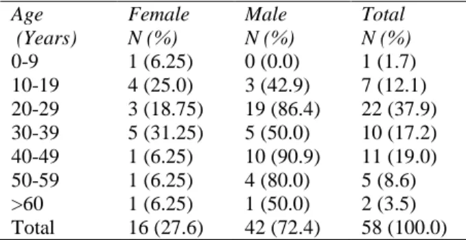
Table 1 Age and sex distribution of leprosy cases (n=58).
Age (Years)
Female N (%)
Male N (%)
Total N (%)
0-9 1 (6.25) 0 (0.0) 1 (1.7)
10-19 4 (25.0) 3 (42.9) 7 (12.1) 20-29 3 (18.75) 19 (86.4) 22 (37.9) 30-39 5 (31.25) 5 (50.0) 10 (17.2) 40-49 1 (6.25) 10 (90.9) 11 (19.0) 50-59 1 (6.25) 4 (80.0) 5 (8.6) >60 1 (6.25) 1 (50.0) 2 (3.5) Total 16 (27.6) 42 (72.4) 58 (100.0)
Table 2 Summary of clinical diagnosis (Ridley and Jopling) in the study subjects (n=58)
Type of leprosy
Tuberculoid (TT) 14 (24.1)
Borderline tuberculoid (BT) 22 (37.9) Mid borderline (BB) 2(3.4) Borderline lepromatous (BL) 5(8.6)
Lepromatous (LL) 3 (5.2)
Indeterminate (IL) 12 (20.7)
Paucibacillary 48 (82.8)
Multibacillary 10 (17.2)
Positive family history 6 (10.3) Nerve involvement
Single nerve 26 (44.8)
Two nerve 3 (5.2)
Multiple nerves 7 (12.1)
Not involved 22 (37.9)
Site of biopsy
Head & neck 4 (6.9)
Abdomen 8 (13.8)
Upper extremities 22 (38.0)
Lower extremities 24 (41.4)
Table 3 Clinical presentation of various type of leprosy (N=58).
Clinical presentations N (%)
Hypopigmented area 4 (6.9)
Plaques 46(79.3)
Papulonodule 5 (8.6)
Altered sensation 57 (98.3)
Nerves involvement 36 (62.1)
Deformities 4 (6.8)
Trophic ulcer 1 (1.7)
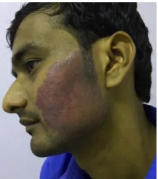
Figure 1 Borderline tuberculoid lesion over face.
Figure 3 Lepromatous leprosy histology.
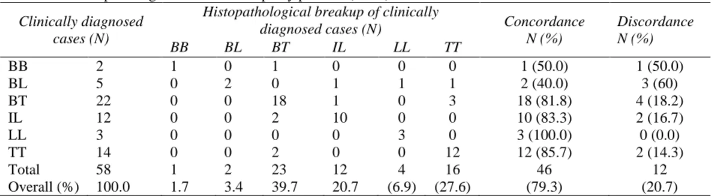
Overall, clinicopathological concordance in the present study was 79.3%. Maximum diversity was observed in BL group 60% and BB 40%, the polar forms i.e. LL and TT had a disparity of 0% and 14.3 %, respectively.
It was observed that clinical diagnosis and histopathological diagnosis of different types of
leprosy were correlating well as shown in Table
4 (p= 0.000).
Figure 2 Borderline tuberculoid leprosy with posterior auricular nerve involvement.
Figure 4 Tuberculoid leprosy histology.
Discussion
Table 4 Clinicopathological correlation leprosy patients (n=58). Clinically diagnosed
cases (N)
Histopathological breakup of clinically
diagnosed cases (N) Concordance N (%)
Discordance N (%)
BB BL BT IL LL TT
BB 2 1 0 1 0 0 0 1 (50.0) 1 (50.0)
BL 5 0 2 0 1 1 1 2 (40.0) 3 (60)
BT 22 0 0 18 1 0 3 18 (81.8) 4 (18.2)
IL 12 0 0 2 10 0 0 10 (83.3) 2 (16.7)
LL 3 0 0 0 0 3 0 3 (100.0) 0 (0.0)
TT 14 0 0 2 0 0 12 12 (85.7) 2 (14.3)
Total 58 1 2 23 12 4 16 46 12
Overall (%) 100.0 1.7 3.4 39.7 20.7 (6.9) (27.6) (79.3) (20.7)
IL: indeterminate, TT: tuberculoid, BT: borderline tuberculoid (BT), BB: mid borderline, BL: borderline lepromatous, and LL: lepromatous leprosy.
leading cause of physical disabilities which contribute to intense social stigma, resulting in discrimination of the patient and their families. Diagnosis of leprosy is based on different clinical parameters which involves detailed examination of skin lesions and peripheral nerves. Demonstration of acid-fast bacilli in slit skin smears by Ziehl-Neelsen staining also helps in diagnosis of leprosy. But a reliable diagnosis is obtained by good histopathological diagnosis and demonstration of acid fast bacilli.
The study included 58 patients ranging from 9-70 years which showed higher preponderance in males 42 (72.4%) and lower in females16 (27.6%) with male to female ratio of 2.6:1. This reflects increased consultations by male as opposed to females. Similar results were found in a study conducted by Bhushan et al.5 there were 63(72.3%) males and 39(27.7%) females; the ratio was 2.61:1. Similar results were found in a study by Raveneet et al.6 where most common age group affected was 21-30 years in 19 (31.7%) and male predominance was seen in 43 (71.7%) of the cases; and by Giridhar M et al.7 who reported 76 (77.6%) male cases as compared to 22 (22.4%) females. Maximum number of cases was in the age group of 21-30 years i.e. 41 (41.8%). Manandhar et al.8 explained these results due to better awareness amongst males than their counterparts.
Age of the youngest patients was 9 years and oldest was 70 years. Theses finding are similar to the finding of other studies.7,8 Increased number of cases in older age group and decreased cases in children indicates decreasing incidence of leprosy.
In the present study 82.8%, cases were of paucibacillary and 17.2% of multibacillary type of leprosy. Almost similar results was found in a study by Giridhar et al.7 who reported 74.5% of paucibacillary and 25.5% of multibacillary type. This study was in contrast to retrospective study done by Tiwari et al.9 who found 80.5% MB cases and 19.4% paucibacillary cases. This difference can be attributed to regional variation and different immune status in study population. In the present study positive family history (10.1%) was comparable with the study of Salodkar and Kalla10 who reported it in 9.5% of cases.
Among the clinical presentation hypopigmented patches with sensory loss and involvement of nerve was the most common presentation in 25 (43.1%) followed by erythematous plaques/papules/nodules in 17 (29.3%).
Clinical spectrum of leprosy cases in the present study revealed maximum cases in BT 22 (37.9%) followed by TT 14(24.1%)
According to our study, the most common clinicohistopathological subtype was LL and least common was BL with their histopathology (100% and 40% respectively). The polar forms i.e. LL and TT had a disparity of 0% and 21.4% respectively.
Among 58 patients in whom skin biopsy was performed, overall clinicohistopathological concordance was seen in 46(79.3%) of cases. The maximum concordance was observed in LL 3 (100%) followed TT 12 (85.7%), IL 10 (83.3%), BT 18(81.8%), BB 1 (50%) and BL 2(40%).
The maximum disparity was observed in BL 3 (60%) and BB 1 (50%).
Maximum concordance is seen in polar form, because they are stable and showed a fixed histopathology, while borderline group have different histopathology in different site and lesions.
In another study by Bhusan et al reported concordance was maximum in LL(100%) and 83.3% in BT and 50% in BB.5 Similarly, Jerath
et al.11 conducted study in 120 patients and
found TT (74.5%) and IL (88.8%). In the present study histological diagnosis of leprosy was established in 95% of cases. In 3 cases where leprosy was clinically suspected, one was diagnosed as Berhardt`s syndrome, in remaining 2 hypopigmented area cleared itself. This may
be due to over diagnosis and misinterpretation as leprosy as many skin diseases presenting with hypopigmented patches. Almost similar results (98%) were observed by Giridhar et al.7 Similarly in a study by Singh et al.12 histological diagnosis of leprosy was established in 93.7% out of 111 cases.
Conclusion
Considering the data of present study, we can conclude that maximum correlation is seen with LL subtype of leprosy. However, in cases of BB and BL subtype of leprosy, histopathology shows ambiguity. The discordance between clinical and histopathological diagnosis was noticed because clinical diagnosis was made on the lines of Ridley and Jopling classification even when histopathological examination was not done. However, during histopathological exam other factors were also taken in to account like type of lesion, selection of cases, nature and depth of biopsy, quality of section, immunological and treatment status of the patients at the time of biopsy. If biopsies were taken at an earlier stage of disease, discordance between clinical and histopathological diagnosis will be more. Therefore, skin biopsies should be taken from classical lesion in order to correlate with clinical diagnosis, which directly influences the proper treatment and helps in eradication of the disease.
Limitations: As this is a hospital-based study,
the results may not reflect the real status of leprosy and treatment in the field setting. Conflict of interest: This study has no conflict of interest to declare by any author.
References
1. Mohite RV, Mohite VR, Durgawale PM. Differential trend of leprosy in rural and urban area of Western Maharashatra. Indian J Lepr. 2013;85:11-8.
2. Abulafia J, Vignale RA, Leprosy: Pathogenesis updated. Int J Dermatol. 1999;38:321-4.
3. Jopling WH, McDougall AC, Editors. Handbook of Leprosy. Fifth edition. Mumbai (India): CBS Publishers; 2008. 4. World Health Organization. Chemotherapy
of leprosy for control programs. WHO Tech Rep Ser675. Geneva: WHO;1982.
5. Bhusan P, Sardana K, Korrane RV, Chaudary M, Manjul P. Diagnosing multibacillary leprosy: A comparative evaluation of diagnostic accuracy of slit skin smear, bacterial index of granuloma and WHO operational classification. Indian J Dermatol Venereol Leprol. 2008; 74: 322-6. 6. Badhan R, Kundal RK, Raj RT, Bahl RK,
Bal MS. A clinicopathological correlation study of leprosy in a tertiary care teaching
institute in Northwest Punjab, India. Am J Med Sci Med. 2014;2:99-108.
7. Giridhar M, Arora G, Lajpal K, Chahal KS. Clinico-pathological concordance in Leprosy - A clinical, histopathological and bacteriological study of 100 cases. Indian J Lepr. 2012;84:217-25.
8. Manandhar U, Adhikari RC, Sayami G. Clinico-histopathological correlation of skin biopsies in leprosy. J Pathol Nepal. 2013;3:452-8.
9. Tiwari PK, Kar HK, Sharma PK, Gautam RK, Arora TC, Naik H et al. Epidemiological trends of leprosy in an urban leprosy centre. Indian J Lepr. 2011;83:201-8.
1. Kalla G, Salodkar A, Kachhawa D. Clinical and histopathological correlation in leprosy. Int J Lepr. 2000;68:184-5.
2. Jerath VP, Desai SR. Diversities in clinical and histopathological classification of leprosy. Lepr India. 1982;54:130-4.
3. Singh PA, Agarwal R, Misra V, Gupta SC,
Bajaj AK. Clinico-histopathological