HIGH-RISK HUMAN PAPILLOMAVIRUS TESTING OF PHYSICIAN- AND SELF-COLLECTED SPECIMENS FOR CERVICAL CANCER SCREENING AMONG FEMALE
SEX WORKERS IN NAIROBI, KENYA
Jie Ting
A dissertation submitted to the faculty of the University of North Carolina at Chapel Hill in partial fulfillment of the requirements for the degree of the Doctor of Philosophy in the
Department of Epidemiology.
Chapel Hill 2013
ii © 2013 Jie Ting
iii ABSTRACT
JIE TING: High-risk human papillomavirus testing of physician- and self-collected specimens for cervical cancer screening among female sex workers in Nairobi, Kenya
(Under the direction of Jennifer S. Smith, PhD, MPH)
A cervical cancer screening program based on high-risk human papillomavirus (hrHPV) testing of self-collected specimens (hrHPV self-testing) may help increase screening access in low-resource settings, thus reducing invasive cervical cancer (ICC) incidence in these regions. Little is known, however, about the performance of hrHPV testing with physician- collected versus self-collected specimens for cervical cancer screening among high-risk women in low-resource settings. In addition, to determine if a screening strategy is optimal for a given setting, the costs and benefits of each screening strategy must also first be compared.
From 2009-2011, 344 female sex workers (FSW) in Nairobi participated in a study to compare hrHPV physician- versus self-testing for cervical cancer screening. Participants must have been between 18-50 years, had an intact uterus, and were not in the second trimester of pregnancy or later.
iv
strategies (conventional cytology, hrHPV physician- and self-testing) for a once-in-a-lifetime cervical cancer screening. At a lower “willingness-to-pay” upper limit (number of
colposcopies willing to conduct to detect a case of ≥CIN2) of <15 colposcopies per case of ≥CIN 2 detected, conventional cytology was the optimal strategy for our FSW population, given the available information.
v
ACKNOWLEDGEMENTS
This dissertation and all which it entailed particularly owe their thanks to Dr. Jennifer Smith. She was always supportive and solicitous of my betterment as a researcher, and as an individual. She encouraged me endlessly, and continued to believe when I found it difficult to do so. I am deeply indebted to her, and I hope my future undertakings do her mentorship and professionalism justice. This work and myself have also benefited greatly from members of my dissertation committee, to whom I wish to express my most sincere and unreserved thanks: Dr. Michael Hudgens for his expertise and advice on the statistical demands of this work, Dr. Evan Myers for his continued patience in coaching me in a new skill, Dr. Charlie Poole for his encouragement, guidance and emphasis on the highest standards of
vi
TABLE OF CONTENTS
LIST OF TABLES ... viii
LIST OF FIGURES ... ix
LIST OF ABBREVIATIONS ... x
CHAPTER 1. SPECIFIC AIMS ... 1
CHAPTER 2. BACKGROUND AND SIGNIFICANCE ... 3
CHAPTER 3. METHODS ... 14
CHAPTER 4. HIGH-RISK HUMAN PAPILLOMAVIRUS mRNA TESTING OF PHYSICIAN- AND SELF-COLLECTED SPECIMENS FOR CERVICAL LESIONS DETECTION IN HIGH-RISK WOMEN, KENYA... 25
Overview ... 25
Introduction ... 26
Materials and Methods ... 27
Results ... 30
Discussion ... 32
CHAPTER 5. IMPACT OF UNCERTAINTY IN RELATIVE TEST PERFORMANCE BETWEEN CYTOLOGY, PHYSICIAN- AND SELF-COLLECTED HIGH-RISK HPV TESTING ON ESTIMATED SCREENING EFFICIENCY IN KENYA: A SIMULATION ... 43
Overview ... 43
vii
Materials and Methods ... 45
Results ... 49
Discussion ... 51
CHAPTER 6. CONCLUSIONS ... 60
Summary of Findings ... 60
Public Health Significance ... 61
Limitations ... 63
Future Research Directions ... 66
APPENDIX…… ... 68
viii
LIST OF TABLES
Table 2.1 Comparison of sensitivity and specificity of the high-risk HPV mRNA
and high-risk HPV DNA testing to detect ≥CIN 2………..11 Table 2.2 Comparison of sensitivity and specificity high-risk HPV DNA testing
of physician- and self-collected specimens to detect of ≥CIN 2……….……13 Table 3.1 Interpretation of APTIMA HPV Assay result for the detection of
high-risk HPV mRNA in female sex workers, Kenya……….24 Table 4.1 Sociodemographic and sexual behavior characteristics of 344 female
sex workers in Kenya, 2009-2011………...36 Table 4.2 Performance of high-risk HPV mRNA testing of physician- and self-
collected specimens for the detection of cytological high-grade cervical lesions in 344 female sex workers in Kenya, 2009-2011………38 Table 4.3 High-risk HPV mRNA testing results of physician- and self-collected
specimens stratified by age and cytology in 344 female sex workers in Kenya, 2009-2011………39 Table 4.4 Association of potential risk factors with hrHPV mRNA positivity among
279 female sex workers with normal cytology in Kenya, 2009-2011…………....40 Table 5.1 Parameter estimates, distributions, confidence intervals utilized in the
simulation model of cervical cancer screening efficiency of female sex workers in Kenya………...55 Table 5.2 Estimated bounds of sensitivity and specificity of high-risk HPV mRNA
testing of physician- and self-collected specimens for ≥CIN 2 detection among female sex workers in Kenya………...56 Table 5.3 Approximate prevalence of ≥CIN 2, stratified by age, among female
ix
LIST OF FIGURES
Figure 5.1 Decision model for cervical cancer screening in female sex workers
x
LIST OF ABBREVIATIONS APD Adjusted prevalence difference
ASCUS Atypical squamous cells of undetermined significance CI Confidence interval
CIN Cervical intraepithelial neoplasia CLD Confidence limit difference DAG Directed acyclic graph
ELISA Enzyme-linked immunosorbent assay FSW Female sex workers
HIV Human immunodeficiency virus HPA Hybridization protection assay HPV Human papillomavirus
HR High-risk
HSIL High-grade squamous intraepithelial lesion ICC Invasive cervical cancer
LSIL Low-grade squamous intraepithelial lesion NPV Negative predictive value
PCR Polymerase chain reaction PPV Positive predictive value RLU Relative light units S/CO Signal-to-cutoff
CHAPTER 1. SPECIFIC AIMS Specific Aim 1
Aim 1.1: To compare the sensitivity and specificity of high-risk HPV (hrHPV) mRNA testing of physician- and self-collected specimens to detect cytological high-grade squamous intraepithelial lesions or more severe (≥HSIL) in a population of female sex workers (FSW) in Nairobi, Kenya aged 18-50 years.
Hypothesis: We hypothesize that the sensitivity of hrHPV mRNA testing of physician-collected specimens for ≥HSIL would be higher than that of self-physician-collected specimens. Specificity of hrHPV mRNA testing of physician- and self-collected specimens would be similar.
Aim 1.2: To examine the risk factors for hrHPV mRNA positivity in physician- and self-collected specimens in our population of FSW in Kenya.
Specific Aim 2
2
Specific Aim 2.2: To evaluate the potential efficiency (measured as number of colposcopies required to detect one case of ≥CIN 2) of a once-in-a-lifetime cervical cancer screening in our population of FSW in Kenya. We consider three different screening strategies:
CHAPTER 2.
BACKGROUND AND SIGNIFICANCE Human Papillomavirus and Cervical Cancer
Invasive cervical cancer (ICC) is the third most common cancer in women
worldwide, the second most common cancer in women in less-developed countries, and the leading cancer in women in sub-Saharan Africa. In Eastern Africa, the estimated annual incidence of ICC is still the highest in the world (43/100,000) (1).
Human papillomavirus (HPV) is one of the most common sexually transmitted infections (STIs) worldwide. HPV infection of the cervix is well established as the primary etiologic factor in ICC carcinogenesis (2). In women, HPV is also responsible for other anogenital cancers, including vulvar and vaginal cancers (3). The clinical classification of HPV types is according to their oncogenic potential. Among the 35 or so HPV types that infect the female genital tract, 14 are considered high-risk types (types 16, 18, 31, 33, 35, 39, 45, 51, 52, 56, 58, 59, 66 and 68 (4). Women with a persistent infection with one of these hrHPV types were shown to have increased risk for developing severe cervical dysplasia or ICC (4, 5). HPV types 16 and 18 alone accounted for 60-70% of ICC worldwide. Low-risk HPV types (types 6, 11, 26, 30, 32, 34, 40, 42-44, 53-55, 57, 61, 64, 67, 69-73, 81-86, 89 and JC 9710) on the other hand are weakly associated with ICC and precancer (6).
4
(hrHPV) infection was 44% in women attending family planning clinics in Nairobi (mean age 35 years, where prevalence of cytologically-confirmed high-grade squamous
intraepithelial lesion or more severe (≥HSIL) was 7% (8). Among FSW in Mombasa (median age 28 years), prevalence of hrHPV infection was 56%, and that of ≥HSIL was 3% (9).
Constraints to Cytology-Based Cervical Cancer Screening
The lower incidence and mortality of ICC in developed countries is attributed to the implementation of effective conventional cytology screening programs (10-13). Such lower incidence and mortality rates are, however, not observed in low-resource regions such as Eastern Africa (1). Although facilities for opportunistic screening may be available (14), there are still insufficient infrastructure and resources to implement and effectively maintain screening programs (15). In Kenya in 2001-2002, only 3% of women were estimated to have had a Pap smear in the last three years (4% in urban areas, 2% in rural areas) (16), compared to 40% in all countries and 19% in less-developed countries (14).
Potential barriers to seeking a pelvic examination for cervical cancer screening include cultural reticence as well as residence in remote areas where healthcare services are not easily assessable (15, 17). Often, these barriers result in late use of screening services by women at high risk of cervical cancer, or by those already presenting with advanced disease (17). Frequent screenings can be costly and satisfactory coverage and follow-up of women with abnormal smears for treatment is difficult to attain (18).
5
undetermined significance (ASCUS) and low-grade squamous intraepithelial lesion (LSIL) cytological interpretations, the quality reviewer interpretation of LSIL concurred with the original interpretation in 68% of cases, and only 47% for HSIL (19). Finally, Pap smears have low sensitivity for detecting ≥CIN 2, with an average sensitivity of 50% (range 30-80%) (20, 21).
On the other hand, compared with conventional cytology, molecular HPV testing provides an objective test outcome and is highly reproducible (22). High test reproducibility has implications on the consistency of the number of true and false positives and negatives over time, given that other factors remained constant. The objectivity of test outcome also means that the benefits of screening are not unequally distributed based on the expertise, consistency or availability of cytopathologists. HrHPV testing is also more sensitive at detecting ≥CIN 2, although generally less specific, compared with conventional cytology (23). Higher sensitivity means higher negative predictive value (NPV) for ≥CIN 2 over a longer period of time, as risk of developing ≥CIN 2 is low following a negative hrHPV test result, thus potentially allowing for decreased number of screening visits and cost (24). The lower specificity of hrHPV testing compared with conventional cytology is because most HPV infections are transient, and only a minority of women will develop high-grade lesions or more severe (25).
Although prophylactic HPV vaccines are currently available for primary prevention of oncogenic HPV types (26, 27), secondary prevention by way of effective screening will remain an essential component of screening programs (28). Screening is especially relevant in low-resource countries such as Kenya, where screening strategies adopted in more
6
alternative screening methods such as HPV testing, which is less subject to inconsistencies and potentially allows for larger intervals between screenings, should be considered (31-35).
High-risk HPV mRNA Testing
In previous studies from Europe and North America, the sensitivity of HPV DNA testing to detect ≥CIN 2 was far superior to that of conventional cytology (96% versus 53%), although somewhat less specific (91% versus 96%) (23). Recent evidence show that
molecular HPV testing based on mRNA detection could improve specificity for ≥CIN 2, compared with DNA detection (36).
HPV infection establishes itself at the basal epithelium of the cervix (37). Here, the early proteins E6 and E7 are expressed at low levels for viral genome maintenance and cell proliferation. As differentiation of the cells of the basal epithelium occurs, the HPV virion undergoes genome amplification, virus assembly and eventual release. Expression patterns of the virion also shift from early to late genes. Should genetic or epigenetic changes occur to cause progression to precursor lesions, the expression of E6/E7 is deregulated, resulting in the overexpression of these E6/E7 oncogenes. The E6/E7 mRNA expression of hrHPV types is thus vital to the development and progression of ICC (38). HrHPV E6/E7 mRNA
7
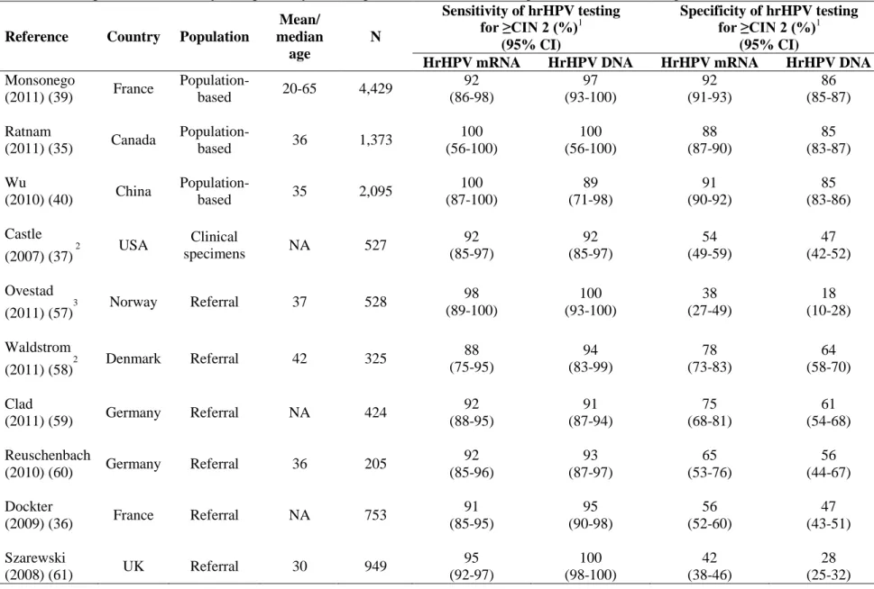
Three population-based studies, from France (39), Canada (35) and China (40), have so far been conducted to compare the performance of hrHPV mRNA testing with that of hrHPV DNA testing (Table 2.1). In these studies, the hrHPV mRNA testing had comparably high sensitivity for the detection of ≥CIN 2 compared with hrHPV DNA testing. HrHPV mRNA testing also appeared to be generally more specific than hrHPV DNA testing for the detection of ≥CIN 2. Similar sensitivity and higher specificity of hrHPV mRNA testing compared with that of hrHPV DNA testing for ≥CIN 2 have also been observed in women referred for colposcopy.
These data demonstrate the feasibility of hrHPV E6/E7 mRNA testing as a valuable biomarker for detecting ≥CIN 2, comparing favorably against the more widely used hrHPV DNA testing and cytology. The potential of hrHPV mRNA testing should therefore be further explored in variable populations and settings. We are not aware of studies of hrHPV mRNA testing as a possible tool for cervical cancer screening in Africa.
Self-Collected Specimens for High-Risk HPV and Cervical Lesion Detection
8
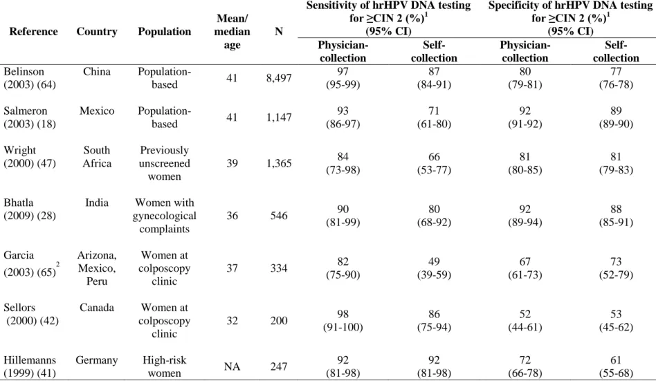
In previous studies, sensitivity of hrHPV testing of physician-collected specimens (hrHPV physician-testing) to detect ≥CIN 2 ranged from 82 to 97%, while that of self-collected specimens ranged from 49 to 92% (Table 2.2). Specificity ranged from 52 to 92% using physician-collected specimens, and 53-89% using self-collected specimens. In all previous studies, sensitivity of hrHPV physician-testing for ≥CIN 2 was generally higher than that of testing. Specificity, however, appeared similar in hrHPV physician- and self-testing across all studies.
Four previous studies have evaluated hrHPV physician- and self-testing (15, 45-47) in African populations. Only one of these studies, conducted on previously unscreened women from South Africa, compared hrHPV physician- and self-testing to detect ≥CIN 2 (47) (Table 2.2). In this study, the sensitivity of hrHPV self-testing for ≥CIN 2 was similar to that of conventional cytology (66% and 68%, respectively), but lower than that of physician-testing (84%). Specificity of hrHPV physician- and self-testing, as well as of conventional cytology for ≥CIN 2 were similar (83%, 85% and 88%, respectively).
Acceptability of self-collection has been surveyed (15, 28, 48, 49). In Rakai, Uganda, women favored self-collection performed during home visits over collection through a pelvic examination (>85% versus 50% acceptability) (15, 28). Self-collection was also generally accepted in Thailand and China, although some women had reservations about the safety of the device (48, 49).
Benefits and Cost Implications of Different Cervical Cancer Screening Strategies in Low-Resource Settings
9
require screening of large populations to generate a measurable effect. Also, these trials may not be able to adequately assess all types screening strategies (50). Simulation modeling can consider differences in population characteristics, test sensitivity and specificity, costs, and benefits of different tests or strategies. Results of modeling can potentially inform specific policy questions regarding the optimal cervical cancer prevention strategy for a particular setting, as well as the affordability of each setting for a screening strategy and follow-up re-screening or treatment (50). Relatively few studies have assessed re-screening costs and benefits of different cervical cancer screening strategies, including hrHPV-based testing, in low-resource countries (51-55), and to date, only one study, from South Africa, has evaluated screening costs and benefits of hrHPV self-testing in a low-resource setting (52).
10
frequent screening and more aggressive follow-up strategies. Previous studies also found that the decision among cytology, hrHPV-based testing, and visual inspection in low-resource settings will be most affected by the following factors: whether or not screening and
treatment can be accomplished in fewer visits, costs required for each test, and test sensitivity (50).
11
Table 2.1 Comparison of sensitivity and specificity of the high-risk HPV mRNA and high-risk HPV DNA testing to detect ≥CIN 2
Reference Country Population
Mean/ median
age
N
Sensitivity of hrHPV testing for ≥CIN 2 (%)1
(95% CI)
Specificity of hrHPV testing for ≥CIN 2 (%)1
(95% CI)
HrHPV mRNA HrHPV DNA HrHPV mRNA HrHPV DNA
Monsonego
(2011) (39) France
Population-
based 20-65 4,429
92 (86-98) 97 (93-100) 92 (91-93) 86 (85-87) Ratnam
(2011) (35) Canada
Population-based 36 1,373
100 (56-100) 100 (56-100) 88 (87-90) 85 (83-87) Wu
(2010) (40) China
Population-based 35 2,095
100 (87-100) 89 (71-98) 91 (90-92) 85 (83-86) Castle (2007) (37)
2 USA Clinical
specimens NA 527
92 (85-97) 92 (85-97) 54 (49-59) 47 (42-52) Ovestad
(2011) (57)3 Norway Referral 37 528
98 (89-100) 100 (93-100) 38 (27-49) 18 (10-28) Waldstrom
(2011) (58)2 Denmark Referral 42 325
88 (75-95) 94 (83-99) 78 (73-83) 64 (58-70) Clad
(2011) (59) Germany Referral NA 424
92 (88-95) 91 (87-94) 75 (68-81) 61 (54-68) Reuschenbach
(2010) (60) Germany Referral 36 205
92 (85-96) 93 (87-97) 65 (53-76) 56 (44-67) Dockter
(2009) (36) France Referral NA 753
91 (85-95) 95 (90-98) 56 (52-60) 47 (43-51) Szarewski
(2008) (61) UK Referral 30 949
12 Table 2.1 continued
HrHPV: high-risk human papillomavirus; ≥CIN 2: cervical intraepithelial neoplasia 2 or more severe; CI: confidence interval; NA: not available 1
Unless otherwise indicated, hrHPV mRNA testing was performed using the APTIMA HPV Assay (Hologic Gen-Probe, CA), which detects 14 hrHPV types (16,18, 31, 33, 35, 39, 45, 51, 52, 56, 58, 59, 66 and 68) (36), and hrHPV DNA testing was performed using the Hybrid Capture 2 High-Risk HPV DNA Test (Qiagen, CA), which detects 13 hrHPV types (16, 18, 31, 33, 35, 39, 45, 51, 52, 56, 58, 59 and 68) (62)
2HrHPV DNA testing was performed using the Linear Array HPV Genotyping Test (Roche Diagnostics, IN), which detects 13 hrHPV types (16, 18, 31, 33, 35,
39, 45, 51, 52, 56, 58, 59 and 68) and 24 low-risk HPV types (6, 11, 26, 40, 42, 53, 54, 55, 61, 62, 64, 66, 67, 69, 70, 71, 72, 73,81, 82, 83, 84, IS39 and CP6108) (63)
3HrHPV DNA testing was performed using the Amplicor Human Papillomavirus Test (Roche Diagnostics, IN), which detects 13 hrHPV types (16, 18, 31, 33,
35, 39, 45, 51, 52, 56, 58, 59 and 68) (63)
13
Table 2.2 Comparison of sensitivity and specificity high-risk HPV DNA testing of physician- and self-collected specimens to detect ≥CIN 2
Reference Country Population
Mean/ median
age
N
Sensitivity of hrHPV DNA testing for ≥CIN 2 (%)1
(95% CI)
Specificity of hrHPV DNA testing for ≥CIN 2 (%)1
(95% CI) Physician-collection Self- collection Physician-collection Self- collection Belinson (2003) (64)
China
Population-based 41 8,497
97 (95-99) 87 (84-91) 80 (79-81) 77 (76-78) Salmeron (2003) (18)
Mexico
Population-based 41 1,147
93 (86-97) 71 (61-80) 92 (91-92) 89 (89-90) Wright (2000) (47) South Africa Previously unscreened women
39 1,365 84
(73-98) 66 (53-77) 81 (80-85) 81 (79-83) Bhatla (2009) (28)
India Women with gynecological
complaints
36 546 90
(81-99) 80 (68-92) 92 (89-94) 88 (85-91) Garcia (2003) (65)2
Arizona, Mexico, Peru Women at colposcopy clinic
37 334 82
(75-90) 49 (39-59) 67 (61-73) 73 (52-79) Sellors (2000) (42)
Canada Women at colposcopy
clinic
32 200 98
(91-100) 86 (75-94) 52 (44-61) 53 (45-62) Hillemanns (1999) (41)
Germany High-risk
women NA 247
92 (81-98) 92 (81-98) 72 (66-78) 61 (55-68)
HrHPV: high-risk human papillomavirus; ≥CIN 2: cervical intraepithelial neoplasia 2 or more severe; CI: confidence interval
1Unless otherwise indicated, hrHPV DNA testing was performed using the Hybrid Capture 2 High-Risk HPV DNA Test (Qiagen, CA), which detects 13 hrHPV
types (16, 18, 31, 33, 35, 39, 45, 51, 52, 56, 58, 59 and 68) (62)
2HPV DNA testing performed by polymerase chain reaction (PCR) amplification using the PGMY 09/11 L1 consensus primer system, followed by detection of
27 HPV types (6, 11, 16, 18, 26, 31, 33, 35, 39, 40, 42, 45, 51-59, 66, 68, 73, 82, 83 and 84) (65)
CHAPTER 3. METHODS Study design
Study population and recruitment process
Between August 2009 and March 2011, FSW attending the Korogocho clinic for sexually transmitted diseases in Nairobi were invited to participate in a cervical cancer screening study. The clinic, jointly managed by the University of Nairobi, provides medical care including free cervical cancer screening and treatment for sexually transmitted
infections (STIs) for FSW in the Korogocho slum area. The clinic also provides counseling on the risks of commercial sex work and ways by which these risks may be reduced.
Women were informed of the study by community peer leaders during “baraza” public meetings. Potential participants were advised that participation was completely
voluntary, and that their care at the clinic would not be affected should they choose to decline participation. Participants must be between 18-50 years of age. Women were not eligible if they had undergone hysterectomy or were in the second trimester of pregnancy or later. A total of 350 FSW aged 18-49 years provided written informed consent and were subsequently enrolled.
Specimen collection and processing
At the clinic during screening, participating women were administered a
15
each participant in private self-collected a cervico-vaginal specimen for hrHPV mRNA testing, using the APTIMA Cervical Specimen Collection and Transport cytobrush (Hologic Gen-Probe, San Diego, CA) according to pictorial instructions. The cytobrush was then swirled in the APTIMA specimen transport medium (STM) and discarded. The participant then underwent a pelvic examination, during which a physician collected one cervical sample for hrHPV mRNA testing, using a Cervex-Brush(Rovers Medical Devices, The
Netherlands), which was then swirled in the PreservCyt medium (Hologic Gen-Probe, San Diego, CA) and discarded. The physician collected a second cervical sample to conduct a conventional Pap smear. The physician- and self-collected specimens were stored at -20ºC until their transport to Hologic Gen-Probe in San Diego for hrHPV mRNA and STI testing. Blood samples were also drawn from each participant for HIV testing and CD4 count assessment in the University of Nairobi.
HrHPV mRNA testing
HrHPV mRNA testing of physician- and self-collected specimens was conducted using the APTIMA HPV Assay (Hologic Gen-Probe, San Diego, CA) by Hologic Gen-Probe in San Diego, according to manufacturer’s instructions, without knowledge of the Pap smear or other study results. The APTIMA HPV Assay is a qualitative nucleic acid amplification test which detects E6/E7 mRNA of 14 hrHPV types (16, 18, 31, 33, 35, 39, 45, 51, 52, 56, 58, 59, 66, 68), but does not identify the HPV type. The assay involves three main steps, all taking place in a single tube. First, target mRNA capture by target-specific oligomers linked to magnetic microparticles; second, amplification of the isolated target mRNA using
16
by chemiluminescent-labeled probed in the Hybridization Protection Assay (HPA). The hybridized probe signals are then measured in relative light units (RLU). An internal control added to all samples at the target capture step minimizes the risk of false-negative results (66).
Before specimen testing, physician-collected specimens in PreservCyt solution were transferred to a tube containing APTIMA STM (Self-collected specimens were already in APTIMA STM). The APTIMA STM lyses the cells, releasing mRNA, and protects them from degradation. When the APTIMA HPV Assay is performed, the Assay captures only single-stranded nucleic acids to ensure that only hrHPV mRNA, but not DNA, is detected. The sequence-specific regions of the capture oligomers hybridizes to specific regions of the target hrHPV mRNA. The capture oligomer-hrHPV mRNA complex is then captured out of solution by lowering the temperature of the reaction to room temperature. The capture oligomer also contains a string of deoxyadenosine residue. The lowering of the temperature allows hybridization between the deoxyadenosine region on the capture oligomer and the poly-deoxythymidine molecules attached to magnetic particles. The captured target hrHPV mRNA bound to the magnetic particles are pulled to the side of the reaction tube by magnets. The supernatant is aspirated, and the particles are washed to remove residual specimen that potentially act as amplification inhibitors (66).
17
contained a promoter sequence for T7 RNA polymerase. From the DNA copy template, T7 RNA polymerase generates multiple copies of hrHPV mRNA amplicon (66).
In the third step, the hrHPV mRNA amplicon are detected by HPA, using single-stranded nucleic acid probes complementary to the amplicon, and which contain
chemiluminescent labels. These labeled probes hybridize to the amplicon. Hybridized probes are differentiated from unhybridized ones using the Selection Reagent, which inactivates the label on the unhybridized probes (66).
Light emitted from the labeled RNA-DNA hybrids is measured as photon signals in a luminometer and reported as RLU. Results of the assay are interpreted based on the analyte signal-to-cutoff (S/CO). An internal control, added during the target capture, monitors the target capture, amplification and detection steps. The signal emitted by the internal control is distinguished in each step from the HPV signal by the different light emission kinetics from probes with different labels. The hrHPV mRNA amplicon is detected using probes with slower light emission (glower), while the internal control amplicon is detected using a probe with a more rapid light emission (flasher). HrHPV mRNA testing results are interpreted as positive, negative or invalid, as determined by the internal control RLU and the analyte S/CO (Table 3.1) (66).
18
inactivated HPV-negative and HPV-positive cultured cells at 25 cells per mL in buffered solution (66).
STI testing
The physician- and self-collected specimens were also tested for Chlamydia
trachomatis and Neisseria gonorrhoeae with the APTIMA Combo 2 assay, for Trichomonas vaginalis with the APTIMA TV assay and for Mycoplasma genitalium with the APTIMA research use only assay, using the same target capture, TMA and HPA steps as hrHPV mRNA detection. STI testing was also performed according to the manufacturer’s
instructions, without knowledge of the Pap smear or other study results. Serum was tested for HIV antibodies by enzyme-linked immunosorbent assay (ELISA), with positive results confirmed by a second ELISA. Peripheral blood CD4 cells were enumerated for HIV-seropositive women.
Cervical cytology
19
a colposcopy-directed biopsy. In the event of histological ≥CIN 2, women received standard care and treatment at Kenyatta National Hospital. Women who had <CIN 2 were considered disease negative for statistical analyses.
Statistical methods
For specific aim 1.1: Sensitivity and specificity hrHPV mRNA physician- and self-testing specimens for ≥HSIL
Of the 350 FSW recruited, 6 women were missing at least one hrHPV testing result (specimens missing for 1 woman, self-collected specimens from 5 women invalid for hrHPV mRNA testing) and were excluded from subsequent analyses, resulting in a final sample size of 344. Median unbiased estimates and their mid-P 95% confidence intervals (CI) were computed for sensitivity, specificity, positive predictive value (PPV) and negative predictive value (NPV) of hrHPV mRNA testing of physician- and self-collected specimens for the detection of ≥HSIL (67).
For specific aim 1.2: Risk factors for hrHPV positivity in physician- and self-collected specimens
20
For specific aim 2.1: Estimation of bounds of sensitivity and specificity of hrHPV testing for ≥CIN 2
Using data on sensitivity and specificity of hrHPV testing for ≥HSIL from our FSW study (70) and those of ≥HSIL for ≥CIN 2 from the South African study (47), we estimated sensitivity and specificity bounds of hrHPV physician- and self-testing in our study of FSW for ≥CIN 2 detection. The estimation is described in detail in the Appendix.
For specific aim 2.2 A. Decision model
We developed a decision model using TreeAge ProTM 2012 (TreeAge Software Inc., Williamstown, MA, USA) to estimate the potential efficiency of a once-in-a-lifetime cervical cancer screening among Kenyan FSW using three screening strategies: conventional
cytology, hrHPV physician- and self-testing (Figure 1). Screening efficiency was defined as the number of colposcopies required to detect one case of ≥CIN 2. This definition was not based on total screening cost, as we did not have the resources to estimate costs.
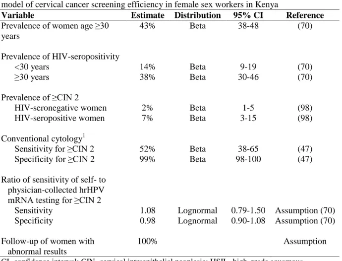
21 B. Model parameters
We obtained estimates of population age distribution, HIV prevalence and of the sensitivity and specificity of hrHPV physician- and self-testing for ≥HSIL detection from our FSW study in Kenya (70). We obtained estimates for sensitivity and specificity of ≥HSIL for ≥CIN 2 detection from published data on unscreened South African women (median age 39 years) (47). The prevalence of ≥HSIL was 4.1% (95% confidence interval, CI: 2.3-6.6%) in our FSW study (70) and 3.1% (95% CI: 2.3-4.1%) in the South African study (47).
Prevalence of ≥CIN 2 in the South African study was 4.1% (95% CI: 3.2-3%) (47) .
Using data on sensitivity and specificity of hrHPV testing for ≥HSIL from our FSW study (70) and those of ≥HSIL for ≥CIN 2 from the South African study (47), we estimated sensitivity and specificity bounds of hrHPV physician- and self-testing for ≥CIN 2 detection in our study of FSW. The estimation is described in detail in the Appendix.
We parameterized sensitivity as a function of specificity and of the diagnostic odds ratio (72) to take into account the inverse relationship between sensitivity and specificity. We used the ratio of sensitivity of hrHPV physician- to self-testing for ≥HSIL in our FSW study to reflect the difference in sensitivity of hrHPV physician- and self-testing for ≥CIN 2. The same was done to reflect the different in specificity. The performance of hrHPV physician- relative to self-testing for ≥HSIL in our study of FSW was consistent (higher sensitivity with physician- compared with self-testing, similar specificity) with that of previous studies comparing hrHPV DNA physician- and self-testing for ≥CIN 2 in resource-low settings (18, 28, 47, 64).
22
hrHPV mRNA testing of physician- and self-collected specimens for ≥CIN 2 were first sampled from uniform distributions. We specified the upper and lower limit of these uniform distributions with our estimated upper and lower bound for sensitivity and specificity. These sampled values were then ascribed a beta distribution, where the mean was the sampled value and the standard deviation was the mean of the approximate standard error of the lower and upper bound estimate. Parameter estimates and distributions used in our model are shown in Table 1.
Monte Carlo simulation
Monte-Carlo simulations were performed to calculate the probability of each screening strategy being optimal at different levels of willingness-to-pay. Probabilistic sensitivity analysis was first performed, by randomly drawing a value from each parameter distribution (screening test sensitivity and specificity), thereby allowing for the evaluation of the combined uncertainty about the parameters in the model (73). This procedure was
repeated 10,000 times ( =10,000).
For each set of parameters drawn, 1,000 observations ( =1,000) were generated to account for the random variation in individual-level outcomes (e.g. age, HIV-serostatus and whether or not a woman had ≥CIN 2) (74). For each of these trials of =1,000 observations, the number of colposcopies performed and the number of women whose ≥CIN 2 was
detected by colposcopy were estimated for each screening strategy, and the net benefit ( ) is calculated across a range of willingness-to-pay limit.
23
(where is the total cost and the total effect of each option), and is calculated across a range of the decision-maker’s willingness to pay for one unit gain in health outcome,
. An option is cost-effective if and only if the for a given > 1 (75). In assessing the of different screening strategies for our FSW study, represents the number of
colposcopies performed the number of women whose ≥CIN 2 was detected by colposcopy and the number of colposcopy the decision maker is willing to conduct to detect one case of ≥CIN 2.
For each trial of =1,000 observations, the screening strategy with the highest was identified. The proportion of the =10,000 interations in which a screening strategy had the highest at a given was then used to construct an acceptability curve for each
24
Table 3.1 Interpretation of APTIMA HPV Assay result for the detection of high-risk HPV mRNA (66) in female sex workers, Kenya
APTIMA HPV Assay result
Criteria Positive Analyte S/CO1 ≥0.05
Internal control ≤2,000,000 RLU Analyte ≤13,000,000 RLU Negative Analyte S/CO <0.05
Internal control ≤2,000,000 RLU
Internal control ≥ Internal control cutoff2
Invalid Analyte S/CO <0.05 and Internal control < Internal control cutoff OR Internal control >2,000,000 RLU
OR Analyte >13,000,000 RLU
HPV: human papillomavirus; S/CO: signal-to-cutoff; RLU: relative light unit 1
Analyte S/CO = analyte RLU/analyte cutoff, where analyte cutoff = (mean analyte RLU of the valid negative calibrator replicates) + (0.09 x mean analyte RLU of the valid positive calibrator replicates) 2
CHAPTER 4.
HIGH-RISK HUMAN PAPILLOMAVIRUS mRNA TESTING OF PHYSICIAN- AND SELF-COLLECTED SPECIMENS FOR CERVICAL LESIONS DETECTION IN
HIGH-RISK WOMEN, KENYA Overview
Little is known about the performance hrHPV physician- and self-testing or risk factors for hrHPV mRNA positivity in physician- versus self-collected specimens. We compared the performance of hrHPV mRNA physician- and self-testing to detect ≥HSIL and examined risk factors for hrHPV mRNA positivity in FSW in Nairobi.
From 2009-2011, 344 FSW participated in this cross-sectional study. Women self-collected a cervico-vaginal specimen. A physician conducted a pelvic examination to obtain a cervical specimen. Physician- and self-collected specimens were tested for hrHPV mRNA and sexually transmitted infections using APTIMA nucleic acid amplification assays. Cervical cytology was conducted using physician-collected specimens and classified according to the Bethesda criteria.
26
positivity in both physician- and self-collected specimens appeared higher in women who were younger (<30 years), had Trichomonas vaginalis or Mycoplasma genitalium infections, or had >8 years of educational attainment.
Self-collected specimens for hrHPV mRNA testing appeared to have similar sensitivity and specificity as physician-collected specimens for the detection of ≥HSIL among high-risk women.
Introduction
Successful implementation of Papanicolaou (Pap) smear screening programs has drastically reduced ICC incidence and mortality in developed countries (11, 13). However, Pap screening programs have been difficult to implement in low-resource settings due to limited infrastructure and access to trained cytopathologists and clinicians (30).
Consequently, a region with low screening coverage such as Eastern Africa still has among the highest estimated annual incidence of ICC in the world (34/100,000) (1).
27
acceptability of self-collection has generally been positive in various geographical settings worldwide (77).
Very few studies have evaluated hrHPV self-testing testing for cervical cancer
screening in low-resource settings and, to date, all have used HPV DNA testing (28, 47, 78). Recently developed diagnostic testing allows for the detection of hrHPV mRNA, which may be a more specific marker than hrHPV DNA for clinically significant disease (39), and has not yet been implemented in a high-risk, low-resource setting.
We present here results comparing the performance of hrHPV mRNA physician- and self-testing to detect high-grade cervical lesions in high-risk FSW in Kenya. We also
examined risk factors for hrHPV mRNA positivity in our population of FSW.
Materials and Methods Study population
From August 2009 to March 2011, FSW attending the Korogocho clinic in Nairobi, Kenya were invited to participate in this study to compare the performance of physician- and self-collected specimens for cervical cancer screening with hrHPV mRNA testing. The clinic provides counseling and medical care including screening and treatment for cervical cancer as well as STIs for FSW in the Korogocho slum area.
Women were informed of the study by community peer leaders during “baraza” public meetings. Women were not eligible if they had undergone hysterectomy or were in the second trimester of pregnancy or later. A total of 350 FSW aged 18-49 years provided
28
At screening, participating women were administered a questionnaire to collect sociodemographic, reproductive, and sexual behavior data. Of the 350 FSW recruited, 6 women were missing hrHPV mRNA testing results and were excluded from subsequent analyses, resulting in a final sample size of 344.
Sample collection and laboratory analyses
Each woman self-collected a cervico-vaginal specimen using the APTIMA Cervical Specimen Collection and Transport cytobrush (Hologic Gen-Probe Incorporated, San Diego, CA) according to pictorial instructions. The cytobrush was then swirled in the APTIMA specimen transport medium and then discarded. During a pelvic examination, the physician collected one cervical sample from each woman using a Cervex-Brush (Rovers Medical Devices, The Netherlands), which was then swirled in the PreservCyt (Hologic Gen-Probe Incorporated, San Diego, CA) medium and then discarded. The physician then collected a second cervical sample to conduct a conventional Pap smear.
Cytological smears were evaluated at the University of Nairobi and classified
29
treatment at Kenyatta National Hospital. Women who had <CIN 2 were considered disease negative for statistical analyses.
HPV and STI testing
The physician- and self-collected specimens were transported to Hologic Gen-Probe in San Diego for HPV and STI testing. Laboratory testing for HPV in our study was by the APTIMAHPV Assay (Hologic Gen-Probe Incorporated, San Diego, CA) which qualitatively detects E6/E7 mRNA of 14 hrHPV types (16, 18, 31, 33, 35, 39, 45, 51, 52, 56, 58, 59, 66, 68). The hrHPV mRNA assay comprises three main steps, namely target capture,
transcription-mediated amplification (TMA) of the target, and finally target detection by hybridization with complementary probes linked to chemiluminescent labels (66).
The physician- and self-collected specimens were also tested for Chlamydia
trachomatis and Neisseria gonorrhoeae with the APTIMA Combo 2 assay, for Trichomonas vaginalis with the APTIMA TV assay and for Mycoplasma genitalium with the APTIMA research use only assay, using the same target capture, TMA and hybridization steps as hrHPV mRNA detection. All assays were performed according to the manufacturer’s instructions, without knowledge of the Pap smear or other study results.
30 Statistical analyses
Agreement between hrHPV positivity in physician- and self-collected specimens was measured by the kappa statistic. Median unbiased estimates and their mid-P 95% confidence intervals (CI) were computed for sensitivity, specificity, positive predictive value (PPV) and negative predictive value (NPV) of hrHPV testing of physician- and self-collected specimens for the detection of ≥HSIL (67). Potential risk factors for hrHPV positivity in physician- and self-collected specimens were determined, and directed acyclic graphs (DAG) (68) were analyzed with online software (69) to identify minimally sufficient sets of adjustment
variables to reduce confounding in binomial regression estimates of the prevalence difference of hrHPV positivity between categories of potential risk factors. All statistical analyses were performed using SAS 9.2.
Results
Participant characteristics
Overall prevalence of hrHPV was similar in physician- (30%) and self-collected specimens (29%) (Table 4.1). HrHPV prevalence in both physician- and self-collected specimens was slightly higher in women <30 years than in older women. Prevalence of any abnormal cytology (≥ASCUS) in the population was 19%, and was similar in women ≥30 years (21%) than in younger women (17%).
Performance of hrHPV mRNA testing of physician- and self-collected specimens
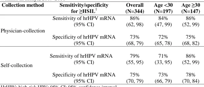
31
that of self-testing (N=13 versus N=12) (Table 4.3). This ≥HSIL case, which was negative by hrHPV self-testing, was also disease negative by histology. Overall specificity for ≥HSIL appeared similar in both hrHPV physician- and self-testing (Table 4.2).
The NPV for hrHPV mRNA testing of physician- and self-collected specimens was 97-99% overall and in both age groups. The PPV was 12-13% overall, and varied by age group, being somewhat lower in women <30 years (9-10%) than in older women (17-18%). The agreement between hrHPV physician- and self-testing was =59% (95% CI: 49-68) overall, =76% (95% CI: 32-100) in women with ≥HSIL and =55% (95% CI: 45-65) in women with <HSIL.
Of the 15 women with ≥HSIL, 14 underwent colposcopy-directed biopsy. One
woman could not be traced and was lost to follow-up. Twelve had histological ≥CIN 2, and 2 were considered disease negative (both had normal histology). Of the 12 ≥CIN 2 cases, 10 were hrHPV positive in both physician- and self-collected specimens. The remaining 2 ≥CIN 2 cases were hrHPV negative in both physician- and self-collected specimens.
Risk factors for hrHPV mRNA positivity in physician- and self-collected specimens with normal cervical cytology
32
The 95% confidence limit differences (difference between upper and lower 95% confidence limits, CLD) (79) for the APDs were generally between 0.19 and 0.25 (Table 4.4). The least precise APD estimates (95% CLD >0.25) in both physician- and self-collected specimens were those relating to HIV-seropositivity, C.trachomatis/ N.
gonorrhoeae, T. vaginalis and M. genitalium positivity, to the number of regular sexual partners, and to frequency of condom use with regular sexual partners.
Discussion
Physician- and self-collection for hrHPV testing demonstrated a high sensitivity for the detection of ≥HSIL. HrHPV self-testing in our population of FSW with high hrHPV prevalence appeared to have similar sensitivity and specificity for ≥HSIL as that of hrHPV physician-testing. We also found that prevalence of hrHPV positivity in both physician- and self-collected specimens was somewhat higher in women who were <30 years, had T. vaginalis or M. genitalium infection, or had higher educational attainment.
The prevalence of hrHPV positivity in our study (30%) was lower than that of hrHPV DNA positivity in another FSW population in Mombasa (56%) (9) which had higher HIV prevalence than in our study. HrHPV positivity in our study was also lower than hrHPV DNA positivity in a similar FSW cohort in Nairobi (54%) which had a lower median age (23 years) than our study (80). Compared with non-FSW populations in Africa, hrHPV positivity in our study was higher than hrHPV DNA positivity in studies which had a higher median age (47) or lower HIV prevalence (15) than in our FSW study.
33
physician- and self-collected specimens for ≥CIN 2 (41). However, the small number of women with ≥HSIL (N=15) in our study made comparing sensitivity estimates of hrHPV physician- and self-testing in our population of high-risk FSW somewhat difficult. Previous studies using HC2 hrHPV DNA testing in Africa (47) and in other settings (28, 78) have also found higher overall sensitivity for ≥CIN 2 using physician- compared with self-collected specimens. Our finding of similar overall specificity of hrHPV testing for ≥HSIL when using physician- or self-collected specimens was also consistent with previous studies that used HC2 testing (28, 47, 78).
Prevalence of ≥HSIL in our study (4%) was similar to what was found in other African studies (9, 47). We found that prevalence of ≥HSIL in women <30 years in our study was similar to that in women ≥30 years (4% versus 5%) and was thus not notably different in this group of high-risk FSW in Kenya. The comparability of ≥HSIL detection by hrHPV physician- and testing in women <30 years in our study (Table 4.3) suggests that self-collected specimens for hrHPV testing could also be a viable option for cervical cancer screening in high-risk FSW populations 18-29 years of age in Kenya.
HrHPV self-testing in our study also detected as many ≥CIN 2 cases as did that of hrHPV physician-testing. Our results suggest that in low-resource areas where Pap screening is not routinely available, hrHPV self-testing can be used to identify high-risk women at high risk of ≥CIN 2.
34
more precise hrHPV positivity estimates in women who had higher educational attainment was, on the other hand, in contrast to previous studies on FSW populations, where lower educational attainment strongly predicted hrHPV DNA positivity (81, 84).
Women with M. genitalium or T. vaginalis infection also appeared to have higher APD of hrHPV mRNA positivity, although the estimates were imprecise (95% CLD range: 0.38 to 0.49). Nevertheless, higher hrHPV DNA positivity had been found in women with T. vaginalis infection in earlier studies (85, 86). Data on M. genitalium infection as a risk factor for HPV infection are scarce. There was no evidence of increased hrHPV DNA positivity with M. genitalium infection in women with normal cervical cytology (87). On the other hand, among women with abnormal cytology, higher hrHPV DNA positivity was found in those who had a mycoplasma infection (88). Inconsistencies between our results and those of earlier studies could be due to differences in populations, methods of assessing and
categorizing variables, hrHPV test (DNA versus mRNA) and sampling variability. Our study has several advantages that improved the validity of the comparison of hrHPV physician- and self-testing. Firstly all cervical smears were independently read by two cytopathologists, followed by consensus of reviewing cytopathologists for discrepant cases to ensure accurate cytological diagnoses. Secondly, a woman’s physician- and self-collection of specimens were performed on the same day, enabling direct comparison of hrHPV mRNA testing results of these two sample types.
.One limitation of our study was that histological results were obtained only for
35
potential for verification bias (89). Although cytological ≥HSIL and LSIL often correspond to the histologic diagnoses of, respectively, ≥CIN 2 and CIN 1, previous studies estimated that at least 70% of ≥HSIL cases and up to 30% of LSIL cases have ≥CIN 2 (90).
In terms of public health ramifications, our results of the performance of self- versus physician-collected specimens for hrHPV testing were from a high-risk population of FSW in a resource poor setting, and therefore not necessarily generalizable to lower risk populations. Also, one feasibility concern of using self-collected specimens for HPV testing in primary screening is that a woman who tested HPV positive may not return for follow-up screening or treatment (47). Other commonly reported issues include difficulty in using the brush for self-collection or in understanding the instructions, and contamination of the self-collection brush (77). Despite potential limitations, findings from a meta-analysis (44) supported an increased usage of self-collection in epidemiological studies. Future research should address if self-collection will improve screening coverage in underserved women in low-resource settings.
36
Table 4.1 Sociodemographic and sexual behavior characteristics of 344 female sex workers in Kenya, 2009-2011
Characteristic Overall (N=344) Age <30 (N=197) Age ≥30 (N=147) n median or
%
median or % median or % Age (years) (range) 28 (18-49) 25 (18-29) 35 (30-49) HrHPV mRNA (physician-collection)
Negative 241 70.1 69.0 71.4
Positive 103 29.9 31.0 28.6
HrHPV mRNA (self-collection)
Negative 246 71.5 70.0 73.5
Positive 98 28.5 30.0 26.5
Cervical cytology
Normal 279 81.1 82.7 79.0
ASCUSa 14 4.1 3.0 5.4
LSIL 36 10.5 10.7 10.2
≥HSIL 15 4.3 3.6 5.4
HIVb
Seronegative 259 76.0 86.3 61.8
Seropositive 82 24.0 13.7 38.2
CD4 count/mm3c (range) 478 (152-1391) 476 (160-1269) 492 (152-1391) Sexually transmitted infections
Chlamydia 13 3.8 6.1 0.7
Gonorrhea 8 2.3 3.0 1.4
Trichomonas vaginalis 25 7.3 6.1 8.8
Mycoplasma genitalium 44 12.8 15.7 8.8
Education (years) (range) 8 (0-16) 8 (0-16) 8 (0-15) Marital statusb
Single/never married 150 43.7 54.6 29.2
Divorced/widowed/separated 190 55.4 44.4 70.1
Married/cohabitating 3 0.9 1.0 0.7
37 Number of sexual clients per week
(range) 10 (2-40) 10 (2-40) 10 (2-40) Number of regular sexual partners
(range) 1 (0-10) 1 (0-10) 1 (0-7) Condom use with sexual clientsb
Most of the time/always 253 73.8 76.0 73.7
Sometimes/half the time 70 20.4 19.4 21.8
Never/rarely 20 5.8 4.6 7.5
Condom use with regular sexual partnersd
Most of the time/always 60 24.6 19.5 32.6
Sometimes/half the time 21 8.6 10.7 5.3
Never/rarely 163 66.8 69.8 62.1
Charge per transaction (Ksh) (range)e 200 (50-5,000) 250 (50-5,000) 200 (50-1,500) HrHPV: High-risk HPV; ASCUS: atypical squamous cells of undetermined significance; LSIL: low-grade squamous intraepithelial lesion; HSIL: high-low-grade squamous intraepithelial lesion; Ksh: Kenyan shillings
a
Includes atypical glandular cells of undetermined significance (AGUS) (n=2) b
Numbers do not add up to total due to missing values: HIV serostatus (n=3); marital status (n=1); condom use with sexual clients (n=1)
c
Among HIV seropositive women (n=82) d
Among women with regular sexual partners only (n=244) e
38
Table 4.2 Performance of high-risk HPV mRNA testing of physician- and self-collected specimens for the detection of cytological high-grade cervical lesions in 344 female sex workers in Kenya, 2009-2011
Collection method Sensitivity/specificity
for ≥HSIL1 Overall
(N=344) Age <30 (N=197) Age ≥30 (N=147) Physician-collection
Sensitivity of hrHPV mRNA (95% CI) 86% (62, 98) 84% (47, 99) 86% (52, 99) Specificity of hrHPV mRNA
(95% CI) 73% (68, 79) 72% (65, 78) 75% (68, 82) Self-collection
Sensitivity of hrHPV mRNA (95% CI) 79% (55, 95) 71% (33, 95) 86% (52, 99) Specificity of hrHPV mRNA
(95% CI) 75% (70, 79) 73% (66, 79) 78% (70, 84) HrHPV: high-risk HPV; 95% CI: 95% confidence interval
1
39
Table 4.3 High-risk HPV mRNA testing results of physician- and self-collected specimens stratified by age and cytology in 344 female sex workers in Kenya, 2009-2011
Cytology
HrHPV mRNA test result
N (hrHPV+ in physician-collection, hrHPV+ in self-collection)
Overall Age <30 Age ≥30
Normal 279 (68, 61) 163 (44, 41) 116 (24, 20)
ASCUSa 14 (8, 10) 6 (3, 4) 8 (5, 6)
LSIL 36 (14, 15) 21 (8, 9) 15 (6, 6)
≥HSILb
15 (13, 12) 7 (6, 5) 8 (7, 7)
Total 344 (103, 98) 197 (61, 59) 147 (42, 39)
HrHPV: High-risk HPV; ASCUS: atypical squamous cells of undetermined significance; LSIL: low-grade squamous intraepithelial lesion; HSIL: high-low-grade squamous intraepithelial lesion
a
Includes atypical glandular cells of undetermined significance (AGUS) (n=2) b
40
Table 4.4 Association of potential risk factors with hrHPV mRNA positivity among 279 female sex workers with normal cytology in Kenya, 2009-2011
Risk factors
Normal (N=279)a
HrHPV mRNA positivity (physician-collection)
(N=68)
HrHPV mRNA positivity (self-collection)
(N=61)
n n CP APD (95% CI) b n CP APD (95% CI) b
Age (years)
≥30 116 24 0.21 0 20 0.17 0
<30 163 44 0.27 0.06 (-0.04,0.16) 41 0.25 0.08 (-0.02,0.17)
HIVc
Seronegative 227 53 0.23 0 46 0.20 0
Seropositive 51 15 0.29 0.01 (-0.17,0.20) 15 0.29 0.11 (-0.08,0.29)
STId
Chlamydia/gonorrhea
Negative 261 60 0.23 0 55 0.21 0
Positive 18 8 0.44 0.01 (-0.29,0.28) 6 0.33 0.10 (-0.18,0.37)
Trichomonas vaginalis
Negative 259 60 0.23 0 56 0.22 0
Positive 20 8 0.40 0.19 (-0.06,0.43) 5 0.25 0.09 (-0.12,0.31)
Mycoplasma genitalium
Negative 244 57 0.23 0 51 0.21 0
Positive 35 11 0.31 0.09 (-0.11,0.29) 10 0.29 0.06 (-0.13,0.25)
Education (years) e
≤8 208 47 0.23 0 41 0.20 0
>8 71 21 0.30 0.06 (-0.06,0.19) 20 0.28 0.09 (-0.03,0.20)
Marital statusf
Single/never married 122 37 0.30 0 31 0.25 0
Divorced/widowed/separated 156 31 0.20 -0.09 (-0.20,0.01) 30 0.19 -0.03 (-0.14,0.08)
41 Age at first sexual intercourse
(years)g
≥16 174 44 0.25 0 39 0.22 0
<16 105 24 0.23 -0.02 (-0.13,0.08) 22 0.21 -0.02 (-0.11,0.08)
Number of sexual clients per week e
≤10 158 40 0.25 0 34 0.21 0
>10 121 28 0.23 -0.01 (-0.11,0.09) 27 0.22 0.04 (-0.05,0.14)
Number of regular sexual partnersh
≤1 227 54 0.24 0 47 0.21 0
>1 52 14 0.27 0.02 (-0.11,0.15) 14 0.27 0.05 (-0.08,0.18)
Condom use with sexual clientsi
≥Most of the time 202 47 0.23 0 44 0.21 0
<Most of the time 76 21 0.28 0.06 (-0.06,0.17) 17 0.22 0.01 (-0.09,0.12) Condom use with regular sexual
partnersj
≥Most of the time 43 9 0.21 0 10 0.23 0
<Most of the time 155 42 0.27 0.12 (-0.02,0.28) 36 0.23 0.05 (-0.10,0.19) Charge per transaction (Ksh) e
≤200 160 35 0.22 0 29 0.18 0
>200 119 33 0.28 0.06 (-0.06,0.16) 32 0.27 0.05 (-0.06,0.16)
HrHPV: high-risk HPV; STI: sexually transmitted infection; Ksh: Kenyan shillings; CP: crude prevalence; AP: adjusted prevalence; APD: adjusted prevalence difference; CI: confidence interval
a
Analyses restricted to women with normal cervical cytology and valid specimens for hrHPV mRNA testing (n=279) b
Each minimally sufficient adjustment set identified by directed acyclic graph (DAG) c
One woman missing HIV serostatus; estimates adjusted for age, STI, average number of sexual clients per week, number of regular sexual partners, condom use with sexual clients and condom use with regular sexual partners
42 Table 4.4 continued
d
Estimates adjusted for age, other STI, average number of sexual clients per week, number of regular sexual partners, condom use with sexual clients and condom use with regular sexual partners
e
Estimates adjusted for age f
One woman missing marital status; the three women presently married were categorized under divorced/widowed/separated; estimates adjusted for age
g
Estimates adjusted for age and education h
Estimates adjusted for age and marital status i
One woman missing data on condom use with sexual clients; estimates adjusted for age, HIV serostatus, STI, education, marital status jAmong women with ≥1 regular sexual partners only (n=198); estimates adjusted for age, HIV serostatus, STI, education, marital status
CHAPTER 5.
IMPACT OF UNCERTAINTY IN RELATIVE TEST PERFORMANCE BETWEEN CYTOLOGY, PHYSICIAN- AND SELF-COLLECTED HIGH-RISK HPV TESTING ON
ESTIMATED SCREENING EFFICIENCY IN KENYA: A SIMULATION Overview
The costs and benefits of each cervical cancer screening strategy must be considered to determine the optimal screening strategy for a low-resource setting. We estimated the potential efficiency (measured as colposcopies required per ≥CIN 2 detected) of a once-in-a-lifetime cervical cancer screening among female sex workers (FSW) in Kenya using three strategies: conventional cytology, high-risk (hr) HPV physician- and self-testing.
We estimated bounds of sensitivity and specificity of hrHPV physician- and self-testing for ≥CIN 2 from our study of FSW in Kenya and from published South African data. We constructed a decision model of FSW in Kenya, and performed probabilistic sensitivity analyses to identify the proportion of simulations where a given screening strategy was optimal at a given “willingness-to-pay” (number of colposcopies willing to conduct to detect a case of ≥CIN2) limit.
The estimated sensitivity bounds for ≥CIN 2 of hrHPV physician-testing
44
all three strategies being optimal was ≤50%. Above 40 colposcopies per ≥CIN 2 detected, the probability of conventional cytology being optimal decreased to 10-20%.
At a willingness-to-pay limit of <15 colposcopies per ≥CIN 2 detected, conventional cytology was the optimal screening strategy, given the available information. At a higher willingness-to-pay limits, the probability that hrHPV testing being optimal exceeded that of conventional cytology. However, due to relative imprecision of the sensitivity and specificity of hrHPV testing for ≥CIN 2, more data (e.g extending the model to include costs and
estimated impact of each screening strategy on cervical cancer mortality) is likely required to determine which screening strategy is most efficient at higher willingness-to-pay limits.
Introduction
45
Identification of the most efficient screening strategy requires comparison of the impact of different screening tests’ sensitivity and specificity for ≥CIN 2 screening benefits and costs (74, 93). Here we compare the screening efficiency of three cervical cancer screening strategies, as well as the uncertainty surrounding the choice among them, in a female sex worker study from Kenya (70). We define screening efficiency as the number of colposcopies required to detect a case of ≥CIN 2. Screening strategies considered are conventional cytology, hrHPV testing of collected specimens (hrHPV physician-test) and that of self-collected specimens (hrHPV self-physician-test).
We first estimate bounds of sensitivity and specificity of hrHPV physician- and self-testing for ≥CIN 2 detection from our study of female sex workers (FSW) (70) and other published data from South Africa (47). We then identify the optimal screening strategy, in terms of screening efficiency at a given “willingness-to-pay” (number of colposcopies willing to conduct to detect one ≥CIN 2) limit, for a once-in-a-lifetime cervical cancer screening in a female sex worker population in Kenya.
Materials and Methods Study population
46
each woman were collected by a physician for hrHPV mRNA testing and for conventional cytology. Women with ≥HSIL were immediately referred to colposcopy and directed-biopsy upon indication. The physician- and self-collected specimens were tested for hrHPV mRNA by the APTIMAAssay (Hologic Gen-Probe Incorporated, San Diego, CA).
Decision model
We developed a decision model using TreeAge ProTM 2012 (TreeAge Software Inc., Williamstown, MA, USA) to estimate the potential efficiency of a once-in-a-lifetime cervical cancer screening among Kenyan FSW using three screening strategies: conventional
cytology, hrHPV physician- and self-testing (Figure 5.1). Screening efficiency was defined as the number of colposcopies required to detect one case of ≥CIN 2. This definition was not based on total screening cost, as we did not have the resources to estimate costs.
Furthermore, recommendations of screening strategy need not necessarily be based on cost-effectiveness findings (71). Colposcopy referrals were used as a surrogate for screening program cost in our analysis. We also assume that the cost of each screening test is approximately equivalent. We constructed a decision model by which women who had ≥HSIL at cytological screening or who tested hrHPV mRNA positive in either the physician- or self-collected specimens were referred to colposcopy. We modeled whether a woman with or without ≥CIN 2 would be referred for colposcopy, under each screening strategy.
Model parameters
47
FSW study in Kenya (70). We obtained estimates for sensitivity and specificity of ≥HSIL for ≥CIN 2 detection from published data on unscreened South African women (median age 39 years) (47). The prevalence of ≥HSIL was 4.1% (95% confidence interval, CI: 2.3-6.6%) in our FSW study (70) and 3.1% (95% CI: 2.3-4.1%) in the South African study (47).
Prevalence of ≥CIN 2 in the South African study was 4.1% (95% CI: 3.2-5.3%) (47) . Using data of i) hrHPV testing to detect ≥HSIL and ii) ≥CIN 2 diagnosis in women with ≥HSIL from our FSW study (70), as well as iii) cytological testing (≥HSIL) to detect ≥CIN 2 from the South African study (47), we estimated sensitivity and specificity bounds of hrHPV physician- and self-testing for ≥CIN 2 detection in our study of FSW. The estimation is described in detail in the Appendix.
We parameterized sensitivity as a function of specificity and of the diagnostic odds ratio (72) to take into account the inverse relationship between sensitivity and specificity. We used the ratio of sensitivity of hrHPV physician- to self-testing for ≥HSIL in our FSW study to reflect the difference in sensitivity of hrHPV physician- and self-testing for ≥CIN 2. The same was done to reflect the different in specificity. The performance of hrHPV physician- relative to self-testing for ≥HSIL in our study of FSW was consistent (higher sensitivity with physician- compared with self-testing, similar specificity) with that of previous studies comparing hrHPV DNA physician- and self-testing for ≥CIN 2 in resource-low settings (18, 28, 47, 64).
48
distributions with our estimated upper and lower bound for sensitivity and specificity. These sampled values were then ascribed a beta distribution, where the mean was the sampled value and the standard deviation was the mean of the approximate standard error of the lower and upper bound estimate. Parameter estimates and distributions used in our model are shown in Table 1.
Monte Carlo simulation
Monte-Carlo simulations were performed to calculate the probability of each screening strategy being optimal at different levels of willingness-to-pay. Probabilistic sensitivity analysis was first performed, by randomly drawing a value from each parameter distribution (screening test sensitivity and specificity), thereby allowing for the evaluation of the combined uncertainty about the parameters in the model (73). This procedure was
repeated 10,000 times ( =10,000).
For each set of parameters drawn, 1,000 observations ( =1,000) were generated to account for the random variation in individual-level outcomes (e.g. age, HIV-serostatus and whether or not a woman had ≥CIN 2) (74). For each of these trials of =1,000 observations, the number of colposcopies performed and the number of women whose ≥CIN 2 was
detected by colposcopy were estimated for each screening strategy, and the net benefit ( ) is calculated across a range of willingness-to-pay limit.
49
outcome, . An option is cost-effective if and only if the for a given > 1 (75). In assessing the of different screening strategies for our FSW study, represents the number of colposcopies performed, the number of women whose ≥CIN 2 was detected by colposcopy and the number of colposcopy the decision maker is willing to conduct to detect one case of ≥CIN 2.
For each trial of =1,000 observations, the screening strategy with the highest was identified. The proportion of the =10,000 interations in which a screening strategy had the highest at a given was then used to construct an acceptability curve for each
screening strategy by plotting these proportions on the -axis and the corresponding on the -axis (Figure 5.2) (75). Since we defined willingness-to-pay as the number of colposcopies willing to conduct to detect one ≥CIN 2, this is also equivalent to the inverse of the positive predictive value (PPV) for ≥CIN 2 among women with ≥HSIL or positive hrHPV physician- or self-testing result.
Results
Bounds of sensitivity and specificity of hrHPV testing for ≥CIN 2
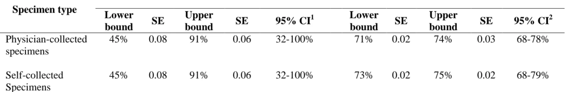
The estimated sensitivity bounds of hrHPV physician-testing for ≥CIN 2 (45-91%) in our study of FSW were similar to that of self-testing (45-9%). The estimated specificity bounds were also similar for physician- (71-74%) and self-testing (73-75%), and were much narrower than those of sensitivity (Table 5.2).
50
CLD of the 95% confidence limit for the true specificity was also similar for physician- and self-testing (CLD=0.10 for physician- and CLD=0.11 for self-collected specimens) (Table 5.2), and are lower than those for sensitivity.
Comparison of screening efficiency and associated uncertainty
Figure 5.2 shows an acceptability curve: the proportion of simulations that an
individual screening strategy was optimal (i.e, having the highest at a given willingness-to-pay limit. Given the available information, at a willingness-willingness-to-pay limit of <15
colposcopies per case of ≥CIN 2 detected, conventional cytology has >80% probability of being optimal. At a willingness-to-pay of between 20 and 30-40 colposcopies per case of ≥CIN 2 detected, the probability of all three strategies being optimal was between 8-50%. Above 40 colposcopies per case of ≥CIN 2 detected, the probability of conventional cytology being optimal decreased to between 10-20%. Also, above this willingness-to-pay limit, the probability of hrHPV physician-testing being optimal increased to between 55-60%, while that of hrHPV self-testing remained at between 25-30%.
Prevalence of ≥CIN 2 by age