CAP Laboratory Improvement Programs
Molecular Biomarkers for the Evaluation of Colorectal
Cancer
Guideline From the American Society for Clinical Pathology, College of American
Pathologists, Association for Molecular Pathology, and American Society of
Clinical Oncology
Antonia R. Sepulveda, MD, PhD,1Stanley R. Hamilton, MD,2Carmen J. Allegra, MD,5Wayne Grody, MD, PhD,6 Allison M. Cushman-Vokoun, MD, PhD,7William K. Funkhouser, MD, PhD,8Scott E. Kopetz, MD, PhD,3Christopher Lieu, MD,9 Noralane M. Lindor, MD,10Bruce D. Minsky, MD,4Federico A. Monzon, MD,11Daniel J. Sargent, PhD,12Veena M. Singh, MD,13
Joseph Willis, MD,14Jennifer Clark, SCT, MB(ASCP)cm,15Carol Colasacco, MLIS,16R. Bryan Rumble, MSc,17 Robyn Temple-Smolkin, PhD,18Christina B. Ventura, MT(ASCP),16and Jan A. Nowak, MD, PhD19
Objectives.—To develop evidence-based guideline rec-ommendations through a systematic review of the litera-ture to establish standard molecular biomarker testing of colorectal cancer (CRC) tissues to guide epidermal growth factor receptor (EGFR) therapies and conventional che-motherapy regimens.Methods.—The American Society for Clinical Pathology, College of American Pathologists, Association for Molec-ular Pathology, and American Society of Clinical Oncology convened an expert panel to develop an evidence-based guideline to establish standard molecular biomarker testing and guide therapies for patients with CRC. A comprehensive literature search that included more than 4,000 articles was conducted.
Results.—Twenty-one guideline statements were estab-lished.
Conclusions.—Evidence supports mutational testing for EGFR signaling pathway genes, since they provide clini-cally actionable information as negative predictors of benefit to anti-EGFR monoclonal antibody therapies for targeted therapy of CRC. Mutations in several of the biomarkers have clear prognostic value. Laboratory ap-proaches to operationalize CRC molecular testing are presented.
(Arch Pathol Lab Med.2017;141:625–657; doi: 10.5858/ arpa.2016-0554-CP)
M
olecular testing to select targeted and conventional therapies for patients with colorectal cancer (CRC) has been the focus of a number of recent studies and is becoming standard practice for management of patients with CRC. Molecular markers that predict response to a specific therapy or treatment regimen are known as predictive biomarkers.1Monoclonal antibody therapies that target the epidermal growth factor receptor (EGFR) bind theAccepted for publication November 11, 2016.
Published as an Early Online Release February 6, 2017.
Supplemental digital content is available for this article at archivhesofpathology.org in the May 2017 table of contents.
From the 1Department of Pathology and Cell Biology, Columbia
University, New York, NY; Departments of2Pathology,3Gastrointestinal
(GI) Medical Oncology, and4Radiation Oncology, University of Texas MD
Anderson Cancer Center, Houston; 5Division of Hematology and
Oncology, University of Florida Medical Center, Gainesville; 6
Depart-ments of Pathology and Laboratory Medicine, Pediatrics, and Human Genetics, UCLA Medical Center, Los Angeles, CA; 7Department of
Pathology and Microbiology, University of Nebraska Medical Center, Omaha;8Department of Pathology and Laboratory Medicine, University of
North Carolina School of Medicine, Chapel Hill;9Division of Medical
Oncology, University of Colorado Denver School of Medicine, Denver;
10Department of Medical Genetics, Mayo Clinic, Scottsdale, AZ;11Castle
Biosciences, Friendswood, TX;12Department of Health Sciences Research,
Mayo Clinic, Rochester, MN;13Biocept, San Diego, CA;14Department of
Pathology, Case Western Reserve University, Cleveland, OH; 15ASCP
Institute for Science, Technology, and Policy, American Society for Clinical Pathology, Washington, DC;16Laboratory and Pathology Quality Center,
College of American Pathologists, Northfield, IL;17American Society of
Clinical Oncology, Alexandria, VA;18Association for Molecular Pathology,
Bethesda, MD; and19Department of Pathology and Laboratory Medicine,
Roswell Park Cancer Institute, Buffalo, NY.
This guideline was developed through collaboration between the American Society for Clinical Pathology, College of American Pathologists, Association for Molecular Pathology, American Society of Clinical Oncology, and American Society for Investigative Pathology and has been jointly published by invitation and consent in theAmerican Journal of Clinical Pathology,Archives of Pathology & Laboratory Medicine, Journal of Molecular Diagnostics, and
Journal of Clinical Oncology. It has been edited in accordance with standards established at theAmerican Journal of Clinical Pathology. Copyright 2017 American Society for Clinical Pathology, College of American Pathologists, Association for Molecular Pathology, American Society of Clinical Oncology, and American Society for Investigative Pathology.
EGFR extracellular domain, blocking EGFR signaling pathways. Anti-EGFR monoclonal antibodies have been the main targeted therapies for CRC that require knowledge of the mutational status of genes in the pathway as predictive biomarkers of response to these therapies.2–4 Initial clinical trial data demonstrated that patients with CRC carrying activating mutations ofKRASaffecting exon 2 codons 12 and 13 did not benefit from anti-EGFR monoclonal antibody therapy.2–4 Subsequent studies de-scribed other mutations in genes of the EGFR signaling pathways involving other exons of KRAS and in NRAS,
BRAF,PIK3CA, andPTENthat may affect response of CRC to anti-EGFR antibody therapies. Guidelines addressing the molecular testing of EGFR pathway genes beyond KRAS
have not been established and are needed in clinical practice.
The DNA mismatch repair (MMR) status of CRC may have predictive value in some clinical settings. While testing of CRC for MMR has been recommended for all patients with CRC as a workup test to evaluate for possible Lynch syndrome,5 guidelines for the use of MMR as a predictive biomarker of response to therapy have not been reported. Recent molecular biomarker data have shown the impor-tance of microsatellite instability (MSI) testing, a marker of deficient mismatch repair (dMMR), for the selection of patients for immunotherapy (see section on emerging biomarkers below).
Alterations of a number of critical genes in CRC development and progression such as dMMR and BRAF
activating mutations have been shown to affect prognosis, as measured by several metrics of tumor progression or survival.6–8 The utility of incorporating prognostic bio-markers in the management of patients with CRC has not been well defined in clinical practice. Defining the utility of information gathered from prognostic molecular biomark-ers for clinical management of patients with CRC is warranted.
The postgenome era and the emphasis on precision genomic-based medicine are providing enormous amounts of new data and many promising new molecular cancer biomarkers that may emerge as molecular diagnostic tools that can be used to enhance successful treatment of patients with CRC and other cancers. Laboratories and regulatory agencies are faced with challenges to rapidly and efficiently provide new test results for the management of patients with cancer. Laboratory testing of molecular biomarkers involves the selection of assays, type of specimens to be tested, timing of ordering of tests, and turnaround time for testing results. Recent years have shown that a plethora of technical approaches can effectively be used as long as test specificity and sensitivity meet the clinical needs. While earlier testing approaches were focused on one or a few testing targets, the current need for multiple molecular markers from potentially minute tumor samples is leading to greater use of gene panels such as targeted next-generation sequencing (NGS) cancer panels, which can assay from a few to hundreds of genes and amplicons with known mutational hotspots in cancer.
There is a need for current evidence-based recom-mendations for the molecular testing of CRC tissues to guide EGFR-targeted therapies and conventional chemo-therapy regimens. Therefore, the current recommenda-tions were developed through collaboration of four societies: American Society for Clinical Pathology
(ASCP), College of American Pathologists (CAP), Asso-ciation for Molecular Pathology (AMP), and American Society of Clinical Oncology (ASCO). This guideline follows well-established methods used in their develop-ment as well as for regular updates, such that new advances in the molecular testing for clinical manage-ment of CRC can be integrated in future updates of the guideline in a timely manner.
PANEL COMPOSITION
The ASCP, the CAP Pathology and Laboratory Quality Center (the Center), AMP, and ASCO convened an expert panel consisting of practicing pathologists, on-cologists, geneticists, and a biostatistician with expertise and experience in molecular biomarker testing and targeted therapies for CRC. The ASCP, CAP, AMP, and ASCO jointly approved the appointment of the project, cochairs, and expert panel members. In addition, a methodologist experienced in systematic review and guideline development consulted with the panel throughout the project.
Conflict of Interest Policy
Prior to acceptance on the expert or advisory panel, potential members completed a joint guideline conflict of interest (COI) disclosure process, whose policy and form (in effect July 2011) require disclosure of material financial interest in, or potential for benefit of significant value from, the guideline’s development or its recommendations 12 months prior through the time of publication. The potential members completed the COI disclosure form, listing any relationship that could be interpreted as constituting an actual, potential, or apparent conflict. All project participants were required to disclose conflicts prior to beginning and continuously throughout the project’s timeline. Disclosed conflicts of the expert panel members are listed in Appendix 1 and Appendix 2.
The ASCP, CAP, AMP, and ASCO provided funding for the administration of the project; no industry funds were used in the development of the guideline. All panel members volunteered their time and were not compen-sated for their involvement, except for the contracted methodologist. Please see the Supplemental Digital Content (SDC) at www.archivesofpathology.org in the May 2017 table of contents for full details on the COI policy.
OBJECTIVE
The scope of the project was to develop an evidence-based guideline to help establish standard molecular biomarker testing, guide targeted therapies, and advance personalized care for patients with CRC. The panel addressed the following key questions:
1. What biomarkers are useful to select patients with CRC for targeted and conventional therapies?
2. How should tissue specimens be processed for biomark-er testing for CRC management?
3. How should biomarker testing for CRC management be performed?
4. How should molecular testing of CRC be implemented and operationalized?
MATERIALS AND METHODS
This evidence-based guideline was developed following stan-dards as endorsed by the Institute of Medicine.9 A detailed
description of the methods and systematic review (including the quality assessment and complete analysis of the evidence) can be found in the SDC.
Literature Search and Selection
A comprehensive search for literature was performed in MEDLINE using the OvidSP (August 1, 2013) and PubMed (September 17, 2013) interfaces. The initial MEDLINE search encompassed the publication dates of January 1, 2008, through August 1, 2013 (OvidSP), and January 1, 2008, through September 17, 2013 (PubMed). A supplemental literature search was performed using Scopus (September 25, 2013) to identify relevant articles published between January 1, 2008, and September 25, 2013, in journals not indexed in MEDLINE. The literature search of the electronic databases involved two separate searches in each database, the first using Medical Subject Headings (MeSH) terms and keywords for the concepts
‘‘colorectal cancer,’’ ‘‘biomarkers,’’ ‘‘treatment,’’ and‘‘treatment outcomes’’ and the second using terms for the concepts
‘‘colorectal cancer,’’ ‘‘biomarkers,’’ and ‘‘laboratory methods.’’
Limits were set for human studies published in English, and a publication filter was applied to exclude lower levels of evidence such as letters, commentaries, editorials, and case reports. The Ovid search was rerun on February 12, 2015, to identify articles published since August 1, 2013.
In addition to the searches of electronic databases, an Internet search of international health organizations, the National Guidelines Clearinghouse, and Guidelines International Network was conducted for existing relevant guidelines or protocols. Guidelines were included if they were published since 2008 in English. The proceedings of the meetings of the ASCO and ASCO-Gastrointestinal Cancers Symposium, European Society for Medical Oncology, and the American Association for Cancer Research from 2012 and 2013 were also searched for relevant abstracts.
A focused examination of all systematic reviews retrieved by the initial literature search and retained after full-text review was performed to identify primary research studies not already included. In addition, recommendations from the expert panel were reviewed, and the reference lists of all articles deemed eligible for inclusion were scanned for relevant reports. The results of all searches were combined and deduplicated.
Detailed information regarding the literature search strategy can be found in the SDC.
Eligible Study Designs
Practice guidelines, consensus documents, systematic reviews, meta-analyses, randomized controlled trials, comparative studies, reviews, and evaluation studies were eligible for inclusion. In addition to journal articles, the search identified meeting abstracts.
Inclusion Criteria
Published studies were selected for full-text review if they met each of the following criteria:
1. Patients with colorectal or rectal cancer with a pathology diagnosis of adenocarcinoma or adenocarcinoma with neuro-endocrine differentiation, either primary or metastatic 2. Patients of all ages
3. Patients with cancer of any invasive stage (T1-T4)
4. Biomarker testing such as KRAS (Kirsten rat sarcoma viral oncogene homolog), DNA MMR/MSI, BRAF (V-raf murine sarcoma viral oncogene homolog B1), NRAS (neuroblastoma RAS viral [v-ras] oncogene homolog),PIK3CA (phosphatidyli-nositol-4,5-bisphosphate 3-kinase, catalytic subunit alpha),
PTEN (phosphatase and tensin homolog), MLH1 (MutL homolog 1) methylation, or gene expression profiles
5. Comparative studies 6. Human studies
7. Studies published in English
Exclusion Criteria
1. All other tumor primaries and types (ie, noncolorectal or nonrectal cancers, tumor types other than adenocarcinoma or adenocarcinoma with neuroendocrine differentiation)
2. Patients with noninvasive tumors (ie, intraepithelial, dysplasia, in situ, polyps without carcinoma)
3. Studies of colorectal cancers without biomarker testing, novel biomarkers—for example, VEG-F (vascular endothelial growth factor), XRCC1 (X-ray repair complementing defective repair in Chinese hamster cells 1), IGF (insulin-like growth factor), ERCC (excision repair cross-complementing rodent repair deficiency, complementation group 1), micro-RNA, TYMS (thymidylate synthetase), GCC (guanylyl cyclase C), LINE (long interspersed nucleotide element) methylation, CIMP (CpG island methylator phenotype), HER2 (V-erb-b2 erythroblastic leukemia viral oncogene homolog 2), CIN (chromosomal instability) status LOH (loss of heterozygosity), and germline (genetics only) testing
4. Non-English-language articles 5. Animal studies
6. Studies published prior to 2002
7. Noncomparative studies, letters, commentaries, or editorials 8. Studies that did not address at least one of the defined inclusion
criteria
9. Studies with fewer than 50 patients per comparison arm
Outcomes of Interest
The primary outcomes of interest included survival outcomes and performance characteristics of laboratory testing assays. Survival outcomes included overall survival (OS), disease-free survival (DFS), progression-free survival (PFS), recurrence-free survival, time to recurrence, and response to therapy (eg, complete and partial response). Laboratory data and test performance characteristics included percent mutation, concordance of testing methods, sensitivity of testing methods, specificity of testing methods, concordance of detected mutations between primary and metastatic mutations (number [%] of cases with mutations versus number of cases with no mutations in the gene of interest), and concordance of mutations (synchronous primary versus metastatic, metachronous primary versus metastatic, between synchronous metastases, between metachronous metastases).
Quality Assessment
An assessment of the quality of the evidence was performed for all retained studies following application of the inclusion and exclusion criteria by the methodologist. Using this method, studies deemed to be of low quality would not be excluded from the systematic review but would be retained and their methodologic strengths and weaknesses discussed where relevant. Studies would be assessed by confirming the presence of items related to both internal and external validity, which are all associated with methodologic rigor and a decrease in the risk of bias. The quality assessment of the studies was performed by determining the risk of bias by assessing key indicators, based on study design, against known criteria. (Refer to the SDC for detailed discussion of the quality assessment.)
For strength of the evidence, the panel considered the level of evidence, as well as its quantity and quality of included studies. The level of evidence was based on the study design as described
in Table 1.10 In general, level I and II evidence is considered
number of studies and number of cases included for each outcome in the recommendation. The quality of studies reflects how well the studies were designed to eliminate bias and threats to validity.
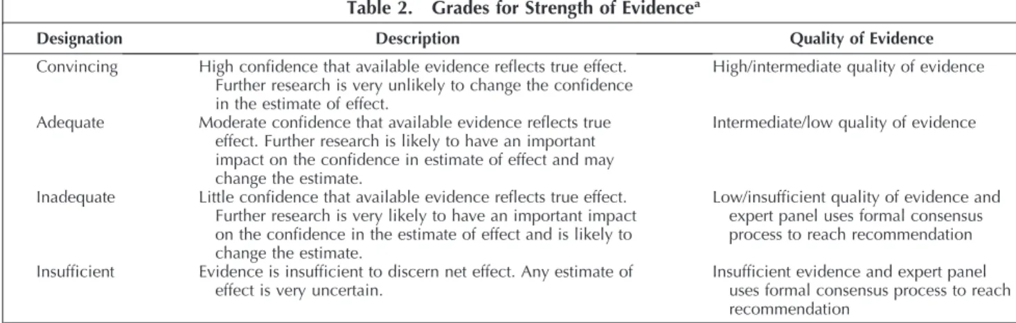
The appropriateness of the study design and data collected, relevance and clarity of findings, and adequacy of conclusions were evaluated. Each study was assessed individually (refer to the SDC for individual assessments and results) and then summarized by study type. Components such as generalizability and applicability were also considered when determining the strength of evidence. A summary of the overall quality of the evidence was given considering the evidence in totality. Ultimately, the designation (ie, rating or grade) of the strength of evidence is a judgment by the expert panel of its level of confidence that the evidence from the studies informing the recommendations reflects true effect.Table 2
describes the grades for strength of evidence.11
Assessing the Strength of Recommendations
Development of recommendations requires that the panel review the identified evidence and make a series of key judgments (using procedures described in the SDC). Grades for strength of recommendations were developed by the CAP Pathology and Laboratory Quality Center and are described inTable 3.11
Guideline Revision
This guideline will be reviewed every 4 years or earlier in the event of publication of substantive and high-quality evidence that could potentially alter the original guideline recommendations. If necessary, the entire panel will reconvene to discuss potential changes. When appropriate, the panel will recommend revision of the guideline to the ASCP, CAP, AMP, and ASCO for review and approval.
Disclaimer
Practice guidelines and consensus statements reflect the best available evidence and expert consensus supported in practice. They are intended to assist physicians and patients in clinical decision making and to identify questions and settings for further research. With the rapid flow of scientific information, new evidence may emerge between the time a practice guideline or consensus statement is developed and when it is published or read.
Guidelines and statements are not continually updated and may not reflect the most recent evidence. Guidelines and statements address only the topics specifically identified therein and are not applicable to other interventions, diseases, or stages of diseases. Furthermore, guidelines and consensus statements cannot account for individual variation among patients and cannot be considered inclusive of all proper methods of care or exclusive of other treatments. It is the responsibility of the treating physician or other health care provider, relying on independent experience and knowledge, to determine the best course of treatment for the patient. Accordingly, adherence to any practice guideline or consensus statement is voluntary, with the ultimate determination regarding its application to be made by the physician in light of each patient’s individual circumstances and preferences. The ASCP, CAP, AMP, and ASCO make no warranty, express or implied, regarding guidelines and statements and specifically exclude any warranties of merchantability and fitness for a particular use or purpose. The ASCP, CAP, AMP, and ASCO assume no respon-sibility for any injury or damage to persons or property arising out of or related to any use of this statement or for any errors or omissions.
RESULTS
A total of 4,197 studies met the search term requirements. A total of 123 articles were included for data extraction. Excluded articles were available as discussion or background references. The panel convened 14 times (11 teleconference webinars and three face-to-face meetings) from July 27, 2013, through September 24, 2015, to develop the scope, draft recommendations, review and respond to solicited feedback, and assess the quality of evidence that supports the final recommendations. Additional work was completed via electronic mail. An open comment period was held from March 30, 2015, through April 22, 2015, during which draft recommendations were posted on the AMP website. Twenty-one guideline statements had an agreement rang-ing from 60% to 94% for each statement from the open-comment period participants (refer to Outcomes in the SDC for full details). The website received a total of 248 Table 1. Levels of Evidencea
Level Description
Level I Evidence derived from systematic reviews of appropriate level II studies and/or clinical practice guidelines
Level II Evidence derived from randomized controlled trials
Level III Evidence derived from comparative studies (eg, prospective cohort studies, retrospective cohort studies) Level IV Evidence without a comparator (eg, case reports, case series, narrative reviews)
aData derived from National Health and Medical Research Council.10
Table 2. Grades for Strength of Evidencea
Designation Description Quality of Evidence
Convincing High confidence that available evidence reflects true effect. Further research is very unlikely to change the confidence in the estimate of effect.
High/intermediate quality of evidence
Adequate Moderate confidence that available evidence reflects true effect. Further research is likely to have an important impact on the confidence in estimate of effect and may change the estimate.
Intermediate/low quality of evidence
Inadequate Little confidence that available evidence reflects true effect. Further research is very likely to have an important impact on the confidence in the estimate of effect and is likely to change the estimate.
Low/insufficient quality of evidence and expert panel uses formal consensus process to reach recommendation
Insufficient Evidence is insufficient to discern net effect. Any estimate of effect is very uncertain.
Insufficient evidence and expert panel uses formal consensus process to reach recommendation
comments. Teams of three to four expert panel members were assigned three to five draft recommendations to review all comments received and provide an overall summary to the rest of the panel. Following panel discussion and the final quality of evidence assessment, the panel members determined whether to maintain the original draft recom-mendation as is, revise it with minor language change, or consider it as a major recommendation change. The expert panel modified eight draft statements based on the feedback during the open-comment period and the considered judgment process. Resolution of all changes was obtained by majority consensus of the panel using nominal group technique (rounds of email discussion and multiple edited recommendations) among the panel members. The final recommendations were approved by the expert panel with a formal vote. The panel considered the risks and benefits throughout the whole process in their considered judgment process. Formal cost analysis or cost-effectiveness was not performed.
Each organization instituted a review process to approve the guideline. The ASCP assigned the review of the guideline to a Special Review Panel. For the CAP, an independent review panel (IRP) representing the Council on Scientific Affairs was assembled to review and approve the guideline. The IRP was masked to the expert panel and vetted through the COI process. The AMP approval process required the internal review of an independent panel led by the Publications and Communications Committee chair and Executive Committee approval. The ASCO approval process required the review and approval of the Clinical Practice Guidelines Committee.
GUIDELINE STATEMENTS
1. Recommendation.—Patients with CRC being consid-ered for anti-EGFR therapy must receive RAS mutational testing. Mutational analysis should includeKRASandNRAS
codons 12 and 13 of exon 2, 59 and 61 of exon 3, and 117 and 146 of exon 4 (‘‘expanded’’or‘‘extended’’RAS) (Table 4).
Aberrant activation of EGFR signaling pathways in CRC is primarily associated with activating mutations of genes in the mitogen-activated protein kinase and phosphatidylino-sitol-3-kinase (PI3K) pathways. Together,KRAS, NRAS, and
BRAFmutations have been reported to occur in more than half of all CRC cases, and KRAS or NRAS and BRAF
mutations are inversely associated, with a small proportion
of individual CRCs showing co-occurrence ofRASandRAF
mutations.3,12
Cetuximab and panitumumab are antibodies that bind to the extracellular domain of EGFR, blocking the binding of EGF and other EGFR endogenous ligands, thereby blocking EGFR signaling. Earlier studies reported the effects of anti-EGFR antibody treatment independent ofKRAS status.13–16 However, it was later reported that targeted EGFR therapies with cetuximab or panitumumab improve PFS and OS in patients with metastatic CRC with wild-typeKRASbut not for patients with mutatedKRAS.2,3,17In these earlier studies, only mutations ofKRASexon 2 were considered. Based on the available clinical trial data in 2009, ASCO recommended that patients with metastatic CRC who are candidates for anti-EGFR antibody therapy should have their tumor tested forKRASmutations in a Clinical Laboratory Improvements Amendments ’88 (CLIA)–accredited laboratory.2
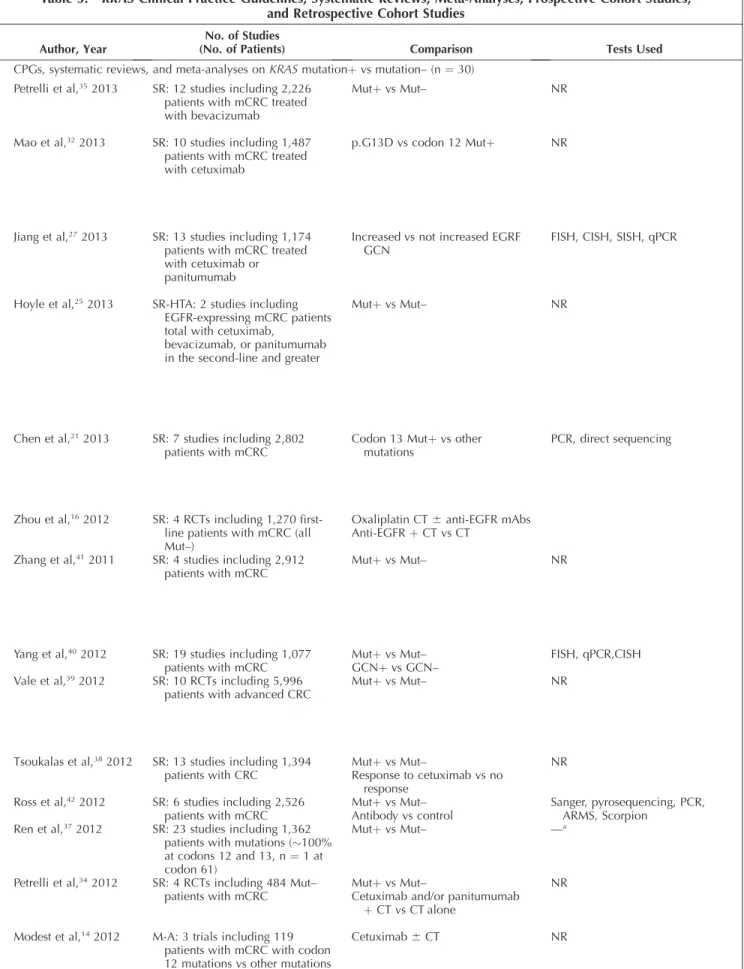
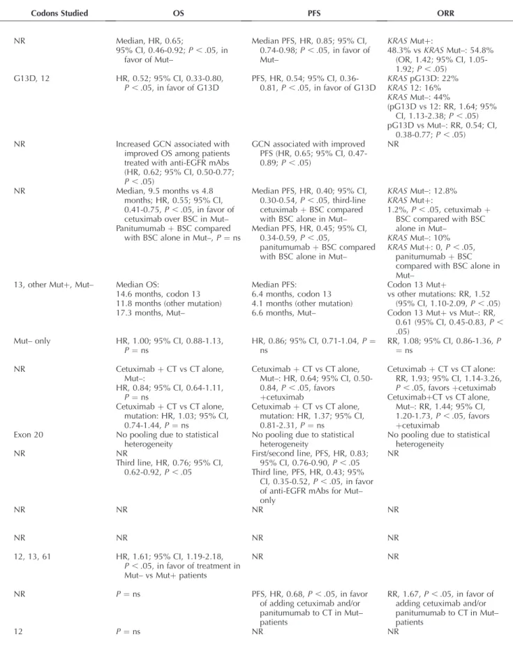
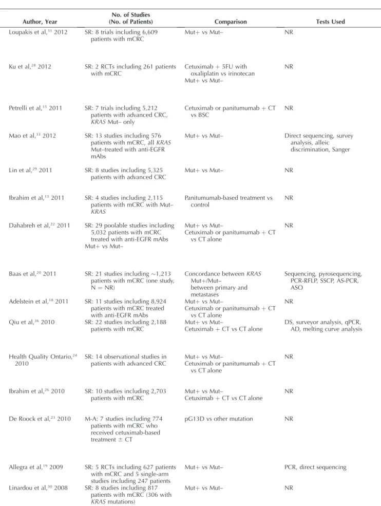
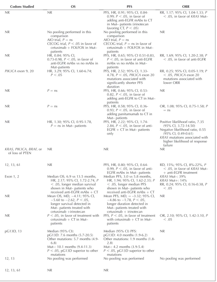
A large body of evidence was available to guide the recommendation in the current guideline forRAStesting in colorectal cancers (Table 5 and Supplemental Table 14). From 2008 to 2015, there were 311 primary studies that included 74,546 patients and reported treatment outcomes for patients with RAS mutations compared with non-mutated/wild type.12–16,18–45The most common comparison of anti-EGFR antibody treatment outcomes was between
K RA S m u t a t i o n v e r s u s K RA S n o nm u t a t e d / w il d type.18–20,22,24–26,28–31,33–42 Some studies also compared the effects of adding an anti-EGFR inhibitor to KRAS non-mutated/wild-type patients versus chemotherapy alone.18,22,24,26,28,36–38 A few studies reported anti-EGFR antibody treatment outcomes for the following compari-sons: KRAS G13D versus codon 12 mutations,32 KRAS codon 13 mutations versus other mutations,21 and G13D versus other exon 2 mutations.23
The reported anti-EGFR therapy outcomes in these studies were pooled survival,1 3 – 1 6 , 2 1 – 2 7 , 2 9 , 3 2 – 3 7 , 3 9 , 4 1 pooled PFS,13,15,16,18,21–27,29,31–36,39,41and pooled objective response rate (ORR).13,15,16,18,21,22,25,26,30–36,41Thirteen studies reported signif-icant differences between comparators.15,21,23–27,32,33,35–37,39The systematic review literature of data on anti-EGFR therapy outcomes is presented in Supplemental Table 14. Five of these studies detected a significant pooled survival advantage of anti– EGFR-treated patients forKRASnonmutated/wild type com-pared withKRASmutation.21,33,35,37,39Three studies detected an advantage for patients with nonmutated tumors given anti-EGFR treatment compared with KRAS mutation-positive Table 3. Grades for Strength of Recommendationa
Designation Recommendation Rationale
Strong recommendation Recommend for or against a particular molecular testing practice for colorectal cancer (can includemustorshould)
Supported by convincing or adequate strength of evidence, high or intermediate quality of evidence, and clear benefit that outweighs any harms
Recommendation Recommend for or against a particular
molecular testing practice for colorectal cancer (can includeshouldormay)
Some limitations in strength of evidence (adequate or inadequate) and quality of evidence (intermediate or low), balance of benefits and harms, values, or costs, but panel concludes that there is sufficient evidence and/or benefit to inform a recommendation Expert consensus opinion Recommend for or against a particular
molecular testing practice for colorectal cancer (can includeshouldormay)
Serious limitations in strength of evidence (inadequate of insufficient), quality of evidence (intermediate or low), balance of benefits and harms, values, or costs, but panel consensus is that a statement is necessary No recommendation No recommendation for or against a particular
molecular testing practice for colorectal cancer
Insufficient evidence or agreement of the balance of benefits and harms, values, or costs to provide a recommendation
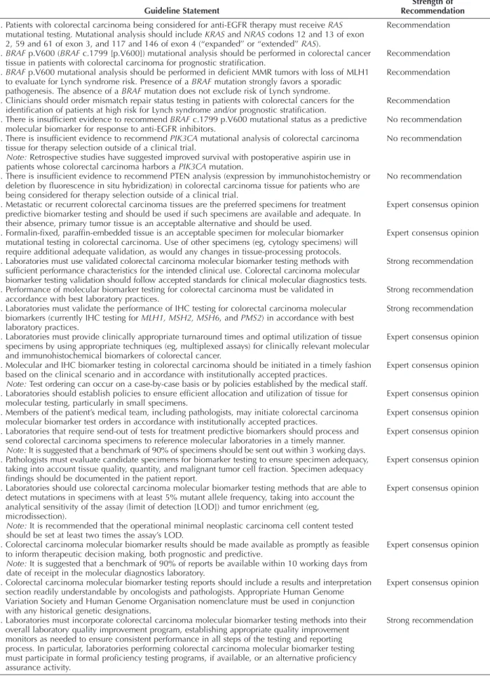
Table 4. Guideline Statements and Strength of Recommendations
Guideline Statement
Strength of Recommendation
1. Patients with colorectal carcinoma being considered for anti-EGFR therapy must receiveRAS
mutational testing. Mutational analysis should includeKRASandNRAScodons 12 and 13 of exon 2, 59 and 61 of exon 3, and 117 and 146 of exon 4 (‘‘expanded’’ or ‘‘extended’’RAS).
Recommendation
2a.BRAFp.V600 (BRAFc.1799 [p.V600]) mutational analysis should be performed in colorectal cancer tissue in patients with colorectal carcinoma for prognostic stratification.
Recommendation
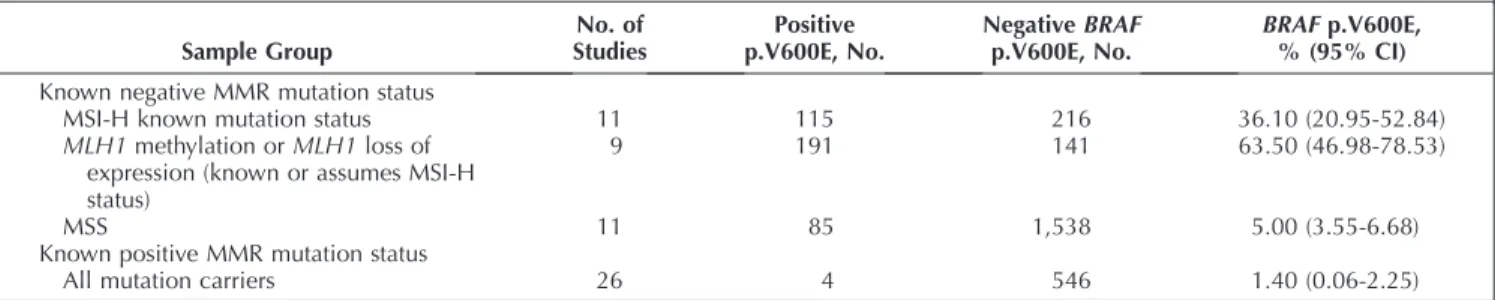
2b.BRAFp.V600 mutational analysis should be performed in deficient MMR tumors with loss of MLH1 to evaluate for Lynch syndrome risk. Presence of aBRAFmutation strongly favors a sporadic pathogenesis. The absence of aBRAFmutation does not exclude risk of Lynch syndrome.
Recommendation
3. Clinicians should order mismatch repair status testing in patients with colorectal cancers for the identification of patients at high risk for Lynch syndrome and/or prognostic stratification.
Recommendation
4. There is insufficient evidence to recommendBRAFc.1799 p.V600 mutational status as a predictive molecular biomarker for response to anti-EGFR inhibitors.
No recommendation
5. There is insufficient evidence to recommendPIK3CAmutational analysis of colorectal carcinoma tissue for therapy selection outside of a clinical trial.
No recommendation
Note:Retrospective studies have suggested improved survival with postoperative aspirin use in patients whose colorectal carcinoma harbors aPIK3CAmutation.
6. There is insufficient evidence to recommend PTEN analysis (expression by immunohistochemistry or deletion by fluorescence in situ hybridization) in colorectal carcinoma tissue for patients who are being considered for therapy selection outside of a clinical trial.
No recommendation
7. Metastatic or recurrent colorectal carcinoma tissues are the preferred specimens for treatment predictive biomarker testing and should be used if such specimens are available and adequate. In their absence, primary tumor tissue is an acceptable alternative and should be used.
Expert consensus opinion
8. Formalin-fixed, paraffin-embedded tissue is an acceptable specimen for molecular biomarker mutational testing in colorectal carcinoma. Use of other specimens (eg, cytology specimens) will require additional adequate validation, as would any changes in tissue-processing protocols.
Expert consensus opinion
9. Laboratories must use validated colorectal carcinoma molecular biomarker testing methods with sufficient performance characteristics for the intended clinical use. Colorectal carcinoma molecular biomarker testing validation should follow accepted standards for clinical molecular diagnostics tests.
Strong recommendation
10. Performance of molecular biomarker testing for colorectal carcinoma must be validated in accordance with best laboratory practices.
Strong recommendation
11. Laboratories must validate the performance of IHC testing for colorectal carcinoma molecular biomarkers (currently IHC testing forMLH1, MSH2, MSH6, andPMS2) in accordance with best laboratory practices.
Strong recommendation
12. Laboratories must provide clinically appropriate turnaround times and optimal utilization of tissue specimens by using appropriate techniques (eg, multiplexed assays) for clinically relevant molecular and immunohistochemical biomarkers of colorectal cancer.
Expert consensus opinion
13. Molecular and IHC biomarker testing in colorectal carcinoma should be initiated in a timely fashion based on the clinical scenario and in accordance with institutionally accepted practices.
Expert consensus opinion
Note:Test ordering can occur on a case-by-case basis or by policies established by the medical staff. 14. Laboratories should establish policies to ensure efficient allocation and utilization of tissue for
molecular testing, particularly in small specimens.
Expert consensus opinion
15. Members of the patient’s medical team, including pathologists, may initiate colorectal carcinoma molecular biomarker test orders in accordance with institutionally accepted practices.
Expert consensus opinion
16. Laboratories that require send-out of tests for treatment predictive biomarkers should process and send colorectal carcinoma specimens to reference molecular laboratories in a timely manner.
Expert consensus opinion
Note:It is suggested that a benchmark of 90% of specimens should be sent out within 3 working days. 17. Pathologists must evaluate candidate specimens for biomarker testing to ensure specimen adequacy,
taking into account tissue quality, quantity, and malignant tumor cell fraction. Specimen adequacy findings should be documented in the patient report.
Expert consensus opinion
18. Laboratories should use colorectal carcinoma molecular biomarker testing methods that are able to detect mutations in specimens with at least 5% mutant allele frequency, taking into account the analytical sensitivity of the assay (limit of detection [LOD]) and tumor enrichment (eg, microdissection).
Expert consensus opinion
Note:It is recommended that the operational minimal neoplastic carcinoma cell content tested should be set at least two times the assay’s LOD.
19. Colorectal carcinoma molecular biomarker results should be made available as promptly as feasible to inform therapeutic decision making, both prognostic and predictive.
Expert consensus opinion
Note:It is suggested that a benchmark of 90% of reports be available within 10 working days from date of receipt in the molecular diagnostics laboratory.
20. Colorectal carcinoma molecular biomarker testing reports should include a results and interpretation section readily understandable by oncologists and pathologists. Appropriate Human Genome Variation Society and Human Genome Organisation nomenclature must be used in conjunction with any historical genetic designations.
Expert consensus opinion
21. Laboratories must incorporate colorectal carcinoma molecular biomarker testing methods into their overall laboratory quality improvement program, establishing appropriate quality improvement monitors as needed to ensure consistent performance in all steps of the testing and reporting process. In particular, laboratories performing colorectal carcinoma molecular biomarker testing must participate in formal proficiency testing programs, if available, or an alternative proficiency assurance activity.
Strong recommendation
patients given chemotherapy alone.24,26,36 Twenty of the included studies pooled PFS,13,15,16,18,21–27,29,31–36,39,41 with 19 reporting significant differences between compara-tors.13,15,18,21–27,29,31–36,39,41Fourteen papers detected a significant PFS advantage for adding an anti-EGFR inhibitor to chemo-therapy forKRAS nonmutated/wild-type patients compared with chemotherapy alone.13,15,18,22,24–26,29,31,33,34,36,39,41Sixteen of the included papers pooled ORR,13,15,16,18,21,22,25,26,30–36,41with 14 reporting significant differences between compara-tors.15,18,21,22,25,26,30–36,41Eight studies detected ORR advantages for adding an anti-EGFR inhibitor to chemotherapy for patients with nonmutated/wild-type tumors compared with chemo-therapy alone,18,25,26,30,33,34,36,41 and four detected an ORR advantage for KRAS nonmutated/wild-type patients over mutation patients.22,31,32,35Survival advantages (OS and PFS, ORR) for G13D mutations over codon 12 and G13D over other mutations were reported in two studies23,32and codon 13 over otherKRASmutations.21
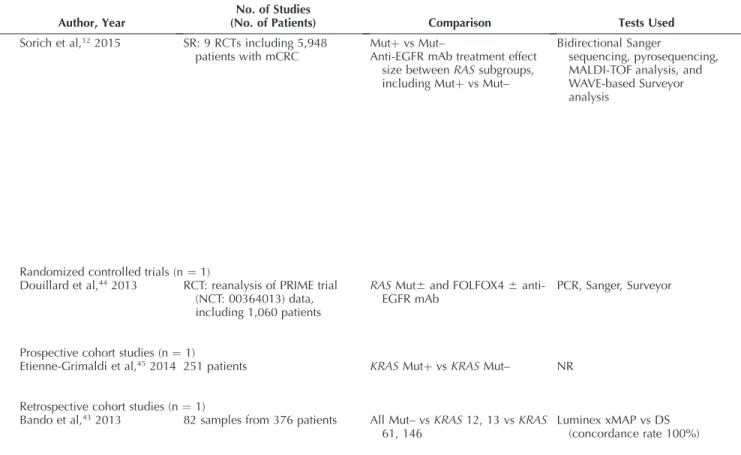
Recent studies showed conclusive evidence that in addition to mutations inKRASexon 2, otherRASmutations inKRASexons 3 and 4 andNRASexons 2, 3, and 4 were also associated with nonresponse of metastatic CRC to anti-EGFR monoclonal antibody therapy.12,44,46 Douillard et al44 published a reanalysis of the Panitumumab Randomized Control Trial in Combination with Chemotherapy for Metastatic Colorectal Cancer to Determine Efficacy (PRIME) trial, reporting that patients with anyRASmutations were associated with inferior PFS and OS with panitumumab-FOLFOX4 treatment, which was consistent with the findings previously reported for patients with KRAS
mutations in exon 2. Subsequently, a meta-analysis of nine randomized clinical trials provided further evidence that not allKRASexon 2 nonmutated/wild-type tumors benefit from anti-EGFR monoclonal antibody treatment in metastatic CRC.12Patients with colorectal cancers that areKRASexon 2 nonmutated/wild type but harborRASmutations in KRAS
exons 3 and 4 or NRAS exons 2, 3, and 4 also have significantly inferior anti-EGFR treatment outcomes benefit compared with those without anyRASmutations (Table 5 and Table 6). RAS mutations occur mostly at exon 2, followed by mutations in exons 3 and 4 (Table 7). The results suggest that ‘‘extended’’ or ‘‘expanded’’ RAS
mutation testing (KRASexons 2, 3, and 4 andNRASexons 2, 3, and 4) must be performed before the administration of an anti-EGFR monoclonal antibody therapy.12In summary, current evidence indicates that both cetuximab and pan-itumumab should only be prescribed for patients with metastatic CRCs that are nonmutated/wild type for all knownRAS-activating mutations.12
This recommendation is supported by 34 stud-ies,1 2 – 1 6 , 1 8 – 4 5 , 4 7 co mp rising 29 syst ema tic stu d -ies,1 2 , 1 3 ,1 5 , 1 6 , 1 8 –2 2 , 2 4 – 4 2 ,4 7 two meta-analyses,14 , 2 3 one randomized controlled trial,44 one prospective cohort study,45and one retrospective cohort study.43
Of the 29 systematic reviews,12,13,15,16,18–22,24–42,47 only three reported using a multidisciplinary panel,19,25,30 and only one reported taking patient preferences into ac-count,37 although 13 examined important patient sub-types.12,15,16,18,21,22,24,27,30,33,37,39,40 All but one had well-described and reported methods sections.42Seven did not report on conflict of interest.13,15,16,34,38,41,42Only nine rated the quality of the included evidence, and these same nine were the only reviews that reported on the strength of the included evidence.16,18,21,22,24,25,32,37,39 None of the studies included a plan for updating. None of the systematic
reviews reported industry funding, two reported no funding,16,31 and 11 did not report on the source of funding, if any.13,15,26,32,34–36,38,41,42,47Two of these system-atic reviews were deemed to have a low risk of bias,24,3714 were deemed to have a low to moderate risk of bias,12,16,18,19,21,22,25,27,29,30,32,35,39,47 12 were deemed to have a moderate risk of bias,13,15,20,26,28,31,33,34,36,38,40,41 and one was deemed to have a high risk of bias.42
Of the two meta-analyses obtained,14,23 both had well-reported and reproducible methods sections, both described the planned pooling a priori, and both discussed the limitations of their analyses. Neither was based on a systematic review of the literature, and neither did a quality assessment of the included studies. One reported nonin-dustry funding,23and the other reported industry funding.14 One was deemed to have a low to moderate risk of bias,23 and the other was deemed to have a moderate risk of bias.14 The single randomized controlled trial did not report on any details of the randomization, including blinding, the expected effect size and power calculation, and the length of follow-up.44It did report on differences in baseline patient characteristics. This trial did report at least partial industry funding and was deemed to have a low to moderate risk of bias.44
The single prospective cohort study reported a balance between treatment and assessment groups, reported on baseline characteristics, and made adjustments in the analysis when differences were found.45It reported nonin-dustry funding and was deemed to have a low risk of bias.45 The single retrospective cohort study reported that the treatment and assessment groups were in balance and also reported on baseline patient characteristics.43 It did not report that adjustments were made in the analysis to account for differences, where differences were found. This study reported nonindustry funding and was deemed to have a low risk of bias.43
All of the evidence that supported this recommendation was assessed, and none was found to have methodologic flaws that would raise concerns about their findings.
2a. Recommendation.—BRAF p.V600 (BRAF c.1799 [p.V600]) position mutational analysis should be performed in CRC tissue in selected patients with colorectal carcinoma for prognostic stratification.
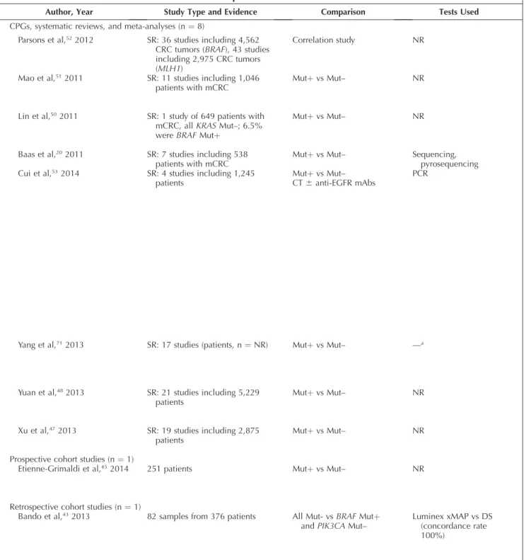
BRAFactivating mutations occur in about 8% of advanced disease patients with CRC47,48and in approximately 14% of patients with localized stage II and III CRC.8,49 As such, mutations inBRAFconstitute a substantial subset of patients with CRC. The key questions related toBRAFmutations are whether patients whose cancers carry aBRAFmutation have a poorer outcome compared with BRAFmutation-negative tumors and whether the presence of a mutation predicts benefit from or lack thereof to anti-EGFR therapy.
Four systematic reviews20,50–52 and three systematic reviews that included meta-analyses47,48,53 pertaining to the prognostic and predictive value of BRAF mutations in patients with CRC were identified through our systematic review process (Table 8and Supplemental Table 14). These studies revealed that patients with advanced CRC who possess a BRAF mutation have significantly poorer out-comes as measured by PFS and OS and have a decreased response rate to anti-EGFR therapy relative to those with nonmutated BRAF. Poorer OS was also demonstrated for those patients with earlier stage II and III CRC having a
Table 5. KRASClinical Practice Guidelines, Systematic Reviews, Meta-Analyses, Prospective Cohort Studies, and Retrospective Cohort Studies
Author, Year
No. of Studies
(No. of Patients) Comparison Tests Used
CPGs, systematic reviews, and meta-analyses onKRASmutationþvs mutation– (n¼30) Petrelli et al,352013 SR: 12 studies including 2,226
patients with mCRC treated with bevacizumab
Mutþvs Mut– NR
Mao et al,322013 SR: 10 studies including 1,487
patients with mCRC treated with cetuximab
p.G13D vs codon 12 Mutþ NR
Jiang et al,272013 SR: 13 studies including 1,174
patients with mCRC treated with cetuximab or panitumumab
Increased vs not increased EGRF GCN
FISH, CISH, SISH, qPCR
Hoyle et al,252013 SR-HTA: 2 studies including
EGFR-expressing mCRC patients total with cetuximab,
bevacizumab, or panitumumab in the second-line and greater
Mutþvs Mut– NR
Chen et al,212013 SR: 7 studies including 2,802
patients with mCRC
Codon 13 Mutþvs other mutations
PCR, direct sequencing
Zhou et al,162012 SR: 4 RCTs including 1,270
first-line patients with mCRC (all Mut–)
Oxaliplatin CT6anti-EGFR mAbs Anti-EGFRþCT vs CT
Zhang et al,412011 SR: 4 studies including 2,912
patients with mCRC
Mutþvs Mut– NR
Yang et al,402012 SR: 19 studies including 1,077
patients with mCRC
Mutþvs Mut– GCNþvs GCN–
FISH, qPCR,CISH
Vale et al,392012 SR: 10 RCTs including 5,996
patients with advanced CRC
Mutþvs Mut– NR
Tsoukalas et al,382012 SR: 13 studies including 1,394
patients with CRC
Mutþvs Mut–
Response to cetuximab vs no response
NR
Ross et al,422012 SR: 6 studies including 2,526
patients with mCRC
Mutþvs Mut– Antibody vs control
Sanger, pyrosequencing, PCR, ARMS, Scorpion
Ren et al,372012 SR: 23 studies including 1,362
patients with mutations (~100% at codons 12 and 13, n¼1 at codon 61)
Mutþvs Mut– —a
Petrelli et al,342012 SR: 4 RCTs including 484 Mut–
patients with mCRC
Mutþvs Mut–
Cetuximab and/or panitumumab þCT vs CT alone
NR
Modest et al,142012 M-A: 3 trials including 119
patients with mCRC with codon 12 mutations vs other mutations
Table 5. Extended
Codons Studied OS PFS ORR
NR Median, HR, 0.65;
95% CI, 0.46-0.92;P,.05, in favor of Mut–
Median PFS, HR, 0.85; 95% CI, 0.74-0.98;P,.05, in favor of Mut–
KRASMutþ:
48.3% vsKRASMut–: 54.8% (OR, 1.42; 95% CI, 1.05-1.92;P,.05)
G13D, 12 HR, 0.52; 95% CI, 0.33-0.80,
P,.05, in favor of G13D
PFS, HR, 0.54; 95% CI, 0.36-0.81,P,.05, in favor of G13D
KRASpG13D: 22%
KRAS12: 16%
KRASMut–: 44%
(pG13D vs 12: RR, 1.64; 95% CI, 1.13-2.38;P,.05) pG13D vs Mut–: RR, 0.54; CI,
0.38-0.77;P,.05)
NR Increased GCN associated with
improved OS among patients treated with anti-EGFR mAbs (HR, 0.62; 95% CI, 0.50-0.77;
P,.05)
GCN associated with improved PFS (HR, 0.65; 95% CI, 0.47-0.89;P,.05)
NR
NR Median, 9.5 months vs 4.8
months; HR, 0.55; 95% CI, 0.41-0.75,P,.05, in favor of cetuximab over BSC in Mut– PanitumumabþBSC compared
with BSC alone in Mut–,P¼ns
Median PFS, HR, 0.40; 95% CI, 0.30-0.54,P,.05, third-line cetuximabþBSC compared with BSC alone in Mut– Median PFS, HR, 0.45; 95% CI,
0.34-0.59,P,.05,
panitumumabþBSC compared with BSC alone in Mut–
KRASMut–: 12.8%
KRASMutþ:
1.2%,P,.05, cetuximabþ BSC compared with BSC alone in Mut–
KRASMut–: 10%
KRASMutþ: 0,P,.05, panitumumabþBSC compared with BSC alone in Mut–
13, other Mutþ, Mut– Median OS:
14.6 months, codon 13 11.8 months (other mutation) 17.3 months, Mut–
Median PFS: 6.4 months, codon 13 4.1 months (other mutation) 6.6 months, Mut–
Codon 13 Mutþ
vs other mutations: RR, 1.52 (95% CI, 1.10-2.09,P,.05) Codon 13 Mutþvs Mut–: RR,
0.61 (95% CI, 0.45-0.83,P, .05)
Mut– only HR, 1.00; 95% CI, 0.88-1.13,
P¼ns
HR, 0.86; 95% CI, 0.71-1.04,P¼ ns
RR, 1.08; 95% CI, 0.86-1.36,P
¼ns
NR CetuximabþCT vs CT alone,
Mut–:
HR, 0.84; 95% CI, 0.64-1.11,
P¼ns
CetuximabþCT vs CT alone, mutation: HR, 1.03; 95% CI, 0.74-1.44,P¼ns
CetuximabþCT vs CT alone, Mut–: HR, 0.64; 95% CI, 0.50-0.84,P,.05, favors
þcetuximab
CetuximabþCT vs CT alone, mutation: HR, 1.37; 95% CI, 0.81-2.31,P¼ns
CetuximabþCT vs CT alone: RR, 1.93; 95% CI, 1.14-3.26,
P,.05, favorsþcetuximab CetuximabþCT vs CT alone,
Mut–: RR, 1.44; 95% CI, 1.20-1.73,P,.05, favors þcetuximab
Exon 20 No pooling due to statistical
heterogeneity
No pooling due to statistical heterogeneity
No pooling due to statistical heterogeneity
NR NR
Third line, HR, 0.76; 95% CI, 0.62-0.92,P,.05
First/second line, PFS, HR, 0.83; 95% CI, 0.76-0.90,P,.05 Third line, PFS, HR, 0.43; 95%
CI, 0.35-0.52,P,.05, in favor of anti-EGFR mAbs for Mut– only
NR
NR NR NR NR
NR NR NR NR
12, 13, 61 HR, 1.61; 95% CI, 1.19-2.18,
P,.05, in favor of treatment in Mut– vs Mutþpatients
NR NR
NR P¼ns PFS, HR, 0.68,P,.05, in favor
of adding cetuximab and/or panitumumab to CT in Mut– patients
RR, 1.67,P,.05, in favor of adding cetuximab and/or panitumumab to CT in Mut– patients
Table 5. Continued
j
j
Author, Year
No. of Studies
(No. of Patients) Comparison Tests Used
Loupakis et al,312012 SR: 8 trials including 6,609
patients with mCRC
Mutþvs Mut– NR
Ku et al,282012 SR: 2 RCTs including 261 patients
with mCRC
Cetuximabþ5FU with oxaliplatin vs irinotecan Mutþvs Mut–
NR
Petrelli et al,152011 SR: 7 trials including 5,212
patients with advanced CRC,
KRASMut– only
Cetuximab or panitumumabþCT vs BSC
NR
Mao et al,332012 SR: 13 studies including 576
patients with mCRC, allKRAS
Mut–treated with anti-EGFR mAbs
Mutþvs Mut– Direct sequencing, survey
analysis, alleic discrimination, Sanger
Lin et al,292011 SR: 8 studies including 5,325
patients with advanced CRC
Mutþvs Mut– NR
Ibrahim et al,132011 SR: 4 studies including 2,115
patients with mCRC with Mut–
KRAS
Panitumumab-based treatment vs control
NR
Dahabreh et al,222011 SR: 29 poolable studies including
5,032 patients with mCRC treated with anti-EGFR mAbs Mutþvs Mut–
Mutþvs Mut–
Cetuximab or panitumumabþCT vs CT alone
NR
Baas et al,202011 SR: 21 studies including~1,213
patients with mCRC (one study, N¼NR)
Concordance betweenKRAS
Mutþ/Mut– between primary and metastases
Sequencing, pyrosequencing, PCR-RFLP, SSCP, AS-PCR, ASO
Adelstein et al,182011 SR: 11 studies including 8,924
patients with mCRC treated with anti-EGFR mAbs
Mutþvs Mut–
Cetuximab or panitumumabþCT vs CT alone
NR
Qiu et al,362010 SR: 22 studies including 2,188
patients with mCRC
Mutþvs Mut–
CetuximabþCT vs CT alone
DS, surveyor analysis, qPCR, AD, melting curve analysis
Health Quality Ontario,24
2010
SR: 14 observational studies in patients with advanced CRC
Mutþvs Mut–
Cetuximab or panitumumabþCT vs CT alone
NR
Ibrahim et al,262010 SR: 10 studies including 2,703
patients with mCRC
Mutþvs Mut–
CetuximabþCT vs CT alone
NR
De Roock et al,232010 M-A: 7 studies including 774
patients with mCRC who received cetuximab-based treatment6CT
pG13D vs other mutation NR
Allegra et al,192009 SR: 5 RCTs including 627 patients
with mCRC and 5 single-arm studies including 247 patients
Mutþvs Mut– PCR, direct sequencing
Linardou et al,302008 SR: 8 studies including 817
patients with mCRC (306 with
KRASmutations)
Table 5. Continued, Extended
Codons Studied OS PFS ORR
NR NR PFS, HR, 0.91; 95% CI,
0.84-0.99;P,.05, in favor of adding anti-EGFR mAbs to CT in Mut– patients (irinotecan favoring CT,P,.05)
RR, 1.17; 95% CI, 1.04-1.33;P
,.05, in favor ofKRASMut–
NR No pooling performed in this
comparison AIO trial,P¼ns
CECOG trial,P,.05 in favor of cetuximabþFOLFOX in Mut– patients
No pooling performed in this comparison
AIO trial,P¼ns
CECOG trial,P¼ns in favor of cetuximabþFOLFOX in Mut– patients
NR
NR HR, 0.84; 95% CI,
0.73-0.98,P,.05, in favor of anti-EGFR mAbs vs no mAbs in Mut–patients
PFS, HR, 0.65; 95% CI 0.51-0.83,
P,.05, in favor of anti-EGFR mAbs vs no mAbs in Mut– patients
RR, 1.69; 95% CI, 1.20-2.38;P
,.05, in favor of anti-EGFR
PIK3CAexon 9, 20 HR, 3.29; 95% CI, 1.60-6.74;
P,.05
PFS, HR, 2.52; 95% CI, 1.33-4.78,P,.05,PIK3CAexon 20 mutations associated with significantly shorter PFS duration
RR, 0.25; 95% CI, 0.05-1.19;P
,.05,PIK3CAexon 20 mutations associated with lower ORR
NR P¼ns PFS, HR, 0.66; 95% CI,
0.53-0.82,P,.05, in favor of adding anti-EGFR to CT in Mut– patients
NR
NR P¼ns PFS, HR, 0.58; 95% CI,
0.36-0.93;P,.05, in favor of adding panitumumab to CT in Mut– patients
OR, 1.08; 95% CI, 0.75-1.58;P
¼ns
NR HR, 1.30; 95% CI, 0.95-1.78,
P¼ns in Mut– patients
PFS, HR, 2.22; 95% CI, 1.74-2.84,P,.05, in favor of anti-EGFRþCT in Mut– patients only
Positive likelihood ratio, 7.35 (95% CI, 3.72-14.50) Negative likelihood ratio, 0.55
(95% CI, 0.49-0.61)
KRASmutations associated with higher likelihood of response failure
KRAS,PIK3CA,BRAF, or of loss of PTEN
NR NR NR
12, 13, 61 NR PFS, HR, 0.80; 95% CI,
0.64-0.99,P,.05, in favor of anti-EGFR mAbs in Mut– patients
RD, 15%; 95% CI, 8%-22%,P
,.05, in favor ofKRASMut– þanti-EGFR treatment
Exon 1, 2 Median OS, 6.9 vs 13.5 months,
HR, 2.17; 95% CI, 1.72-2.74,P
,.05, longer median survival shown in Mut– patients who received anti-EGFR mAbsþCT
Median PFS, 3.0 vs 5.8 months, HR, 1.94; 95% CI, 1.62-2.33;P
,.05, longer median PFS shown in Mut– patients who received anti-EGFR mAbsþCT
KRASMut–: 39%
KRASMutþ: 14%
RR, 0.24; 95% CI, 0.16-0.38,P
,.05
NR Mean OS, MD,4.11; 95% CI,
5.60 to2.62,P,.05, longer survival detected in Mut– patients treated with cetuximabþirinotecan
Mean PFS, MD,¼–3.32; 95% CI, 4.86 to1.78,P,.05, longer duration detected in Mut– patients treated with cetuximabþirinotecan
NR
NR P,.05, in favor of treatment with
cetuximabþCT in Mut– patients
PFS,P,.05, in favor of treatment with cetuximabþCT in Mut– patients
OR, 2.10; 95% CI, 1.42-3.10,P
,.05
pG13D, 13 Median (95% CI):
pG13D: 7.6 months (5.7-20.5) Other mutations: 5.7 months
(4.9-6.8)
Mut–: 10.1 months (9.4-11.3)
P,.05, pG13D superior to other mutations
Median (95% CI) PFS: pG13D: 4.0 months (1.9-6.2) Other mutations: 1.9 months
(1.8-2.8)
Mut–: 4.2 months (3.9-5.4)
P,.05, pG13D superior to other mutations
NR
12, 13 No pooling was performed No pooling was performed No pooling was performed
patients rather than a harbinger of an increased rate of relapse. Finally, while outcomes in advanced disease patients with BRAF mutations were poorer relative to nonmutation patients, the data were consistent with a modest beneficial impact from the use of anti-EGFR agents relative to those patients whose tumors contained a RAS
mutation.55In summary, patients with CRC that contains a
BRAF mutation have a worse outcome relative to non-mutation patients. Selected patients for BRAF mutation testing include patients with metastatic disease, since these patients have particularly poor outcomes. It is important to know the BRAF c.1799 (p.V600) mutation status of a Table 5. Continued
Author, Year
No. of Studies
(No. of Patients) Comparison Tests Used
Sorich et al,122015 SR: 9 RCTs including 5,948
patients with mCRC
Mutþvs Mut–
Anti-EGFR mAb treatment effect size betweenRASsubgroups, including Mutþvs Mut–
Bidirectional Sanger
sequencing, pyrosequencing, MALDI-TOF analysis, and WAVE-based Surveyor analysis
Randomized controlled trials (n¼1)
Douillard et al,442013 RCT: reanalysis of PRIME trial
(NCT: 00364013) data, including 1,060 patients
RASMut6and FOLFOX46 anti-EGFR mAb
PCR, Sanger, Surveyor
Prospective cohort studies (n¼1)
Etienne-Grimaldi et al,452014 251 patients KRASMutþvsKRASMut– NR
Retrospective cohort studies (n¼1)
Bando et al,432013 82 samples from 376 patients All Mut– vsKRAS12, 13 vsKRAS
61, 146
Luminex xMAP vs DS (concordance rate 100%)
5FU, fluorouracil; AD, allelic discrimination; AIO, German AIO colorectal study group; ARMS, amplification refractory mutation system; AS-PCR, allele-specific polymerase chain reaction; ASO, allele-specific oligonucleotide;BRAF, proto-oncogene B-Raf/v-Raf murine sarcoma viral oncogene homolog B; BSC, best supportive care; CECOG, Central European Cooperative Oncology Group; CI, confidence interval; CISH, chromogenic in situ hybridization; CPG, clinical practice guideline; CRC, colorectal cancer; CT, chemotherapy; DS, direct sequencing; EGFR, epidermal growth factor receptor; FISH, fluorescence in situ hybridization; FOLFOX, folinic acid (leucovorin calcium), 5-fluorouracil, and oxaliplatin; FOLFOX4, folacin, 4-fluorouracil, oxaliplatin; GCN, gene copy number; HR, hazard ratio; HTA, health technology assessment;KRAS, Kirsten rat sarcoma viral oncogene homolog; M-A, meta-analysis; mAbs, monoclonal antibodies; MALDI-TOF, matrix-assisted laser desorption/ionization-time of flight; mCRC, metastatic colorectal cancer; MD, mean difference; Mut–, mutation negative or wild type; Mutþ, mutation positive; NR, not reported; NRAS, neuroblastomaRASviral (v-ras) oncogene homolog;ns, nonsignificant; OR, odds ratio; ORR, objective response rate; OS, overall survival; PCR, polymerase chain reaction; PCR-RFLP, polymerase chain reaction–restriction fragment length polymorphism; PFS, progression-free survival;PIK3CA, phosphatidylinositol-4,5-bisphosphate 3-kinase catalytic subunit alpha; PRIME, Panitumumab Randomized Control Trial in Combination with Chemotherapy for Metastatic Colorectal Cancer to Determine Efficacy; PTEN, phosphatase and tensin homolog; qPCR, quantitative polymerase chain reaction; RCT, randomized controlled trial;RAS, rat sarcoma viral oncogene homolog; RD, risk difference; RFS, recurrence-free survival; RR, response rate; SISH, silver in situ hybridization; SR, systematic review; SSCP, single-strand conformation polymorphism; xMAP, multiplex assay.
a Tests used by Ren et al37: hybridization, PCR, direct sequencing, topographic genotyping, AS-PCR, tissue transglutaminase enzyme,
high-performance liquid chromatography, pyrosequencing, capillary sequencing.
Table 6. Outcomes ofRASMutations and Anti-EGFR Therapy12
Characteristic
Overall Survival Progression-Free Survival HR (95% CI) PValue HR (95% CI) PValue
RASnm vsRASmutation,RASnm superior 0.72 (0.56-0.92) ,.01 0.60 (0.48-0.76) ,.001
KRASexon 2 mutant vs newRASmutant ns ns
KRASnm exon 2, anti-EGFR vs no anti-EGFR 0.90 (0.83-0.98) ns 0.68 (0.58-0.80) ,.001
KRASexon 2 mutant, anti-EGFR vs no anti-EGFR 1.05 (0.95-1.17) ns 1.14 (0.95-1.36) ns
RASnm, anti-EGFR vs no anti-EGFR 0.87 (0.77-0.99) ,.04 0.62 (0.50-0.76) ,.001
AnyRASmutant, anti-EGFR vs no anti-EGFR 1.08 (0.97-1.21) ns 1.12 (0.94-1.34) ns
patient’s CRC since standard therapy is not adequate for patients with metastatic disease and BRAF mutation. For these patients, some studies suggest the use of FOLFIR-INOX (folinic acid [leucovorin calcium], 5-fluorouracil,
irinotecan hydrochloride, and oxaliplatin) as first-line therapy, followed by enrollment in a clinical trial.56 Furthermore, early clinical trials data suggest that the combination of aBRAF plus EGFR inhibitor appears to be Table 5. Continued, Extended
Codons Studied OS PFS ORR
KRAS/NRAS12, 13, 59, 61, 117, 146
RASMut– vsRASMutþ: HR, 0.72 (95% CI, 0.56-0.92;P
,.01)RASMut– superior
KRASexon 2 mutant vs newRAS
mutant:P¼ns
RASMut–, EGFR vs no anti-EGFR: HR, 0.87 (95% CI, 0.77-0.99;P,.04)
KRASexon 2 Mut–, anti-EGFR vs no anti-EGFR: HR, 0.90 (95% CI, 0.83-0.98;P¼ns)
AnyRASmutant, anti-EGFR vs no anti-EGFR: HR, 1.08 (95% CI, 0.97-1.21;P¼ns)
KRASexon 2 mutant, anti-EGFR vs no anti-EGFR: HR, 1.05 (95% CI, 0.95-1.17;P¼ns)
RASMut– vsRASMutþ: HR, 0.60 (95% CI, 0.48-0.76;P
,.001)RASMut– superior
KRASexon 2 mutant vs newRAS
mutant:P¼ns
RASMut–, EGFR vs no anti-EGFR: HR, 0.62 (95% CI, 0.50-0.76;P,.001)
KRASexon 2 Mut–, anti-EGFR vs no anti-EGFR: HR, 0.68 (95% CI, 0.58-0.80;P,.001) AnyRASmutant, anti-EGFR vs no
anti-EGFR: HR, 1.12 (95% CI, 0.94-1.34;P¼ns)
KRASexon 2 mutant, anti-EGFR vs no anti-EGFR: HR, 1.14 (95% CI, 0.95-1.36;P¼ns)
NR
KRAS/NRAS12, 13, 61, 117, 146
Mut6and anti-EGFR mAb6: 26 months vs 20.2 months HR, 0.78 (95% CI, 0.62-0.99;P
,.05) in favor of Mut– andþ anti-EGFR mAb
Mut6and anti-EGFR mAb6: 10.1 months vs 7.9 months HR, 0.72 (95% CI, 0.58-0.90;P
,.05) in favor of Mut– andþ anti-EGFR mAb
NR
KRAS12, 13 NR RR, 2.40 (95% CI, 1.27-4.55;P
,.05), RFS shorter inKRAS
Mutþpatients with stage III tumors
NR
KRAS12,13,61,146 All Mut–: 13.8 months (9.2-18.4) vsKRASMutþ: 8.2 months (5.7-10.7;P,.05)
All Mut–: 6.1 months (3.1-9.2) vs
KRASMutþ: 2.7 months (1.2-4.2;P,.05)
All Mut–: 38.8% vsKRAS
Mutþ: 4.8%,P,.05
Table 7. Prevalence of NewRASMutations Across Studiesa
Study
NewRAS
Total,b%
KRAS
Exon 3,b%
KRAS
Exon 4,b%
NRAS
Exon 2,b%
NRAS
Exon 3,b%
NRAS
Exon 4,b%
Codons 59, 56 Codons 117, 146 Codons 12, 13 Codons 59, 61 Codons 117, 146
OPUS 26.3 5.9 9.3 6.8 5.1 0.8
PICCOLO 9.8 NRc 3.7d 6.3e NRc NE
20020408 17.6 4.8c 5.0 4.2 3.0c 1.1
20050181 20.5 4.6 7.9 2.3 5.8 0.0
PRIME 17.4 3.7c 5.6 3.4 4.1c 0.0
FIRE-3 16.0 4.3c 4.9d 3.8 2.0c 0.0
PEAK 20.1 4.1 7.7 5.4 5.9 0.0
COIN 8.4 2.1c NE 0.9f 3.0c NE
CRYSTAL 14.7 3.3 5.6 3.5 2.8 0.9
Summary (95% CI)g 19.9 (16.7-23.4) 4.3 (3.3-5.5) 6.7 (5.7-7.9) 3.8 (3.0-4.8) 4.8 (3.4-6.8) 0.5 (0.2-1.2)
CI, confidence interval; COIN, Combination Chemotherapy With or Without Cetuximab as First-Line Therapy in Treating Patients With Metastatic Colorectal Cancer Trial; CRYSTAL, Cetuximab Combined with Irinotecan in First-Line Therapy for Metastatic Colorectal Cancer Trial; FIRE-3, Folinic Acid and Irinotecan (FOLFIRI) Plus Cetuximab vs FOLFIRI Plus Bevacizumab in First-Line Treatment Colorectal Cancer (CRC) Trial; NE, not evaluated; NR, evaluated but not reported; OPUS, Effect of Roflumilast on Exacerbation Rate in Patients With Chronic Obstructive Pulmonary Disease (BY217/M2-111) Trial; PEAK, Panitumumab Plus mFOLFOX6 vs Bevacizumab Plus mFOLFOX6 for First-Line Treatment of Metastatic Colorectal Cancer (mCRC) Patients With Wild-Type Kirsten Rat Sarcoma-2 Virus (KRAS) Tumors Trial; PICCOLO, Panitumumab and Irinotecan vs Irinotecan Alone for Patients With KRAS Wild-Type, Fluorouracil-Resistant Advanced Colorectal Cancer Trial; PRIME, Panitumumab Randomized Trial in Combination With Chemotherapy for Metastatic Colorectal Cancer to Determine Efficacy Trial.
aModified from Sorich et al12by permission of Oxford University Press on behalf of the European Society for Medical Oncology. bNewRASmutations are reported as a proportion of theKRASexon 2 nonmutated/wild-type group.
cKRASandNRAScodon 59 mutation was not evaluated. dKRAScodon 117 mutation was not evaluated.
eExon 3 codon 61 mutations in addition to the exon 2 mutations. fOnlyNRASmutation G12C evaluated.
Table 8. BRAFClinical Practice Guidelines, Systematic Reviews, Meta-Analyses, Prospective Cohort Studies, and Retrospective Cohort Studies
Author, Year Study Type and Evidence Comparison Tests Used
CPGs, systematic reviews, and meta-analyses (n¼8)
Parsons et al,522012 SR: 36 studies including 4,562
CRC tumors (BRAF), 43 studies including 2,975 CRC tumors (MLH1)
Correlation study NR
Mao et al,512011 SR: 11 studies including 1,046
patients with mCRC
Mutþvs Mut– NR
Lin et al,502011 SR: 1 study of 649 patients with
mCRC, allKRASMut–; 6.5% wereBRAFMutþ
Mutþvs Mut– NR
Baas et al,202011 SR: 7 studies including 538
patients with mCRC
Mutþvs Mut– Sequencing,
pyrosequencing
Cui et al,532014 SR: 4 studies including 1,245
patients
Mutþvs Mut– CT6anti-EGFR mAbs
PCR
Yang et al,712013 SR: 17 studies (patients, n¼NR) Mutþvs Mut– —a
Yuan et al,482013 SR: 21 studies including 5,229
patients
Mutþvs Mut– NR
Xu et al,472013 SR: 19 studies including 2,875
patients
Mutþvs Mut– NR
Prospective cohort studies (n¼1)
Etienne-Grimaldi et al,452014 251 patients Mutþvs Mut– NR
Retrospective cohort studies (n¼1)
Bando et al,432013 82 samples from 376 patients All Mut- vsBRAFMutþ
andPIK3CAMut–
Luminex xMAP vs DS (concordance rate 100%)
BRAF, proto-oncogene B-Raf/v-Raf murine sarcoma viral oncogene homolog B; CI, confidence interval; CPG, clinical practice guideline; CRC, colorectal cancer; CT, chemotherapy; DS, direct sequencing; EGFR, epidermal growth factor receptor; HR, hazard ratio;KRAS, Kirsten rat sarcoma viral oncogene homolog; mAbs, monoclonal antibodies; mCRC, metastatic colorectal cancer;MLH1, mutL homolog 1; Mut–, mutation negative or wild type; Mutþ, mutation positive; NR, not reported;ns, nonsignificant; ORR, objective response rate; OS, overall survival; PCR, polymerase chain reaction; PFS, progression-free survival;PIK3CA, phosphatidylinositol-4,5-bisphosphate 3-kinase catalytic subunit alpha; RR, response rate; RFS, recurrence-free survival; SR, systematic review; xMAP, multiplex assay.
a Yang et al71: adenovirus-PCR pyrosequencing, allele-specific PCR, DS, PCR amplification, quantitative PCR, Sanger, real-time PCR,
effective in this population.57–59Data in support of molecular testing forBRAFc.1799 (p.V600) mutations in CRC continue to emerge from clinical trials. A recent publication of the PETACC-8 (oxaliplatin, fluorouracil, and leucovorin with or without cetuximab in patients with resected stage III colon cancer randomised phase 3) trial reported that trials in the adjuvant setting should consider mismatch repair, BRAF, andKRAS status for stratification, since BRAF p.V600 and
KRASmutations were associated with shorter DFS and OS
in patients with microsatellite-stable colon cancer but not in those with tumors with MSI.60,61
This recommendation is supported by seven systematic reviews,20,47,48,50–53 three of which included meta-analy-sis.47,48,53 None of the systematic reviews reported the composition of their panel, so multidisciplinary panel representation could not be confirmed, and none reported patient representation on the panel. All but the systematic review reported by Baas et al20 reported examining important patient subgroups. All of the systematic reviews Table 8. Extended
Codons Studied OS PFS ORR
BRAFp.V600E,MLH1 NR NR NR
V600E NR NR BRAFMutþ: 0BRAF
Mut–: 36.3%;P,.05; RR, 0.14; 95% CI, 0.04-0.53
V600E Shorter duration inBRAFMutþ
patients, difference 28 weeks,P
,.05
PFS, shorter duration in
BRAFMutþpatients, difference 18 weeks,P
,.05
NR
V600E NR NR NR
V600E NR NR Mutþvs Mut– (allKRAS
Mut–): RR, 0.43 (95% CI, 0.16-0.75;P,.05) in favor of Mut– Mut6vs CT6
anti-EGFR mAbs (allKRAS
Mut–): RR, 0.38 (95% CI, 0.20-0.73;P,.05) in favor of Mut– Mutþand CT6
anti-EGFR mAbs;P¼ns
Mut– andKRASMut– and CT6anti-EGFR mAbs: RR, 1.48 (95% CI,
1.28-1.71;P,.05) in favor ofBRAFMut– with CTþanti-EGFR mAbs
V600E, 599, 466, 469 (7 studies)
BRAFMut6: HR, 2.74 (95% CI, 1.79-4.19;P,.05) in favor of
BRAFMut–
(8 studies)
BRAFMut6:
HR, 2.59 (1.67, 4.03;P
,.05) in favor ofBRAF
Mut–
BRAFMut–: 46.4%
BRAFMut: 18.5%
P,.05 in favor of
BRAFMut–
V600E HR, 0.35 (95% CI, 0.29-0.42;P
,.05) in favor ofBRAFMut–
HR, 0.38 (95% CI, 0.29-0.51;P,.05) in favor of
BRAFMut–
RR, 0.31 (95% CI, 0.18-0.53;P,.05) in favor ofBRAFandKRAS
Mut– V600E, K601E (1 study),
D549C (1 study)
HR, 2.85 (95% CI, 2.31-3.52;P
,.05) in favor ofBRAFMut–
HR, 2.98 (95% CI, 2.07-4.27;P,.05) in favor of
BRAFMut–
ORR, 0.58 (95% CI, 0.35-0.94;P,.05) in favor ofBRAFMut–
BRAFp.V600E NR Shorter RFS inKRASMut–
andBRAFMut– patients with stage III tumors (P
,.05)
600 All Mut–: 13.8 months (95% CI,
9.2-18.4) vsBRAF/PIK3CAMut: 6.3 months (95% CI, 1.3-11.3;
P,.05)
All Mut–: 6.1 months (95% CI, 3.1-9.2) vsBRAF/
PIK3CAMutþ: 1.6 months (95% CI, 1.5-1.7;P,.05)
All Mut–: 38.8% vs