
The role of BCR destabilization in antigen processing
Jin Hyang Kim
A dissertation submitted to the faculty of the University of North Carolina at Chapel Hill in partial fulfillment of the requirements for the degree of Doctor of Philosophy in the Department of Microbiology and Immunology, School of Medicine
Chapel Hill 2006
Approved by
Advisor: Barbara Vilen
Reader: Stephen Clarke
Reader: Jeffrey Frelinger
ABSTRACT
JIN HYANG KIM: The role of BCR destabilization in antigen processing
(Under the direction of Barbara Vilen)
The B cell antigen receptor (BCR) relays extracellular signals and delivers antigen to the
endocytic compartment for processing and presentation. Concurrently, BCR ligation
destabilizes Ig-α/Ig-β from μ-heavy chain (μm). Here, we describe that receptor
destabilization represents a physical separation of μm from Ig-α/Ig-β. Sucrose gradient
fractionation localized Ig-α/Ig-β to GM1-containing lipid microdomains in the absence of
μm. Confocal and transmission electron microscopy studies revealed colocalization of unsheathed μm with clathrin-coated vesicles (CCVs). Further, μm failed to associate
with CCVs when receptor destabilization was inhibited, suggesting that unsheathing of
μm is required for clathrin-mediated endocytosis. In summary, we found that antigen stimulation physically separates Ig-α/Ig-β from μm, facilitating concomitant signal
transduction and antigen delivery to the endocytic compartment. In addition, we
demonstrate the molecular basis and the role of BCR destabilization in antigen processing.
transmembrane domain. Receptors expressing an Y587F mutation transduced signals
that were comparable to wild type receptors, yet failed to dissociate μm from Ig-α/Ig-β.
Further, receptors harboring the Y587F mutation failed to associate with CCVs, resulting
in diminished antigen in the lysosome-associated membrane protein-1 positive
(LAMP-1+) compartments and impaired antigen presentation. Thus, the transmembrane tyrosine
of μm mediates destabilization of the BCR complex and the association of antigen with
CCVs. Our data also indicate that association of receptors with CCVs is required for
antigen presentation. Together, we demonstrate that BCR destabilization reveals a novel
DEDICATION
ACKNOWLEDGMENTS
I would like to thank my advisor Dr. Barbara Vilen for her excellent mentorship
throughout my graduate study. I admire her passion and ability in training and teaching
graduate students to be professional scientists. Specifically, I consider her training in
rigorous thinking, critical interpretation and evaluation of scientific data to be a valuable
lesson that I will continue to develop throughout my scientific career. I would also like to
thank my committee members Dr. Jeffrey Frelinger, Stephen Clarke, Jack Griffith, and
JoAnn Trejo for their encouragement and guidance throughout my project. I especially
thank Dr. Jack Griffith and Dr. Alexandr Makhov of the UNC-CH TEM core facility for
their continuous support throughout my TEM study. I acknowledge Dr. Tony Purdue of
the TEM facility at the UNC-CH Biology department for allowing me to use the electron
microscope and for his technical expertise in TEM. I also thank Dr. Lorraine Cramer for
her effort to establish the TEM technique in our lab and for her training. Lastly, I would
like to thank our lab members, Dr. Michelle Kilmon, Jennifer Rutan, Mileka Gilbert,
Diane Carnathan, and Nikki Wagner for their constant support, feedback, and most
TABLE OF CONTENTS
LIST OF TABLES………viii
LIST OF FIGURES……….ix
LIST OF ABBREVIATIONS………..………xi
Chapter I. Introduction and background………..………..1
1.1.Role of the BCR during development and humoral immune response………..1
1.2.Structure and surface expression of the BCR complex………..1
1.3.BCR-mediated signal transduction……….6
1.4.BCR-mediated antigen processing and presentation………..9
1.5.Endocytic motif……….11
1.6.BCR-mediated signals regulate trafficking of receptor-antigen complexes through the endocytic pathway………...13
1.7.Early events in BCR-mediated antigen endocytosis……….14
1.8.BCR desensitization and destabilization………...15
1.9.Role of BCR destabilization in maintaining tolerance……….17
(Independent trafficking of Ig-α/Ig-β and μm is facilitated by the dissociation of the BCR complex)
Introduction………..20
Materials and Methods……….22
Results………..26
Discussion………52
III. The role of BCR destabilization in antigen processing………..…..…...63
(The transmembrane tyrosine of μ-heavy chain is required for BCR destabilization and entry of antigen into clathrin-coated vesicles) Introduction………...………..64
Materials and Methods………...….66
Results………...………..69
Discussion………87
IV. Conclusions……….92
LIST OF TABLES
Table Page 1. Assessment of subcellular localization of μm and Ig-α
on native membrane sheets in M12g3r cells………..40
2. Assessment of subcellular localization of μm, Ig-α
and CCVs on native membrane sheets in M12g3r cells………46
3. Assessment of subcellular localization of μm and Ig-α
and on native membrane sheets in A20WT and A20Y/F cells………..74
4. Assessment of subcellular localization of μm and CCVs
LIST OF FIGURES
Figure page
Chapter II
2.1. Antigen stimulation induces BCR destabilization in K46μ cells.………...27
2.2. Destabilized Ig-α/Ig-β resides in lipid microdomains in the absence of μm ……….……….28
2.3. M12g3r cells fail to express surface Ig-α/Ig-β in the absence of transfected μm………..………..31
2.4. Herbimycin inhibits BCR destabilization in M12g3r cells…...………32
2.5. Herbimycin inhibits BCR-mediated signal transduction………33
2.6. Ig-β fails to cocap with μm following antigen stimulation………34
2.7. Gold-conjugated PC10BSA induces tyrosine phosphorylation………...37
2.8. Antigen stimulation induces a physical dissociation of μm from Ig-α/Ig-β…………..………38
2.9. Antigen stimulation colocalizes mm with CCVs………43
2.10. BCR ligation induced the association of antigen-μm, but not Ig-α with CCVs………..44
2.11. BCR destabilization facilitate the translocation of antigen-bound μm to CCVs………50
2.13. Lipid rafts do not colocalize with CCVs………..56
2.14. Antigen stimulation fails to induce the aggregation
of lipid rafts-associated molecules……….58
Chapter III
3.1. Surface expression level of human IgM in A20 cells……….67
3.2. Mutation of the transmembrane tyrosine of μm
inhibits BCR destabilization………...70
3.3. Mutation of the transmembrane tyrosine of μm
blocked dissociation of μm from Ig-α/Ig-β………72 3.4. A20Y/F cells induce BCR-mediated signal transduction ………..77
3.5. The association of antigen-bound μm with CCV
requires the transmembrane tyrosine of μm ………..79
3.6. The transmembrane tyrosine of μm is required
for antigen processing……….84
3.7. The transmembrane tyrosine of μm is required
for antigen presentation ……….85
3.8. Surface MHC class II expression is comparable
between A20WT and A20Y/F cells………86
3.9.Exposure of the transmembrane region of μm alone
is insufficient to induce the association of receptors with CCVs………...89
LIST OF ABBREVIATIONS
Abbreviations
BCR = B cell antigen receptor
BLNK = B cell linker protein
CART = conserved antigen receptor transmembrane
CCVs = clathrin-coated vesicles
CTB = cholera toxin B subunit
ER = endoplasmic reticulum
ERK = extracellular-signal-regulated kinase
ITAM = immunoreceptor tyrosine-based activation motif
LAB = linker for activation of B cells
LAMP-1+ = lysosome-associated membrane protein-1 positive
MHC = major histocompatibility complex
MIIC = MHC class II peptide-loading compartments
NP = nitrophenyl
PAGE = poly acrylamide gel electrophoresis
PC = phosphorylcholine
Y/F = Y587F
YXXØ motifs =X: any amino acid, Ø: bulky hydrophobic residue
1. 1. Role of the B cell antigen receptor (BCR) during development and humoral
immune response
The BCR is a multi-chain immune recognition receptor that serves multiple functions
throughout B cell development (1). At early stages of development, the pre-BCR complex
triggers proliferative expansion, allelic exclusion and developmental progression to the
immature stage (2-4). The first tolerance checkpoint occurs when immature B cells express a
functional BCR. Cells expressing self-reactive BCR are negatively selected upon excessive
BCR crosslinking to induce cell death via apoptosis or receptor editing as a means to
eliminate autoreactivity (3). Negative selection eliminates more than 90% of total B cells
generated in the bone marrow and allows approximately 10% of B cells to exit to the
periphery, where naïve, transitional B cells continue to mature (5). At the mature stage of B
cell development, the BCR serves as a specific receptor that binds foreign antigens. Upon
ligation, the BCR initiates the humoral immune response by relaying intracellular signals and
by focusing antigens to the endosomal system (6). This results in processing and presentation
of antigenic peptides in the context of major histocompatibility complex (MHC) class II
molecules to cognate helper CD4+T cells. The ultimate outcome of B cell activation is a
generation of plasma cells that produce antigen-specific antibodies. To accomplish this
variety of functions, B cells control the quality of surface BCR expression and the functions
of BCR by utilizing a complex, yet organized network of receptor-mediated signal
transduction.
The BCR complex is composed of the antigen binding subunit, membrane immunoglobulin
(mIg) and the signaling subunit, disulfide-linked Ig-α/Ig-β (7). The association of μm and
Ig-α/Ig-β is non-covalent (8). The antigen binding subunit consists of two immunoglobulin
heavy chains (IgH) and two light chains (IgL). Assembly of the BCR complex and surface
expression is regulated by chaperon molecules that regulates transport of properly assembled
receptors from the endoplasmic reticulum (ER) to the cell surface (9, 10). Germ
line-transcribed, newly synthesized μ-heavy chains (μm) and light chains are assembled into mIg
and retained by calnexin in the ER (11). Calnexin-mediated ER retention is a checkpoint for
quality control, thereby preventing misfolded or aberrantly glycosylated proteins from
reaching the cell surface (12). For the assembly of multi-chain receptor complexes such as
MHC class I and class II molecules, T cell receptor (TCR)-CD3 complexes, and the BCR
complexes, a single chain of the receptor complex is held by calnexin until the other subunits
associate (13-15). Specifically, calnexin binds the transmembrane region of μm of the BCR
complex until Ig-α/Ig-β assembles onto μm (16). In the absence of Ig-α/Ig-β, mIg remains
calnexin bound in the ER. Thus, Ig-α/Ig-β releases μm from the calnexin-binding site and
allows subsequent processing of μm. Upon release of calnexin, the fully assembled BCR
complex travels quickly to the cell surface via the Golgi apparatus.
The association of the BCR complex is mediated by protein-protein interactions between
transmembrane regions of μm and Ig-α/Ig-β (17). The transmembrane region of μm exhibits
a conserved antigen receptor transmembrane (CART) motif that shares sequence
motif in associating with other integral transmembrane proteins in the hydrophobic
environment of the lipid bilayer. The CART motif of μm is characterized by polar patches
containing polar amino acids (tyrosine, serine, threonine) and aromatic, hydrophobic amino
acids (phenylalanine, valine, leucine) near the carboxy- terminal end of the transmembrane
domain. Modeling of μm using the α-helical wheel predicts that the conserved CART
residues align on one side of the helix (18). This orientation likely provides a protein-protein
interacting face for Ig-α/Ig-β (19). Mutagenesis studies reveal a functional role for the CART
motif in BCR assembly and function (10, 19-25). Although some studies disagree, the
general consensus is that surface expression of mIg and its function in signal transduction
and antigen presentation depends on the association of Ig-α/Ig-β through the
carboxy-terminal polar patches of the CART motif.
Mutagenesis studies of μm provide evidence that μm associates with Ig-α/Ig-β through the
transmembrane regions. Most studies focus on the conserved polar residues near the putative
carboxy-terminus of the transmembrane region and the three amino acid residues of the
cytoplasmic domain of μm. Shaw et al. uses a panel of B cell clones transfected with wild
type and mutated forms of μm to define a complete correlation between intact signaling of
the receptor and the ability of the μm to associate with Ig-α/Ig-β (22). The cytoplasmic
domain of μm is composed of short, positively charged amino acids (Lys-Val-Lys; KVK)
and has no intrinsic enzymatic activity. However, deletion of KVK completely abolishes the
ability of B cells to induce signal transduction, evidenced by failure to mobilize intracellular
Ca2+ mobilization (22). This indicates that the presence of the cytoplasmic tail of μm is
critical for proper signal transduction, although the exact sequence is dispensable. A
non-conservative mutation of polar residues, tyrosine (Y587) and serine (S588) within the
carboxy-terminal region of μm to valines (YS to VV) results in a complete loss of the
intracellular Ca2+ response (11, 22). This indicates that removal of hydroxyl groups from
tyrosine and serine disrupts the signaling ability of receptors. Importantly, the inability of
these mutants (deletion of KVK and substitution of YS to VV) to mobilize intracellular Ca2+
correlates with failure of μm to associate with Ig-α/Ig-β and to induce protein
phosphorylation (23). Coincident with lack of Ca2+ flux, these mutants also fail to activate
cognate T cells following incubation with antigen (22). These findings suggest that
signal-competence and the ability of B cells to present antigen to cognate T cells correlate with
association with Ig-α/Ig-β.
A more subtle change, tyrosine (Y587) to phenylalanine (Y to F) in the carboxy-terminal
polar patch retains the ability to mobilize intracellular Ca2+ and induce protein
phosphorylation (22, 26). Consistently, Y to F mutation of μm maintains the association with
Ig-α/Ig-β, however, despite intact Ig-α/Ig-β, cells bearing these mutated receptors fail to
present antigen to T cells (26). This indicates that the elimination of a single hydroxyl group
at tyrosine 587 of μm maintains the association of μm with Ig-α/Ig-β and the ability to
transduce BCR-signals. In contrast, this single point mutation eliminates antigen endocytosis,
targeting of endocytosed antigen to intracellular compartments, and peptide loading onto
Surface expression of mIg requires association with Ig-α/Ig-β (27, 28). Introducing IgH and
IgL into non-lymphoid cells fails to promote expression of surface mIg (29). However,
surface mIgM expression occurs when Ig-α/Ig-β are provided. Interestingly, receptors
harboring a deletion of the KVK or a substitution of YS to VV are expressed at high levels
on the cell surface, despite the lack of Ig-α/Ig-β association (11, 22, 25). This suggests that
these mutations cause intrinsic changes such as converting the BCR from an integral
transmembrane protein to a GPI (glycosyl-phosphatidyl-linositol)-linked protein in the case
of the KVK deletion (25). In the case of the YS to VV mutation, surface mIgM expression in
the absence of Ig-α/Ig-β is presumed to be due to spontaneous release from calnexin (26). It
is thought that conferring hydrophobicity at tyrosine and serine residues alters the
glycosylation of μm, resulting in reduced avidity for calnexin. This relieves the restraint on
μm required for ER-retention and leads to spontaneous surface expression.
1. 3. BCR-mediated signal transduction
One of the major functions of the BCR is to transduce intracellular signals upon receptor
crosslinking (2-4, 30-32). Antigen ligation activates the src-family protein kinase, Lyn which
rapidly phosphorylates the cytoplasmic tail of Ig-α/Ig-β at tyrosine residues within the
immunoreceptor tyrosine-based activation motif (ITAM). Phosphorylated ITAMs create
docking sites for tandem Src-homology (SH) 2 domains of the cytosolic protein tyrosine
kinase, Syk. Recruitment of Syk to the ITAMs of Ig-α/Ig-β is a key event that amplifies
(Btk), activates PLCγ, resulting in the cleavage of phosphatidylinositol-4,5-biphosphate
(PIP2) to inositol-1,4,5-triphosphate (IP3) and diacylglycerol (DAG). Parallel to PLCγ
activation, PI3K is recruited to the plasma membrane. Both PLCγ and PI3K act on the same
substrate, PIP2, however, in contrast to PLCγ, PI3K phosphorylates PIP2 to generate
phosphatidylinositol-3,4,5-triphosphate (PIP3). Accumulation of PIP3 provides a membrane
binding site for the pleckstrin homology (PH) domain of both PLCγ and Btk, creating a
positive-reinforcement loop. Activation of these cascades elevates and prolongs the
intracellular Ca2+ flux, promoting nuclear translocation of nuclear factor κB (NF-κB),
nuclear factor of activated T cells (NFAT), activated c-Jun amino-terminal kinase (JNK), p38,
and extracellular-signal-regulated kinase (ERK) (33). In addition, signal strength such as
affinity or avidity of antigen is reflected at the level of intracellular Ca2+ elevation by
oscillating spikes of varying duration, interval and amplitude (34). Ultimately, this influences
gene transcription that regulates many B cell responses, including proliferation, plasma cell
differentiation, antibody secretion and the induction of tolerance.
The membrane location of BCR-mediated signal transduction is controversial, however,
considerable evidence suggests that lipid rafts exist in the plasma membrane of living cells
and play an important role in receptor-mediated signal transduction (7, 35). Lipid rafts are
enriched in cholesterol and glycosphingolipids that contain saturated fatty acid side-chains
(e.g. GM1 gangliosides), thus they appear as relatively ordered membrane patches (36). This
structural feature allows lipid rafts to be resistant to solubilization by non-ionic detergent and
(CTB) which binds to GM1 (35). An important biological feature of lipid rafts is selective
partitioning of signaling molecules. Positive signaling proteins such as Lyn, G proteins, Ras
GTPase, and adaptor protein linker for activation of B cells (LAB) constitutively partition
into lipid rafts through the modification of saturated fatty acids (37, 38). In contrast, certain
phosphatases such as CD45, CD22, and SH2 domain-containing protein tyrosine
phosphatase-1 (SHP-1) are excluded from lipid rafts in resting cells (37, 39). Due to a rigid,
ordered structure, lipid rafts have been thought to function as scaffolds for membrane
trafficking and signal transduction.
In mature, resting B cells, most BCR complexes are detected outside of lipid rafts. However,
crosslinking of the BCR induces translocation into lipid rafts (37, 40). Although the regulator
of lipid raft entry remains unknown, receptors within lipid rafts are readily phosphorylated by
lipid raft-associated Lyn at the ITAMs of Ig-α/Ig-β (37, 39). This amplifies the signaling
cascade by recruiting other proteins including PI3K and PLCγ to the lipid rafts, where PIP2 is
enriched (41). It has been proposed that initial recruitment of signaling proteins to lipid rafts
triggers formation of multiple signaling molecule complex (Btk, Vav, Rac, and F-actin)
termed ‘signalosome’ that stabilizes and prolongs receptor-mediated signals (4). Although it
is accepted that lipid rafts regulate the duration of BCR-mediated signals, it remains unclear
if lipid rafts are the only sites that facilitate BCR-signal transduction (42, 43). Disruption of
lipid rafts with pharmacological reagent does not affect BCR-mediated intracellular Ca2+
influx (44). In addition, blocking of lipid rafts has no effect on ERK activation in human B
cells (43). These findings indicate that some BCR-signal transduction occurs independently
1. 4. BCR-mediated antigen processing and presentation
In addition to signal transduction, the BCR captures soluble, multivalent antigen and
internalizes for transport to the endocytic pathway (45). B cells are good antigen-presenting
cells because the BCR focuses 1000-fold less antigen than fluid phase-mediated uptake (46).
In addition, the BCR processes antigen and expresses peptide-MHC class II complexes
within 1 hr of antigen exposure (26). This efficient antigen binding to the BCR ensures rapid
and targeted responses to antigens (47). Endocytosis of antigen-bound BCR complexes
occurs mainly through clathrin-coated vesicles (CCVs) (48). Clathrin is a intracellular protein
that forms a lattice around protein cargo localized on the plasma membrane (49, 50). Three
sets of clathrin heavy and light chains trimerize into a clathrin triskelion that serves as a
building block for CCVs (51). Endocytosed BCR-antigen complexes transit to the early
endosome, where some of the unoccupied receptors recycle back to the surface (52). After
transit to the early endosome, internalized antigen-BCR complexes are delivered to late
endosomes where the complexes persist for prolonged periods of times (53). Antigen
internalized via fluid phase endocytosis is rapidly delivered to lysosomes and degraded by
lysosomal proteases (53). Antigen delivered to late endosomes by the BCR is processed into
peptides by capthepsin (54). Newly synthesized MHC class II molecules are retained in the
ER by association with chaperone invariant chain (Ii) and directly delivered to the endocytic
pathway, where the antigen-BCR complex is localized (55, 56). As the MHC class II-Ii
complex progresses through the endocytic pathway, proteases degrade Ii, leaving the CLIP
(class II-associated invariant chain peptide) fragment of Ii in the peptide-binding cleft of the
class II and load processed antigenic peptides (58, 59) . The Bulk of peptide loading occurs
in the late endocytic vesicles, termed MHC class II-peptide loading compartment (MIIC)
and/or other subcompartments such as the early endocytic class II-containing vesicles
(CIIVs) (60). Peptide-MHC class II (pMHC II) complexes then move to the cell surface to
present the peptide to cognate CD4+ helper T cells (53). The majority of unbound peptides
move to the lysosome and are degraded by lysosome-associated proteases (47, 53). The
persistence of antigen-BCR complexes in the late endocytic vesicles correlates with
prolonged surface expression of pMHC II (53). The nature of antigen or strength of
BCR-signal does not change the itinerary of endocytosed antigen through the endocytic pathway,
but receptor crosslinking accelerates the internalization and targeting of antigen (61).
Antigen presentation by the BCR is critical for B cells to act as immune effectors.
Specifically, the immune response to most protein antigens requires the recognition of
peptide-MHC class II complexes on B cells by CD4+ T cells (6). This T-dependent antigen
response is initiated in spatially organized lymphoid tissues, such as lymph node and spleen.
The initial antigen recognition tethers T cell onto the cellular contact site with the B cell, and
this focal point develops into elaborate cellular synapse by which intracellular information is
exchanged (35). Antigen-stimulated T cells produce cytokines and upregulate co-stimulatory
molecules such as B7.1 and CD40 ligand (CD40L). The local cytokine milieu and
co-stimulatory molecules then activate B cells and trigger proliferative clonal expansion and
differentiation into antibody-secreting plasma cells. In lymphoid tissue, this interaction is
responsible for short-lived plasma cell differentiation and the development of the germinal
plasma cells and memory cells (6). Thus, BCR-mediated targeting of antigens to the
endosomal system is essential for initiating humoral immune responses.
1. 5. Endocytic motif
Clathrin-mediated endocytosis is an integrated series of molecular events that include
clathrin-coat formation, vesicle invagination, vesicle fission, and uncoating in the cytoplasm
(51). Studies have discovered that multiple accessory and adaptor proteins are implicated in
protein–protein and protein-lipid interactions between the endocytic apparatus and the
cytoskeletal elements (62-64). Analysis of clathrin formation in living cells provides a
unifying model that the clathrin adaptor complex, AP-2, plays a central role in sorting protein
cargo and forming a stable platform on the plasma membrane to initiate clathrin cage
assembly (65). From these platforms, clathrin triskelion and cargo proteins are transferred
into forming vesicles that rapidly move laterally and then move towards the cytoplasm
through actin polymerization (49). Progressively, vesicles invaginate and are released into
the cytoplasm as free clathrin-coated cages. The invagination and fission of vesicles appear
to require the GTPase-activity of dynamin, an accessory molecule that regulate actin
polymerization (66). Following endocytosis, uncoating of clathrin occurs rapidly through the
catalytic activity of Hsc70 and ATPase-harboring protein, auxilin (49). While clathrin coat
components are released into the cytoplasm for recycling, protein cargo is transported into
the endocytic pathway.
The AP-2 complex is composed of 4 subunits and serves as a bridge between the
subunit and the clathrin/accessory molecules via α and β subunits (51). Studies suggest that
phosphorylation of the μ2 subunit is necessary to induce conformational changes in the AP-2
complex, allowing the binding of protein cargo (67). Upon phosphorylation, the AP-2
complex recognizes protein cargo at short linear amino acid sequences, called endocytic
sorting motifs or trafficking motifs. Well-characterized canonical sorting motifs are
tyrosine-based YXXØ motifs (X: any amino acid, Ø: bulky hydrophobic residue) and
di-leucine (LL) motifs that bind the μ2 and/or the β subunits of AP-2 complex (50, 68). Other
non-canonical endocytic motifs such as ubiquitin have also been suggested (69). Ubiquitin is
a 76-amino acid polypeptide and can be conjugated to lysine side chain of target proteins.
Poly-ubiquitination regulates turnover of intracellular proteins by tagging a signal for
degradation via the 26S proteasome. In contrast, single or oligo-ubiquitination acts as an
endocytic sorting signal for protein that guides proteins to the endosomal system (69-74).
For the BCR, the ITAMs (YXXL) of Ig-α/Ig-β have been thought to be the endocytic sorting
motif due to their structural resemblance with a canonical tyrosine-based motif (75-78).
While no direct evidence is available to show the interaction of ITAM and AP-2 complex,
this idea is challenged by the studies showing that the phosphorylated form of the
tyrosine-based sorting motif (PO4-YXXØ) has a significantly reduced affinity to the μ2 subunit of the
AP-2 complex (79). Given that BCR crosslinking induces phosphorylation at the tyrosine
residues of the ITAMs, it is likely that activated BCR complexes are inhibited from binding
the AP-2 complex. In addition, evidence for a role of the ITAM in receptor internalization
and subsequent antigen processing/presentation has been contradictory. Some reports suggest
tyrosine of the ITAM has no effect on receptor internalization (76, 77). Others report that
cells harboring mutations within the BCR at sites other than the ITAM fail to present
peptides to T cells despite intact ITAM motifs (26). These data argue against the idea that the
ITAM motifs of Ig-α/Ig-β are the endocytic sorting motif for antigen entry into the endocytic
pathway. This raises the possibility that a novel endocytic motif regulates BCR entry into the
endocytic pathway.
1. 6. BCR-mediated signals regulate trafficking of receptor-antigen complexes through
the endocytic pathway
The two functions of the BCR are signal transduction by Ig-α/Ig-β and endocytosis of
antigen by μm. Although seemingly independent, these functions are interrelated such that
BCR-induced signals accelerate endocytosis of antigen and guide the trafficking of antigen
through the endosomal system (77, 80, 81). BCR crosslinking recruits cytoplasmic Syk to the
plasma membrane, where Syk promotes and stabilizes the formation of tightly aggregated
BCR-antigen complexes (82). Lyn activation is required for the internalization of aggregated
BCR-antigen complexes and for phosphorylation of clathrin heavy chain during endocytosis
(82, 83). The valency of antigen influences the rate of internalization, as crosslinking with
polyvalent antigen prolongs the BCR aggregates on the plasma membrane (84). This
concomitantly increases the duration of protein tyrosine kinase activation, slows down
receptor internalization, and reduces movement of antigen to the late endosomes.
BCR-mediated signals also increase cellular F-actin and change the organization of actin filaments
to promote receptor internalization and trafficking (85). Perturbing actin filaments does not
early endosome to the late endosomes/lysosomes (85). This suggests that BCR signals target
actin filaments to regulate and guide the trafficking of antigen-receptor complexes through
the endosomal system. Collectively, these findings show how signaling molecules intersect
the process of internalization and movement of BCR-antigen complexes to the endocytic
pathway.
Following endocytosis, phosphorylation of Ig-α/Ig-β ITAM tyrosines is required for the
access of antigen to MHC class II peptide-loading compartments (MIIC), as chimeric
receptors expressing either subunit alone fail to reach MIIC, yet internalize normally (76, 78).
Specifically, proper sorting of antigen to the MIIC requires the recruitment of Syk, B cell
linker protein (BLNK), Vav, and Grb2 to phosphorylated ITAM of Ig-α (86). In addition,
BCR-mediated signals reorganize, fuse, and acidify the transient MIIC-like compartments
into invariant chain (Ii)- and MHC class II-rich large vesicles (81). These vesicles become
the primary site for processing of endocytosed antigen and loading of peptides. These studies
demonstrate an important role for BCR-mediated signal transduction in the targeting and
processing of antigen within the endocytic pathway.
1. 7. Early events in BCR-mediated antigen endocytosis
Although BCR-mediated signals regulate antigen processing, the initial events that guide the
entry of antigen-bound receptors to the endocytic pathway remain poorly understood. Early
studies suggested that endocytosis occurred via CCVs (48). Subsequently, lipid-mediated
receptor internalization is proposed when it is found that the BCR translocates into lipid rafts
class II peptide-loading compartments (MIIC) (37, 40). This study suggests that upon
receptor translocation, lipid rafts act as platforms for the entry of the BCR into the endocytic
pathway. However, from this study, it is not shown if the BCR directly internalizes from lipid
rafts or if the associated BCR moves laterally in the membrane and internalizes by a
raft-independent mechanism. A unifying model that links lipid raft- and clathrin-mediated
receptor endocytosis comes from a study showing that BCR-signals in lipid rafts modify
clathrin through Lyn-dependent phosphorylation (83). Inducible phosphorylation of clathrin
correlates with BCR uptake. This suggests that BCR-mediated antigen endocytosis occurs
through a subset of clathrin that is constitutively associated with lipid rafts. These
conclusions are questioned, however, by a study showing that the extent of BCR endocytosis
is not affected by the disruption of lipid rafts (44). This study demonstrates that the bulk of
antigen-bound receptors are internalized through a pathway independent of lipid rafts.
Furthermore, this study suggests that raft-associated receptors have a different intracellular
itinerary than the majority of receptors internalized through CCVs (44). An independent
study shows that although lipid rafts are uniformly distributed across the plasma membrane,
few associates with CCVs, suggesting that CCVs and lipid rafts localize to separate sites on
the plasma membrane (87). While the precise mechanism of BCR-mediated endocytosis
remains to be elucidated, these studies show that BCR-mediated signal transduction and early
endocytic events are interrelated.
1. 8. BCR desensitization and destabilization
Upon initial antigen encounter, naïve B cells transduce robust signals, but become less
desensitization (88). Desensitization of B cells is characterized at the molecular level as an
inability to modulate intracellular Ca2+ and to induce phosphorylation of receptor-proximal
kinases and Ig-α/Ig-β. For many receptors, desensitization is either due to rapid
internalization of receptors or recruitment of negative signaling regulators to the receptors
(89, 90). This results in a reduced availability of receptors for extracellular ligands or muting
of signaling cascades at the receptor level. However, BCR desensitization does not correlate
with reduced surface receptor expression level, inability of μm to bind antigen, or qualitative
changes in BCR-signaling cascades (91). At a desensitizing dose of antigen, approximately
25% of the surface receptor pool is occupied by antigen but the remaining 75% of receptors
bind antigen but fail to induce a phospho-tyrosine response (88). This indicates that for some
self-antigens, surface receptor level is maintained during desensitization. In addition, despite
diminished induction in tyrosine phosphorylation, Lyn remains competent to bind and be
activated by exogenous phospho-ITAM-containing peptides, suggesting Lyn from
desensitized cells is functional but not activated (88). These observations indicate that
activation is disrupted downstream of antigen binding of μm, but upstream of activation of
ITAM and receptor-proximal kinase. Thus, desensitized cells remain competent to bind
antigen through μm, but fail to subsequently transmit signals to Ig-α/Ig-β, hence failing to
activate downstream effectors. This suggests that the phenotype of unresponsiveness is at the
level of the receptor complex.
This hypothesis is tested in a study demonstrating that receptor desensitization also
destabilizes the BCR complex (91). This is shown by a 2-fold decrease in the amount of
transduction and the activation of Syk and Lyn (91). Importantly, destabilized Ig-α/Ig-β
heterodimers remain responsive to anti-Ig-β stimulation, consistent with the observations that
function of Ig-α/Ig-β ITAM and receptor-proximal kinases in desensitized cells remain intact
(88). This suggests that antigen ligation with BCR induces the quantitative and/or qualitative
changes in interaction between μm and Ig-α/Ig-β and BCR destabilization is a possible
mechanism that induced the unresponsiveness of desensitized BCR. However, from this
study, it is not known if the BCR complex becomes transiently destabilized by antigen
stimulation, hence representing biochemical instability or if the receptor subunits (μm and
Ig-α/Ig-β) physically dissociated.
1. 9. Role of BCR destabilization in maintaining tolerance
The nature of antigen and the availability of T cell help determine the fate of B cells
following antigen encounter. Under conditions where foreign antigens bind the BCR and
cognate T cell help is available, B cells undergo activation, proliferation, and differentiation
to antibody-secreting plasma cells. However, binding of the BCR with self-antigen induces
receptor desensitization, and lack of cognate T cell help maintains the cellular
unresponsiveness of B cells. This unresponsive state is termed anergy (92). While the role of
BCR destabilization in antigen targeting, B cell activation and antibody secretion is currently
not known, its role in anergy is suggested by several studies. In many immunoglobulin (Ig)
transgenic mouse models of B cell tolerance, B cells exhibit reduced tyrosine kinase
activation, increased basal Ca2+ level, and constitutively elevated Erk (93-97). In these
desensitization contributes to the unresponsive state of autoreactive B cells. In the in vitro
model of BCR desensitization, antigen binding to only 25% of the surface receptor pool
desensitizes the remaining BCR pool (~75%) that presumably lacks antigen. This suggests
that receptor desensitization functions “in-trans” such that unbound receptors become
unresponsive when a small number of receptors are ligated. This idea is tested in a study by
Vilen et al., where a panel of B cells expressing both competent and
signal-incompetent receptors is used to mimic antigen-bound and unbound receptors during
desensitization (98). The study shows that coaggregation of signal competent and signal
incompetent receptors prevents ITAM phosphorylation, Syk activation, Ca2+flux, and the
upregulation of the B cell activation marker, CD69. This study proposes that the inclusion of
desensitized receptors within functional BCR aggregates disrupts the spatial organization of
receptor-aggregate, thereby inhibiting the activation of key signaling molecules. Although it
remains to be tested whether BCR signaling is trans-modulated by desensitized receptors in
vivo, this study suggests that destabilization-mediated receptor desensitization plays a key
Chapter II. Biophysical nature of BCR destabilization
; Independent trafficking of Ig-α/Ig-β and μm is facilitated
by the dissociation of the BCR complex.
(adopted from the publication by Jin Hyang Kim, Lorraine Cramer, Heather Mueller, Bridget
Introduction
The humoral immune response is initiated upon antigen binding to the B cell antigen receptor
(BCR). The BCR, composed of membrane immunoglobulin noncovalently associated with
the Ig-α/Ig-β heterodimer, functions to transduce intracellular signals and to accelerate
antigen targeting to the endocytic pathway (47, 80, 99). These functions are interrelated as
BCR-induced signals promote efficient targeting of antigen into the late endosome through
the activation of Syk and BLNK (77, 86). In addition, BCR-mediated signals induce
transient formation of antigen-processing compartments and promote acidification of the late
endosome (81, 100). These studies demonstrate an important role for BCR-mediated signal
transduction in the targeting and processing of antigen in the endocytic pathway.
The initial events that guide entry of antigen-bound receptors to the endocytic pathway
remain poorly understood. Early studies suggested that endocytosis occurred via CCVs,
however subsequent studies showed that raft-associated BCR and the glycosphingolipid GM1
were targeted to the class II peptide-loading compartments (37, 48). This indicates that GM1
-rich lipid rafts may act as platforms for the entry of receptors into the endocytic pathway.
Another study linked raft- and clathrin-mediated endocytosis, showing that Lyn
phosphorylated clathrin heavy chain (83). This proposes that the endocytosis of receptor
occurs through a subset of clathrin that is constitutively associated with lipid rafts. These
conclusions were questioned however when it was shown that the extent of BCR endocytosis
was not affected by the disruption of lipid rafts, demonstrating that the bulk of antigen-bound
receptors are internalized through a pathway independent of lipid rafts (44). Furthermore,
the majority of receptors internalized through CCVs. An independent study showed that
although lipid rafts were uniformly distributed across the plasma membrane, few associated
with CCVs (87). While the precise mechanism of BCR-mediated endocytosis remains to be
clarified, these studies show that BCR-mediated signal transduction and early endocytic
events are interrelated.
We previously showed that BCR-mediated signal transduction destabilized Ig-α/Ig-β from μ
-heavy chain (μm), as evidenced by a reduced association of Ig-α/β with μm following BCR
ligation (91). However, it remained unclear if destabilization represented a physical
dissociation of the receptor complex, how the destabilized BCR components partitioned on
the plasma membrane, and what role BCR destabilization played in targeting antigens to the
endocytic pathway. Herein we demonstrate that the BCR complex physically dissociates
upon antigen stimulation. Analysis of destabilized receptors showed Ig-α/Ig-β in GM1-rich
microdomains in the absence of μm. In contrast, antigen-bound μm associated with CCVs
in the absence of Ig-α/Ig-β. Further, conditions that prevented BCR destabilization also
inhibited the association of antigen-bound μm with CCVs. These data show that physical
dissociation of the receptor complex allows μm and Ig-α/β to traffic independently on the
Materials and Methods
Cell Lines. The K46μ and M12g3r cell lines express nitrophenyl (NP)- or
phosphorylcholine (PC)-specific mouse IgM and were maintained as previously described
(101).
Antibodies and Reagents. The monoclonal antibodies b-7-6 (anti-μ), HM79 (anti-Ig-β),
anti-Ig-α, anti-Lyn and anti-Syk have been previously described (88, 91). Primary
antibodies include goat anti-mouse IgM (Jackson ImmunoResearch), mouse anti-clathrin
heavy chain (Ab-1, Oncogene Research Products), mouse anti-phosphotyrosine (Ab-2,
Oncogene Science), unconjugated or biotin-conjugated cholera toxin B (List Biological), and
rabbit anti-cholera toxin (Sigma). Gold (10 nm)-conjugated antigens (NP8BSA, PC10BSA)
were prepared as previously described (102). Herbimycin (Calbiochem) was used either at 5
μM for 16 hrs or at 100 μM for 30 minutes. At these concentrations of inhibitor, viability
remained >95% , Lyn-phosphorylation was completely inhibited, and BCR destabilization
was inhibited (91).
Isolation of Detergent-Insoluble Microdomains. K46μ cells (20-30x106) were stimulated
with NP8BSA (1 μg/10x106cells/ml) for the indicated times, resuspended to a final
concentration of 108 cells/ml in ice-cold MES buffer containing 0.1% Triton-X 100 and
incubated 30 minutes on ice. Cells were dounce homogenized and lysates were mixed with
80% sucrose/MES to a final concentration of 40% sucrose. Samples were overlaid with
MES/30% sucrose, followed by MES/5% sucrose, and then spun for 20 hrs at 37,000 rpm.
Immunofluorescence Staining. M12g3r cells were stimulated with PC10BSA (1μg/ml) for
5-10 minutes and then stained with goat anti-mouse IgM and Cy3-anti-goat IgG. Cells were
spun onto poly-D-lysine (Sigma)-coated coverslips, fixed with 3% paraformaldehyde and then
stained with biotinylated anti-Ig-β followed by streptavidin-Alexa 488 or streptavidin-Alexa
647. To assess the association of μm or Ig-β with lipid rafts, cells were further stained for
GM1 with cholera toxin B, rabbit anti-cholera toxin B and anti-rabbit IgG-Alexa 647. To
assess the association of receptor component with clathrin, cells were permeabilized with
100% ice-cold MeOH then stained with mouse anti-clathrin and anti-mouse IgG-Alexa 488.
Samples were observed using the Zeiss Axioplan 2 fluorescence microscope and digitally
deconvolved using SlideBook (Intelligent Imaging Innovation). The percent colocalization
between the μm, Ig-β and clathrin was determined by statistical analysis of a masked area of
the image.
Plasma Membrane Sheets and Transmission Electron Microscopy. Cells grown on
poly-D-lysine coated coverslips were stimulated with gold-conjugated antigens for the
indicated times. Plasma membrane sheets were prepared as described (102, 103).
Unstimulated cells were fixed with paraformaldehyde prior to staining. Samples were
examined using the Zeiss EM 10C and images obtained at a magnification of 25,000. The
criteria used to assess the association of μm and Ig-α/Ig-β was established using membranes
from the IgM/α chimeric cell line. On these membranes, the average distance between 10
nm (μm) and 5 nm (Ig-α) gold particles was found to be 11 ± 2 nm (SEM; standard error of
mean). To be conservative, 10 nm and 5 nm gold particles separated by greater than 30 nm
/Ig-β was established by measuring the actual distance from 10 nm gold particles (μm) to the
nearest 5 nm gold particles (Ig-α) and displayed as mean ± SEM. The association of
receptor components with CCVs was defined by the presence of μm or Ig-α in vesicles that
stained specifically for clathrin, and that exhibited a prototypic polyhedral structure.
Analysis of BCR Endocytosis. The kinetics of internalization of PC10BSA-HRP was
determined using a colorimetric assay that quantitates HRP activity (104). Briefly, M12g3r
cells were either non-treated or herbimycin-treated (5 μΜ) for 16 hr at 37°C, then labeled
with PC10BSA-HRP (1μM) for 30 minutes on ice. After washing, cells were incubated in
37°C to allow receptor internalization. At each corresponding time-point, internalization
was stopped with ice-cold buffer. The HRP activity remaining on cell surface was measured
Results
Antigen stimulation localizes Ig-α/Ig-β to lipid microdomains in the absence of μm.
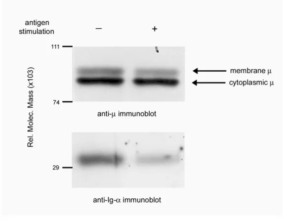
Antigen ligation of the BCR destabilizes Ig-α/Ig-β from μm as evidenced by the inability to
coprecipitate stoichiometric amounts of Ig-α/Ig-β with μm (91). As shown in Figure 2.1 and
previously described, immunoprecipitation of the BCR complex from antigen stimulated
K46μ cells failed to coprecipitate stoichiometric amounts of Ig-α/β (lane 1 compared to lane
2) despite precipitation of comparable amounts of μm. Quantitation of the amount of Ig-α
relative to the amount of μm revealed a 2-fold decrease in Ig-α/β. Given the dual role of the
BCR in signal transduction and receptor-mediated endocytosis, we reasoned that receptor
destabilization might provide a mechanism for antigen to rapidly enter the endocytic pathway
while simultaneously allowing Ig-α/Ig-β to sustain signal transduction. This predicts that
BCR destabilization represents a physical separation of μm from Ig-α/Ig-β thereby allowing
independent trafficking on the plasma membrane.
Antigen ligation of the BCR was previously shown to induce translocation of the receptor
complex to lipid rafts (37). Although this study showed both Ig-α and μm in lipid rafts, it
was unclear if the levels of μm relative to Ig-α were stoichiometric. To determine if BCR
destabilization was evident in lipid microdomains, we fractionated unstimulated and antigen
stimulated cells on a sucrose gradient (Figure 2.2A). Further, we simultaneously
immunoprecipitated the BCR complex from unstimulated cells (Figure 2.2B; lane 7), to
define the stoichiometry of sheathed receptors, thus providing a means to accurately
determine immunoblot exposure times. Antigen-stimulated K46μ cells showed induced
(fractions 10 and 11) (Figure 2.2A). Immunoblot analysis revealed that fraction 5 contained
lipid microdomains as evidenced by the presence of GM1 and Lyn (Figure 2.2A; bottom panel
and 2.2B middle panel). In addition, fraction 5 showed the translocation of Ig-α, but not μm,
within 1 minute of antigen stimulation (Figure 2.2B; top and bottom panels). The amount of
Ig-α recovered from fraction 5 was similar to the amount immunoprecipitated from 5x10⁶
unstimulated cells (Figure 2.2B; anti-Ig-α blot, lane 2 compared to lane 7). However, the
amount of μm in fraction 5 was undetectable compared to the amount expected from 5x10⁶
sheathed receptors (Figure 2.2B; anti-μ blot, lanes 2-6 compared to lane 7). The data show
Ig-α in lipid microdomains in the absence of μm, suggesting that BCR destabilization
Figure 2.1. Antigen stimulation induces BCR destabilization in K46μ cells. Anti-μ and
anti-Ig-α immunoblots of anti-μ immunoprecipitates. K46μ cells were either unstimulated
(lane 1) or stimulated with NP8BSA (1μg/10x106 cells/ml) for 50 minutes (lane 2). Cells
Figure 2.2. Destabilized Ig-α/Ig-β resides in lipid microdomains in the absence of μm.
(A) Cell lyates from unstimulated and antigen (NP8BSA)-stimulated (7 minutes) K46μ cells
were separated on sucrose gradients and immunoblotted for tyrosine-phosphorylated proteins
(upper panel) and GM1 (lower panel). (B) Anti-μ, anti-Lyn and anti-Ig-α immunoblots from
GM1-containing lipid microdomains (fraction 5). Lane 1: unstimulated-, lane 2-6: NP8
BSA-stimulated cells, lane 7; anti-μ immunoprecipitate from unstimulated, unfractionated K46μ
Ig-β fails to cocap with μm following antigen stimulation.
To assess if physical dissociation was evident in live cells, we examined unstimulated and
antigen-stimulated B cells for their membrane distribution of Ig-β and μm following
anti-μ−induced capping. We reasoned that if antigen ligation induced a physical separation of
the receptor complex, subsequent capping of μm would leave 50-75% of Ig-α/Ig-β outside
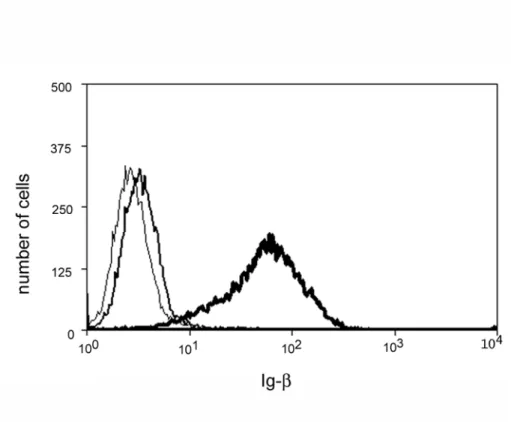
the cap (91). The analysis was performed on M12g3r cells since they fail to express surface
Ig-α/Ig-β in the absence of transfected μm (Figure 2.3). Within 30 minutes of antigen
stimulation, M12g3r cells showed a loss of coprecipitated Ig-α/β, and treating cells with the
src-kinase inhibitor, herbimycin, prevented this effect (Figure 2.4). Under this condition of
herbimycin treatment (100 μM for 30 minutes), Lyn-phosphorylation were completely
inhibited and Syk-phosphorylation was partially inhibited (Figure 2.5). This suggests that
BCR-mediated signals regulate receptor destabilization, consistent with previous studies (91).
Immunofluorescence staining of unstimulated cells showed a 40% colocalization of μm and
Ig-β (Figure 2.6; top panels). In addition, lipid microdomains stained for GM1 were evenly
distributed on membrane. Following antigen stimulation the amount of Ig-β that colocalized
with μm was reduced to only 14% (Figure 2.6; middle panels). This observation was not
due to an unexpected consequence of antibody-induced capping, Ig-α/Ig-β anti-μ maintained
the colocalization of μm and Ig-β when cells were treated with herbimycin to inhibit
destabilization (Figure 2.6; bottom panels). In addition, inhibiting kinase activation induced
μm and Ig-β to co-cap independent of GM1 aggregation. This suggests that receptor
destabilization occurs outside lipid microdomains. Since the lipid microdomains did not
visibly aggregate upon antigen stimulation, the capped μm/ Ig-α/Ig-β complexes (herbimycin
we cannot exclude the possibility that association of the BCR with a small fraction of lipid
microdomains is sufficient to induce the dissociation of 50-75% of Ig-α/Ig-β from μm during
BCR destabilization. Nonetheless, the 3-fold decrease in the colocalization of Ig-β and μm
following antigen stimulation supports the notion that BCR destabilization represents a
Figure 2.3. M12g3r cells fail to express surface Ig-α/Ig-β in the absence of transfected
μm. Expression of Ig-β on M12.4 (thin line) compared to M12g3r (thick line) cells. M12.4
cells stained with anti-μ are shown as the negative control (gray line). A representative of 2
Figure 2.4. Herbimycin inhibits BCR destabilization in M12g3r cells. Anti-μ and
anti-Ig-α immunoblots of anti-μ immunoprecipitates from M12g3r cells. Cells were either
unstimulated (lane 1) or stimulated with PC10BSA (1μg/5x106 cells/ml) for 30 minutes (lane
2) or treated with herbimycin at 100 μM for 30 minutes followed by antigen stimulation.
Cells were lysed in 1% Chaps and the BCR complex was immunoprecipitated with anti-μ.
Figure 2.5. Herbimycin inhibits BCR-mediated signal transduction.
Anti-phosphotyrosine immunoblots of anti-Lyn (upper, left panel) or anti-Syk (upper, right panel)
immunoprecipitates. M12g3r cells were either untreated (lanes 1 and 2 of each panel) or
herbimycin-treated (lane 3 of each panel) and stimulated with PC10BSA (1μg/5x106 cells/ml)
to assess BCR sensitivity. Anti-Lyn (bottom, left panel) and anti-Syk (bottom, right panel)
immunoblots were done sequentially. The data are representative of 2 experiments and has
Figure 2.6. Ig-β fails to cocap with μm following antigen stimulation.
M12g3r cells were either fixed prior to staining (top panels) or stimulated with PC10BSA for
10 minutes (middle panels) or pretreated with herbimycin as in Figure 4, followed by antigen
stimulation (bottom panels). Cells were stained for μm (Cy3), Ig-β (Alexa 488) and GM1
(Alexa 647). Approximately 50% of all cells showed good capping and comparable staining
19-Destabilization of the BCR complex represents a physical dissociation of Ig-α/Ig-β from
μm. The data strongly suggest that Ig-α/Ig-β dissociated from μm following antigen
stimulation. However, we could not formally exclude the possibility that Ig-α/Ig-β was
re-expressed outside the anti-μ-induced cap during stimulation and staining. Toaddress this,
we assessed the location of Ig-α/Ig-β relative to μm immediately post-stimulation using
native membrane sheets viewed by transmission electron microscopy (TEM). This method
afforded greater resolution than confocal microscopy, and unlike ultrathin sectioning,
allowed the analysis of large pieces of native membrane (102, 103). In addition, it allowed a
more accurate assessment of the kinetics of destabilization. To induce receptor
destabilization and simultaneously label antigen-bound μm, we prepared gold-conjugated
PC10BSA and tested its ability to induce tyrosine phosphorylation following BCR
crosslinking of M12g3r cells. As shown in Figure 2.7, the level of tyrosine phosphorylation
induced by gold-conjugated PC10BSA was comparable to that induced by unconjugated
antigen. Native membranes prepared from M12g3r cells that were fixed prior to the addition
of gold-conjugated antigen exhibited colocalization of 10 nm (gold-conjugated antigen) and
5 nm (Ig-α) gold particles, confirming the association of μm and Ig-α in unstimulated cells
(Figure 2.8A, Table 1). The percent colocalization was determined by the frequency of 10
nm particles that associated with 5 nm particles (see Materials and Methods for the criteria of
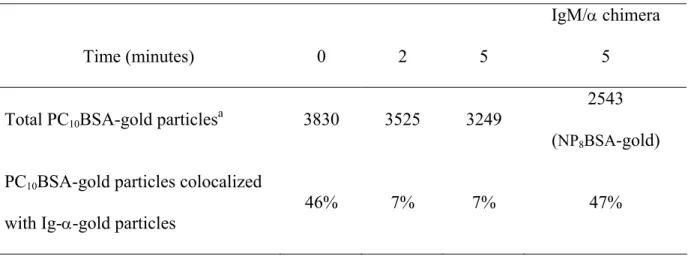
assessment). Of the 3830 PC10BSA-gold particles (10 nm) counted, 46% were associated
with Ig-α-gold particles (5 nm), with an average distance of 18 ± 2 nm separating small and
large gold particles. In contrast, cells stimulated for 5 minutes showed 7% colocalization,
with the average distance of 218 ± 5 nm separating the receptor components (Figure 2.8B,
the distance separating the receptor components, confirm a physical dissociation of the BCR
complex within 5 minutes of receptor ligation.
To establish if the low frequency of μm that associated with Ig-α in resting cells (46%)
reflected the limitations of staining the BCR complex or constitutive dissociation of resting
receptors, we determined the frequency of μm/Ig-α association in cells expressing a chimeric
BCR. The chimeric receptor contained a NP-specific extracellular domain fused to Ig-α
cytoplasmic tail (IgM/α) and was predicted to show 100% colocalization of μm and
Ig-α (105). However, only 47% of chimeric μm colocalized with Ig-α (Figure 2.8C, Table 1).
Thus, the relatively low frequency of μm/Ig-α association in unstimulated cells reflected the
inability to label the components of the BCR at saturating levels. This finding is consistent
with the confocal microscopy study where 40% of μm colocalized with Ig-β (Figure 2.6).
Nonetheless, the 6-fold decrease in the colocalization of μm with Ig-α clearly indicates a
dissociation of the receptor complex following ligation. The Ig-α staining was specific in
Figure 2.7. Gold-conjugated PC10BSA induces tyrosine phosphorylation.
Anti-phosphotyrosine immunoblot of M12g3r cells either unstimulated or stimulated for 1 minute
with unconjugated or gold (10 nm)-conjugated PC10BSA (0.5 μg/5x106 cells/ml). The data
Figure 2.8. Antigen stimulation induces a physical dissociation of μm from Ig-α/Ig-β.
(A) Unstimulated M12g3r cells were fixed prior to μm staining or, (B) stimulated with
PC10BSA-gold (10 nm, large arrows) for 5 minutes prior to the preparation of native
membranes then stained for Ig-α (5 nm, small arrows). (C) Cells expressing chimeric BCR
were stimulated with NP8BSA-gold (10 nm, large arrows) for 5 minutes prior to the
preparation of native membrane then stained for Ig-α (5 nm, small arrows). (D) Cells were
stained with unrelated isotype matched antibody, Her2/Neu (5 nm), after preparing native
display higher magnification of association/dissociation of μm and Ig-α. Arrowheads in (A)
Table 1. Assessment of subcellular localization of μm and Ig-α on native membrane
sheets in M12g3r cells.
Time (minutes) 0 2 5
IgM/α chimera
5
Total PC10BSA-gold particlesa 3830 3525 3249
2543
(NP8BSA-gold)
PC10BSA-gold particles colocalized
with Ig-α-gold particles
46% 7% 7% 47%
Cells were either fixed prior to μm staining (t = 0) or stimulated with PC10BSA-gold (10 nm)
or NP8BSA-gold (10 nm). Native membrane sheets were prepared and stained on the
intracellular side for Ig-α (5 nm). Membrane localization of gold particles was analyzed by
TEM at 25,000x.
aGold particles (10 nm) were counted from at least 55 different native membrane sheets
Antigen-bound μm associates with clathrin-coated vesicles following BCR ligation.
Previous studies suggested that endocytosis of antigen-bound receptors occurred through
CCVs (48). To assess if receptor destabilization played a role in this process, we viewed
intact cells using confocal microscopy for the distribution μm, Ig-β and clathrin following
antigen stimulation. We chose a timepoint of 5-10 minutes based on the EM study (Figure
2.8 A-B). Images of Z-axis planes from unstimulated cells revealed that μm and Ig-β were
evenly distributed on the plasma membrane, while clathrin was distributed throughout the
cytoplasm with a slightly higher concentration at the membrane (Figure 2.9; upper panels).
In unstimulated cells, only 5% of μm and 3% of Ig-β colocalized with CCVs. Following
antigen stimulation, this pattern changed dramatically such that 68% of μm became
clathrin-associated in large foci at the membrane (Figure 2.9; lower panels). However, the majority
of Ig-β remained distributed over the plasma membrane with only 2% of Ig-β associating
with CCVs, similar to the findings in Figure 2.6 middle panels. These data indicate that
antigen stimulation promotes the association of μm with CCVs, but not the association of
Ig-β.
To further assess the distribution of μm, Ig-α/Ig-β and clathrin in native membranes, we
analyzed native membrane sheets by TEM. As shown in Figure 2.10A and Table 2, 14% of
PC10BSA-gold(10 nm) resided within CCVs (5 nm) in native membranesfrom resting cells.
Within five minutes of BCR ligation, the distribution of μm changed dramatically, revealing
a 4-fold increase in the association of PC10BSA-gold (10 nm) with clathrin (5 nm) (Figure
2.10B, Table 2). The staining for clathrin heavy chain and antigen was specific sincea
membrane (Figure 2.10C, 5D). These data show that antigen-bound μm associates with
Figure 2.9. Antigen stimulation colocalizes μm with CCVs. Cells were fixed prior to
staining (upper panels) or stimulated with PC10BSA for 10 minutes (lower panels) then
stained for μm (Cy3), Ig-β (Alexa 647) and clathrin (Alexa 488). The percent colocalization
of μm or Ig-β with CCVs was quantitated from 15 cells obtained from 3 independent
experiments. Approximately 50% of all cells showed good capping and comparable staining
Figure 2.10. BCR ligation induces the association of antigen-μm, but not Ig-α, with
clathrin-coated vesicles. (A) Cells were fixed, then stained with PC10BSA-gold (10 nm,
large arrows) and clathrin (5 nm, small arrows) or, (B) stimulated for 5 minutes prior to
preparation and staining of native membranes. Inset displays a higher magnification of μm
association with CCVs. The large and small arrows represent some of the large and small
gold particles. (C) Cells were prepared as in (A), and then stained with an isotype matched,
polyclonal mouse IgG antibody (5 nm). Arrows depict 10 nm gold particles. (D) Cells
were stimulated with BSA-gold (10 nm) for 5 minutes prior to the preparation of native
membranes, and then stained for clathrin (5 nm). Arrows depict 5 nm particles staining
CCVs. (E) Cells were fixed, and then stained forIg-α (12 nm, large arrows), and clathrin (5
nm, small arrows) or, (F) stimulated with PC10BSA for 5 minutes prior to the preparation and
staining of native membranes. (G) Cells were fixed, and then stained for Ig-α (5 nm, small
arrows) and clathrin (10 nm, arrow heads), or (F) stimulated with PC10BSA for 5 minutes
prior to the preparation and staining of native membranes. The data are representative of 5-10
Table 2. Assessment of subcellular localization of μm, Ig-α and CCVs on native
membrane sheets in M12g3r cells.
Time (minutes) 0 2 5 10 20
Total PC10BSA-gold particlesa 1750 1833 3212 1995 2064
PC10BSA-gold particles
Colocalized with CCVs
14% 55% 47% 45% 54%
Total Ig-α gold particlesb 987 7556 2812 1283 2147
Ig-α-gold particles
Colocalized with CCVs
20% 16% 23% 13% 11%
Cells were either fixed prior to μm staining (t = 0) or stimulated with PC10BSA-gold (10 nm)
or unconjugated antigen. Native membrane sheets were prepared and stained on the
intracellular side for clathrin (5 nm) and/or Ig-α (12 nm). Membrane localization of gold
particles was analyzed by TEM at 25,000x.
aGold particles (10 nm) were counted from 14-39 different native membrane sheets obtained
from 2-7 different experiments.
bGold particles (12 nm) were counted from 18-76 different native membrane sheets obtained
Ig-α fails to translocate into clathrin-coated vesicles following BCR ligation.
Collectively, the data demonstrate a physical separation of Ig-α/Ig-β from μm following
antigen ligation of the BCR. The findings that antigen-bound μm associates with CCVs
while Ig-α resides in lipid microdomains suggest that μm associates with clathrin in the
absence of Ig-α/Ig-β. To determine if antigen stimulation induced comparable recruitment of
Ig-α into CCVs, we stained native membranes for clathrin (5 nm) and Ig-α (12 nm). In
resting cells, 20% of Ig-α associated with CCVs (Figure 2.10E, Table 2). However, there
was no increased association of Ig-α with CCVs following antigen stimulation (Figure 2.10F,
Table 2). These findings were not due to the inability of the anti-Ig-α antibody to recognize
phosphorylated protein since this antibody immunoprecipitated phosphorylated Ig-α (88). In
addition, the density of Ig-α staining in membranes from unstimulated cells compared to
antigen-stimulated cells did not change. The failure to colocalize was not due to the
inability of12 nm gold particles (Ig-α) to enter the clathrin lattice, sincereversing the gold
particle size did not change the results (Figure 2.10G-H). This suggests that although some
Ig-α constitutively colocalizes with clathrin, antigen-ligation of the BCR does not induce
Unsheathed μm preferentially associates with clathrin-coated vesicles.
The selective association of μm, but not Ig-α, with CCVs suggests that destabilization may
play a role in entry of μm into CCVs. However, the low basal association of Ig-α with
CCVs, regardless of antigen stimulation, raised the possibility that a small pool of BCR
entered CCVs as an intact complex. To resolve this, we assessed the frequency at which
μm and Ig-α colocalized within the same CCVs. CCVs containing PC10BSA-gold (10 nm)
particles were identified by their prototypic polyhedral structure. In resting cells, 10% of the
PC10BSA-gold (10 nm) particles found within CCVs were associated with Ig-α(5 nm)
(Figure 2.11A). Following antigen stimulation, there was a 4-fold increase in the
association of μm with CCVs (Table 2) however; only 3-4% of the μm were associated with
Ig-α (Figure 2.11A). This indicates that unsheathed μm preferentially enters CCVs.
Destabilization of Ig-α/Ig-β is required for μm association with clathrin-coated vesicles.
To further assess the role of BCR destabilization in antigen entry to CCVs, we treated cells
with a kinase inhibitor to prevent BCR destabilization (Figure 2.4)(91). We reasoned that if
BCR destabilization was required for clathrin-mediated endocytosis, inhibiting
destabilization would diminish the translocation of μm to CCVs. As shown in Figure 2.11B,
μm failed to associate with CCVs in herbimycin-treated cells, suggesting that BCR
destabilization facilitates the movement of antigen-bound μm to CCVs. It remained possible
that lack of μm association with CCVs was simply due to failure of receptor internalization.
To directly assess if B cell receptors clear the membrane in the absence of kinase activation,
we monitored the clearance of HRP-tagged antigen in untreated and herbimycin-treated
herbimycin-treated cells occurred with comparable kinetics and magnitude. Further,
herbimycin treatment did not affect receptor clearance as detected by flow cytometry (Figure
2.11D). Collectively, conditions that prevented BCR destabilization also inhibited the
association of μm with CCVs, suggesting that receptor destabilization is required for the




![Crystal structure of (methanol κO)[5,10,15,20 tetrakis(2 aminophenyl)porphyrinato κ4N]zinc(II)–chloroform–methanol (1/1/1)](data:image/gif;base64,R0lGODlhAQABAIAAAP///wAAACH5BAEAAAAALAAAAAABAAEAAAICRAEAOw==)