THE PROTEASE/ANTIPROTEASE BALANCE DETERMINES INFLUENZA SUSCEPTIBILITY AND CAN BE MODIFIED BY OXIDANTS AND
ANTIOXIDANTS
Megan Garcia Meyer
A dissertation submitted to the faculty of The University of North Carolina at Chapel Hill in partial fulfillment of the requirements for the degree of Doctor of Philosophy in the Department of Microbiology and Immunology
Chapel Hill 2015
iii ABSTRACT
Megan Garcia Meyer: The protease/antiprotease balance determines influenza susceptibility and can be modified by oxidants and antioxidants
(Under the direction of Ilona Jaspers)
The respiratory epithelium functions as a central orchestrator to initiate and organize responses to inhaled stimuli. Proteases and antiproteases are secreted from the respiratory epithelium and are involved in respiratory homeostasis. Modifications to the protease/antiprotease balance can lead to the development of lung diseases such as emphysema or chronic obstructive pulmonary disease. Furthermore, altered
protease/antiprotease balance, in favor for increased protease activity, is associated with increased susceptibility to respiratory viral infections such as influenza virus. We
demonstrated that inhaled oxidants, such as cigarette smoke, alters intracellular regulation and extracellular modifications of a key respiratory antiprotease, secretory leukocyte protease inhibitor (SLPI). Additionally, we established that SLPI is a novel anti-influenza biomarker that restricts influenza infection in vivo and in vitro. Furthermore, we
investigated the effect of nutritional antioxidants, such as sulforaphane (SFN), on SLPI expression and found that SFN induced SLPI expression and secretion using in vivo and in vitro models. Finally, we detailed that smokers have increased secretions of an
influenza-activating protease, transmembrane protease serine 2 (TMPRSS2) and that SFN supplementation reduced TMPRSS2 secretion, which protected against influenza
infection in vitro. Taken together, these studies establish the integral role of the
iv
v
ACKNOWLEDGEMENTS
There have been many friends, family, colleagues, and mentors that have helped in my professional and scientific development. First, I would like to thank my mentor, Ilona Jaspers, for her guidance and support. Ilona was always willing to meet with me to flesh out new ideas, hypotheses, and experimental designs and provided valuable insight on during my dissertation research. Ilona also encouraged and supported my career goals, and I am extremely grateful of the freedom I was given to develop both my research and professional skills. Moreover, “team Jaspers” was a significant contributor to my graduate school experience. I want to thank the past and current members of “team Jaspers”, especially Rebecca Bauer, Kelly Chason, Claire Chehrazi, Philip Clapp, Billy Fischer, Ellen Glista-Baker, Katie Horvath, Shannon Jones, Blanche Letang, Loretta Muller, Erica Pawlak, and Adam Speen for making research a fun and collaborative environment. I’d especially like to thank Missy Brighton for all of the primary cell culture assistance and being an awesome lab “fairy”. I would also like to thank my other colleagues at the CEMALB as well as the EPA for their scientific and professional assistance.
vi
Freeman, for the constant support and professional development. I also want to thank NHLBI for funding, which supported three years of my dissertation research.
Lastly, I would like to thank all my friends and family for their encouragement over the years. Thanks to my undergraduate mentor, Chis Thompson, for inspiring me to pursue my PhD. Times spent in the Thompson lab cultivated my interest in scientific research and exposed me to the fascinating world of microbiology and immunology. Many thanks to my immediate family for all of the support, love, and laughs over the years. I want to especially thank my parents for instilling in me the importance of education, engaging my curiosity, and allowing me to ask questions about
vii
TABLE OF CONTENTS
LIST OF TABLES ... xi
LIST OF FIGURES ... xii
LIST OF ABBREVIATIONS ... xiv
CHAPTER 1 ...1
1.1 Respiratory epithelium serves as a central “orchestrator” to initiate and coordinate respiratory responses ...2
1.1.1 Barrier and mechanical functions of the respiratory epithelium ... 2
1.1.2 Receptor-mediated interactions of the respiratory epithelium ... 3
1.2 Epithelial cell-derived secreted host defense mediators exhibit novel antimicrobial effects ...5
1.2.1 Lysozyme and lactoferrin: the most abundant host defense mediators found in the respiratory tract ... 5
1.2.2 Lung lipid mediators: surfactant proteins ... 6
1.2.3 Respiratory cationic peptides: cathelicidins and defensins ... 7
1.2.4 Proteases and antiproteases of the lung: a delicate enzymatic balance ... 9
1.3 Respiratory viruses infect respiratory epithelial cells and cause respiratory exacerbations ...20
1.3.1 Influenza virus components and viral life cycle ... 21
viii
1.4 Cigarette smoking is a risk factor for influenza infection ...23
1.4.1 Cigarette smoking decreases respiratory host defense responses to influenza infection ... 23
1.4.2 Cigarette smoking induces oxidative stress on the respiratory epithelium ... 24
1.4.3 Cigarette smoke modulates the protease/antiprotease balance 25 1.4.4 Cigarette smokers are immunosuppressed ... 26
1.5 Nutritional supplementation can decrease oxidative stress and protect against IAV infection ...27
1.5.1 The effects of vitamins and trace elements on IAV infection in vivo... 27
1.5.2 Antioxidants are an attractive strategy to induce host defense and protect against infection ... 29
1.5.3 Sulforaphane (SFN), a nutritional antioxidant, modulates the protease/antiprotease balance and protects against IAV infection ... 30
1.6 Summary ...31
CHAPTER 2 ...40
2.1 Overview ...40
2.2 Introduction ...42
2.3 Materials and methods ...44
2.4 Results ...49
2.5 Discussion ...53
CHAPTER 3 ...65
3.1 Overview ...65
ix
3.3 Materials and methods ...70
3.4 Results ...75
3.5 Discussion ...80
CHAPTER 4 ...91
4.1 Overview ...91
4.2 Introduction ...93
4.3 Materials and methods ...96
4.4 Results ...101
4.5 Discussion ...104
CHAPTER 5 ...116
5.1 Overview ...116
5.2 Introduction ...117
5.3 Materials and methods ...120
5.4 Results ...124
5.5 Discussion ...127
CHAPTER 6 ...135
6.1 Modifications to the antiprotease shield: implications for infection ...135
6.2 Targeting the antiprotease shield: mechanisms to prevent infection and/or chronic lung disease ...137
6.3 Using sulforaphane (SFN) to induce SLPI expression and secretion: in vivo supplementation ...140
x
6.5 The effects of inhaled oxidants on the protease/antiprotease balance: implications for chronic lung disease and respiratory
infection ...146 6.6 Mitigating the effects of inhaled oxidants: policy and
xi
LIST OF TABLES
Table 1-1. Target substrates, sources, and activity of respiratory
proteases. ... 33
Table 1-2. Target substrates, sources, and activity of respiratory antiproteases. ... 34
Table 1-3. Genome and structure, target cell type, and sensitivity to protease/antiprotease balance of respiratory viruses. ... 36
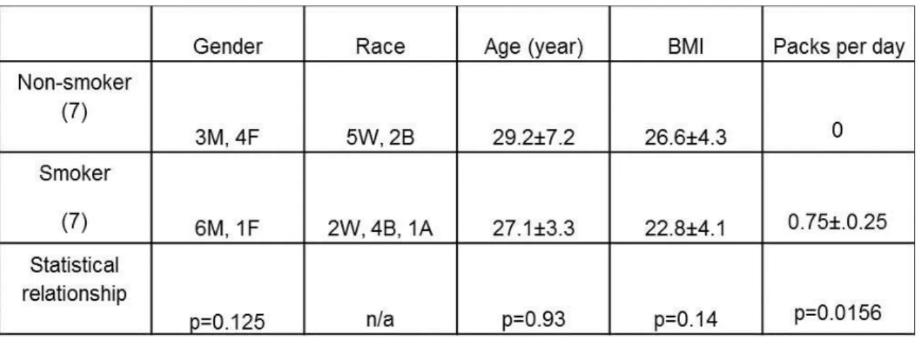
Table 2-1. Subject characteristics and smoking status... 58
Table 4-1. Subject characteristics. ... 108
Table 4-2. BSH-associated side effects. ... 109
xii
LIST OF FIGURES
Figure 1-1. Respiratory epithelial cells function as an “orchestrator” to initiate, coordinate, and respond to inhaled
stimuli. ...37 Figure 1-2. SFN dampens inflammation, alleviates oxidative stress,
and alters the protease/antiprotease balance. ...38 Figure 1-3. The protease/antiprotease balance regulates IAV
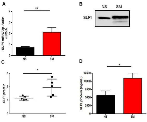
infection and can be modulated by SFN supplementation...39 Figure 2-1. SLPI mRNA and protein secretion from in vitro and in
vivo samples. ...59 Figure 2-2. Transcriptional regulation of SLPI in bronchial
epithelial cells. ...60 Figure 2-3. Expression of slpi in wild-type (WT) and stat1-/- mice. ...61 Figure 2-4. STAT1 mRNA and protein levels in NEC from NS and
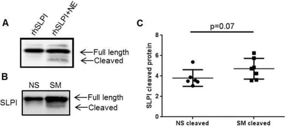
SM. ...62 Figure 2-5. Extracellular posttranslational modification of SLPI. ...63 Figure 2-6. SLPI activity against NE in NLF. ...64 Figure 3-1. SLPI is induced during H3N2 infection in NEC and
during H1N1 and H3N2 infection in BEAS-2B cells. ...85 Figure 3-2. Decreasing SLPI expression increases H1N1 and
H3N2 infection in BEAS-2B cells. ...86 Figure 3-3. rhSLPI treatment protects against H3N2 replication. ...87 Figure 3-4. SLPI modulates RIG-I expression during influenza
infection. ...88 Figure 3-5. Knocking down SLPI expression increases viral entry...89 Figure 3-6. Elevated baseline SLPI NLF protein levels are
correlated with reduced markers of IAV replication in vivo. ...90 Figure 4-1. Study design. ...111 Figure 4-2. BSH supplementation stimulates Nrf2 and phase II
xiii
Figure 4-3. BSH supplementation induces secreted SLPI levels in
NLF. ...113 Figure 4-4. SFN supplementation alters secreted SLPI protein in
differentiated NEC. ...114 Figure 4-5. SLPI expression is regulated by Nrf2 and can be
induced with SFN supplementation in vitro. ...115 Figure 5-1. Nasal secretions from smokers increase HA cleavage
and IAV replication...131 Figure 5-2. TMPRSS2 is elevated in NLF from smokers in vivo. ...132 Figure 5-3. SFN supplementation in NEC decreases TMPRSS2
expression/secretion in vitro. ...133 Figure 5-4. SFN supplementation in NEC decreases intracellular
TMPRSS2 mRNA, extracellular TMPRSS2 protein levels, and
markers of IAV replication in vitro. ...134 Figure 6-1. SLPI NLF levels from SM correlate with markers of
cigarette smoking. ...152 Figure 6-2. SLPI NLF levels from SM do not correlate with
xiv
LIST OF ABBREVIATIONS
AJ Adherens junction α-1AT α-1 antitrypsin
AQI Air quality index
AR Androgen receptor
ARE Antioxidant response element
ARDS Acute respiratory distress syndrome
AUC Area under the curve
BSH Broccoli shake homogenate
CAM Complementary and alternative medicine
CLR C-type lectin receptor
COPD Chronic obstructive pulmonary disease
CSE Cigarette smoke extract
DAMP Damage associated molecular pattern
DESC Differentially expressed in squamous cell carcinoma
ECM Extracellular matrix
xv
ETS E-twenty-six
GJ Gap junction
HA Hemagglutinin
HAT Human airway trypsin
HBD Human beta defensin
hCAP18 Human cationic antimicrobial protein 18
HIV Human immunodeficiency virus
HO-1 Heme oxygenase-1 (HO-1)
HRV Human rhinovirus
HPV Human papillomavirus
IAV Influenza A virus
IFN Interferon
IL-1β Interleukin-1β
IL-6 Interleukin-6
IL-8 Interleukin-8
IRF Interferon regulatory factor
xvi
JAK Janus kinase
Keap1 Kelch-like ECH-associated protein 1
LAIV Live attenuated influenza virus vaccine
LPS Lipopolysaccharide
M Matrix protein
MAPK Mitogen-activated protein kinase
MERS-CoV Middle East respiratory syndrome coronavirus
MMP Matrix metalloproteinase
NBS Nasal biopsy
NCCAM National Center for Complementary and Alternative Medicine
NE Neutrophil elastase
NEC Nasal epithelial cell
NF-κB Nuclear factor kappa-light-chain-enhancer of activated B cells
NHANES National Health and Nutrition Examination Survey
NK Natural killer cell
NLF Nasal lavage fluid
xvii
Nrf2 Nuclear factor (erythroid-derived 2)- like 2
NS Non-smoker
NS1 Non-structural protein 1
NS2 Non-structural protein 2
NQO1 NADPH quinone oxidoreductase 1
PAMP Pattern associated molecular pattern
PAR2 Protease-activated receptor 2
PB Polymerase basic protein
PRR Pattern recognition receptor
RCL Reactive center loop
RIG-I Retinoic acid-inducible gene I
RLR Retinoic acid-inducible gene I-like receptor
RLU Relative luciferase unit
ROS Reactive oxygen species
rSLPI Recombinant SLPI
rhSLPI Recombinant human SLPI
xviii
SARS-CoV Severe acute respiratory syndrome coronavirus
Serpin Serine protease inhibitor
SFN Sulforaphane
SLPI Secretory leukocyte protease inhibitor
SM Smoker
SP Surfactant protein
STAT Signal transducer and activator of transcription
VLP Virus-like particle
TCID50 Tissue-culture infectious dose
TIMP Tissue inhibitor of metalloproteinase
TLR Toll-like receptor
TJ Tight junction
TMPRSS2 Transmembrane protease seine 2
TNF-α Tumor necrosis factor-α
TTSP Type II transmembrane serine protease
WAP Whey acidic protein domain
1 CHAPTER 1
Introduction
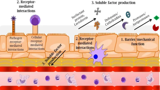
Nearly 40 different types of cells have been identified in the respiratory tract, including neutrophils, dendritic cells, macrophages, natural killer (NK) cells, B cells, and T cells (1). Due to this diverse population of cells, a central “orchestrator” is necessary for coordinating an appropriate immune response to inhaled stimuli. The respiratory epithelium is one of the first sites within the respiratory tract to be exposed to inhaled stimuli and functions as a focal “orchestrator” to organize and initiate appropriate responses in three distinct ways (Figure 1-1). First, the respiratory epithelium forms a physical barrier through tightly regulated cell-cell interactions. These interactions promote basic biologic functions, protect against invading pathogens, and establish mechanical strength. Second, epithelial cells express receptors and ligands that coordinate cellular responses and activate immune cells. These processes are important for detecting foreign stimuli, communicating among different cells, and guiding immune responses. Third, respiratory epithelial cells secrete soluble factors such as cytokines, chemokines, and host defense mediators such as mucins, lactoferrin, defensins, and
2
the role of secreted soluble factors, such as the protease/antiprotease balance, in response to oxidants, antioxidants, and viral infection. These findings will elucidate the impact of this balance in the context of a respiratory viral infection and further highlight how respiratory epithelial cells are master regulators of respiratory function.
1.1 Respiratory epithelium serves as a central “orchestrator” to initiate and coordinate respiratory responses
1.1.1 Barrier and mechanical functions of the respiratory epithelium
The respiratory epithelium is comprised of basal, secretory, and ciliated cells that vary in number and density throughout the respiratory tract (2, 3). Playing an integral role in the respiratory structure, basal cells firmly attach to the basement membrane (4). Secretory cells are comprised of goblet cells, which secrete mucus, a gel-like network that is important for trapping foreign objects (5). Ciliated columnar cells are the most numerous cell type of the respiratory tract that arise from either basal or secretory cells (3). These cells express mechanical organelles called cilia, which are extracellular machinery proteins that clear foreign substances trapped in the mucus matrix.
3
respiratory epithelium through the connective network and strength of desmosomes (9). AJ function similarly to desmosomes, serving as an adhesive between adjacent cells. Located below TJ, AJ serve as critical epithelial linkers to cytoskeleton actin filaments (10). Lastly, GJ form clusters of channels that link neighboring cells and allow for ion, secondary messenger, and metabolite transport (11).
Beyond barrier protection, the respiratory epithelium is equipped with mechanical strategies to protect against foreign inhaled insults. The respiratory epithelium is coated with mucus, a thin viscoelastic gel that allows for hydration of the epithelium. Mucus is comprised of proteins called mucins that form the structural network of the mucus lining (12, 13). Mucins are large glycoproteins that are encoded by MUC genes and provide the viscoelastic properties of mucus. To date, over 10 mucin genes have been detected, with MUC5AC and MUC5B being the most prevalent mucins found in human sputum secretions (14). Mucus is not only important for hydration but also is a vital component for the mucocillilary escalator, a mechanical process that traps and clears foreign
particles. The escalator function is achieved by ciliary movement and beating, which pushes the mucus and trapped foreign particles out of the respiratory tract. However, elevated MUC levels can be detrimental for respiratory health since increased MUC5AC and MUC5B are increased in chronic obstructive pulmonary disease (COPD) and asthma patients (15, 16). In sum, physical and mechanical properties of the respiratory
epithelium promote respiratory homeostasis and protect against injurious stimuli.
1.1.2 Receptor-mediated interactions of the respiratory epithelium
4
immune cells. One mechanism of detection involves pattern recognition receptors
(PRRs), which are receptors that recognize conserved microbial motifs, termed pathogen associated molecular patterns (PAMPs), and endogenous ligands produced from stressed cells, termed damage associated molecular patterns (DAMPs) (17). Respiratory epithelial cells possess a wide variety PRRs such as toll-like receptors (TLRs), nod-like receptors (NLRs), C-type lectin receptors (CLRs), and retinoic acid-inducible gene I-like receptors (RLRs). CLRs and some TLRs serve as extracellular sensors, while NLRs, RLRs, and some TLRs act as intracellular sensors (18-20). Furthermore, epithelial cells express various receptors such as integrins and adhesion molecules that bind to inhaled stimuli, such as respiratory viruses, and allow for infection (21, 22). Once foreign stimuli are detected, respiratory epithelial cells communicate with neighboring cells and immune cells through extracellular receptors and ligands (23). These physical signals are important for mounting and controlling immune responses.
5
summary, the respiratory epithelium serves an important “orchestrator” to promote barrier and mechanical function, moderate receptor-mediated interactions, and produce soluble factors that contribute to respiratory host defense.
1.2 Epithelial cell-derived secreted host defense mediators exhibit novel antimicrobial effects
1.2.1 Lysozyme and lactoferrin: the most abundant host defense mediators found in the respiratory tract
In addition to the production and secretion of cytokines and chemokines,
epithelial cells secrete host defense mediators that possess broad antimicrobial properties and are imperative for the early, innate responses to infection. Two of the most abundant host defense mediator found in respiratory nasal secretions or sputum samples are lysozyme and lactoferrin (26, 27). Lysozyme is a 14 kilodalton (kDa) enzyme that cleaves bacterial cell wall components, such as peptidogycan, which results in bacterial cell lysis. Lysozyme possesses antimicrobial activity against many Gram-positive microbes, but is less effective against Gram-negative bacteria (28). When challenged with group B streptococci, transgenic mice that over-expressed lysozyme in the lung cleared the bacterial infection and had enhanced rates of survival compared to wild type mice (29). Lysozyme is induced by enveloped viruses; however, viruses such as influenza A virus (IAV) antagonize lysozyme function by inhibiting lysozyme release from cells (30, 31).
6
sequestering iron, a required nutrient for microbial growth (32). Additionally, lactoferrin binds to lipopolysaccharide (LPS), a bacterial cell wall component, which results in bacterial permeabilization (33). Lactoferrin also prevents viral entry either by blocking host extracellular receptors or by directly binding to the viruses to prevent entry (34). Moreover, lactoferrin alters immune responses. Shin et al. reported that mice treated with lactoferrin before intranasal infection with IAV had an attenuated rate of
pro-inflammatory cytokines such as interleukin 6 (IL-6), leukocyte infiltrate, and lung destruction compared to control animals (35). These findings indicate that while lysozyme and lactoferrin exhibit anti-viral properties, lactoferrin may possess greater anti-IAV properties than lysozyme.
1.2.2 Lung lipid mediators: surfactant proteins
Lung surfactant proteins (SP) are an additional class of secreted respiratory proteins that are comprised of four different subtypes: SP-A, SP-B, SP-C, and SP-D. SP can form multimers to increase accessibility and affinity to immune cells and pathogens, similar to antibody function (36). SP-B and SP-C are hydrophobic and regulate alveolar surface tension, while SP-A and SP-D are hydrophilic and mainly contribute to epithelial host defense responses (37). SP-A and SP-D range in size from 26-43 kDa and possess many carbohydrate-rich domains, which are important for agglutination and
7
phagocytosis and clearance of apoptotic cells (43, 44). Of note, genetic alterations to the SP-A gene is associated with increased risk for mechanical ventilation and acute
respiratory failure during IAV pandemics (45). Together, these studies illuminate the potent respiratory anti-IAV effects of SP-A and SP-D.
1.2.3 Respiratory cationic peptides: cathelicidins and defensins
Cathelicidins are cationic peptides secreted from neutrophil and macrophage granules as well as epithelial cells. The only cathelicidin found in humans is the 18kDa protein human cationic antimicrobial protein 18 (hCAP18) (46). hCAP18 binds and neutralizes bacterial LPS (47). Additionally, hCAP18 can be cleaved, resulting in the generation of LL-37, a 4.5kDa antimicrobial peptide that contains two leucine resides on its N-terminal domain. LL-37 stimulates various intracellular signaling cascades, such as the NF-κB and mitogen-activated protein kinase (MAPK) pathways, involved in
inflammation and proliferation (48-50).
8
infected with IAV infection had increased H2O2 release and neutrophil extracellular trap
formation (55).
Defensins (α, β, θ) are an additional family of highly cationic peptides that exert both antibacterial and anti-viral properties (56, 57). α-defensins are produced by
leukocytes, β-defensins (HBDs) are primarily produced by epithelial cells and θ-defensins are found in non-human primates (58). While over eight HBDs have been identified, only HBD1-3 have been isolated from human tissue. HBD1 is constitutively expressed and is primarily expressed in the epithelium of urinary and respiratory tracts (59, 60). HBD2 and HBD3 are expressed in the skin and mouth as well as the
gastrointestinal and respiratory tracts (61). HBD2 and HBD3 are induced in response to inflammatory stimuli in an NF-κB-dependent manner (62, 63).
9
defensins protect against viral infection by disrupting viral membrane permeability, blocking viral entry, and modulating cellular immune responses to viral infection.
1.2.4 Proteases and antiproteases of the lung: a delicate enzymatic balance In addition to antimicrobial peptides, proteases and antiproteases are key components of respiratory host defense. In the healthy lung, proteases maintain tissue homeostasis and their activities are regulated by antiproteases. Elevated net protease activity is associated with lung destruction and the development of chronic lung diseases such as emphysema and COPD (71, 72). Lung destruction is induced by proteolytic degradation of extracellular matrix (ECM) components, including collagens, laminins, and elastin, with type I collagen serving as the major target (73). Cysteine proteases, matrix metalloproteinases (MMPs), and serine proteases are prevalent proteases found in the lung, with serine proteases serving as the most predominant class. Moreover, each family has unique target substrates, cellular sources, and active sites (Table 1-1).
10
syndrome coronavirus (SARS-CoV), and cathepsin B is induced during IAV infection in mice (78, 79).
MMPs are a family of metal endopeptidases that target ECM components. Secreted by immune cells and epithelial cells, MMPs possess a common prodomain and catalytic domain. Because MMPs are synthesized as inactive zymogens, the signal domain must be cleaved for MMPs activity. The catalytic domain contains a conserved zinc binding site that confers protease activity. MMP1, 2, 3, 7, 8, 9, 12, and 13 are the main MMPs found in the respiratory tract and are induced by growth factors, reactive oxygen species, and pro-inflammatory cytokines (80-82). MMP1, 8, and 13 cleave collagen, while MMP2 and 9 act as gelatinases and also degrade elastin and type IV collagen (83, 84).
Additionally, increased MMP activity is associated with inflammation. Baseline MMPs levels such as MMP1, 2, 3, 8, and 9 are elevated in smokers and patients with COPD (85). Moreover, MMP9 cleaves pro-inflammatory cytokines such as interleukin-8 (IL-8), resulting in the release of a modified, more potent neutrophil chemotactic
11
Serine proteases make up the largest portion of respiratory proteases (92). While neutrophil elastase (NE), proteinase 3, and cathepsins G are neutrophil-derived proteases, other serine proteases such as type II transmembrane serine proteases (TTSPs) are
produced and secreted by the respiratory epithelium. TTSPs are a family of proteases that contain an N-terminal transmembrane domain, a stem region, and a C-terminal serine protease domain that contains the conserved catalytic triad composed of histidine,
aspartic acid, and serine (93). Protease activity is attained through the nucleophilic serine, which attacks the substrate’s carbonyl functional group. This activity generates an acyl intermediate, transfers a proton from the positively charged histidine to the substrate, and allows for the hydrolysis and cleavage of the substrate (94). TTSPs are synthesized as a single chain, inactive pro-enzyme and require cleavage following a basic arginine or lysine (95). The catalytic domains are located on the terminal extracellular region to permit direct exposure to the extracellular environment.
TTSPs are comprised of four subfamilies: the human airway trypsin-like
protease/differentially expressed in squamous cell carcinoma (HAT)/(DESC) subfamily that include HAT, DESC1, and HAT-like 1-5; the hepsin/transmembrane protease serine (TMPRSS) subfamily contains hepsin, TMPRSS2-5 and 13, mosaic serine protease large-form, and enteropeptidase; the corin protease, and the matriptase subfamily that includes matriptase 1-3, and polyserase-1 (94). While TTSPs are transmembrane proteases, release of the extracellular domains has been reported for several of the TTSPs such as
12
Matriptase cleaves and activates protease-activated receptor 2 (PAR2), hepatocyte growth factor, and urokinase, indicating that matriptase plays a role not only in ECM degradation but also in epithelial cell differentiation and remodeling (98, 99). In the respiratory epithelium, matriptase is expressed on the basolateral side of the epithelial cells. Upon activation, matriptase migrates to the apical side of the epithelium, where it can be secreted into the lumen of the respiratory tract. Recent studies have revealed that intracellular matriptase localizes to the plasma membrane and endosomes, allowing for interaction and activation of the IAV virion (100, 101). Moreover, secreted matriptase cleaves hemagglutinin (HA) protein on the IAV virion, including H1, but not H2 or H3, which activates the virion and allows for enhanced rates of replication (100, 101).
HAT cleaves fibrinogen, activates PAR2, and urokinase (101). In sputum samples from patients with chronic airway disorders such as bronchitis or asthma, secreted HAT levels are elevated (96). Moreover, Chokki, et al. demonstrated that HAT enhanced MUC5AC gene expression, resulting in increased mucus production (102). HAT is produced and secreted by ciliated epithelial cells, and is not found in basal or goblet cells of the respiratory tract (103). HAT activates the SARS-CoV viral spike protein necessary for host cell entry (104). Moreover, HAT cleaves the monobasic site of the HA protein required for HA activation, enhancing IAV replication (105-107). Cleavage of HA by HAT has been shown to occur at the cell surface either during attachment and entry into host cells or during budding and shedding of virions from an infected cell (106).
13
full length 70kDa protein, or a variety of smaller, truncated forms, depending on tissue site and localization (97, 109). TMPRSS2 mRNA levels are elevated in prostate cancer cell lines and nearly two-fifths of prostate cancer patients express duplication of the TMPRSS2 gene (110, 111). Increased levels of TMPRSS2 in prostate cancer cells are found as a gene fusion product of TMPRSS2 with an E-twenty-six (ETS) transcription factor, such as ERG and ETV (112). This fusion event generates a C-terminally truncated TMPRSS2 protein attached to an N-terminally truncated ERG/ETV protein, which induces migration and invasion in non-tumorigenic epithelial prostate cells (113). The TMPRSS2-ERG/ETV gene fusion is regulated by androgen receptor (AR) signaling and is associated with a high rate of prostate cancer recurrence and/or severe disease (114-116).
Additionally, TMPRSS2 is implicated in the activation of respiratory viruses. Cells expressing TMPRSS2 show enhanced replication of human metapneumovirus as well as increased activation and replication of SARS-CoV (117, 118). Bottcher et al. reported that cells stably expressing TMPRSS2 resulted in the monobasic cleavage of HA, which elevated IAV replication (105, 107). In recent reports, TMPRSS2 has been shown to preferentially cleave H1N1, and to a lesser degree, H3N2. Hatesuer et al. revealed that viral titers in H1N1 infected TMPRSS2-deficient mice were significantly reduced compared to wild type animals (119). Furthermore, this group reported that while H3N2 replicated at similar levels in both knockout and wild type mice, the knockout mice had increased survival and body weight post-H3N2 infection (119).
14
not lethal H3N2 infection, while another group showed that TMPRSS2 knockout mice were protected against both H1N1 and H3N2 infection (120, 121). These opposing findings suggest that HA activation not only depends on HA structure, but can vary amongst different viral strains bearing the same HA subtype. Despite these differences, there is substantial evidence implicating TTSPs in IAV pathogenesis.
Antiproteases are a broad class of proteins that inhibit proteases and modulate immune responses in the lung. Respiratory antiproteases are comprised of four families: tissue inhibitors of metalloproteinases (TIMPs), serpins, trappin-2/elafin, and secretory leukocyte protease inhibitor (SLPI). Each antiprotease family has unique target
substrates, cellular sources, and antiprotease function (Table 1-2). TIMPs are produced by alveolar epithelial cells and alveolar macrophages (122). TIMPs inhibit MMP activity through the formation of an N-terminal reactive ridge domain that inserts into the active site of the target MMP (123, 124). In the context of viral infection, over-expressing TIMP-1 blocks RSV syncytia formation (125) . However, since MMP activity is not implicated in viral activation and infection, there are limited studies that investigate the direct anti-viral effects of TIMPs.
15
for the N-terminal portion of the RCL to insert into the protease active site. At this time, the protease and serpin remain covalently linked, the protease structure is
conformationally changed, and the protease activity is significantly reduced (128). This action leads to the permanent deactivation of both serpin and protease, often described as a “suicide” event.
SerpinA1 encodes the most abundant antiprotease in the lung, alpha-1 antitrypsin (α-1AT). Point mutations of α-1AT generates a mis-folded version of this protein, resulting in an α-1AT deficiency (129). α-1AT deficient individuals are at a high risk for developing liver disease, emphysema, and COPD (130, 131). Respiratory epithelial cells secrete α-1AT in both the apical and basolateral compartments of the cell (132).
Additionally, serpins have been shown to block viral entry of enveloped viruses such as human immunodeficiency virus (HIV) and herpes simplex virus (133, 134). Smee et al. showed that in vitro serpin antithrombin III treatment inhibited IAV infection nearly 100 fold more than ribavirin, a nucleoside inhibitor used to halt IAV replication (135). The authors found that this effect was dependent on viral HA with decreasing efficacy in order of H1N1>H3N2>H5N1 (135). These data suggest that that serpin activity may parallel the TTSP activity, which is dependent on amino acid sequence, protein structure, and/or HA substrate accessibility.
16
antiprotease active site, and N-terminal region, which allows for interactions with ECM proteins (137). Elafin specifically inhibits NE and preoteinase-3, and is secreted from tracheal epithelial cells, Clara cells, type II cells, and alveolar macrophages (138, 139).
Trappin-2 and elafin function as alarm antiproteases, as both are produced in response to inflammatory stimuli (140). In respiratory epithelial cells, elevated elafin levels are protective against recombinant NE treatment and products derived from activated human neutrophils in vitro (141). The elafin promoter is regulated by
extracellular signal-regulated kinase, a component of the MAPK pathway and Rel-A, a mediator involved in NF-κB signaling (142, 143). In addition to functioning as an alarm antiprotease, elafin possesses antimicrobial effects. Ghosh et al. revealed that
recombinant elafin was protective during HIV infection only when elafin was
pre-incubated with HIV, suggesting that elafin directly interacts with HIV virion and prevents viral infection (144). Interestingly, this group found that secreted elafin levels were elevated in the cervico-vaginal lavages of negative women compared to HIV-positive women, albeit not statistically significant, suggesting that elafin secretions may be decreased after HIV infection. Similarly, after HRV infection, elafin is down-regulated in subjects with COPD (145). Furthermore, elafin has been identified as a biomarker for acute respiratory distress syndrome (ARDS) (146). These findings suggest that elafin functions as an alarm mediator and may be decreased after viral infection.
17
boomerang (149). Similar to the trappin family, SLPI contains a cysteine-rich WAP C-terminal domain that is responsible for protease inhibition as well as the N-C-terminal domain that stabilizes the protease/antiprotease complex and exerts antimicrobial activity (150). The C-terminal active site, which confers antiprotease activity, is comprised of leucine and methionine residues (151, 152).
The main function of SLPI is to inhibit serine proteases such as NE (its main target), cathepsin G, elastase, chymase (153, 154). SLPI further contributes to respiratory host defense by modulating inflammation and inducing wound healing. SLPI exerts cellular anti-inflammatory effects by preventing the degradation of regulatory
components of the NF-κB pathway and by directly competing for NF-κB binding sites in the promoter regions of pro-inflammatory cytokines such as IL-8 and tumor necrosis factor-α (TNF-α) (155-157). SLPI is also implicated in wound healing as reported by Ashcroft, et al. who investigated the role of SLPI in a mouse model of dermal wounds. Using this model, the authors found that SLPI knockout mice had impaired wound healing due to increased NE activity (158).
18
exerts fungicidal and fungistatic activity towards Aspergillus fumigatus and Candida albicans that is also dependent on the N-terminal domain of SLPI (163).
In the context of viral infections, SLPI inhibits HIV, human papillomavirus (HPV), and respiratory viruses. McNeeley et al. first detailed the anti-HIV activity of SLPI in human saliva (164). The authors demonstrated that SLPI was highly expressed in the saliva and inhibited HIV infection in monocytes and T cells in dose dependent
fashion. Further studies revealed that SLPI exerts anti-HIV properties not through direct interaction with the virus, but rather through interacting with the target host cell (165). However, this group found that despite decreasing viral load, SLPI did not alter the infectivity of progeny virions, suggesting that SLPI inhibits HIV at the early stages of viral infection (166).
19
Although SLPI contributes to respiratory host defense by decreasing
inflammation and infection, SLPI can also induce pro-inflammatory responses. Mulligan et al. demonstrated that post-translational modifications of SLPI increased IL-8 secretion, neutrophil recruitment, and vascular permeability (172). Extracellular SLPI can be post-translationally modified by respiratory proteases such as cathepsin L, MMP12, chymase, and NE, resulting in cleavage of SLPI (76, 87, 173, 174). Cleaved SLPI levels are elevated in COPD patients during respiratory infection, which renders SLPI
non-functional (72, 145, 175). Moreover, we have shown that in NLF from smokers, SLPI is post-translationally processed and cleaved (Chapter 2) (176). Oxidative stress, which can be derived from reactive oxygen intermediates or environmental exposures such as cigarette smoke, modifies the active site and decreases the antiprotease properties of SLPI (177, 178). While SLPI exerts powerful protective effects in the respiratory tract, post-translational alterations and/or oxidative modifications can drastically shift the activity of SLPI towards potentially damaging effects.
The protease/antiprotease balance is a delicate interaction of enzymes and
20
component for respiratory homeostasis but also is a powerful determinant of respiratory viral pathogenesis.
1.3 Respiratory viruses infect respiratory epithelial cells and cause respiratory exacerbations
Respiratory epithelial cells are the primary targets for respiratory viral infection based on extracellular ligands and residues that are found on these cells. More than 200 antigenically different viruses infect respiratory epithelial cells (179). These viruses include coronaviruses, rhinoviruses, metapneumoviruses, enteroviruses, adenoviruses, respiratory syncytial viruses, measles virus, parainfluenza viruses, and influenza viruses. These respiratory viruses differ in genome composition, viral structure, target cell type, and sensitivity to components of the respiratory protease/antiprotease balance (Table 1-3).
Upon viral infection, respiratory epithelial cells replicate viral genomes and propagate infectious virions for productive viral infection. Additionally, respiratory epithelial cells mount intracellular immune responses to warn neighboring cells as well as orchestrate recruitment of immune cells to sites of infection (180). The respiratory
21
context of a parainfluenza or influenza infection could elucidate mechanisms and strategies to prevent viral infections.
We are interested in investigating the role of the protease/antiprotease balance in the context of an influenza infection since influenza infects 2-5 million people worldwide each year and can cause worldwide pandemics, as seen with the recent 2009 H1N1 strain (181). Moreover, in the US alone, over 20,000 people die due to influenza infection and related complications (182). Influenza remains a significant public health burden due to yearly virus mutations and reassortment and the ability of the virus to infect a wide range of hosts. Despite large-scale vaccination programs and the development of anti-viral therapeutics, influenza-associated morbidity and mortality rates have not changed in recent years (183). As such, examining strategies, such as modulating the
protease/antiprotease balance, may serve as alternative target to prevent influenza infection.
1.3.1 Influenza virus components and viral life cycle
Influenza is comprised of three subtypes: influenza A, B, and C. All subtypes are able to infect humans, but IAV is the most common due to the large host range.
22
RNA segments. For viral egress, the viral neuraminidase protein cleaves the interaction between sialic acid residues located on newly formed virions and host cell glycoproteins.
The matrix (M) gene encodes two proteins, M1 and M2. M1 is important for viral structure, forming a layer between the viral core and viral envelope, while M2 is critical for viral entry into host cytoplasm. Upon low pH, M2 becomes activated, inserts into the endosomal membrane, functioning as an ion channel to induce fusion and uncoating. The last IAV gene includes the non-structural gene (NS) that encodes two proteins, NS1 and NS2 (186). NS1 regulates mRNA splicing and translation and is involved in virus evasion from host anti-viral responses. NS2 aggregates viral RNA and mediates export of newly synthesized viral proteins from the nucleus to allow for viral progeny assembly (187).
1.3.2 HA must be proteolytically cleaved for productive IAV infection
For IAV to infect cells, the viral membrane protein HA must be proteolytically cleaved and activated by respiratory serine proteases. HA resides on the virion as a fusion-inactive precursor (HA0), and upon proteolytic cleavage, a fusion-active trimer with disulfide-linked HA1 and HA2 subunits, is generated (105, 107, 188, 189). After entry into the cell, the virion enters endosomal compartments and upon acidification of the endosome, viral and host membranes fuse together. The membrane fusion allows for viral entry into the cell, so viral replication can initiate (190).
23
by ubiquitous intracellular furin-like serine proteases (192, 193). This difference in HA cleavage motifs increases susceptibility for systemic, wide-spread infection to sites such as the central nervous system rather than localized, restricted infection to sites such as the respiratory tract. As such, investigating strategies to block protease-induced HA
activation and increase antiprotease activity may serve as a potential approach to protect against IAV infection.
1.4 Cigarette smoking is a risk factor for influenza infection
1.4.1 Cigarette smoking decreases respiratory host defense responses to influenza infection
24
1.4.2 Cigarette smoking induces oxidative stress on the respiratory epithelium
We and others speculate that the increased risk for infection in smokers is tightly linked to increased rates of oxidative stress (201, 202). The respiratory epithelium is constantly being exposed to inhaled insults, which results in the generation of reactive oxygen species, free radicals, and peroxides (203). While these byproducts are
indispensable for many biological processes, too many of these byproducts can result in oxidative stress. Oxidative stress has been associated with the development of disorders including cancer, cardiovascular diseases, neurodegenerative diseases, and chronic inflammation (204, 205).
As such, cells are equipped with various mechanisms, such as antioxidant responses, to block these damaging effects (206, 207). One mechanism involves nuclear factor (erythroid-derived 2)- like 2 (Nrf2). Nrf2 is a key transcription factor involved regulating intracellular redox status (208, 209). In the absence of inducers, Nrf2 is sequestered in the cytoplasm by kelch-like ECH-associated protein 1 (Keap1) (210). However, upon stimulation, Nrf2 is released from Keap1, and Nrf2 translocates into the nucleus and binds to antioxidant response elements (ARE). ARE binding sites are located on the promoter regions of phase II enzymes such as NADPH quinone oxidoreductase 1 (NQO1) and heme oxygenase-1 (HO-1) (211). The Nrf2-dependent phase II response has been implicated in the development of chronic lung diseases since antioxidant capacity and Nrf2 protein levels are decreased in the lungs of COPD patients (212).
Nrf2-25
deficient mice exposed to cigarette smoke developed emphysema-like qualities and were unable to induce antiproteases such as SLPI (214). SLPI contains ARE regions on its promoter, suggesting that oxidative stress responses modulate SLPI regulation. We have shown that intracellular SLPI is alternatively regulated in the respiratory epithelium of smokers, further supporting the findings that oxidative stress modifies respiratory host defense responses (Chapter 2) (176). These data demonstrate that Nrf2 is a central mediator that regulates respiratory epithelial responses to oxidative stress. Moreover, it is evident that cigarette smoke impairs both Nrf2 signaling and antiproteases regulation and function in the lung.
1.4.3 Cigarette smoke modulates the protease/antiprotease balance
Cigarette smoking shifts the protease/antiprotease balance, in favor for increased protease expression and activity, which increases susceptibility to viral infection (72, 215, 216). Long-term cigarette smoke exposure in mice, in tandem with RSV infection,
resulted in elevated levels of lung inflammation and protease expression (217). Gualano et al. reported increased protease expression and activity in the lungs of cigarette
exposed, IAV infected mice (218). Other groups have further expanded these findings, demonstrating that Nrf2-deficient mice exposed to cigarette smoke infected with IAV had increased inflammation as well as elevated rates of mortality compared to wild type, cigarette exposed infected mice (219). These studies implicate the role of the
protease/antiprotease balance during viral infection in the context of cigarette smoke exposure.
26
examined the effects of smoking on respiratory infections in humans. Single nucleotide polymorphisms in genes involved in the protease/antiprotease such as SerpinA1,
SerpinE2, MMP9, and MMP12 have been identified as candidate genes implicated in the development of COPD or emphysema, chronic lung conditions that are associated with increased risk for respiratory infection (220-222). Additionally, we have demonstrated that exposure to inhaled oxidants, such as ozone gas, increases TMPRSS2 and HAT release and decreases SLPI secretion. Furthermore, we found that this altered
protease/antiprotease balance resulted in elevated levels of HA cleavage and increased viral entry into ozone-exposed epithelial cells (171). These findings highlight the effects of oxidant exposure on the protease/antiprotease balance in the context of viral infection using relevant human models.
1.4.4 Cigarette smokers are immunosuppressed
In addition to increased oxidative stress and altered protease/antiprotease balance, smokers are deficient in many dietary vitamins and minerals (223). In a national study of over 7800 healthy adults including smokers or nonsmokers, Wei et al. found that smokers had lower serum levels of vitamin C, alpha- and beta-carotene, and lutein (224).
27
1.5 Nutritional supplementation can decrease oxidative stress and protect against IAV infection
Nutritional supplementation could serve as a low-cost, convenient strategy to reduce oxidative stress, restore the protease/antiprotease balance, and protect against infection. Additionally, nutritional supplementation could function as an intervention in nutritionally deficient populations such as smokers (225). Potential nutritional
antioxidants that have been investigated include vitamins, trace elements, and
antioxidants. Though vitamins have been shown to reduce inflammation and oxidative stress, research regarding the anti-viral properties of vitamins in humans remains tenuous. The link between respiratory infection and vitamins C and D have been researched for decades, with many studies revealing mixed results (227-230).
1.5.1 The effects of vitamins and trace elements on IAV infection in vivo
Isolated in the 1930s, vitamin C is one of the most famous nutritional supplements thought to prevent respiratory infection. However, a thorough meta-analysis recently conducted for the Cochrane Library by Hemila et al. examined twenty-nine trials, with over 11,000 study subjects enrolled in a supplementation study of 0.2g/day or more of vitamin C, found that there was no evidence that vitamin C reduced the incidence or severity of the common cold (227). The anti-viral effects of vitamin D have also resulted in inconclusive or negative results. Sparked by the seasonality of influenza, researchers in the 1980s proposed that the decrease in sun exposure contributed the influenza infection. Sun exposure triggers vitamin D production in the skin and lack of exposure decreases vitamin D levels and can lead to vitamin D deficiency. However, randomized
28
reduce incidence or duration of upper respiratory tract infection (228). These findings suggest that vitamin C or D supplementation do not significantly reduce the incidence or severity of respiratory virus infection.
Contrary to vitamin C and D, minerals such as zinc and selenium possess potent anti-viral properties. A double-blind placebo controlled trial using zinc lozenges before or after HRV challenge revealed that zinc treatment significantly reduced clinical scores of HRV infection (231). Furthermore, an Intervention Review on zinc treatment for the common cold was conducted for the Cochrane Library. Singh et al. analyzed data from nearly 20 trials and over 1,700 participants and found that zinc was associated with significant reduction in the duration of common cold symptoms (232). The anti-viral effects of zinc are thought to be mediated by decreasing receptors necessary for respiratory virus attachment, demonstrating the broad anti-viral properties of zinc in response to respiratory virus infection (233).
Another effective anti-viral trace element includes selenium (234-236). Selenium is necessary for the formation of nearly 25 selenoproteins, including glutathione
29
these supplements as daily interventions due to potential cellular toxicity, especially for trace elements such as selenium.
1.5.2 Antioxidants are an attractive strategy to induce host defense and protect against infection
Antioxidants have garnered much attention due to the inhibition of respiratory oxidative stress and inflammation (240). Antioxidants are a broad class of molecules that can be found in a variety of foods and exert potent host defense effects. One potent class of antioxidants includes flavonoids. Flavonoids are nonessential, polyphenolic
phytonutrients that occur naturally in many plant-based foods and possess profound effects on human health, including decreasing oxidative stress (241). Over 8,000
flavonoid compounds have been identified, and they can be broken down into six groups: flavonols, flavones, isoflavones, flavanones, anthocyanidins, and flavan-3-ols (242). Flavan-3-ols (or commonly referred to as flavanols or catechins) are the most common flavonoid consumed in the American diet and can be found in red wine, tea, apples, grapes, and chocolate (243).
30
corroborated these findings showing that oral administration of EGCG in mice infected with influenza had nearly a 50% decrease in viral titers and a 50% increase in survival rates (248). Using in vitro human models, we have also shown that EGCG blocks influenza entry and viral replication in differentiated NEC (249). As such, antioxidant supplementation with flavan-3-ols may serve as an attractive anti-viral therapy.
1.5.3 Sulforaphane (SFN), a nutritional antioxidant, modulates the protease/antiprotease balance and protects against IAV infection
Another potent antioxidant is SFN, an isothiocyanate that induces respiratory host defense by decreasing oxidative stress and inflammation. SFN is derived from the
hydrolysis of glucosinolates found in cruciferous vegetables such as broccoli, brussel sprouts, and cabbage. Using in vitro models of respiratory epithelial cells, SFN
supplementation enhances Nrf2 activity, induces cellular antioxidants, such as HO-1 and NQO1, and inhibits pro-inflammatory cytokine release (250-253). Furthermore, Riedl et al. reported that in vivo SFN supplementation enhanced respiratory phase II enzyme expression, indicating that nutritional SFN supplementation induces respiratory antioxidant responses (254).
Beyond its antioxidant function, SFN may have important functions in
31
TMPRSS2 expression (259). Recent reports from Schultz et al. have confirmed and expanded these findings showing that Nrf2 negatively regulates AR transactivation of androgen response genes such as TMPRSS2 (260). The authors found that nuclear factor (erythroid-derived 2)- like 1, a cytoplasmic transactivator of AR, is sequestered in the nucleus when Nrf2 is induced. These data show that SFN increases SLPI secretion and decreases TMPRSS2 expression in a variety of models (Figure 1-2).
SFN also exerts broad antimicrobial effects. SFN supplementation of alveolar macrophages from COPD patients increases clearance of Pseudomonas aeruginosa (261). Further, SFN elevates bacterial detection and induces phagocytosis in alveolar
macrophages from COPD patients, which is dependent on Nrf2 activation (261).
Additionally, SFN is protective against respiratory viruses such as IAV. We have shown that SFN decreases IAV replication by reducing IAV entry into respiratory epithelial cells (249). Furthermore, our group recently showed that sulforaphane-containing broccoli sprouts significantly decreased IL-6 and markers of viral replication in NLF from smokers post-LAIV inoculation (262). These results indicate that SFN may be a safe, low-cost intervention for decreasing influenza infection in susceptible populations such as smokers. We hypothesize that the anti-IAV effects of SFN may be mediated in a two pronged approach: 1) by inducing SLPI into the nasal mucosa, which protects against influenza entry into respiratory epithelial cells and 2) by decreasing TMPRSS2 secretion, which decreases HA activation and IAV replication (Figure 1-3).
1.6 Summary
32
HA-activation and SLPI, a serine antiprotease found highly expressed in the respiratory tract. Moreover, oxidants, such as cigarette smoke, and antioxidants, such as SFN, directly and indirectly alter the protease/antiprotease balance. In the subsequent chapters, I will describe how protease/antiprotease balance is modified by oxidants and nutritional antioxidants, which determines influenza susceptibility. These results will help to identify potential targets and therapeutics to limit respiratory IAV infection, especially in
33 Proteases in the
respiratory
tract Target substrates
Sources in the
lung Activity
Cathepsin B, K, L, and S
Elastin, laminin, antiproteases, MHC II, respiratory viruses
(75-78) Bronchial epithelial cells, macrophages, dendritic cells (74, 263) Cysteine proteases with cysteine serving
as a nucleophile for target motifs
MMP1, 2, 3, 7, 8, 9, 12, and 13
Collagen, elastin, inflammatory cytokines such as
IL-1β and IL-8, defensins, antiproteases (83, 84,
264) Epithelial cells, alveolar macrophages, monocytes, neutrophils, mast cells, eosinophils (265-267) Metal endopeptidases with conserved zinc
binding site
Neutrophil-derived proteases
(NE, cathepsin G, proteinase 3)
Elastin, collagen, fibronectin, laminin,
proteoglycans, immunoglobulins,
complement components, T cell
receptors. antiproteases (174, 176, 268-273) Mature neutrophils (274)
Serine proteases with conserved catalytic
triad of serine, aspartic acid, and
histidine
TTSPs
Fibrinogen, urokinase, respiratory
viruses (98, 99, 104, 105, 117)
Epithelial cells (93)
Serine proteases with conserved catalytic
triad of serine, aspartic acid, and
34 Antiproteases in
the respiratory
tract Target substrates
Sources in the
lung Activity
TIMPs MMPs (124)
Alveolar epithelial cells,
alveolar macrophages
(122)
Insert reactive ridge N-terminal domain into MMP active site, inhibit apoptosis (123,
275)
Serpin
Serine and cysteine proteases, caspases, bacteria, viruses (126, 127, 133, 134,
276)
Epithelial cells, alveolar macrophages (132, 277, 278)
Insert RCL into the active site of the targeted protease, inhibit LPS-induced inflammation, block microbial infection (128, 279) Trappin/Elafin
NE and proteinase-3, enveloped viruses
(280)
Tracheal epithelial cells,
Clara cells, type II cells,
alveolar macrophages
(139)
Contain WAP domain for antiprotease activity, inhibit viral
infection (138, 144, 281, 282)
SLPI
NE, cathepsin G, chymase, bacteria,
and viruses (153, 154)
Epithelial cells, neutrophils, macrophages
(149, 176)
Contain WAP domain for antiprotease activity, antimicrobial
effects due to N-terminal domain and high cationicity, block
inflammatory cascades, inhibit viral
infection (138, 155-157, 169, 171, 281,
35 Respiratory virus
Genome;
structure Target cell
Require proteolytic cleavage by secreted respiratory proteases? Sensitive to secreted respiratory anti-proteases? Adenovirus dsDNA; Non-enveloped Epithelial cells using clathrin-dependent entry mechanisms (283)
No No
Rhinoviruses
+ssRNA; Non-enveloped
Epithelial cells of the upper
airway expressing ICAM-1 (21)
No No
Enteroviruses +ssRNA; Non-enveloped Epithelial cells expressing ICAM-1 or DAF
(284)
No No
Coronaviruses +ssRNA; Enveloped
Epithelial cells expressing ACE2 or DPP4
(285, 286)
Yes- cleave S protein (78,
104, 118, 287-290)
Yes (291, 292)
Measles viruses -ssRNA; Enveloped
Epithelial cells expressing nectin
4 (293, 294)
No No
Metapneumoviruses -ssRNA; Enveloped
36 Respiratory syncytial viruses -ssRNA; Enveloped Epithelial cells expressing sulfated glycosamino-glycans (295)
No No
Parainfluenza viruses
-ssRNA; Enveloped
Epithelial cells expressing 2,3 or
2,6 linked sialic acid residues (296) Yes- cleave F protein (297, 298) Yes (169, 298, 299)
Influenza viruses -ssRNA; Enveloped
Epithelial cells expressing 2,3 or
2,6 linked sialic acid residues (300) Yes- cleave HA protein (101, 105, 107, 118-121, 188, 290, 301, 302) Yes (169-171, 303)
37
1. Respiratory epithelial cells form a physical and mechanical barrier to protect against respiratory stimuli. 2. Respiratory epithelial cells express receptors and ligands that interact with neighboring epithelial cells, respiratory immune cells, and inhaled stimuli such as respiratory pathogens. 3. Respiratory epithelial cells secrete soluble factors such as cytokines, chemokines, and antimicrobial peptides to contribute to respiratory host defense.
38
SFN enters the cell and induces Nrf2 release from its cytoplasmic repressor, Keap1. Nrf2 translocates into the nucleus and binds ARE regions on various cytoprotective promoters. SLPI contains ARE sites on its promoter and SFN induces Nrf2-dependent activation of SLPI. Additionally, SFN decreases TTSP expression such as TMPRSS2.
39
Figure 1-3. The protease/antiprotease balance regulates IAV infection and can be modulated by SFN supplementation.
40 CHAPTER 2
The regulation and activity of secretory leukoprotease inhibitor (SLPI) is altered in smokers*
2.1 Overview
A hallmark of cigarette smoking is a shift in the protease/antiprotease balance, in favor of protease activity. However, it has recently been shown that smokers have increased expression of a key antiprotease, secretory leukoprotease inhibitor (SLPI), yet the mechanisms involved in SLPI transcriptional regulation and functional activity of SLPI remain unclear. We examined SLPI mRNA and protein secretion in differentiated nasal epithelial cells (NEC) and nasal lavage fluid (NLF) from non-smokers and smokers and demonstrated that SLPI expression is increased in NEC and NLF from smokers. Transcriptional regulation of SLPI expression was confirmed using SLPI promoter reporter assays followed by chromatin immunoprecipitation. The role of STAT1 in regulating SLPI expression was further elucidated using WT and stat1-/-mice. Our data
*
Megan Meyer, Rebecca N. Bauer, Blanche D. Letang, Luisa Brighton, Elizabeth Thompson, Rosalia C. M. Simmen, James Bonner, Ilona Jaspers. First published in American Journal of Physiology Lung Cellular and Molecular Physiology. February 2014, Vol. 306, No. 3, p. L269-276. DOI: 10.1152/ajplung.00290.2013. Reproduced with permission from American Physiological Society.
I conceptualized and designed the study; performed experiments; analyzed and
41
demonstrate that STAT1 regulates SLPI transcription in epithelial cells and slpi protein in the lungs of mice. Additionally, we reveal that NEC from smokers have increased
42 2.2 Introduction
Cigarette smoking is a significant public health burden and has been linked with various cancers, heart disease, infection, and respiratory pathologies (304, 305). Each year in the United States, cigarette smoking results in over $100 billion lost to cover healthcare and indirect costs (306). In the context of the respiratory mucosa, a hallmark of cigarette smoking is a shift in protease/antiprotease balance, in favor of protease expression and activity, resulting in increased inflammation and pathology (72, 215, 216, 269, 307, 308). As a potential balance to the increased protease expression and activity, recent studies indicate that a key antiprotease secretory leukoprotease inhibitor (SLPI) is elevated in smokers compared to non-smokers (309). Moreover, in patients with chronic obstructive pulmonary disease (COPD) and in patients with COPD and secondary bacterial infection, SLPI levels are elevated in the respiratory tract of these individuals (71, 145). However, the mechanisms mediating this induction of SLPI in the respiratory tract of smokers and patients with COPD are not known.
43
In addition to transcriptional regulation, extracellular SLPI can be post-translationally cleaved by respiratory proteases such as cathepsins, matrix
metalloproteinases, chymase, neutrophil elastase (NE), which can dramatically reduce SLPI activity (76, 87, 173, 174). Since increased protease levels are associated with smoking, this suggests that SLPI is cleaved and less active in smokers (72, 215, 216). Because cleaved SLPI can be pro-inflammatory, we believe examining extracellular SLPI cleavage and activity is important to understand the pathophysiology associated with smoking (145, 172).
44 2.3 Materials and methods
Study subjects and nasal lavage fluid (NLF) collection
14 healthy young adults (nine male and 5 female), seven non-smokers (29.2±7.2 years old) and seven smokers (27.1±3.3 years old), as characterized by our previous studies, were recruited to participate in this study (197). Informed consent was obtained from all subjects and the protocol was approved by the UNC Biomedical Institutional Review Board. Table 2-1describes the demographic and cigarette smoking status of the participants. Nasal lavage was performed as previously described (255). NLF was filtered, centrifuged, and cell-free NLF supernatants were stored at -80C.
Differentiated human nasal epithelial cells (NEC) and bronchial epithelial cell line.
45 qRT-PCR
Total RNA was extracted using TRIzol. First-strand cDNA synthesis and qRT-PCR were performed using commercially available primers and probes (Applied Biosystems, Life Technologies, Carlsbad, CA) for SLPI, STAT1, and β-Actin. Gene-specific mRNA levels were normalized to β-Actin mRNA levels.
Western blotting
Cell lysates and apical washes from NEC as well as lung homogenates from wild type (WT) and stat1-/-mice were harvested. In some experiments, 7.5ug of recombinant human SLPI (rhSLPI, R&D, Minneapolis, MN ) was incubated with 0.15U neutrophil elastase (NE, Enzo Life Sciences, Farmingdale, NY) at 37C for 30 minutes prior to analyses by Western blotting. All samples were separated by 15% SDS-PAGE and
transferred to nitrocellulose. Proteins were detected using specific antibodies (Santa Cruz, Dallas, TX) to SLPI and STAT1 (1:1,000) or β-actin (1:2,000), which served as a loading control. Antigen-antibody complexes were incubated with horseradish
peroxidase-conjugated secondary antibody and were detected using chemiluminescence.
SLPI ELISA
NLF was collected as described above and analyzed for SLPI protein using commercially available ELISA (R&D).
SLPI transcriptional activation
46
To measure SLPI transcriptional activation, a plasmid containing luciferase E under the control of the SLPI promoter, containing 1385 bp of the 5’ regulatory region on the porcine SLPI gene, was constructed (313). Cells were transfected with 1μg SLPI-Luc and 500 ng TK-Renilla for 24 hours. Cells were treated with 25μM AG490 (Sigma Aldrich, St. Louis, MO), a JAK/STAT inhibitor, for 4 hours and/or 1ng/mL recombinant human IFN-γ (Calbiochem, Billerica, MA) for 2 hours. Cell lysates were harvested and subjected to dual luciferase assay (Promega, Madison, WI).
Chromatin immunoprecipitation (ChIP)
BEAS-2B cells were seeded on 10cm dishes overnight and were processed for ChIP using a ChIP-IT Express Kit (Active Motif, Carlsbad, CA). Chromatin was
immunoprecipitated with mouse anti-human phospho-STAT1 monoclonal antibody (Cell Signaling, Beverly, MA) or mouse anti-human RNA polymerase II IgG (Active Motif). Antibody-bound protein-DNA complexes were recovered using protein G-coated magnetic beads, and the DNA was analyzed by PCR. The oligonucleotide primers were designed to amplify the -500 to -700 region of the SLPI promoter, which contained a STAT1 binding site and were as follows: 5-CCTGAACCCTACTCCAAGCA -3 and 5- AGAAAGACACTTGCCCAGGA -3 (forward and reverse 179 bp).
Wild type (WT) and stat1-/-mice
Male wild type (WT) and stat1-/- (KO) mice bred on a 129S6/Sv/Ev background were purchased (Taconic Laboratories, Germantown, NY). Mice were housed in a
47
NIH guidelines and approved by the North Carolina State University IACUC committee. Mice were euthanized, lungs were harvested and either flash frozen in liquid nitrogen or fixed with formalin for histological evaluation.
Immunohistochemistry
Formalin-fixed lungs were embedded in paraffin, cut into sections, and prepared as previously described (197). Slides were incubated with SLPI antibody (Santa Cruz, 1:200) followed by incubation with a biotin-labeled anti-rabbit antibody, washed, incubated with avidin-biotin complex, and washed. The signal was then detected with DAB (3, 3’-diaminobenzidine), washed, and evaluated under light microscopy.
Anti-NE assay using NLF from non-smokers and smokers
SLPI activity was measured by examining the ability of SLPI to halt cleavage of NE-specific chromogenic substrate (272). Briefly, rhNE (3.5uM) was incubated with NLF samples from non-smokers and smokers for 20 minutes. After incubation, NE-specific chromogenic substrate (MeOSuc-AAPV-pNA, Sigma, 20mM) was mixed into each sample and absorbance at 405nm was measured. No inhibitor was used as a positive control. Anti-NE activity was determined by comparing anti-NE activity of samples to the anti-NE activity of no inhibitor and was expressed as percent activity.
Statistical analysis
Data are presented as mean (±S.E.M.) for normally distributed data.
48
49 2.4 Results
Nasal epithelial cells (NEC) from smokers have increased SLPI expression in vitro and in vivo.
Recent data demonstrate that in the airway of smokers have increased SLPI expression and secretion, yet the mechanisms mediating this phenomenon remain unclear (71, 145). To examine SLPI levels in respiratory epithelial cells from non-smokers and smokers, we used an in vitro model of differentiated primary nasal epithelial cells (NEC), as previously described (255). Intracellular and extracellular secreted SLPI levels were measured in NEC from non-smokers and smokers. Figure 2-1A-C reveal that NEC from smokers have increased SLPI mRNA and secreted SLPI protein compared to non-smokers. These data reveal that, at baseline, NEC from smokers express significantly higher levels of SLPI compared to non-smokers. To confirm and expand our in vitro findings, we collected nasal lavage fluid (NLF) from non-smokers and smokers and analyzed the NLF for SLPI proteins levels by ELISA. Figure 2-1D indicates that SLPI levels are significantly higher in NLF from smokers compared to non-smokers. These data demonstrate that SLPI mRNA expression and SLPI secretion is increased in the nasal mucosa of smokers in vitro and in vivo.
STAT1 regulates SLPI transcriptional activation and binds to the SLPI promoter.