Genetic Variation between Dengue Virus
Type 4 Strains Impacts Human
Antibody Binding and Neutralization
Emily N. Gallichotte,1,5,*Thomas J. Baric,2Usha Nivarthi,1Matthew J. Delacruz,1Rachel Graham,2Douglas G. Widman,2 Boyd L. Yount,2Anna P. Durbin,3Stephen S. Whitehead,4Aravinda M. de Silva,1and Ralph S. Baric1,2,*
1Department of Microbiology and Immunology, University of North Carolina at Chapel Hill School of Medicine, Chapel Hill, NC, USA
2Department of Epidemiology, University of North Carolina at Chapel Hill School of Public Health, Chapel Hill, NC, USA
3Johns Hopkins Bloomberg School of Public Health, Baltimore, MD, USA
4Laboratory of Viral Diseases, National Institute of Allergy and Infectious Diseases, National Institutes of Health, Bethesda, MD, USA
5Lead Contact
*Correspondence:[email protected](E.N.G.),[email protected](R.S.B.)
https://doi.org/10.1016/j.celrep.2018.10.006
SUMMARY
There are four distinct DENV serotypes, and within
DENV4, there are five distinct genotypes. The impact
of genotypic diversity is not known, nor is it clear
whether infection with one DENV4 genotype results
in protective immunity against the other genotypes.
To measure the impact of DENV4 genetic diversity,
we generated an isogenic panel of viruses containing
the envelope protein from the different genotypes.
We characterized many properties of these viruses
and find that a small number of amino acids changes
within the envelope have disproportionate impacts
on virus biology. Additionally, we observe large
dif-ferences in the ability of DENV4 antibodies, immune
sera, and vaccine sera to neutralize the panel,
sug-gesting that DENV4 immunity might not be equally
protective against all DENV4s. Our results support
the monitoring of changing or emerging DENV
geno-types and their role in escaping pre-existing
neutral-izing antibodies in people who have been vaccinated
or exposed to natural DENV4 infections.
INTRODUCTION
Dengue virus (DENV) is a single-stranded positive sense RNA vi-rus. It is estimated that over one-third of the world’s population is at risk for DENV infection, resulting in almost 400 million infec-tions annually (Bhatt et al., 2013). Infection with DENV can result in a range of symptoms, from subclinical or mild disease, to se-vere DENV hemorrhagic disease and shock syndrome (Halstead,
2015; Katzelnick et al., 2016). There are four genetically and
anti-genically distinct DENV serotypes (DENV1–DENV4), which co-circulate around the world (Weaver and Vasilakis, 2009; Calisher
et al., 1989; Holmes and Twiddy, 2003). Infection with one
sero-type is thought to provide long-term protection against subse-quent infection with the homologous serotype; however, individ-uals are at risk for infection with the remaining three serotypes
(Coloma and Harris, 2015). However, there are rare instances
of reinfection with the homologous serotype (Forshey et al.,
2016; Waggoner et al., 2016), suggesting that homotypic
immu-nity may fail to prevent infection under some conditions (
Katzel-nick et al., 2015).
The four DENV serotypes share approximately 80% homology at an amino acid level across the entire coding region of the genome (Fleith et al., 2016). The envelope glycoprotein is roughly 70% conserved across DENV1–DENV4, containing fully conserved regions with no variation (e.g., fusion loop), and other regions containing highly divergent sequences (Rey et al., 2018). The molecular and evolutionary drivers of variation between and within serotypes remains uncertain (Bennett et al., 2010; Holmes
and Twiddy, 2003). As determined using phylogenetic analyses,
within each serotype, there are multiple genetically distinct genotypes, which are more closely related to each other than they are to the other serotypes (Weaver and Vasilakis, 2009).
DENV4 was first reported in the Philippines and Thailand in 1953, has since spread worldwide, and currently co-circulates with DENV1–DENV3 (Messina et al., 2014). Within DENV4, there are five distinct genotypes (I, II, III, IV, and V) with genotype II being further divided into IIa and IIb (Figure 1) (Chen and Han, 2016). Genotypes I and II currently circulate in human popula-tions throughout the world (Cao-Lormeau et al., 2011; Dash
et al., 2011; Fares et al., 2015; Klungthong et al., 2004).
Conversely, genotype III, IV, and V infections are relatively rare. Genotype III has been detected sporadically in Asia between 1997 and 2015, and genotype V was primarily detected in India in the 1960s, but has been detected as recently as 2009
(Klungthong et al., 2004; Zhao et al., 2010; Shihada et al.,
2017). Genotype IV is sylvatic, with only three known sequences
(Durbin et al., 2013; Rossi et al., 2012), and has not yet been
shown to spillover into humans, although rare cases of transient spillover have been documented for DENV1–DENV3 (Teoh et al.,
2010; Vasilakis et al., 2008b).
In this manuscript, we used reverse genetics to generate a panel of recombinant DENV4 viruses that contain an isogenic backbone and differ only by the genotype sequence of the E pro-tein. We used this panel of viruses to evaluate biological and virological properties associated with the E protein including its impact on neutralization using a well-characterized panel of human monoclonal antibodies, convalescent DENV4 sera, and
ª 2018TheAuthor(s).
vaccine sera from human volunteers. Our data reveal clear and significant antigenic differences among the DENV4 genotypes, which is critical for understanding immunity after natural DENV infection and evaluating vaccine responses.
RESULTS
Design of DENV4 Isogenic Envelope Panel
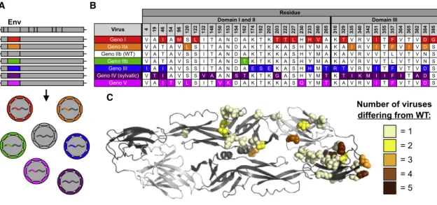
Phylogenetic analyses of DENV4 identifies six groups desig-nated as genotypes I, IIa, IIb, III, IV (sylvatic), and V (Figure 1). As different isolates and genotypes of DENVs demonstrate var-iable growth rates and foci morphology in cell culture, hampering comparative studies of E protein variation, we used reverse ge-netics to construct a panel of recombinant DENV4 viruses. Using our previously described DENV4 molecular clone (genotype IIb)
(Gallichotte et al., 2015), we replaced the wild-type (WT)
enve-lope sequence with that from each of the other genotypes (Table
S1;Figure 2A). All other structural and non-structural proteins
were derived from WT DENV4, resulting in an isogenic panel of viruses that only differ in the E gene sequence (Figure 2A). Sequence analyses across the DENV4 genotype viruses reveal significant amino acid variation in EDIII as well as residues adjoining the hinge region between EDI and EDII (Figures 2B, 2C, andS1). When looking at the representative strain for each genotype, some amino acids differ in only one virus (e.g., posi-tion 132), whereas at other sites (e.g., posiposi-tion 351) the residues are variable across multiple genotypes (Figures 2B and 2C).
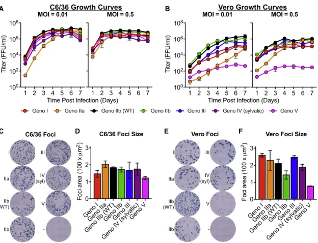
DENV4 Viruses Differ in Growth Kinetics and Foci Morphology
To recover recombinant viruses, full-length cDNAs were assem-bled as previously described (Gallichotte et al., 2015; 2017).
Figure 1. Phylogenetic Relationship of DENV4 Genotypes
DENV4 envelope protein sequences were aligned using neighbor-joining method with 100 replicates based on the multiple sequence alignment. Numbers in parentheses following virus species names indicate the number of sequences repre-sented at that tree position.
Viruses were isolated by electroporating full-length infectious viral RNA into C6/36 cells, and passaging cell-culture supernatant once to produce infectious stocks. When C6/36 insect cells were in-fected in a multi-step viral growth curve (MOI of 0.01), all viruses replicated with similar kinetics and achieved similar peak titers of about 107 ffu/mL after 4 days, with genotype IIa having slightly lower titers at earlier time points (
Fig-ure 3A). Growth curves performed at a
higher MOI in C6/36 cells had similar growth kinetics across the panel, although viruses reached peak titers by day 3 post-infection (Figure 3A). The recombinant DENVs displayed more heterogeneous growth kinetics on Vero cells following both low and high MOI infections
(Figure 3B). Genotype IIb viruses replicated most efficiently, and
genotype V was significantly attenuated in growth, with peak titers 3 logs lower than that of the other genotypes (Figure 3B). While genotype V was highly attenuated in Vero cells, it was the only virus to cause complete syncytia in C6/36 cells (
Fig-ure S2). Although speculative, it is possible that this
demon-strates a virus adaptation for increased growth and spread in insect cells and insects. Syncytia formation has been seen with other strains of DENV (Pierro et al., 2006), but we did not observe syncytia with any of the other DENV4 genotype viruses.
In addition to virus growth, we also compared viral foci morphology (Figures 3C–3F). C6/36 foci were similar across the entire panel (Figures 3C and 3D). Slightly more variation in foci morphology and size was noted in Vero cells across the panel, with genotype V producing the smallest foci; however, all strains produced foci that were clearly defined and visible
(Figures 3E and 3F). The attenuated foci size of genotype V
was consistent with reduced replication in Vero cell growth curves (Figure 3B).
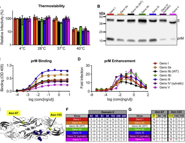
DENV4 E Genotype Viruses Do Not Differ in Thermostability
The large differences in viral replication of the DENV4 panel in mammalian and insect cells (Figure 3) may be a result of different temperature sensitivities, as studies have implicated envelope sequences and virion stability (Lim et al., 2017). A thermostability assay revealed that the DENV4 variants are similarly stable, with little loss of infectivity after incubation at 28C and 37C; how-ever, all viruses lost1 log of infectivity after incubation at 40C
genotype V was the most mature. As the DENV4 variant panel contains differing amounts of pr protein (Figures 4B and 4C), we sought to determine whether the viruses could be enhanced by a non-neutralizing, pr-specific mAb (Smith et al., 2015). An antibody-dependent enhancement (ADE) assay revealed that despite differing levels of pr present on viruses, all viruses are similarly enhanced, although the concentration of antibody needed to achieve peak enhancement and the level of enhance-ment do vary across the viruses (Figure 4D).
The furin cleavage site (located in prM protein) was not altered across the panel, suggesting that the E protein sequence can impact virus maturation (Pierson and Diamond, 2012). At a neutral pH of the released virus, the pr protein sits over the fusion-loop and is predicted to interact with seven amino acids in EDII (Figures 4E and 4F); however, at low pH, during process-ing of the virion, pr can make additional contacts across the en-velope dimer. Under either condition, none of these amino acids were altered in the panel, suggesting that other residues may function to stabilize pr. Interestingly, there is little variability within the paired pr protein sequences of the variant panel
(Figure S3).
Immunoblotting also revealed that the envelope protein of genotype V is slightly smaller than that of the other genotypes
(Figure 4B). The DENV envelope protein contains two
glycosyl-ation sites, one in EDII (Asn-67) and one in EDI (Asn-153) (
Fig-ure 4E). Analysis of the glycosylation site sequences across
the panel revealed that a single amino acid change at residue 153 in genotype V disrupts the N-X-T/S glycosylation motif
(Figure 4G), resulting in a smaller molecular weight envelope
protein (Figure 4B). When looking at all genotype V viruses used in our phylogenetic tree (Figure 1;Table S2), 87.5% have amino acid variability that disrupts the Asn-153 glycosylation site (Table S3), suggesting this disrupted motif is not unique Virus 4 19 46 54 96 120 122 132 148 150 153 154 162 174 182 202 203 222 227 230 233 260 265 310 329 335 340 342 351 355 357 364 365 382 384 385
Geno I V A I A M S L I T A N D A K T K T T L H H V A K T V R V I T F V T V D G
Geno IIa V A T A V L S I T A N D A K T K K A S H Y M A K A I R V I T F V I V D S
Geno IIb (WT) V A T A V S S I T A N D A K T K K A S H Y M A K A V R V V T L V T V N S
Geno IIb V A T A V S S I T A N D T K T K K A S H Y M A K A V R V V T L V T V N S Geno III I A I A V S S I T A N D A E S E K A S H H M T R T V R V I T F V T V D S Geno IV (sylvatic) V T I A V S S V A A N S T K T K G A S H Y M T K T I K M I I F I T A D S Geno V V A I T V L S I T V K D A K T K K A S Q Y M T K A V R V I T F V T V D S
Residue
Domain I and II Domain III
Env
Number of viruses differing from WT:
= 1 = 2 = 3 = 4 = 5
A B
C
Figure 2. Design and Diversity of a Panel of DENV4 Genotypic Variants
(A) Sequences encoding the envelope protein from each genotype were placed into a genotype IIb (WT) infectious clone, resulting in recombinant viruses that are entirely WT except for envelope protein, and sequence encoding envelope protein.
(B) Envelope protein amino acids positions that differ from WT (genotype IIb).
(C) Envelope protein amino acids that differ from WT are mapped on dimer based on number of genotypes differing at that position (PDB: 1OAN).
thanthermostabilitycontributestodifferencesintheabilityofthe virusestoinfectandreplicateinmammalianandinsectcells.
DENV4EVariantsDifferinMaturationStatus, Enhanceability,andGlycosylationPattern
AsDENVmaturationstatemaybeheterogeneousinvitro,the re-combinantpanel allowedus toevaluatetherole ofE protein sequenceonmaturationstatusinanisogenicDENV4backbone. Duringinfection,DENVisassembledwithintheendoplasmic re-ticulumasimmaturevirionscontainingpre-membrane(prM) pro-teins,whichpreventfusionduringviralegress.AsDENVtransits throughthetrans-Golginetwork,pHchangetriggerscleavageof prMby the host proteasefurin. Asthe virus leaves thecell, cleaved prdissociates, leaving fullymature viral particles. In cellculture,furincleavageandprdissociationareinefficient pro-cessesandhighlycelltypedependent,leadingtoheterogonous populationofdifferentiallymatureviruses,containingdifferent amounts of uncleaved pr peptide (Pierson and Diamond, 2012).Asmaturation statuscaninfluence infectivityand anti-bodyneutralization(Mukherjeeetal.,2014),wecomparedthe maturationstatusacrosstheDENV4panelusingimmunoblotting andELISAs(Figures4Band4C).
to the genotype V strain selected within our panel, but appears to be conserved across most genotype V viruses. DENV enve-lope glycosylation can be important for binding to host cell re-ceptors, determining infectivity in different hosts, and binding and neutralization by antibodies (Pokidysheva et al., 2006;
Mondotte et al., 2007; Rouvinski et al., 2015). Therefore,
geno-type V’s conserved lack of the second glycosylation site might impact the virus’s ability to efficiently infect and be transmitted between vertebrate and invertebrate hosts, and be the result of adaptation to a different cellular or host tropism (Bryant et al.,
2007; Lee et al., 2010). Additionally, the lack of this glycosylation
site might contribute to the genotype V C6/36 syncytia pheno-type (Figure S2). Genetic alteration of glycosylation and pr pro-tein sequences, and generation of fully mature and fully imma-ture virus preparations would allow one to determine the role of glycosylation and maturation status on many aspects of virus biology. Glycosylation status and large differences in viral matu-ration state have previously been shown to impact antibody binding and neutralization (Mukherjee et al., 2014); therefore, the binding and neutralization differences we see within this panel might be partially attributable to the variation in glycosyl-ation and maturglycosyl-ation.
Binding of Serotype-Specific and Cross-Reactive mAbs to DENV4 E Genotype Variants
We next measured the binding of a panel of well-characterized DENV4 serotype-specific and DENV cross-reactive mAbs to our DENV4 viruses by ELISA (Figure 5). DENV4 serotype-specific antibodies D4-126 and D4-131 recognize partially overlapping epitopes that have not been fully defined in the EDI/II hinge region (Figures S4A and S4B) (Nivarthi et al., 2017). All DENV4 genotypes bound similarly to D4-126 and D4-131 (Figure 5A). mAb D4-141, which recognizes an EDIII epitope (Figure S4C), also bound all viruses similarly, despite large amount of variation in EDIII across the panel (Figure 5A). The non-human primate mAb 5H2, which binds to a well-defined epitope on EDI, displayed highly variable binding across the panel (Figure 5A). Two amino acids (162 and 174) predicted to be 5H2 contact res-idues were variable across the DENV4 panel (Figure S4A) (
Cock-burn et al., 2012). Genotype III did not bind 5H2 and contains an
amino acid polymorphism at position 174, suggesting that this position is essential for 5H2 binding.
The cross-reactive human mAbs C10 and B7 recognize qua-ternary envelope dimer epitopes (EDEs) that span across the fusion loop of one E monomer into EDIII or EDI of the neighboring
Geno IGeno IIa
Geno IIb (WT) Geno IIbGeno III
Geno IV (sylvatic) Geno V
0 1 2 3
Foci
area (100 x
m
2)
Vero Foci Size
Geno IGeno IIa
Geno IIb (WT) Geno IIbGeno III
Geno IV (sylvatic) Geno V
0 1 2 3
Foci
area (100 x
m
2)
C6/36 Foci Size C6/36 Foci
1 2 3 4 5 6 7 MOI = 0.5
1 2 3 4 5 6 7 100
102 104 106 108
T
iter (FFU/ml)
MOI = 0.01
Time Post Infection (Days) Time Post Infection (Days)
IIa
IIb (WT) I
IIb
III
IV (syl)
V
-
Geno I Geno IIa Geno IIb (WT) Geno IIb Geno III Geno IV (sylvatic) Geno V
I
IIa
IIb (WT)
IIb
III
IV (syl)
V
-
1 2 3 4 5 6 7 MOI = 0.5
1 2 3 4 5 6 7 100
102 104 106 108
T
iter (FFU/ml)
MOI = 0.01
C6/36 Growth Curves
Vero Growth Curves
Vero Foci
A B
C D E F
Figure 3. DENV4 Genotype Viruses Differ in Growth and Foci Morphology
(A and B) Viruses were analyzed for their ability to replicate in (A) C6/36 and (B) Vero cells at multiplicities of infection (MOI) of either 0.01 or 0.5 (mean±SD of biological triplicates).
cell focus reduction neutralization test (FRNT), and a flow cy-tometry-based neutralization assay with U937 cells expressing DC-SIGN, a known DENV attachment factor (Figures 6A, 6B, andS5). We observed a 1- to 2-log difference in antibody neutral-ization titers of the DENV4 serotype-specific antibodies against the DENV4 panel. Importantly, some mAbs were not able to neutralize select genotypes even at the highest concentrations tested, despite robust binding (e.g., D4-126 and genotype III)
(Figures 5A,6A, and6B). Other mAbs have similar neutralization
titers despite lower levels of binding (e.g., 5H2 and genotype V). These results reveal that, for each mAb and virus, the amount of mAb sufficient to bind and/or neutralize varies significantly.
Consonant with the binding results, EDE mAbs C10 and B7 potently neutralize all viruses similarly (Figures 6A and 6B), with the exception of B7 and genotype V, due to its missing glycosyl-ation site. The C10 and B7 epitopes are highly conserved across
Geno IV (sylvatic)
E
prM Geno I Geno IIa Geno IIb (WT) Geno IIb Geno III Geno V 50 kDa
38
25
15
-6 -4 -2 0 2
0 10 20 30
log (conc[ng/ul])
Fold Infection
prM Enhancement
-4 -3 -2 -1 0 1
0.0 0.5 1.0
log (conc[ng/ul])
Binding (OD 405)
prM Binding
4°C 28°C 37°C 40°C
1 10 100
Relative Infectivity (%)
Thermostability A
E F G
Asn-67 Asn-153
Virus 67 68 69 153 154 155
Geno I N I T N D T
Geno IIa N I T N D T
Geno IIb (WT) N I T N D T
Geno IIb N I T N D T
Geno III N I T N D T
Geno IV (sylvatic) N I T N S T
Geno V N I T K D T
Virus 64 66 83 84 103 248 249
Geno I S S K W N Q D
Geno IIa S S K W N Q D
Geno IIb (WT) S S K W N Q D
Geno IIb S S K W N Q D
Geno III S S K W N Q D
Geno IV (sylvatic) S S K W N Q D
Geno V S S K W N Q D
B
C D
Geno I Geno IIa Geno IIb (WT) Geno IIb Geno III Geno IV (sylvatic) Geno V
Figure 4. DENV4 Genotype Viruses Differ in Thermostability, Maturation, and Glycosylation Status
(A) DENV4 viruses were evaluated for their thermostability at 28C, 37C, and 40C (mean±SD of biological triplicates). Relative infectivity is calculated as (ffu/mLtest temperature/ ffu/mL4C)3100.
(B) Viruses were immunoblotted for envelope (E) and precursor (prM) protein present in viral stocks (representative image).
(C) Binding assay using pr-specific mAb 1E16 to detect amount of pr protein present in virus stocks (mean±SD of biological triplicates).
(D) Antibody-dependent enhancement (ADE) assay was performed with DENV4 virus panel and anti-pr mAb 1E16 in U937 cells (mean±SD of biological duplicates).
(E) DENV envelope dimer showing location of putative pr interacting residues (navy) and glycosylation sites (yellow) (PDB: 1OAN). (F) Envelope and putative pr interacting sites (via side-chain interactions) are listed for each genotype.
(G) Amino acid sequences of glycosylation sites 67 and 153 for each genotype. Genotype V lysine at amino acid position 153 (highlighted in red) disrupts glycosylation motif (N-X-T/S).
monomer(FiguresS4AandS4B)(Rouvinskietal.,2015).These mAbs bind all four DENV serotypes, reflecting the highly conserved natureof theepitope across theDENV Eprotein. Consequently,itwasnotsurprisingthatC10andB7boundall genotypeswithintheDENV4panelwithsimilarefficiencies( Fig-ure5B),asthedifferencesbetweengenotypesaresmallerthan thosebetweenserotypes.BindingofB7,however,isdependent onthepresenceofaglycanatposition153inEDI(Rouvinski etal.,2015).TheDENV4genotypeVvirusinthispanel,which lackedthisglycosylation site(Figure4E),failedtobindtothe B7antibody(Figure5B).
NeutralizationofDENV4EGenotypeVariantsby Serotype-SpecificandCross-ReactivemAbs
the DENV4 panel, likely explaining the robust and consistent neutralizing titers (Figures S4A and S4B). Additionally, the range of neutralization titers is smaller for cross-reactive mAbs compared to serotype-specific mAbs, suggesting that the more cross-reactive an antibody is (i.e., the more serotypes it recognizes), the less genotypic diversity matters.
The Neutralization of DENV4 E Genotype Variant Viruses by Human Sera from Natural Infection and Vaccination Convalescent immune sera from people who have recovered from primary DENV4 infections contain strongly neutralizing serotype-specific and weakly neutralizing cross-reactive anti-bodies. We performed neutralization assays with DENV4 conva-lescent immune sera to measure the breadth of neutralization across different DENV4 E variant genotypes (Figures 7A and S6A). While the absolute neutralization titers vary across samples by 1–2 logs, all DENV4 immune sera were able to neutralize all genotypes (Figure 7A). While this suggests that nat-ural infection with any DENV4 genotype elicits antibodies that are neutralizing against other genotypes as well, individuals who have weaker responses may be vulnerable to reinfection due to genotype variation.
Individuals who received a genotype II DENV4 monovalent vaccine developed neutralizing antibodies (Figures 7B and S6B). As seen with natural isolates (Durbin et al., 2013), we also observed a larger spread in neutralization titers with the monovalent vaccine immune sera (>2 logs) compared to the natural infection sera. Additionally, for some vaccine sera, neutralizing antibodies were undetectable against some
geno-types, despite robust neutralization of other strains (e.g., sample 68 does not neutralize genotype IV or V, but potently neutralizes genotype II viruses). Among the currently circulating geno-type I, II, and III viruses, vaccine-matched genogeno-type II viruses were most potently neutralized. To determine whether the differ-ential genotype neutralization is driven by serotype-specific or cross-reactive antibodies, we used depletion techniques to spe-cifically remove cross-reactive antibodies (de Alwis et al., 2012)
(Figures 7C,7D, andS6C–S6E). We find that removing
cross-reactive antibodies minimally alters neutralization titers, sug-gesting that the majority of total neutralization comes from sero-type-specific antibodies, and that the differences in titers across DENV4 genotypes are primarily driven by DENV4 serotype-spe-cific antibodies as well.
We next looked at the DENV4 genotypic neutralizing breadth of individuals that received a tetravalent DENV vaccine. As tetrava-lent vaccination can result in both DENV4 serotype-specific anti-bodies, and strongly neutralizing cross-reactive antianti-bodies, depletion techniques were again used to determine the contribu-tion of each populacontribu-tion of antibodies to total neutralizacontribu-tion. Control depleted sera, containing both serotype-specific and cross-reactive antibodies, differentially neutralized the DENV4 variants, with vaccine-matched genotype II viruses neutralized on average 3- to 20-fold more efficiently than the other genotypes
(Figures 7E andS7A). In addition, some sera failed to neutralize
currently circulating genotype I or III variants. When we removed DENV serotype cross-reactive antibodies, we observed only a small reduction in neutralization titers, indicating that the vac-cine mainly induced serotype-specific neutralizing antibodies
-4 -2 0 2
0.0 0.5 1.0 1.5
log (conc[ng/ul])
Binding (OD 405)
C10
-4 -2 0 2
0.0 0.5 1.0 1.5
log (conc[ng/ul])
Binding (OD 405)
B7
-4 -2 0 2
0.0 0.5 1.0 1.5
log (conc[ng/ul])
Binding (OD 405)
D4-126
-4 -2 0 2
0.0 0.5 1.0
log (conc[ng/ul]) D4-131
-4 -2 0 2
0.0 0.5 1.0 1.5
log (conc[ng/ul])
Binding (OD 405)
D4-141
-4 -2 0 2
0.0 0.5 1.0
log (conc[ng/ul]) 5H2
DENV4 Serotype-Specific MAbs Cross-Reactive MAbs
Geno I Geno IIa Geno IIb (WT) Geno IIb Geno III Geno IV (sylvatic) Geno V
A B
Figure 5. mAbs Differentially Bind DENV4 Genotype Viruses
The existence of phylogenetically and antigenically distinct DENV1–DENV4 serotypes is well accepted in the literature
(Holmes and Twiddy, 2003); however, the role of genetic
diver-sity across genotypes is less well studied. Many common laboratory DENV strains have either been heavily cell culture adapted and/or differ in sequence from contemporary circulating strains (Dowd et al., 2015; Katzelnick et al., 2015, 2017). Addi-tionally, some laboratory, and importantly, vaccine strains, are composed of DENV genotypes that are likely extinct and, conse-quently, do not circulate in human populations (Katzelnick et al., 2017). While CD8+T cells, CD4+T cells, and other mechanisms of cellular immunity are correlated with DENV protective immu-nity (Mathew and Rothman, 2008), and antibodies against DENV NS1 may also alter disease severity (Hertz et al., 2017), neutralizing antibodies represent the best correlate of protection to date (Katzelnick et al., 2016; Buddhari et al., 2014).
Natural DENV infection is thought to provide lifelong protec-tion against symptomatic reinfecprotec-tion with that serotype (
Katzel-nick et al., 2016; Buddhari et al., 2014); however, it is unknown
whether individuals are protected with the same efficacy against all genotypes within the serotype. There are reports of rare, typically asymptomatic, homotypic reinfection in people in Nicaragua and Peru (Forshey et al., 2016; Waggoner et al., 2016), which is potentially driven by genotypic differences be-tween the primary and secondary infecting viruses. Some studies have evaluated the breadth of antibody neutralization against different genotypes elicited by natural infection or vacci-nation (Blaney et al., 2005; Durbin et al., 2013; Katzelnick et al.,
2015; Messer et al., 2012; Vasilakis et al., 2008a). While most
in-dividuals exposed to natural infections or vaccines neutralized multiple genotypes within each serotype, absolute levels of neutralizing antibodies vary depending on the individual and the DENV genotypes used. Indeed, even in the current study, we noted that most individuals exposed to natural infections or a vaccine, developed antibodies that neutralized the most
D4
126 131D4 141D4 5H2 C10 B7
0.01 0.1 1 10
Neut
50
[ng/
l]
MAb Flow-Based Neutralization
Cross-Reactive Serotype-Specific
D4 126
D4 131
D4 141
5H2 C10 B7
0.01 0.1 1 10
FRNT
50
[ng/
l]
MAb Focus Reduction Neutralization
Cross-Reactive Serotype-Specific
Geno I Geno IIa Geno IIb (WT) Geno IIb Geno III Geno IV (sylvatic) Geno V
B A
Figure 6. DENV4 Genotypic Variants Are Differentially Neutralized by Monoclonal Antibodies
(A and B) DENV4 serotype-specific antibodies D4-126, D4-131, D4-141, and 5H2 and DENV cross-reactive antibodies C10 and B7 were evaluated for their ability to neutralize DENV4 genotype viruses in (A) Vero cell focus reduction neutralization test (FRNT) and (B) flow cytometry-based neutralization assay (Neut) (mean± SD of technical duplicates). The y-axes represent the concentration of antibody required to neutralize 50% of infectious virus. The dashed line represents assay limit of detection.
(Figures7FandS7B).Importantly,afterremovingcross-reactive
antibodies,weseeasimilarspreadintitersacrossthepanel, sug-gesting that DENV4 serotype-specific antibodies are driving thedifferentialgenotypicneutralization.WhenallDENV cross-reactive and serotype-specific antibodies were depleted, we completelylostneutralizationagainstallviruses(FigureS7C).
DISCUSSION
DENVisthemostsignificantarthropod-bornevirus,causing sig-nificant morbidity and mortality worldwide. Sanofi-Pasteur’s tetravalentDENVvaccine,Dengvaxia,hasbeenmarketedand usedinhumanpopulations,andtherearetwoadditional com-mercial tetravalent vaccine candidates under evaluation in phaseIIIhumantrials,includingtheNIHtetravalentDENV vac-cine.RecentresultswithDengvaxiademonstratehighvaccine ef-ficacyinpeoplewhoweredengueimmunepriortovaccination (81.9%),and muchpoorerefficacy inpeoplewhowerenaive beforevaccination(52.5%)(Hadinegoroetal.,2015).Moreover, naive individuals who received the vaccine appear to be at greaterriskofdevelopingseveredisease,whenexposedtoa nat-uralDENVinfectionapproximately24monthsormorefollowing thelastdoseofvaccine.Asaresult,Dengvaxiaiscurrently rec-ommendedonlyforuseinpeoplewhohavebeenprimedby nat-uralDENVinfections(Sridharetal.,2018).Dengvaxiastimulated highlevelsofDENV4serotype-specificneutralizingantibodies
(Heneinetal.,2017),andoverallvaccineefficacywashighest
prevalent DENV4 genotypes, but the levels of neutralizing anti-body varied considerably by genotype. In 2008, the World Health Organization (WHO) noted that there is little evidence of anti-genic drift within DENV serotypes that might lead to resistance of certain strains to post-vaccination neutralization, yet they advised that laboratories consider inclusion of multiple virus strains, including laboratory prototype strains and recent clinical isolates when performing neutralization assays (Roehrig et al., 2008). Our results demonstrate the value of including different DENV genotypes when evaluating vaccine responses.
Using reverse genetics, we developed an isogenic panel of DENV4 recombinant viruses that only differ in their E glycopro-tein, which was derived from different genotypes. We recon-structed clinically relevant isolates with E protein genes derived from clinical specimens or low passage history in culture. Our data demonstrate that DENV4 E protein genotypic diversity can impact many aspects of the virus’s cell biology including growth in cells, glycosylation, syncytial formation, and matura-tion. Additionally, as all the viruses can be enhanced, it highlights
the importance of determining the impact of DENV genetic vari-ation on disease enhancement after infection and vaccinvari-ation. The chimeric DENV4 virus panel described here is a powerful tool for initial assessment of the impact of DENV E genotypic variation on virus biology and humoral immunity. As we selected one representative envelope sequence per genotype and utilized a recombinant approach, the viruses here do not capture all E protein sequence diversity within each genotype and some will never actually be encountered by vaccine recipients. Hence, it will be important to evaluate more contemporary DENV4 geno-type I, II, and III strains in future studies, including the use of both natural and recombinantly derived isolates. In agreement with our findings, we note that another study using WT strains of endemic and sylvatic DENV4 viruses demonstrated better neutralization of vaccine-matched endemic genotype II viruses compared to sylvatic viruses using sera from DENV4 monovalent vaccine recipients (Durbin et al., 2013).
Our results demonstrate that infection or vaccination with a single DENV4 genotype stimulates variable levels of neutralizing
A
C
E
B
D
F
Figure 7. DENV4 Polyclonal Immune Sera Have a Range of Neutralization Titers against DENV4 Variants Driven by Serotype-Specific Antibodies
(A and B) Using Vero cell focus reduction neutralization test (FRNT), (A) pooled polyclonal immune sera from DENV4-infected non-human primates (NHPs) or from naturally infected individuals, or from (B) individuals who received the NIH DENV4 monovalent vaccine were evaluated for their ability to neutralize DENV4 genotype viruses.
Growth Curves
C6/36 or Vero cells were seeded in 24-well plates 1 day prior to infection. Viruses were diluted to an MOI of either 0.01 or 0.5, added to cells, and incu-bated for 1 hr at either 32C (C6/36) or 37C (Vero). Inoculum was removed, cells were washed three times with PBS, and growth media were replaced. Media were sampled daily, replaced with fresh media, and immediately frozen at 80C. Samples were titered as described above.
Thermostability Assay
DENV4 viruses were diluted 1:10, then incubated at 4C, 28C, 37C, or 40C for 1 hr, then immediately transferred to 4C for 15 min. Viruses were then titered on Vero cells and immunostained as described above.
Immunoblotting
Virus stocks were diluted in PBS, mixed with sample buffer, and heated at 95C for 10 min. Samples were run on 4%–20% Protean TGX gels and transferred to polyvinylidene difluoride (PVDF) membrane. Membranes were blocked in 5% non-fat dried milk and probed with anti-E (4G2) and anti-prM (1E16) mAbs. Membranes were washed and probed with secondary anti-bodies labeled with HRP and developed using chemiluminescent substrate. Membranes were visualized using a LI-COR C-DiGit Blot Scanner.
Enzyme-Linked Immunosorbent Binding Assay
Plates were coated with anti-E (4G2) and anti-prM (2H2) antibodies in carbon-ate buffer overnight and blocked in 5% non-fat dried milk, and then virus an-tigen was added. Primary antibody was diluted in blocking buffer and added to plates for 1 hr at 37C. Alkaline-phosphate-labeled secondary antibody was added and plates were incubated for 1 hr at 37C. Plates were developed with p-nitrophenyl phosphate substrate and color changes were quantified using Bio-Rad iMark Microplate Absorbance Reader.
ADE Assay
mAbs was diluted 5-fold and mixed with virus previously diluted to result in
15% infection in U937+DC-SIGN cells. Virus:mAb mixtures were incubated at 37C for 45 min, and then added to 53104
U937 cells and incubated at 37C for 2 hr. After incubation, cells were washed with growth media, and then resuspended in fresh growth media. The cells were incubated for 20 hr at 37C, washed in PBS, fixed in 10% phosphate-buffered formalin, and then stained with anti-E mAb 4G2 directly conjugated to Alexa Fluor 488. Cells were analyzed on a Guava easyCyte flow cytometer.
Neutralization Assays
FRNT was performed by seeding Vero cells 1 day prior to infection. mAbs or immune sera were diluted 4-fold and mixed with virus stocks previously diluted to40 ffu/well. Virus:Ab mixtures were incubated at 37C for 1 hr, and then added to cells for 1 hr at 37C. After incubation, overlay media was added and plates were incubated for 3 days. Cells were fixed and immunostained as described as above. Flow cytometry-based neutralization assays were per-formed as described above in ADE assays, except with U937+DC-SIGN cells.
Polyclonal Antibody Depletion Assay
Dynabeads were covalently bound to anti-E mAb 1M7 overnight at 37C. Bead:mAb complex was blocked with 1% BSA in PBS at 37C, and then washed with 0.1 M 2-(N-morpholino)ethanesulfonic acid (MES) buffer. Beads were incubated with BSA (control), purified DENV3 (cross-reactive depletion), or a mix of DENV3 and DENV4 (full depletion) for 1 hr at 37C, and then washed three times with PBS. Bead:mAb:DENV complex was fixed with 2% paraformaldehyde in PBS for 20 min, and then washed four times with PBS. DENV-specific antibodies were depleted from sera by incubating beads with sera diluted 1:10 in PBS for 1 hr at 37C with end-over-end mixing for at least two sequential rounds of depletions. Removal of DENV antibodies was confirmed by ELISA.
Data Analysis and Software
All data were analyzed and graphed using GraphPad Prism v7.0a. Protein structures were visualized using MacPyMOL: PyMOL v1.7.6.2. Replicate infor-mation is included in the figure legends.
antibodiesto differentgenotypes.Currently, thereare insuffi-cientdatatocorrelatelevelsofneutralizingantibodiesto protec-tionfromDENVdisease.Moreover,otherimmunemechanisms involvingTcells, NS1immunity,and Bcell memorymayalso reduce or eliminate clinical disease (Mathew and Rothman, 2008).However,itisworthnotingthatthelicensedtetravalent DENVvaccine(Dengvaxia)hadhighervaccineefficacyagainst vaccine-matchedDENV4genotypeII(83%)virusescompared toco-circulatingDENV4genotypeIviruses(47%)(Rabaaetal., 2017).We propose that targetedsurveillance of changing or emergingDENVgenotypesfollowingvaccinationwillbevaluable inassessingtheinfluenceofDENVgenotypeonthefrequencyof repeatinfectionsandoverallvaccineeffectiveness.
EXPERIMENTALPROCEDURES
PhylogeneticTree
ThetreewasconstructedinGeneiousR11usingtheneighbor-joiningmethod (Jukes–Cantorgeneticdistance)with100replicatesbasedonthemultiple sequencealignment.Theradialphylogramwasvisualizedandrenderedfor publicationusingCLCSequenceViewer7andAdobeIllustratorCC2017.
VirusConstruction
ChimericrecombinantDENV4viruseswereconstructedasdescribedbefore (Gallichotteetal.,2015,2017).Briefly,DNAencodingisogenicenvelopeprotein sequenceswasintroducedintoaquadripartiteDENV4infectiousclonesystem usingsyntheticallyderivedgenesandrecombinantDNAapproaches.Plasmid DNAwasdigestedandligatedtogether,andviralfull-lengthgenomicRNAwas generatedusingT7RNApolymerase.Infectiousgenome-lengthcappedviral RNAtranscriptswereelectroporatedintoC6/36cells,andsupernatantwas harvestedandpassagedontoC6/36cellstomakeviralworkingstocks.
Cells
C6/36cells(ATCC CRL-1660) weregrowninminimumessentialmedium (MEM)at32C. Verocells (ATCCCCL-81) weregrowninDMEM media, U937andU937cells stablyexpressing DC-SIGN(U937+DC-SIGN)were growninRPMImedium1640,andallwereculturedat37C.Allmediawere supplementedwith5%fetalbovineserum(FBS),whichwasreducedto2% duringDENVinfection.Allmediaweresupplementedwith100U/mLpenicillin, 100 mg/mLstreptomycin,and0.25 mg/mLamphotericinB.C6/36andU937/ U937+DC-SIGNmediawereadditionallysupplementedwith non-essential aminoacids, andU937/U937+DC-SIGN mediawasfurthersupplemented withL-glutamineand b-mercaptoethanol.Allcellswereincubatedat5%CO2.
ImmuneSera
DENV4immunenon-humanprimateserumwasobtainedfromBEIResources (NR-41789).Humandengueimmuneserawereobtainedfromapreviously describedDengueTravelercollectionattheUniversityofNorthCarolina. Vac-cineserawereobtainedfromindividualswhoreceivedalive-attenuated mono-valentDENV4ortetravalentvaccine180dayspost-vaccination,asdeveloped bytheNIH,andwereprovidedbyA.P.D.andS.S.W.Allhumanserasamples wereanonymizedandobtainedunderInstitutionalReviewBoardapproval.
ViralTiteringandImmunostaining
SUPPLEMENTAL INFORMATION
Supplemental Information includes seven figures and three tables and can be found with this article online athttps://doi.org/10.1016/j.celrep.2018. 10.006.
ACKNOWLEDGMENTS
This research was supported by National Institute of Allergy and Infectious Dis-eases (NIAID) grants R01s AI107731 (principal investigator [PI], A.M.d.S.) and AI125198 (PI, A.M.d.S.), P01 AI106695 (PI, E. Harris), and U19 AI109761 (PI, R.S.B.) and by grant OPP1104710 from the Bill and Melinda Gates Foundation (PI, A.P.D.). Support was also provided in part by the Intramural Research Pro-gram of the NIAID. E.N.G. was supported by T32 NIH Training Grant AI007419. We thank Stephanie Marcet, Scott Royal, and Prem Lakshmanane for assistance.
AUTHOR CONTRIBUTIONS
Conceptualization, E.N.G., A.M.d.S., and R.S.B.; Investigation, E.N.G., T.J.B, U.N., M.J.D., R.G., D.G.W., and B.L.Y.; Resources, A.P.D., S.S.W., A.M.d.S., and R.S.B.; Writing – Original Draft, E.N.G.; Writing – Review & Editing, E.N.G., A.P.D., S.S.W., A.M.d.S., and R.S.B.; Supervision, A.M.d.S. and R.S.B.
DECLARATION OF INTERESTS
A.M.d.S. has consulted on dengue vaccines for Takeda, Merck, and GSK. R.S.B. has consulted with Takeda on vaccines. A.M.d.S. and R.S.B. are inven-tors on patents filed and issues relevant to dengue vaccines.
Received: June 11, 2018 Revised: August 15, 2018 Accepted: September 28, 2018 Published: October 30, 2018
REFERENCES
Bennett, S.N., Drummond, A.J., Kapan, D.D., Suchard, M.A., Mun˜oz-Jorda´n, J.L., Pybus, O.G., Holmes, E.C., and Gubler, D.J. (2010). Epidemic dynamics revealed in dengue evolution. Mol. Biol. Evol.27, 811–818.
Bhatt, S., Gething, P.W., Brady, O.J., Messina, J.P., Farlow, A.W., Moyes, C.L., Drake, J.M., Brownstein, J.S., Hoen, A.G., Sankoh, O., et al. (2013). The global distribution and burden of dengue. Nature496, 504–507.
Blaney, J.E., Jr., Matro, J.M., Murphy, B.R., and Whitehead, S.S. (2005). Recombinant, live-attenuated tetravalent dengue virus vaccine formulations induce a balanced, broad, and protective neutralizing antibody response against each of the four serotypes in rhesus monkeys. J. Virol.79, 5516–5528.
Bryant, J.E., Calvert, A.E., Mesesan, K., Crabtree, M.B., Volpe, K.E., Silengo, S., Kinney, R.M., Huang, C.Y., Miller, B.R., and Roehrig, J.T. (2007). Glycosyl-ation of the dengue 2 virus E protein at N67 is critical for virus growth in vitro but not for growth in intrathoracically inoculatedAedes aegypti mosquitoes. Virology366, 415–423.
Buddhari, D., Aldstadt, J., Endy, T.P., Srikiatkhachorn, A., Thaisomboonsuk, B., Klungthong, C., Nisalak, A., Khuntirat, B., Jarman, R.G., Fernandez, S., et al. (2014). Dengue virus neutralizing antibody levels associated with protec-tion from infecprotec-tion in thai cluster studies. PLoS Negl. Trop. Dis.8, e3230.
Calisher, C.H., Karabatsos, N., Dalrymple, J.M., Shope, R.E., Porterfield, J.S., Westaway, E.G., and Brandt, W.E. (1989). Antigenic relationships between fla-viviruses as determined by cross-neutralization tests with polyclonal antisera. J. Gen. Virol.70, 37–43.
Cao-Lormeau, V.M., Roche, C., Aubry, M., Teissier, A., Lastere, S., Daudens, E., Mallet, H.P., Musso, D., and Aaskov, J. (2011). Recent emergence of dengue virus serotype 4 in French Polynesia results from multiple introductions from other South Pacific Islands. PLoS One6, e29555.
Chen, R., and Han, G.Z. (2016). Dengue in China: comprehensive phylogenetic evaluation reveals evidence of endemicity and complex genetic diversity. Am. J. Trop. Med. Hyg.94, 198–202.
Cockburn, J.J., Navarro Sanchez, M.E., Goncalvez, A.P., Zaitseva, E., Stura, E.A., Kikuti, C.M., Duquerroy, S., Dussart, P., Chernomordik, L.V., Lai, C.J., and Rey, F.A. (2012). Structural insights into the neutralization mechanism of a higher primate antibody against dengue virus. EMBO J.31, 767–779.
Coloma, J., and Harris, E. (2015). Broad and strong: the ultimate antibody to dengue virus. Nat. Immunol.16, 135–137.
Dash, P.K., Sharma, S., Srivastava, A., Santhosh, S.R., Parida, M.M., Neeraja, M., Subbalaxmi, M.V., Lakshmi, V., and Rao, P.V. (2011). Emergence of dengue virus type 4 (genotype I) in India. Epidemiol. Infect.139, 857–861.
de Alwis, R., Smith, S.A., Olivarez, N.P., Messer, W.B., Huynh, J.P., Wahala, W.M., White, L.J., Diamond, M.S., Baric, R.S., Crowe, J.E., Jr., and de Silva, A.M. (2012). Identification of human neutralizing antibodies that bind to com-plex epitopes on dengue virions. Proc. Natl. Acad. Sci. USA109, 7439–7444.
Dowd, K.A., DeMaso, C.R., and Pierson, T.C. (2015). Genotypic differences in dengue virus neutralization are explained by a single amino acid mutation that modulates virus breathing. MBio6, e01559-15.
Durbin, A.P., Mayer, S.V., Rossi, S.L., Amaya-Larios, I.Y., Ramos-Castaneda, J., Eong Ooi, E., Jane Cardosa, M., Munoz-Jordan, J.L., Tesh, R.B., Messer, W.B., et al. (2013). Emergence potential of sylvatic dengue virus type 4 in the urban transmission cycle is restrained by vaccination and homotypic immunity. Virology439, 34–41.
Fares, R.C., Souza, K.P., An˜ez, G., and Rios, M. (2015). Epidemiological sce-nario of dengue in Brazil. BioMed Res. Int.2015, 321873.
Fleith, R.C., Lobo, F.P., Dos Santos, P.F., Rocha, M.M., Bordignon, J., Strott-mann, D.M., Patricio, D.O., Pavanelli, W.R., Lo Sarzi, M., Santos, C.N., et al. (2016). Genome-wide analyses reveal a highly conserved dengue virus enve-lope peptide which is critical for virus viability and antigenic in humans. Sci. Rep.6, 36339.
Forshey, B.M., Reiner, R.C., Olkowski, S., Morrison, A.C., Espinoza, A., Long, K.C., Vilcarromero, S., Casanova, W., Wearing, H.J., Halsey, E.S., et al. (2016). Incomplete protection against dengue virus type 2 re-infection in Peru. PLoS Negl. Trop. Dis.10, e0004398.
Gallichotte, E.N., Widman, D.G., Yount, B.L., Wahala, W.M., Durbin, A., White-head, S., Sariol, C.A., Crowe, J.E., Jr., de Silva, A.M., and Baric, R.S. (2015). A new quaternary structure epitope on dengue virus serotype 2 is the target of durable type-specific neutralizing antibodies. MBio6, e01461-15.
Gallichotte, E.N., Menachery, V.D., Yount, B.L., Jr., Widman, D.G., Dinnon, K.H., 3rd, Hartman, S., de Silva, A.M., and Baric, R.S. (2017). Epitope addition and ablation via manipulation of a dengue virus serotype 1 infectious clone. MSphere2, e00380-16.
Hadinegoro, S.R., Arredondo-Garcı´a, J.L., Capeding, M.R., Deseda, C., Chotpitayasunondh, T., Dietze, R., Muhammad Ismail, H.I., Reynales, H., Lim-kittikul, K., Rivera-Medina, D.M., et al.; CYD-TDV Dengue Vaccine Working Group (2015). Efficacy and long-term safety of a dengue vaccine in regions of endemic disease. N. Engl. J. Med.373, 1195–1206.
Halstead, S.B. (2015). Pathogenesis of dengue: dawn of a new era. F1000Res. 4, F1000 Faculty Rev-1353.
Henein, S., Swanstrom, J., Byers, A.M., Moser, J.M., Shaik, S.F., Bonaparte, M., Jackson, N., Guy, B., Baric, R., and de Silva, A.M. (2017). Dissecting anti-bodies induced by a chimeric yellow fever-dengue, live-attenuated, tetravalent dengue vaccine (CYD-TDV) in naive and dengue-exposed individuals. J. Infect. Dis.215, 351–358.
Hertz, T., Beatty, P.R., MacMillen, Z., Killingbeck, S.S., Wang, C., and Harris, E. (2017). Antibody epitopes identified in critical regions of dengue virus nonstructural 1 protein in mouse vaccination and natural human infections. J. Immunol.198, 4025–4035.
Holmes, E.C., and Twiddy, S.S. (2003). The origin, emergence and evolu-tionary genetics of dengue virus. Infect. Genet. Evol.3, 19–28.
Rabaa, M.A., Girerd-Chambaz, Y., Duong Thi Hue, K., Vu Tuan, T., Wills, B., Bonaparte, M., van der Vliet, D., Langevin, E., Cortes, M., Zambrano, B., et al. (2017). Genetic epidemiology of dengue viruses in phase III trials of the CYD tetravalent dengue vaccine and implications for efficacy. eLife6, e24196.
Rey, F.A., Stiasny, K., Vaney, M.C., Dellarole, M., and Heinz, F.X. (2018). The bright and the dark side of human antibody responses to flaviviruses: lessons for vaccine design. EMBO Rep.19, 206–224.
Roehrig, J.T., Hombach, J., and Barrett, A.D. (2008). Guidelines for plaque-reduction neutralization testing of human antibodies to dengue viruses. Viral Immunol.21, 123–132.
Rossi, S.L., Nasar, F., Cardosa, J., Mayer, S.V., Tesh, R.B., Hanley, K.A., Weaver, S.C., and Vasilakis, N. (2012). Genetic and phenotypic characteriza-tion of sylvatic dengue virus type 4 strains. Virology423, 58–67.
Rouvinski, A., Guardado-Calvo, P., Barba-Spaeth, G., Duquerroy, S., Vaney, M.C., Kikuti, C.M., Navarro Sanchez, M.E., Dejnirattisai, W., Wongwiwat, W., Haouz, A., et al. (2015). Recognition determinants of broadly neutralizing human antibodies against dengue viruses. Nature520, 109–113.
Shihada, S., Emmerich, P., Thome´-Bolduan, C., Jansen, S., G€unther, S., Frank, C., Schmidt-Chanasit, J., and Cadar, D. (2017). Genetic diversity and new lineages of dengue virus serotypes 3 and 4 in returning travelers, Germany, 2006–2015. Emerg. Infect. Dis.23, 272–275.
Smith, S.A., Nivarthi, U.K., de Alwis, R., Kose, N., Sapparapu, G., Bombardi, R., Kahle, K.M., Pfaff, J.M., Lieberman, S., Doranz, B.J., et al. (2015). Dengue virus prM-specific human monoclonal antibodies with virus replica-tion-enhancing properties recognize a single immunodominant antigenic site. J. Virol.90, 780–789.
Sridhar, S., Luedtke, A., Langevin, E., Zhu, M., Bonaparte, M., Machabert, T., Savarino, S., Zambrano, B., Moureau, A., Khromava, A., et al. (2018). Effect of dengue serostatus on dengue vaccine safety and efficacy. N. Engl. J. Med. 379, 327–340.
Teoh, B.T., Sam, S.S., Abd-Jamil, J., and AbuBakar, S. (2010). Isolation of ancestral sylvatic dengue virus type 1, Malaysia. Emerg. Infect. Dis.16, 1783–1785.
Vasilakis, N., Durbin, A.P., da Rosa, A.P., Munoz-Jordan, J.L., Tesh, R.B., and Weaver, S.C. (2008a). Antigenic relationships between sylvatic and endemic dengue viruses. Am. J. Trop. Med. Hyg.79, 128–132.
Vasilakis, N., Tesh, R.B., and Weaver, S.C. (2008b). Sylvatic dengue virus type 2 activity in humans, Nigeria, 1966. Emerg. Infect. Dis.14, 502–504.
Waggoner, J.J., Balmaseda, A., Gresh, L., Sahoo, M.K., Montoya, M., Wang, C., Abeynayake, J., Kuan, G., Pinsky, B.A., and Harris, E. (2016). Homotypic dengue virus reinfections in Nicaraguan children. J. Infect. Dis.214, 986–993.
Weaver, S.C., and Vasilakis, N. (2009). Molecular evolution of dengue viruses: contributions of phylogenetics to understanding the history and epidemiology of the preeminent arboviral disease. Infect. Genet. Evol.9, 523–540.
Zhao, R., Chinnawirotpisan, P., Klungthong, C., Zhang, C., and Putnak, R. (2010). Evidence for inter- and intra-genotypic variations in dengue serotype 4 viruses representing predominant and non-predominant genotypes co-circu-lating in Thailand from 1977 to 2001. Virus Genes41, 5–13.
(2015).Denguevirusesclusterantigenicallybutnotasdiscreteserotypes. Science349,1338–1343.
Katzelnick,L.C.,Montoya,M.,Gresh,L.,Balmaseda,A.,andHarris,E.(2016). Neutralizingantibodytitersagainstdengueviruscorrelatewithprotectionfrom symptomaticinfectioninalongitudinalcohort.Proc.Natl.Acad.Sci.USA113, 728–733.
Katzelnick,L.C.,andHarris,E.;ParticipantsintheSummitonDengueImmune CorrelatesofProtection(2017).Immunecorrelatesofprotectionfordengue: stateoftheartandresearchagenda.Vaccine35,4659–4669.
Klungthong,C.,Zhang,C.,Mammen,M.P.,Jr.,Ubol,S.,andHolmes,E.C. (2004).Themolecularepidemiologyofdenguevirusserotype4inBangkok, Thailand.Virology329,168–179.
Lee,E.,Leang,S.K.,Davidson,A.,andLobigs,M.(2010).BothEprotein glycansadverselyaffectdenguevirusinfectivitybutarebeneficialforvirion release.J.Virol.84,5171–5180.
Lim,X.X.,Chandramohan, A.,Lim,X.E.,Crowe,J.E.,Jr.,Lok,S.M.,and Anand,G.S.(2017).Epitopeandparatopemapping reveals temperature-dependentalterationsinthedengue-antibodyinterface.Structure25,1391– 1402.e3.
Mathew, A., and Rothman, A.L. (2008). Understanding the contribution ofcellularimmunity todenguediseasepathogenesis.Immunol.Rev.225, 300–313.
Messer,W.B.,Yount,B.,Hacker,K.E.,Donaldson,E.F.,Huynh,J.P.,deSilva, A.M.,andBaric,R.S.(2012).Developmentandcharacterizationofareverse geneticsystemforstudyingdenguevirusserotype3strainvariationand neutralization.PLoSNegl.Trop.Dis.6,e1486.
Messina,J.P.,Brady,O.J.,Scott,T.W.,Zou,C.,Pigott,D.M.,Duda,K.A., Bhatt,S.,Katzelnick,L.,Howes,R.E.,Battle,K.E.,etal.(2014).Globalspread ofdenguevirustypes:mappingthe70yearhistory.TrendsMicrobiol.22, 138–146.
Mondotte,J.A.,Lozach,P.Y.,Amara,A.,andGamarnik,A.V.(2007).Essential roleofdenguevirusenvelopeproteinNglycosylationatasparagine-67during viralpropagation.J.Virol.81,7136–7148.
Mukherjee,S.,Dowd,K.A.,Manhart,C.J.,Ledgerwood,J.E.,Durbin,A.P., Whitehead,S.S.,andPierson,T.C.(2014).Mechanismandsignificanceof celltype-dependentneutralizationofflaviviruses.J.Virol.88,7210–7220.
Nivarthi,U.K.,Kose,N.,Sapparapu,G.,Widman,D.,Gallichotte,E.,Pfaff, J.M.,Doranz,B.J.,Weiskopf,D.,Sette,A.,Durbin,A.P.,etal.(2017).Mapping thehumanmemoryB cellandserumneutralizingantibodyresponses to denguevirusserotype4infectionandvaccination.J.Virol.91,e02041-16.
Pierro,D.J.,Salazar,M.I.,Beaty,B.J.,andOlson,K.E.(2006).Infectiousclone constructionofdenguevirustype2,strainJamaican1409,and characteriza-tionofaconditionalE6mutation.J.Gen.Virol.87,2263–2268.
Pierson,T.C.,andDiamond,M.S.(2012).Degreesofmaturity:thecomplex structureandbiologyofflaviviruses.Curr.Opin.Virol.2,168–175.