HEDGEHOG SIGNALING IN HEPATOCELLULAR CARINOMA
Isaac S. Chan
A dissertation submitted to the faculty of the University of North Carolina at Chapel Hill in partial fulfillment of the requirements for the degree of Doctorate of Philosophy in the Curriculum in Genetics and Molecular Biology.
Chapel Hill 2012
Approved by:
ABSTRACT
ISAAC S. CHAN: Hedgehog Signaling in Hepatocellular Carcinoma (Under the direction of Dr. Anna Mae Diehl and Dr. Albert Baldwin)
ACKNOWLEDGEMENTS
TABLE OF CONTENTS
LIST OF TABLES ... vii
LIST OF FIGURES ... viii
LIST OF ABBREVIATIONS AND SYMBOLS... x
Introduction ... 1
Hepatocellular carcinoma ... 1
Cirrhosis: the pre-neoplastic environment ... 4
The tumor microenvironment ... 7
Hedgehog signaling, an overview... 12
The role of Hedgehog signaling in liver disease ... 16
Over the hedge: Hedgehog signaling in cancer ... 20
Metabolism in physiology and cancer ... 27
Summary ... 32
Works Cited ... 41
Hedgehog signaling antagonist promotes regression of both liver fibrosis and hepatocellular carcinoma in a murine model of primary liver cancer ... 57
Summary ... 57
Introduction ... 58
Methods ... 60
Results ... 64
Discussion ... 68
Hepatocellular Carcinoma ... 95
Summary ... 95
Introduction ... 95
Methods ... 97
Results and Discussion ... 101
Works Cited ... 121
Conclusions and Future Directions ... 123
Summary ... 123
Future Directions ... 133
A student’s perspective on the “War on Cancer” ... 138
LIST OF TABLES
Table 2.1 EFFECT OF GDC-0449 TREATMENT ON INTRAHEPATIC HCC AND
LIST OF FIGURES
Figure 1.1 Masson trichrome stained core liver biopsy of NAFLD with cirrhosis .. ... 33
Figure 1.2 Graphical representation of the liver architecture ... 34
Figure 1.3 Overview of the Hedgehog signaling pathway ... 35
Figure 1.4 Overview of glycolysis ... 37
Figure 1.5 Anaerobic glycolysis ... 39
Figure 1.6 Overview of the Warburg Effect ... 40
Figure 2.1 Increased hepatic Hedgehog (Hh) pathway activity in Mdr2-/- mice. ... 74
Figure 2.2 Hedgehog (Hh) inhibitor, GDC-0449, abrogates effects of Hh signaling within liver parenchyma and HCC nodules. ... 76
Figure 2.3 GDC-0449 treatment reduces fibrosis in Mdr2-/- mice ... 78
Figure 2.4 Effects of Mdr2 deficiency and Hedgehog (Hh) inhibition on hepatic progenitor populations. ... 80
Figure 2.5 Inhibition of Hh signaling decreases osteopontin and osteopontin-responsive (CD44) positive cells in tumors and peri-tumoral tissues of aged Mdr2-/- mice ... 82
Figure 2.6 Hh pathway inhibition decreases in liver tumor volume by magnetic resonance imaging (MRI) and induces histologic features of tumor involution ... 84
Figure 2.S1 Evidence of ongoing liver injury in Mdr2-/- mice ... 87
Figure 2.S2 Systemic treatment of GDC-0449 treatment is well tolerated in Mdr2-/- mice with advanced liver disease and HCC ... 88
Figure 2.S3 Increased hepatic expression of TGFβand PDGFβ in Mdr2-/- mice is reversed by GDC-0449 treatment ... 89
Figure 3.1 Evidence for paracrine Hedgehog signaling between malignant epithelia and tumor stroma in human HCC ... 108
Figure 3.2 Murine HCC stroma is enriched with Hh-dependent glycolytic MF ... 110
Figure 3.3 Paracrine Hh signaling stimulates MF glycolysis ... 112
Figure 3.4 Lactate generated by glycolytic MF fuels lipogenesis in HepG2 cells ... 114
Figure 3.S2 Other malignant hepatoma lines can increase MF Hh and glycolytic
activity. ... 117 Figure 3.S3 Summary of glycolytic metabolism ... 118 Figure 3.S4 MF-derived lactate provides energy source for lipogenesis in other
malignant hepatoma lines ... 119 Figure 3.S5 Schematic of alterations in hepatoma cells due to release of
metabolic end products by glycolytic MF ... 120 Figure 4.1 Coculture with liver MF increases β1-integrin expression in HepG2
LIST OF ABBREVIATIONS
HCC Hepatocellular carcinoma HSC Hepatic stellate cell
MF Myofibroblasts
SHH Sonic hedgehog
CHAPTER 1
INTRODUCTION
1.1. Hepatocellular carcinoma
Hepatocellular carcinoma (HCC) is a primary malignancy of the liver and is the third leading cause of cancer-related deaths worldwide. The incidence of HCC is nearly identical to the number of deaths per year (Jemal, Bray et al. 2011). In 2008, the incidence of HCC was estimated to be 16 cases per 100,000 individuals (Forner, Llovet et al. 2012), and has risen to over 700,000 cases diagnosed each year (Ferlay, Shin et al. 2010).
1.1.1 – Risk factors for HCC
The single largest risk factor for HCC is fibrosis and cirrhosis (Schuppan and Afdhal 2008). Patients with compensated liver cirrhosis have a 1-5% annual risk of HCC and over 56% of patients with HCC have undiagnosed cirrhosis (Forner, Llovet et al. 2012). Because all chronic liver diseases lead to cirrhosis, all are risk factors for HCC. The most common of these is viral hepatitis, caused by either hepatitis B virus, accounting for over half of HCC, or hepatitis C virus, accounting for a third of all cases in the US (Sherman 2010). Given the current positive response rates to antiviral therapy in the US, the incidence of HCC due to viral hepatitis is plateauing (Davis, Alter et al. 2010). Yet unexpectedly, overall incidence has increased by almost 50% in the past two decades, suggesting that other factors are contributing to HCC development (Forner, Llovet et al. 2012).
have five times higher risk for HCC development and in certain studies, diabetes patients are 4.5 times more likely to develop HCC (Adami, Chow et al. 1996; Calle, Rodriguez et al. 2003). While the exact mechanism underlying NAFLD HCC carcinogenesis remains unknown, it is thought that steatoic changes in NAFLD induces inflammatory and wound-healing responses that lead to cirrhosis and HCC. Other risk factors for HCC include alcoholic liver disease: Compared to teetotalers, patients who regularly consume alcohol are 3.6 times more likely to develop HCC on cirrhosis (Ascha, Hanouneh et al. 2010). Thus, regardless of the etiology of the underlying liver disease, most HCCs arise from an established background of cirrhosis.
1.1.2 – Molecular characteristics of HCC
1.1.3 – Diagnosis and treatment of HCC
In patients with increased risk for developing HCC (i.e. cirrhotic patients), the preferred methods for monitoring and diagnosis are ultrasonography (the gold standard) and serum alpha-fetoprotein (AFP) levels. Sensitivity for ultrasonography imaging ranges between 60-80% with a specificity exceeding 90%. However, the accuracy of AFP monitoring has been questioned. The sensitivity of AFP serum monitoring is at best 66% (Marrero, Feng et al. 2009) and even when combined with ultrasonography, detection rates improve by only 8% while considerably raising the number of false positives compared to ultrasonography alone (Zhang and Yang 1999). These results raise the need for the identification of better and more sensitive biomarkers that predict the presence of HCC.
et al. 1996). Despite these outstanding results, liver transplantation is limited as an option due to the shortage of organs available for transplant (Bruix and Sherman 2011). Furthermore, liver transplantation is feasible in only 5% of HCC patients due to underlying liver disease (Rougier, Mitry et al. 2007). Therefore, while surgical procedures are the first line for early stage HCC, many patients fail to qualify.
Currently, no systemic chemotherapies (e.g. doxorubicin) or hormonal therapies (e.g. tamoxifen, flutamide) have been shown to improve overall patient survival (Lai, Wu et al. 1988; Chao, Chan et al. 1996; Nowak, Findlay et al. 2004). The only FDA-approved therapeutic agent for late stage HCC is Sorafenib, an orally administered multikinase inhibitor that inhibits Raf signaling, VEGF, PDGF, and c-Kit (Bruix and Sherman 2011). Sorafenib has been shown to significantly increase overall survival by about three months (10.7 months vs. 7.9 months in placebo group) (Llovet, Ricci et al. 2008). Still, despite progress towards better therapies for HCC patients, more can be done. While this section is not meant to be an exhaustive listing of every HCC treatment tested in clinical trials, it highlights the very limited treatment options that are currently offered. There is a real patient-driven need for novel treatment therapies and understanding the molecular events in the pre-neoplastic environment leading to HCC could reveal new targets for better treatment.
1.2. Cirrhosis: the pre-neoplastic environment
associated with chronic liver injury, leading to progressive accumulation of collagen matrix (Figure 1.1). As a result, liver function becomes impaired.
Liver scar tissue is composed of extracellular matrix comprised primarily of collagen. While several cells in the liver can produce collagen (Sedlaczek, Jia et al. 2001), liver myofibroblasts (MF) are the major producers of collagen in injured livers (Friedman 2008). Liver MF accumulation is stimulated by various injury-associated growth factors/cytokines, including platelet derived growth factor (PDGF), transforming growth factor (TGF) beta, and Hedgehog signaling (Pinzani, Knauss et al. 1991; Okuno, Moriwaki et al. 1997; Omenetti and Diehl 2008). Although different studies have shown that liver MF can be derived from many different sources including portal fibroblasts (Knittel, Kobold et al. 1999), hepatocytes (Zeisberg, Yang et al. 2007), cholangiocytes (Rygiel, Robertson et al. 2008), and bone marrow fibrocytes (Abe, Donnelly et al. 2001), sinusoidal hepatic stellate cells (HSC), which reside in the space of Disse (Figure 1.2), are the major source of MF in the liver (de Leeuw, McCarthy et al. 1984; Friedman 2008).
1.2.1. Cellular interactions contributing to fibrosis and cirrhosis
(Omenetti, Syn et al. 2009). In addition to producing collagen, MF can further contribute to the fibrotic environment by initiating paracrine signaling with liver epithelial cells to promote Hedgehog signaling (Omenetti, Porrello et al. 2008). MF also play an important role in angiogenesis (Ankoma-Sey, Matli et al. 1998; Ankoma-Sey, Wang et al. 2000), matrix remodeling through secretion of MMPs (Han, Yan et al. 2007), and the direct secretion of inflammatory signals such as TGF-β (Bachem, Meyer et al. 1992) or osteopontin (Syn, Choi et al. 2011) to help to sustain the inflammatory response associated with fibrosis.
1.2.3. The role of liver progenitors in injury and repair
also promote the expansion and accumulation of the progenitor population (Lin, Tang et al. 2008).
It is often thought that cancer is wound healing gone awry (Dvorak 1986). In injured livers, molecular signals not present in healthy livers become activated. As mentioned above, liver MF are heavily influential in the fibrogenic repair process via the paracrine signals they receive and secrete. These same signals are also pertinent in cancer. Given that HCCs emerge from this microenvironment, it is likely that cells and signals from the injured liver microenvironment contribute to progression of HCC.
1.3. The tumor microenvironment
1.3.1. A historical understanding of the tumor microenvironment
Early on, study of the tumor microenvironment focused on the ECM and endothelial cells. Specifically, it was thought that both corrupted components of the ECM (Dvorak, Dvorak et al. 1979) or corrupt endothelial cells (Folkman 1971) promoted an environment suitable for angiogenesis. In a study by Fukumura et al., orthotopic liver tumors or spontaneous breast tumors were transplanted or induced in mice expressing GFP under the control of the promoter for the angiogenic factor VEGF. In both tumor models, VEGF-GFP positive cells accumulated in the mesenchymal compartment of the tumors, demonstrating that the VEGF production occurs in fibroblasts, not tumor cells (Fukumura, Xavier et al. 1998). Since these initial studies, our understanding of the tumor microenvironment has broadened. Over the past decade, there is an evolving sense that the many cells that constitute the microenvironment can influence tumors beyond angiogenesis.
1.3.2. The role of the tumor microenvironment
breast tumor cells is required for either cell to migrate, suggesting an almost commensal relationship between the two (Wyckoff, Wang et al. 2004). In another example, elevated VEGF signaling from stromal cells in the tumor microenvironment (Fukumura, Xavier et al. 1998) triggers angiogenesis and the creation of microvessels, which disrupt cellular tight junctions lining the vasculature, and leads to pericyte depletion and worse patient outcomes (Yonenaga, Mori et al. 2005). As a result, a pericyte-depleted microenvironment leads to increased hypoxic and EMT signaling within breast cancer cells, which promotes metastatic behavior (Cooke, LeBleu et al. 2012). Thus, interactions between cells in the tumor microenvironment are not restricted to one-dimensional signaling, but can be reciprocal and multi-layered.
1.3.3. Tumors as wounds that fail to heal
Since dysregulated wound healing is thought to advance malignancy, there has been renewed interest in the role of the cancer associated fibroblast (CAF). It was posited that since fibroblasts and their cousins, myofibroblasts (MF) have major roles in wound healing (i.e. they are key cells involved in matrix remodeling), these cells would also have cancer nourishing properties. One observation supporting this hypothesis is that following normal wound healing, the number of activated fibroblasts and MF decrease (Tomasek, Gabbiani et al. 2002) . Yet in the tumor microenvironment, fibroblasts and MF maintain a constant presence, perhaps as a corollary of organ fibrosis (Kalluri and Zeisberg 2006).
reorganization and remodeling of ECM (i.e. increased fibrotic injury). In a prospective observational study of 439 patients with breast cancer, levels of fibrosis were found to be a potential prognostic parameter. Patients with increased fibrosis had poorer survival, higher tumor recurrence, and higher distant organ metastasis (Hasebe, Sasaki et al. 2002). Additional studies in glioblastoma (Huijbers, Iravani et al. 2010), breast cancer (Levental, Yu et al. 2009), and HCC (Zhao, Cui et al. 2010) suggest that increased ECM stiffness and increased fibrosis accompanies cancer progression and can induce invasive properties of cancerous epithelia. Moreover, the genetic or pharmacologic disruption of fibrosis prevented tumor progression in mouse models of breast cancer (Levental, Yu et al. 2009). Similarly, in a mouse model of pancreatic ductal adenocarcinoma, Olive et al. increased therapeutic delivery by ablating CAFs in the tumor microenvironment, which resulted in depleted desmoplasia surrounding the tumor and improved survival (Olive, Jacobetz et al. 2009). These studies support the idea that CAFs contribute to the tumor microenvironment through dysregulated wound healing and increased fibrosis, which supports a more aggressive tumor phenotype.
1.3.4. The tumor microenvironment in HCC
demonstrated that liver MF can augment hepatocellularcarcinogenesis: Simultaneous subcutaneous injection of malignant hepatocytes with liver MF in mice resulted in a more aggressive xenografts than malignant hepatocytes alone, due to induction of TGF-β and epithelial-to-mesenchymal transitions in tumor cells (Mikula, Proell et al. 2006). Microarray analysis of hepatocytes in co-culture with liver MF revealed increased expression of genes associated with inflammatory cytokines and motility (Coulouarn, Corlu et al. 2012). In the same co-culture, analysis of liver MF reaffirmed previous studies that demonstrated increased angiogenic potential (VEGFA) and matrix remodeling (MMP9). While these studies demonstrate that crosstalk between malignant hepatocytes and liver MF are bidirectional and dynamic, much is still unknown about how liver MF drive hepatocellularcarcinogenesis and how malignant hepatocytes influence liver MF.
1.4. Hedgehog signaling, an overview
Our understanding of Hedgehog signaling came from humble and small beginnings. In 1980, Nüsslein-Volhard and Wieschaus set out to understand the genes regulating
Drosophila development. To accomplish this, they used a chemical screen to generate
mutants with malformed bodies, and as a result, first identified Hedgehog as a critical gene for body polarity and segmentation (Nusslein-Volhard and Wieschaus 1980). The gene mutation corresponded to an abnormal dentricle formation that produced flies which looked like hedgehogs, thus granting it its apropos name. In 1992, three labs independently cloned the Drosophila hh gene and showed that it encodes a secreted peptide, which paved the road for future discoveries of Hedgehog’s role in development and disease (Lee, von Kessler et al. 1992; Mohler and Vani 1992; Tabata, Eaton et al. 1992).
Since then, we have found Hedgehog signaling to be a remarkably conserved pathway, critical in the development and patterning of many multicellular organisms (Ingham and McMahon 2001). Hedgehog is also well known for its role in limb (Riddle, Johnson et al. 1993; Harfe, Scherz et al. 2004), neural tube (Roelink, Porter et al. 1995), and organ development (Motoyama, Liu et al. 1998). The crucial role of Hedgehog signaling is validated by developmental defects that occur when Hedgehog signaling malfunctions. Mutations in the gene encoding SHH ligand in developing mammals can lead to holoencephalopathy (Belloni, Muenke et al. 1996). A similar malformation occurs after exposure to the teratogen cyclopamine, which inhibits Hedgehog signaling activity (Binns, James et al. 1962). Thus, proper Hedgehog signaling is required for normal development.
1.4.1. Hedgehog ligands
ligands are first synthesized as propeptides, cleaved to generate an N-terminal fragment (Lee, Ekker et al. 1994), and undergo further lipid modifications (Porter, Young et al. 1996). The biological purpose for cells to secrete HH ligands into the environment still remain obscure but seem to depend on specific environmental stimuli. For example, the liver and ventral pancreas both emerge from the ventral foregut endoderm, due to signaling from the cardiac mesoderm (Douarin 1975). Deutsch et al. demonstrated that FGF signaling from cardiac mesoderm directs ventral foregut endoderm to express genes for liver development and stimulates SHH ligand expression in order to suppress pancreatic development genes (Deutsch, Jung et al. 2001). The secretion of different mammalian HH ligands is also affected by location specific signals. Pathi et al. found that SHH expression was required for digit duplication and lateralization, whereas IHH was not (Pathi, Pagan-Westphal et al. 2001). However, in mice with constitutively active Hh signaling, IHH ligands, not SHH, are expressed by colonic epithelium (van Dop, Uhmann et al. 2009). Overexpression of DHH and SHH ligands in basal cells of mouse skin results in a common phenotype different from IHH overexpression. While IHH overexpression did not show any overt phenotype, epidermal progenitor cell hyperplasia, loss of epidermal tissue renewal, and spontaneous development of basal carcinoma lesions followed DHH and SHH overexpression, suggesting that these two ligands are responsible for regulating epidermal stem cell fate (Adolphe, Narang et al. 2004). Signals from injured tissue can also drive HH ligand production: Both SHH and IHH ligands are expressed the environments of injured liver (Sicklick, Li et al. 2005), lung (Watkins, Berman et al. 2003), and kidney (Ding, Zhou et al. 2012).
1.4.2. Explaining Patched and Smoothened
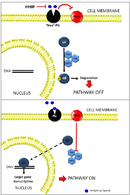
similar affinity and activate the Hh signaling cascade (Figure 1.3). Binding of HH ligands to PTC relieves it of its normal function as an inhibitor of Smoothened (SMO) (Murone, Rosenthal et al. 1999), a 7 trans-membrane domain protein which facilitates Hh signal transduction (Corbit, Aanstad et al. 2005). Although the mechanism by which PTC represses SMO is still unknown, it is thought that PTC inhibits SMO indirectly, possibly through the movement of a SMO-regulating small molecule across the cell membrane. Support for this hypothesis comes from the observation that PTC shares structural similarities to the resistance-nodulation-division (RND) family of bacterial proton-driven transmembrane transporters (Taipale, Cooper et al. 2002). Mutations in the RND-homologous regions of PTC decrease its ability to suppress Hh signaling activity. Furthermore, the Taipale et al. failed to note any specific and direct association between PTC and SMO in Hh-responsive cells, suggesting that a direct physical inhibition of SMO by PTC is unlikely. One candidate for a small molecule regulator of SMO is oxysterols (Dwyer, Sever et al. 2007). Oxysterols induce expression of Hh target genes through indirect activation of SMO. In the model proposed by Dwyer et al., the authors suggest that by regulating SMO exposure to intracellular oxysterols, PTC is able to regulate SMO. Supporting these results is the observation that PTC shares significant homology with NPC1 (which itself has homology with the bacterial RND family), a protein involved in LDL-derived cholesterol transport (Davies, Chen et al. 2000). Other regulators of SMO include vitamin D3 (Bijlsma, Spek et al. 2006). Despite multiple studies addressing this question, further research is needed to fully understand the mechanism behind PTC and SMO interactions.
1.4.3. The glee club: GLI transcription factors
activators, only GLI2 is required for proper Hh signaling transduction and embryonic development (Ding, Motoyama et al. 1998; Bai, Auerbach et al. 2002). Regulation of GLI activity occurs through protein phosphorylation and nuclear localization (Figure 1.3). All GLI proteins are negatively regulated through phosphorylation by PKA, GSK3b, or CK1 (Kaesler, Luscher et al. 2000; Riobo, Lu et al. 2006; Varjosalo, Bjorklund et al. 2008; Pan, Wang et al. 2009). Activated SMO promotes Hh signaling by preventing the phosphorylation and ubiquitination of GLI proteins through a mechanism that seems to involve the AKT antagonism of PKA activity (Riobo, Lu et al. 2006). In mammals, there are conflicting studies on whether phosphorylation of GLI can also influence nuclear-cytoplasmic shuttling (Sheng, Chi et al. 2006). Suppressor of Fused (SUFU), which binds to all three GLI proteins, is thought to act as a negative regulator of GLI nuclear localization (Stone, Murone et al. 1999). The exact mechanism by which SUFU regulates GLI proteins is not yet understood but several studies suggest that SUFU keeps GLI proteins in the cytosol (Dunaeva, Michelson et al. 2003; Merchant, Vajdos et al. 2004), possibly via regulation of a leucine-rich nuclear export signal region on GLI (Kogerman, Grimm et al. 1999) or via recruitment of a histone deacetylase to competitively inhibit GLI binding sites in the nucleus (Cheng and Bishop 2002). SMO counteracts these repressive activities by promoting the rapid ubiquitination of SUFU (Yue, Chen et al. 2009).
1.4.4. Negative feedback control of Hedgehog signaling
similar phenotypes as mice with overexpressed Hh signaling. The GLI3 repressor is formed after cleavage and degradation of the C-terminal domain following sequential phosphorylation by PKA, GSK3b, and CK1 (Tempe, Casas et al. 2006). SHH ligand downregulates the formation of the GLI3-repressor by inhibiting its phosphorylation (Wang, Fallon et al. 2000; Bai, Stephen et al. 2004).
Although certain aspects of Hh signaling remain obscure, what is clear is that environmental context contributes to the activity of Hh signaling. In development, active Hh signaling facilitates the crosstalk between epithelial and mesenchyme cell populations (Ingham and McMahon 2001). Similar patterns of Hh signaling emerge after tissue injury. In the liver, following injury, Hh signaling is triggered, becomes reactivated, and plays an important role in repair. Sustained and dysregulated Hh signaling in the liver leads to fibrosis, cirrhosis, and cancer.
1.5. The role of Hedgehog signaling in liver disease
After liver development, Hedgehog signaling becomes silenced. In both in vitro (e.g. mouse embryonic stem cells vs. well differentiated hepatocytes) and in vivo (e.g. Ptc-lacZ mice) models, qRT-PCR and IHC analysis reveals that the Hh pathway is activated and present in early development but becomes silenced in mature cells (Sicklick, Li et al. 2006). In uninjured livers, there is little to no expression of HH ligands and evidence of Hh signaling activity (Sicklick, Li et al. 2006; Yang, Wang et al. 2008). One possible explanation for suppressed Hh signaling in healthy livers is the high expression of HHIP, a Hh signaling repressor, by sinusoidal cells, observed in both in vitro (Yang, Wang et al. 2008) and in vivo
1.5.1. Hedgehog signaling is required for liver repair and regeneration
Hepatic injury triggers a wound healing response to regenerate the liver. This response is regulated by autocrine and paracrine signals within and between hepatic cells and includes a reactivation of Hh signaling pathway. Transient activation of Hh signaling is required after acute injury for livers to mount a proper wound healing response and once the injury signal is removed, the liver resolves the insult and Hh signaling is down-regulated (Omenetti, Popov et al. 2008). For example, treating mice with a Hh inhibitor after partial hepatectomy (a model of acute liver injury) results in a lack of activated Hh signaling, impaired liver regeneration and epithelial cell proliferation, significantly reduces accumulation of liver MF and progenitors, and results in the death of mice after 72 hours (Ochoa, Syn et al. 2010).
1.5.2. Clinical evidence for Hedgehog signaling in liver disease
co-express alpha smooth muscle actin (ASMA) and Vimentin (VIM), both mesenchymal and myofibroblastic markers. These observations suggest that the liver MF population is Hh-responsive and Hh signaling promotes their hepatic accumulation. Hh-Hh-responsive cells continued to be expressed in HCC, where they surround the tumor nodule (Pereira Tde, Witek et al. 2010). Thus, strong clinical evidence suggests that continuous Hh signaling promotes liver fibrosis and worsen liver injury. These clinical observations have been verified experimentally in mice with haploinsufficiency of ptc. When these mice, which overexpress Hh signaling, are subjected to cholestatic liver injury (Omenetti, Porrello et al. 2008) or hepatotoxic diets (Syn, Jung et al. 2009), liver MF accumulation is enhanced and fibrosis is worsened as compared to WT mice.
1.5.3. Hepatocyte injury initiates Hedgehog signaling in the liver
1.5.4. Hedgehog signaling activation affects multiple liver cell types
While hepatocytes are not themselves Hh responsive, neighboring stromal cells are Hh responsive and include hepatic stellate cells (HSC), hepatic progenitors, immune cells, cholangiocytes, and endothelial cells. These cells activate Hh signaling in response to HH ligands, which sets off a cascade of intracellular events to support the wound healing process. HH ligands activate Hh signaling in HSC, which promotes pro-survival pathways. Reducing the bioavailability of HH ligands in culture significantly reduces proliferation and increases HSC apoptosis (Sicklick, Li et al. 2005). In addition, HSC require active Hh signaling as they undergo EMT to transform into liver MF (Choi, Omenetti et al. 2009). Culture activated HSC upregulate Hh pathway genes (e.g. ptc, smo, and gli2) and downregulate Hh pathway inhibitors (e.g. hhip), whereas pharmacologic inhibition of Hh signaling with SMO antagonists reduces HSC differentiation into MF (Yang, Wang et al. 2008; Choi, Omenetti et al. 2009).
Liver progenitors are also Hh responsive and accumulate after liver injury (Sicklick, Li et al. 2006; Jung, Brown et al. 2008; Ochoa, Syn et al. 2010). HH ligands enhance the growth, proliferation, and viability of liver progenitors (e.g. oval cells, immature ductular cells) that become mobilized to regenerate the liver after injury (Sicklick, Li et al. 2006; Omenetti, Yang et al. 2007), but these responses are blunted with Hh signaling inhibition (Ochoa, Syn et al. 2010). Furthermore, in transgenic mice with unrestrained Hh signaling, the accumulation of injury-related Hh-responsive liver progenitors is enhanced.
and activated cholangiocytes, (Witek, Yang et al. 2009) can also produce additional HH ligands, which further enhance Hh signaling in the injured liver microenvironment. Given the broad impact of activated Hh signaling in cells that respond to liver injury, it is not surprising that Hh signaling inhibition potentially interferes with the activities of fibrosis-causing cells. Treating mice after partial hepatectomy with the Hh inhibitor cyclopamine results in a dramatic reduction in fibrosis (Ochoa, Syn et al. 2010), providing evidence that pharmacologic inhibition of Hh signaling can reverse fibrogenic repair.
Taken as a whole, the result of chronic liver injury is a Hh-rich microenvironment that promotes continuous wound healing, caused in part by sustained Hh signaling. Dysregulated and sustained Hh signaling promotes a pro-fibrotic phenotype, which is an important risk factor for liver cancer. Thus, it is reasonable to hypothesize that Hh signaling is also active in the liver tumor microenvironment. In other solid tumors, increased Hh signaling leads to tumor progression, either in a ligand independent (i.e. malignant epithelia harbor Hh pathway mutations) or ligand dependent (i.e. dysregulated paracrine Hh signaling between the tumor cell and its environment) fashion.
1.6. Over the hedge: Hedgehog signaling in cancer
resulted in BCC lesions (Adolphe, Hetherington et al. 2006). Together, these results suggested that 1) Ptc1 is a tumor suppressor, 2) a two-hit mechanism to Ptc-1 is required for cancer development, and 3) abnormal Hh signaling in the pre-neoplastic setting promotes malignancy.
Since then, there has been mounting evidence that abnormal Hh signaling is involved in multiple cancers. In prostate cancer, high levels of Hh signaling have been observed (Karhadkar, Bova et al. 2004) and a positive correlation exists between the level of Hh signaling activity and the severity and aggressiveness of the disease (Sheng, Li et al. 2004). Elevated Hh signaling has been observed in glioblastomas (Verhaak, Hoadley et al. 2010), pancreatic cancer (Thayer, di Magliano et al. 2003), renal cell carcinoma (Dormoy, Danilin et al. 2009), ovarian cancer (Bhattacharya, Kwon et al. 2008), esophageal cancer (Ma, Sheng et al. 2006), colon cancer (Varnat, Duquet et al. 2009), lung cancer (Watkins, Berman et al. 2003; Yuan, Goetz et al. 2007), melanoma (Stecca, Mas et al. 2007), and liver cancer (Sicklick, Li et al. 2006). Knockdown of Hh signaling in various cancer cell lines appears to decrease cell malignancy.
1.6.1. Ligand independent Hedgehog signaling in cancer
Hetherington et al. 2006). GLI consensus binding sites have been found on cyclin D1 (Yoon, Kita et al. 2002) and overexpression of GLI results in elevation of cell cycle regulator foxm1 (Teh, Wong et al. 2002). Hh signaling also promotes cell survival, another cancer hallmark. In BCC cell lines, binding sites for GLI were found on bcl2, a negative regulator molecule of apoptosis, and transfection of gli1 pcdna corresponded with dose dependent increases of bcl2 activity and decreased cellular apoptosis (Bigelow, Chari et al. 2004). In gastric cancer cells, downregulating GLI activity by blocking SMO resulted in decreased bcl2 expression (Han, Lee et al. 2009). Thus, the association between GLI1 and blc2 appears to be conserved among multiple cancer types.
Hh signaling is also an established effector of EMT, a process involved in tumor invasiveness and metastasis. In ovarian cancer cell lines, overexpression of GLI1 increased cell mobility and invasiveness along with EMT genes such as vimentin and mmp1 (Liao, Siu et al. 2009). Hh inhibition in pancreatic cancer cell lines resulted in downregulation of mesenchymal programming and invasiveness and upregulated epithelial genes such as E-cadherin (Feldmann, Dhara et al. 2007). Notably, overexpression of GLI1 in pancreatic cancer cells resulted in increased invasiveness (Feldmann, Dhara et al. 2007; Inaguma, Kasai et al. 2011). Similar observations are reported in breast cancer cells (Souzaki, Kubo et al. 2011), glioma cells (Wang, Pan et al. 2010), and gastric cancer cells (Yoo, Kang et al. 2008). Together, these studies help to explain the effectiveness of targeting GLI proteins to disrupt tumor xenograft growth of various cancers (Thayer, di Magliano et al. 2003; Lauth, Bergstrom et al. 2007).
1.6.2. Ligand dependent, paracrine Hedgehog signaling in cancer
suggesting that disruption of the paracrine Hh signaling between epithelial tumor cells and stromal cells can prevent carcinogenesis.
1.6.3. Paracrine Hedgehog signaling promotes the hallmarks of cancer
1.6.4. Hedgehog signaling in HCC
The question of how does Hh signaling contribute to liver cancer is still largely unknown. Several studies would suggest that autocrine Hh signaling in liver tumor cells is elevated and is a major pathway responsible for tumor proliferation, viability, chemoresistance and invasion (Sicklick, Li et al. 2006; Cheng, Xu et al. 2009; Chen, Lingala et al. 2011; Chen, Lin et al. 2011; Lu, Zhao et al. 2012). However most of these studies describing the advantages conferred by Hh signaling were performed in cell lines. Furthermore, evidence for overexpression of Hh signaling was found in whole liver tissue and does not account for cell-specific localization. Addressing this, Pereira et al. performed immunostaining on human HCC samples and found that PTC(+) and GLI2(+) cells were localized to the stromal compartment surrounding the tumor nodule, suggesting a paracrine mechanism might exist for liver cancer to advance (Pereira Tde, Witek et al. 2010).
1.6.5. Development of clinical inhibitors of Hedgehog signaling
inhibitor of Hh signaling to be approved for clinical treatment of BCC (Guha 2012). Vismodegib is considered a competitive inhibitor of SMO, and belongs to a class of cycopamine-derivatives that outcompete cyclopamine for binding to SMO. However, to date, reproducible clinical responses to SMO inhibitors have only been reported in BCC (Sekulic, Migden et al. 2012) and medulloblastomas (Rudin, Hann et al. 2009). In fact, a 199 patient Phase II, randomized, double-blind, placebo-controlled study evaluating GDC-0449 as first line therapy for previously untreated metastatic colorectal cancer yielded unsatisfactory results: Not only were there no statistically significant differences in outcomes between the two arms, but the hazard ratio for progression free survival actually favored the placebo group (Rudin 2012). And despite significant responses in a Phase II trial in advanced BCC (overall disease control rate was over 86%), the median duration of response was 7.6 months and median progression free survival was 9.6 months. Acquired resistance is attributed to mutations that alter SMO to prevent the binding of GDC-0449 to SMO (Yauch, Dijkgraaf et al. 2009). Unique challenges remain to determine whether Hh signaling inhibition will provide therapeutic benefit to patients.
promising and with clinical inhibitors against Hh signaling being developed, understanding the mechanism of Hh signaling in HCC could open up new avenues for medical treatment.
1.7. Metabolism in physiology and cancer
necessary to recycle NADH back to NAD+, a required coenzyme for glycolysis. When oxygen is resupplied, lactate is converted back into pyruvate to enter the aerobic respiration.
1.7.1. Cancer metabolism
Over 90 years ago, Otto Warburg first noted that the growth of cancer cells (from human skin, throat, intestine, penis and nose) relies upon enhanced anaerobic glycolysis, even when sufficient oxygen is available for oxidative phosphorylation (Warburg 1956). Warburg recognized that despite aerobic conditions, cancer cells (as opposed to normal cells) produce energy by high rates of glycolysis and lactic acid fermentation, yet have low rates of oxidative phosphorylation in the mitochondria (Figure 1.6). Thus, cancer cells follow a pattern of glycolytic activity that mimics the activity of normal cells under hypoxic (anaerobic) conditions. This observation was later termed the Warburg effect (Racker 1972). Warburg hypothesized that this was due to impaired respiration in the tumor cell (mitochondrial defects), although we now know this is not necessarily the case (Dang 2012). The FDG-PET scan is the direct clinical application of the Warburg Effect. Briefly, a radiolabeled hexokinase substrate, 2-18F-2-deoxyglucose (FDG), is incorporated into the cell by glucose transporters and phosphorylated by hexokinase. In oncology, this technique is used to label and monitor tumors and track metastatic growth on the assumption that glycolytic tumor cells have a high uptake of glucose (Ben-Haim and Ell 2009).
1.7.2. Regulators of the Warburg Effect
decreased detection by FDG-PET, suggesting that disruption of PI3K/AKT signaling impacts tumor glucose transport (Engelman, Chen et al. 2008). Other oncogenes, such as RAS and SRC also increase glucose uptake and the phosphorylation of glycolytic enzymes (Cooper, Esch et al. 1984; Flier, Mueckler et al. 1987). A study by Matoba et al. showed that p53 is coupled to mitochondrial respiration. Specifically, through Synthesis of Cytochrome c Oxidase 2 (SCO2), p53 regulates the cytochrome c oxidase complex, essential for oxidative phosphorylation. In p53 deficient cells, the authors observed a shift towards cellular utilization of glycolytic pathways (Matoba, Kang et al. 2006). The metabolic reprogramming of tumor cells is also attributed to HIF1α, a transcription factor that is stabilized in response to hypoxia. Multiple tumor cells have shown to have elevated HIF1α signaling (Sutter, Laughner et al. 2000) and HIF1α activation suppresses mitochondrial function through activating Pdk1 (Kim, Tchernyshyov et al. 2006; Papandreou, Cairns et al. 2006) and directly increases glycolytic-associated genes hexokinase II (Mathupala, Rempel et al. 2001). HIF1α binding sites have been found on the M2 isoform of pyruvate kinase (PKM2), which further promotes the idea that HIF1α regulates aspects of glycolytic metabolism in cells (Kim, Tchernyshyov et al. 2006).
regulating epigenetic changes (Yang, Xia et al. 2012), increasing tumor growth (Christofk, Vander Heiden et al. 2008), increasing bioavailability of nucleic acids (Ye, Mancuso et al. 2012), and even further altering the metabolism of the cancer cell by promoting lipogenesis (Panasyuk, Espeillac et al. 2012).
1.7.3. The tumor microenvironment and cancer metabolism
Although the amount of information being uncovered about tumor metabolism is growing, questions still remain about how the tumor microenvironment influences the metabolic state of the cancer. Two groups in ovarian and breast cancer have reported that the growth of tumor cells is enhanced by metabolic end-products of the surrounding stroma. In an co-culture model of stromal adipocytes and ovarian cancer cells, Nieman et al. demonstrated that adipocytes directly transfer lipids to ovarian cancer cells to promote in vitro and in vivo growth and migration (Nieman, Kenny et al. 2011). Another group suggested that stromal fibroblasts in breast cancer are glycolytic themselves and secrete lactate into the microenvironment to promote growth and metastasis of breast cancer cells (Bonuccelli, Tsirigos et al. 2010). The idea that metabolic, and specifically, glycolytic end products such as lactate, from stromal cells can support tumorigenesis is intriguing and demonstrates yet another method by which the microenvironment influences the growth of tumors.
1.7.4. The impact of glycolytic end product lactate on tumor progression
release mitogenic and angiogenic factors, indirectly contributing to tumor proliferation and angiogenesis (Jensen, Hunt et al. 1986). The impact of lactate on the tumor microenvironment is reinforced by additional evidence that tumor cells express higher levels of monocarboxylate transporter 1 (MCT1), a lactate importer, than non-malignant cells (Koukourakis, Giatromanolaki et al. 2007; Sonveaux, Vegran et al. 2008). Inhibition of MCT1 causes antitumor effects, including reduced growth and increased necrosis of tumor xenografts, suggesting that targeting lactate associated transporters has therapeutic benefit. Another study which suggests that endothelial cells uptake lactate in the tumor microenvironment to promote angiogenesis would seemingly rule them out as a source of lactate production (Sonveaux, Copetti et al. 2012).
1.7.5. Glycolytic regulation of liver MF
In the pre-neoplastic environment of the liver, the metabolism of hepatic stellate cells is an important regulator of their fibrogenic potential. In recently published work, our lab revealed a previously unsuspected “metabolism-centric” mechanism governing the fate of hepatic stellate cells (Chen, Choi et al. 2012). We demonstrate that during culture (and in injured livers), the trans-differentiation of quiescent HSC into myofibroblasts is mediated via a metabolic switch that favors glucose consumptive processes, and show that this global change in HSC metabolism is controlled by Hedgehog signaling. Hh signaling orchestrates this myofibroblastic reprogramming of HSC by directing HIF1α-dependent induction of glycolytic enzymes, resulting in cellular accumulation of the glycolytic end-product, lactate. In HSC themselves, lactate accumulation was shown to orchestrate global phenotypic changes that cause those cells to become myofibroblastic, thereby enhancing their wound healing capabilities.
an important role in development of fibrosis, and is mediated by Hh signaling. It is possible that hypoxic environments upregulate glucose metabolism in fibrotic and pre-neoplastic livers, which later contribute their end-products to promote tumorigenesis. Thus, the extent of metabolic contribution by liver cancer associated fibroblasts towards HCCarcinogenesis remains largely unknown and highlights the need for further study on the metabolism of liver cancer as a whole.
1.8. Summary
Hepatocellular carcinoma is a disease that afflicts many patients worldwide. Unfortunately for many patients with HCC, therapeutic options are extremely limited, creating a real patient-driven need for better therapies. The largest risk factor for HCC is cirrhosis, and HCC commonly recurs in cirrhotic livers after tumor ablation. The pre-neoplastic cirrhotic microenvironment may promote the outgrowth of malignant hepatocytes, but the mechanisms involved remain obscure. Because deregulated, excessively fibrogenic repair of liver injury causes cirrhosis itself, one possibility is that stromal-epithelial interactions fuel HCC growth. In multiple tumors, communication between stromal cells in the tumor microenvironment and tumor cells nurtures malignant phenotypes. An important signaling pathway that regulates crosstalk between stroma and epithelia is Hedgehog signaling, which plays critical roles in development, wound healing, and cancer. This pathway is especially important in liver regeneration and is a potential candidate for therapeutic targeting in liver cancer. Sustained Hedgehog signaling worsens liver fibrosis and regulates the behaviors of multiple cells involved in wound-healing. Dysregulation of this pathway continues as liver cancer develops, which warrants investigation into a) whether active Hh signaling is required for tumor growth, b) does Hh signaling regulate communication between malignant hepatocytes and Hh-responsive liver MF, and if so, c)
Figure 1.1. Masson trichrome stained core liver biopsy of NAFLD with cirrhosis. Cirrhosis is characterized by the replacement of normal liver parenchyma with fibrous tissue composed of collagenous matrix. Here fibrotic bands are stained blue by trichrome staining. Histologically, cirrhosis contains nodular remnants of normal liver architecture (portal triads and central veins) that are separated by wide scars and thin fibrous septa. Normal liver architecture is shown in the insert. Note the lack of fibrogenic
matrix in normal liver.
Figure 1.2. Graphical representation of the liver architecture. The liver receives blood from the portal vein and the hepatic artery, which run beside the common bile duct and form the portal triad. The hepatic artery receives arterial blood and the portal vein receives blood from the gastrointestinal tract. Blood is passed through the hepatic sinusoids and out of the liver through hepatic venules. The central vein is a branch of the hepatic vein. Hepatocytes are organized into cords that are separated by sinusoids. Endothelial cells line the sinusoids and lie directly adjacent to hepatocytes. The space between the endothelium and hepatocytes is the space of Disse. Hepatic stellate cells occupy this perisinusoidal space. Kupffer cells are specialized macrophages in the liver and contribute to the inflammatory response during wound healing.
Figure 1.3. Overview of the Hedgehog signaling pathway. Hedgehog ligands interact with the Hh receptor, Patched (Ptc) which relieves Ptc-mediated repression of Smoothened (Smo), permitting Smo activation. Activated Smo prevents the phosphorylation and ubiquitination of Glioma (Gli)-family proteins, leading to their accumulation and nuclear localization. In the nucleus, Gli proteins bind to Hh-target genes and regulate the transcription of Hh-associated genes. Factors like Hedgehog-interacting protein (HHIP) competitively inhibit Hh signaling by binding to HH ligands.
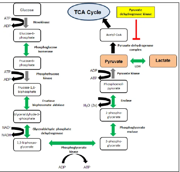
Figure 1.4. Overview of glycolysis. Glycolysis begins when glucose is transported into the cell via glucose transporters. Glucose is metabolized to pyruvate through a series of catalyzed reactions that occur during glycolysis. In aerobic conditions, pyruvate dehydrogenase catalyzes the reaction that converts pyruvate to acetyl CoA which then enters the TCA cycle and is further oxidized in the mitochondria during oxidative phosphorylation. The activity of pyruvate dehydrogenase is regulated by the enzyme pyruvate dehydrogenase kinase. In anaerobic conditions, pyrvuate is coverted to lactate via the enzyme lactate dehydrogenase (LDH) during lactic acid fermentation. Gray arrows
Figure 1.6. Overview of the Warburg Effect. As discussed, cells metabolize glucose to pyruvate during glycolysis. In the presence of oxygen, normal differentiated cells use oxidative phosphorylation to produce ATP. However, cancerous or proliferative cells downregulate their use of oxidative phosphorylation in the mitochondria and instead produce ATP using primarily glycolysis. As a result, lactate is often a side product of the Warburg effect.
(Figure is adapted from Understanding the Warburg Effect: The Metabolic Requirements of
Works Cited
Abe, R., S. C. Donnelly, et al. (2001). "Peripheral blood fibrocytes: differentiation pathway and migration to wound sites." J Immunol 166(12): 7556-7562.
Adami, H. O., W. H. Chow, et al. (1996). "Excess risk of primary liver cancer in patients with diabetes mellitus." J Natl Cancer Inst 88(20): 1472-1477.
Adolphe, C., R. Hetherington, et al. (2006). "Patched1 functions as a gatekeeper by promoting cell cycle progression." Cancer Res 66(4): 2081-2088.
Adolphe, C., M. Narang, et al. (2004). "An in vivo comparative study of sonic, desert and Indian hedgehog reveals that hedgehog pathway activity regulates epidermal stem cell homeostasis." Development 131(20): 5009-5019.
Ankoma-Sey, V., M. Matli, et al. (1998). "Coordinated induction of VEGF receptors in mesenchymal cell types during rat hepatic wound healing." Oncogene 17(1): 115-121.
Ankoma-Sey, V., Y. Wang, et al. (2000). "Hypoxic stimulation of vascular endothelial growth factor expression in activated rat hepatic stellate cells." Hepatology 31(1): 141-148. Apte, U., M. D. Thompson, et al. (2008). "Wnt/beta-catenin signaling mediates oval cell
response in rodents." Hepatology 47(1): 288-295.
Arwert, E. N., E. Hoste, et al. (2012). "Epithelial stem cells, wound healing and cancer." Nat Rev Cancer 12(3): 170-180.
Ascha, M. S., I. A. Hanouneh, et al. (2010). "The incidence and risk factors of hepatocellular carcinoma in patients with nonalcoholic steatohepatitis." Hepatology 51(6): 1972-1978.
Aszterbaum, M., J. Epstein, et al. (1999). "Ultraviolet and ionizing radiation enhance the growth of BCCs and trichoblastomas in patched heterozygous knockout mice." Nat Med 5(11): 1285-1291.
Bachem, M. G., D. Meyer, et al. (1992). "Activation of rat liver perisinusoidal lipocytes by transforming growth factors derived from myofibroblastlike cells. A potential mechanism of self perpetuation in liver fibrogenesis." J Clin Invest 89(1): 19-27. Bai, C. B., W. Auerbach, et al. (2002). "Gli2, but not Gli1, is required for initial Shh signaling
and ectopic activation of the Shh pathway." Development 129(20): 4753-4761. Bai, C. B., D. Stephen, et al. (2004). "All mouse ventral spinal cord patterning by hedgehog
is Gli dependent and involves an activator function of Gli3." Dev Cell 6(1): 103-115. Bailey, J. M., A. M. Mohr, et al. (2009). "Sonic hedgehog paracrine signaling regulates
metastasis and lymphangiogenesis in pancreatic cancer." Oncogene 28(40): 3513-3525.
pancreatic cancer." Clin Cancer Res 14(19): 5995-6004.
Belloni, E., M. Muenke, et al. (1996). "Identification of Sonic hedgehog as a candidate gene responsible for holoprosencephaly." Nat Genet 14(3): 353-356.
Ben-Haim, S. and P. Ell (2009). "18F-FDG PET and PET/CT in the evaluation of cancer treatment response." J Nucl Med 50(1): 88-99.
Bhattacharya, R., J. Kwon, et al. (2008). "Role of hedgehog signaling in ovarian cancer." Clin Cancer Res 14(23): 7659-7666.
Bigelow, R. L., N. S. Chari, et al. (2004). "Transcriptional regulation of bcl-2 mediated by the sonic hedgehog signaling pathway through gli-1." J Biol Chem 279(2): 1197-1205. Bijlsma, M. F., C. A. Spek, et al. (2006). "Repression of smoothened by patched-dependent
(pro-)vitamin D3 secretion." PLoS Biol 4(8): e232.
Binns, W., L. F. James, et al. (1962). "Cyclopian-type malformation in lambs." Arch Environ Health 5: 106-108.
Bonuccelli, G., A. Tsirigos, et al. (2010). "Ketones and lactate "fuel" tumor growth and metastasis: Evidence that epithelial cancer cells use oxidative mitochondrial metabolism." Cell Cycle 9(17): 3506-3514.
Bruix, J. and M. Sherman (2011). "Management of hepatocellular carcinoma: an update." Hepatology 53(3): 1020-1022.
Calle, E. E., C. Rodriguez, et al. (2003). "Overweight, obesity, and mortality from cancer in a prospectively studied cohort of U.S. adults." N Engl J Med 348(17): 1625-1638. Canbay, A., H. Higuchi, et al. (2002). "Fas enhances fibrogenesis in the bile duct ligated
mouse: a link between apoptosis and fibrosis." Gastroenterology 123(4): 1323-1330. Cancer Genome Atlas Research Network (2008). "Comprehensive genomic characterization
defines human glioblastoma genes and core pathways." Nature 455(7216): 1061-1068.
Cancer Genome Atlas Research Network (2011). "Integrated genomic analyses of ovarian carcinoma." Nature 474(7353): 609-615.
Cancer Genome Atlas Research Network (2012). "Comprehensive molecular characterization of human colon and rectal cancer." Nature 487(7407): 330-337. Cardinale, V., Y. Wang, et al. (2011). "Multipotent stem/progenitor cells in human biliary tree
give rise to hepatocytes, cholangiocytes, and pancreatic islets." Hepatology 54(6): 2159-2172.
Chao, Y., W. K. Chan, et al. (1996). "Phase II study of flutamide in the treatment of hepatocellular carcinoma." Cancer 77(4): 635-639.
signaling activation are associated with chemoresistance and invasion of hepatoma subpopulations." J Hepatol 55(4): 838-845.
Chen, Y., S. S. Choi, et al. (2012). "Hedgehog Controls Hepatic Stellate Cell Fate by Regulating Metabolism." Gastroenterology.
Chen, Y. J., C. P. Lin, et al. (2011). "Sonic hedgehog signaling protects human hepatocellular carcinoma cells against ionizing radiation in an autocrine manner." Int J Radiat Oncol Biol Phys 80(3): 851-859.
Cheng, S. Y. and J. M. Bishop (2002). "Suppressor of Fused represses Gli-mediated transcription by recruiting the SAP18-mSin3 corepressor complex." Proc Natl Acad Sci U S A 99(8): 5442-5447.
Cheng, W. T., K. Xu, et al. (2009). "Role of Hedgehog signaling pathway in proliferation and invasiveness of hepatocellular carcinoma cells." Int J Oncol 34(3): 829-836.
Cho, Y. K., J. K. Kim, et al. (2009). "Systematic review of randomized trials for hepatocellular carcinoma treated with percutaneous ablation therapies." Hepatology 49(2): 453-459.
Choi, S. S., A. Omenetti, et al. (2009). "Hedgehog pathway activation and epithelial-to-mesenchymal transitions during myofibroblastic transformation of rat hepatic cells in culture and cirrhosis." Am J Physiol Gastrointest Liver Physiol 297(6): G1093-1106. Christofk, H. R., M. G. Vander Heiden, et al. (2008). "The M2 splice isoform of pyruvate
kinase is important for cancer metabolism and tumour growth." Nature 452(7184): 230-233.
Chuang, P. T. and A. P. McMahon (1999). "Vertebrate Hedgehog signalling modulated by induction of a Hedgehog-binding protein." Nature 397(6720): 617-621.
Cooke, V. G., V. S. LeBleu, et al. (2012). "Pericyte depletion results in hypoxia-associated epithelial-to-mesenchymal transition and metastasis mediated by met signaling pathway." Cancer Cell 21(1): 66-81.
Cooper, J. A., F. S. Esch, et al. (1984). "Phosphorylation sites in enolase and lactate dehydrogenase utilized by tyrosine protein kinases in vivo and in vitro." J Biol Chem 259(12): 7835-7841.
Cooper, M. K., J. A. Porter, et al. (1998). "Teratogen-mediated inhibition of target tissue response to Shh signaling." Science 280(5369): 1603-1607.
Corbit, K. C., P. Aanstad, et al. (2005). "Vertebrate Smoothened functions at the primary cilium." Nature 437(7061): 1018-1021.
Coulouarn, C., A. Corlu, et al. (2012). "Hepatocyte-stellate cell cross-talk in the liver engenders a permissive inflammatory microenvironment that drives progression in hepatocellular carcinoma." Cancer Res 72(10): 2533-2542.
p53 gene in hepatocellular carcinomas from Senegal." Br J Cancer 67(6): 1395-1397.
Dang, C. V. (2012). "Links between metabolism and cancer." Genes Dev 26(9): 877-890. Davies, J. P., F. W. Chen, et al. (2000). "Transmembrane molecular pump activity of
Niemann-Pick C1 protein." Science 290(5500): 2295-2298.
Davis, G. L., M. J. Alter, et al. (2010). "Aging of hepatitis C virus (HCV)-infected persons in the United States: a multiple cohort model of HCV prevalence and disease progression." Gastroenterology 138(2): 513-521, 521 e511-516.
de Leeuw, A. M., S. P. McCarthy, et al. (1984). "Purified rat liver fat-storing cells in culture divide and contain collagen." Hepatology 4(3): 392-403.
Deutsch, G., J. Jung, et al. (2001). "A bipotential precursor population for pancreas and liver within the embryonic endoderm." Development 128(6): 871-881.
Ding, H., D. Zhou, et al. (2012). "Sonic hedgehog signaling mediates epithelial-mesenchymal communication and promotes renal fibrosis." J Am Soc Nephrol 23(5): 801-813.
Ding, Q., J. Motoyama, et al. (1998). "Diminished Sonic hedgehog signaling and lack of floor plate differentiation in Gli2 mutant mice." Development 125(14): 2533-2543.
Dormoy, V., S. Danilin, et al. (2009). "The sonic hedgehog signaling pathway is reactivated in human renal cell carcinoma and plays orchestral role in tumor growth." Mol Cancer 8: 123.
Douarin, N. M. (1975). "An experimental analysis of liver development." Med Biol 53(6): 427-455.
Dunaeva, M., P. Michelson, et al. (2003). "Characterization of the physical interaction of Gli proteins with SUFU proteins." J Biol Chem 278(7): 5116-5122.
Dvorak, H. F. (1986). "Tumors: wounds that do not heal. Similarities between tumor stroma generation and wound healing." N Engl J Med 315(26): 1650-1659.
Dvorak, H. F., A. M. Dvorak, et al. (1979). "Fibrin gel investment associated with line 1 and line 10 solid tumor growth, angiogenesis, and fibroplasia in guinea pigs. Role of cellular immunity, myofibroblasts, microvascular damage, and infarction in line 1 tumor regression." J Natl Cancer Inst 62(6): 1459-1472.
Dwyer, J. R., N. Sever, et al. (2007). "Oxysterols are novel activators of the hedgehog signaling pathway in pluripotent mesenchymal cells." J Biol Chem 282(12): 8959-8968.
Fabris, L., M. Cadamuro, et al. (2007). "Analysis of liver repair mechanisms in Alagille syndrome and biliary atresia reveals a role for notch signaling." Am J Pathol 171(2): 641-653.
Fan, L., C. V. Pepicelli, et al. (2004). "Hedgehog signaling promotes prostate xenograft tumor growth." Endocrinology 145(8): 3961-3970.
Fan, Y., K. G. Dickman, et al. (2010). "Akt and c-Myc differentially activate cellular metabolic programs and prime cells to bioenergetic inhibition." J Biol Chem 285(10): 7324-7333.
Faouzi, S., B. E. Burckhardt, et al. (2001). "Anti-Fas induces hepatic chemokines and promotes inflammation by an NF-kappa B-independent, caspase-3-dependent pathway." J Biol Chem 276(52): 49077-49082.
Feldmann, G., S. Dhara, et al. (2007). "Blockade of hedgehog signaling inhibits pancreatic cancer invasion and metastases: a new paradigm for combination therapy in solid cancers." Cancer Res 67(5): 2187-2196.
Ferlay, J., H. R. Shin, et al. (2010). "Estimates of worldwide burden of cancer in 2008: GLOBOCAN 2008." Int J Cancer 127(12): 2893-2917.
Fiaschi, M., B. Rozell, et al. (2009). "Development of mammary tumors by conditional expression of GLI1." Cancer Res 69(11): 4810-4817.
Flier, J. S., M. M. Mueckler, et al. (1987). "Elevated levels of glucose transport and transporter messenger RNA are induced by ras or src oncogenes." Science 235(4795): 1492-1495.
Folkman, J. (1971). "Tumor angiogenesis: therapeutic implications." N Engl J Med 285(21): 1182-1186.
Forner, A., J. M. Llovet, et al. (2012). "Hepatocellular carcinoma." Lancet 379(9822): 1245-1255.
Friedman, S. L. (2008). "Hepatic stellate cells: protean, multifunctional, and enigmatic cells of the liver." Physiol Rev 88(1): 125-172.
Fujiwara, Y., D. S. Hoon, et al. (2000). "PTEN / MMAC1 mutation and frequent loss of heterozygosity identified in chromosome 10q in a subset of hepatocellular carcinomas." Jpn J Cancer Res 91(3): 287-292.
Fukumura, D., R. Xavier, et al. (1998). "Tumor induction of VEGF promoter activity in stromal cells." Cell 94(6): 715-725.
Goetze, K., S. Walenta, et al. (2011). "Lactate enhances motility of tumor cells and inhibits monocyte migration and cytokine release." Int J Oncol 39(2): 453-463.
Guha, M. (2012). "Hedgehog inhibitor gets landmark skin cancer approval, but questions remain for wider potential." Nat Rev Drug Discov 11(4): 257-258.
Guy, C. D., A. Suzuki, et al. (2012). "Hedgehog pathway activation parallels histologic severity of injury and fibrosis in human nonalcoholic fatty liver disease." Hepatology 55(6): 1711-1721.
Hahn, H., C. Wicking, et al. (1996). "Mutations of the human homolog of Drosophila patched in the nevoid basal cell carcinoma syndrome." Cell 85(6): 841-851.
Hahn, H., L. Wojnowski, et al. (1998). "Rhabdomyosarcomas and radiation hypersensitivity in a mouse model of Gorlin syndrome." Nat Med 4(5): 619-622.
Han, M. E., Y. S. Lee, et al. (2009). "Hedgehog signaling regulates the survival of gastric cancer cells by regulating the expression of Bcl-2." Int J Mol Sci 10(7): 3033-3043. Han, Y. P., C. Yan, et al. (2007). "A matrix metalloproteinase-9 activation cascade by
hepatic stellate cells in trans-differentiation in the three-dimensional extracellular matrix." J Biol Chem 282(17): 12928-12939.
Hanahan, D. and R. A. Weinberg (2011). "Hallmarks of cancer: the next generation." Cell 144(5): 646-674.
Harfe, B. D., P. J. Scherz, et al. (2004). "Evidence for an expansion-based temporal Shh gradient in specifying vertebrate digit identities." Cell 118(4): 517-528.
Hasebe, T., S. Sasaki, et al. (2002). "Prognostic significance of fibrotic focus in invasive ductal carcinoma of the breast: a prospective observational study." Mod Pathol 15(5): 502-516.
Hoshida, Y., A. Villanueva, et al. (2008). "Gene expression in fixed tissues and outcome in hepatocellular carcinoma." N Engl J Med 359(19): 1995-2004.
Hui, A. M., M. Sakamoto, et al. (1996). "Inactivation of p16INK4 in hepatocellular carcinoma." Hepatology 24(3): 575-579.
Huijbers, I. J., M. Iravani, et al. (2010). "A role for fibrillar collagen deposition and the collagen internalization receptor endo180 in glioma invasion." PLoS One 5(3): e9808.
Inaguma, S., K. Kasai, et al. (2011). "GLI1 facilitates the migration and invasion of pancreatic cancer cells through MUC5AC-mediated attenuation of E-cadherin." Oncogene 30(6): 714-723.
Incardona, J. P., W. Gaffield, et al. (1998). "The teratogenic Veratrum alkaloid cyclopamine inhibits sonic hedgehog signal transduction." Development 125(18): 3553-3562. Ingham, P. W. and A. P. McMahon (2001). "Hedgehog signaling in animal development:
Ishizaki, Y., S. Ikeda, et al. (2004). "Immunohistochemical analysis and mutational analyses of beta-catenin, Axin family and APC genes in hepatocellular carcinomas." Int J Oncol 24(5): 1077-1083.
Jedeszko, C., B. C. Victor, et al. (2009). "Fibroblast hepatocyte growth factor promotes invasion of human mammary ductal carcinoma in situ." Cancer Res 69(23): 9148-9155.
Jemal, A., F. Bray, et al. (2011). "Global cancer statistics." CA Cancer J Clin 61(2): 69-90. Jensen, J. A., T. K. Hunt, et al. (1986). "Effect of lactate, pyruvate, and pH on secretion of
angiogenesis and mitogenesis factors by macrophages." Lab Invest 54(5): 574-578. Johnson, R. L., A. L. Rothman, et al. (1996). "Human homolog of patched, a candidate gene
for the basal cell nevus syndrome." Science 272(5268): 1668-1671.
Jung, Y., K. D. Brown, et al. (2008). "Accumulation of hedgehog-responsive progenitors parallels alcoholic liver disease severity in mice and humans." Gastroenterology 134(5): 1532-1543.
Jung, Y., S. J. McCall, et al. (2007). "Bile ductules and stromal cells express hedgehog ligands and/or hedgehog target genes in primary biliary cirrhosis." Hepatology 45(5): 1091-1096.
Jung, Y., R. P. Witek, et al. (2010). "Signals from dying hepatocytes trigger growth of liver progenitors." Gut 59(5): 655-665.
Kaesler, S., B. Luscher, et al. (2000). "Transcriptional activity of GLI1 is negatively regulated by protein kinase A." Biol Chem 381(7): 545-551.
Kalluri, R. and M. Zeisberg (2006). "Fibroblasts in cancer." Nat Rev Cancer 6(5): 392-401. Karhadkar, S. S., G. S. Bova, et al. (2004). "Hedgehog signalling in prostate regeneration,
neoplasia and metastasis." Nature 431(7009): 707-712.
Kim, J. W., I. Tchernyshyov, et al. (2006). "HIF-1-mediated expression of pyruvate dehydrogenase kinase: a metabolic switch required for cellular adaptation to hypoxia." Cell Metab 3(3): 177-185.
Knittel, T., D. Kobold, et al. (1999). "Rat liver myofibroblasts and hepatic stellate cells: different cell populations of the fibroblast lineage with fibrogenic potential." Gastroenterology 117(5): 1205-1221.
Kogerman, P., T. Grimm, et al. (1999). "Mammalian suppressor-of-fused modulates nuclear-cytoplasmic shuttling of Gli-1." Nat Cell Biol 1(5): 312-319.
Kolterud, A., A. S. Grosse, et al. (2009). "Paracrine Hedgehog signaling in stomach and intestine: new roles for hedgehog in gastrointestinal patterning." Gastroenterology 137(2): 618-628.