R E S E A R C H A R T I C L E
Open Access
Accuracy of SIAscopy for pigmented skin lesions
encountered in primary care: development and
validation of a new diagnostic algorithm
Jon D Emery
1,5*, Judith Hunter
2,3, Per N Hall
2, Anthony J Watson
1, Marc Moncrieff
4, Fiona M Walter
5,1Abstract
Background:Diagnosing pigmented skin lesions in general practice is challenging. SIAscopy has been shown to increase diagnostic accuracy for melanoma in referred populations. We aimed to develop and validate a scoring system for SIAscopic diagnosis of pigmented lesions in primary care.
Methods:This study was conducted in two consecutive settings in the UK and Australia, and occurred in three stages: 1) Development of the primary care scoring algorithm (PCSA) on a sub-set of lesions from the UK sample; 2) Validation of the PCSA on a different sub-set of lesions from the same UK sample; 3) Validation of the PCSA on a new set of lesions from an Australian primary care population. Patients presenting with a pigmented lesion were recruited from 6 general practices in the UK and 2 primary care skin cancer clinics in Australia. The following data were obtained for each lesion: clinical history; SIAscan; digital photograph; and digital dermoscopy. SIAscans were interpreted by an expert and validated against histopathology where possible, or expert clinical review of all available data for each lesion.
Results:A total of 858 patients with 1,211 lesions were recruited. Most lesions were benign naevi (64.8%) or seborrhoeic keratoses (22.1%); 1.2% were melanoma. The original SIAscopic diagnostic algorithm did not perform well because of the higher prevalence of seborrhoeic keratoses and haemangiomas seen in primary care. A primary care scoring algorithm (PCSA) was developed to account for this. In the UK sample the PCSA had the following characteristics for the diagnosis of‘suspicious’: sensitivity 0.50 (0.18-0.81); specificity 0.84 (0.78-0.88); PPV 0.09 (0.03-0.22); NPV 0.98 (0.95-0.99). In the Australian sample the PCSA had the following characteristics for the diagnosis of
‘suspicious’: sensitivity 0.44 (0.32-0.58); specificity 0.95 (0.93-0.97); PPV 0.52 (0.38-0.66); NPV 0.95 (0.92-0.96). In an analysis of lesions for which histological diagnosis was available (n = 111), the PCSA had a significantly greater Area Under the Curve than the 7-point checklist for the diagnosis of melanoma (0.83; 95% CI 0.71-0.95 versus 0.61; 95% CI 0.44-0.78; p = 0.02 for difference).
Conclusions:The PCSA could have a useful role in improving primary care management of pigmented skin lesions. Further work is needed to develop and validate the PCSA in other primary care populations and to evaluate the cost-effectiveness of GP management of pigmented lesions using SIAscopy.
Background
Pigmented skin lesions are a common presenting problem in general practice and, while the majority are benign naevi or non-melanocytic lesions (seborrhoeic ker-atoses, haemangiomas), a small minority are malignant
melanomas. Melanoma is a serious skin cancer, responsi-ble for 2% of all cancers and 1% of all cancer deaths in the UK, with about 8,000 new cases and 1,800 deaths a year [1]. Worldwide, the incidence of melanoma is increasing faster than any other solid cancer with an approximate doubling of rates every 10-20 years in countries with Cau-casian populations [2,3].
Pigmented lesions and melanoma pose particular diag-nostic and management challenges for general practi-tioners (GPs) [4]. GPs are less able than dermatologists
* Correspondence: [email protected]
1General Practice, School of Primary, Aboriginal and Rural Health Care,
University of Western Australia, 328 Stirling Highway, Claremont, WA 6010, Australia
Full list of author information is available at the end of the article
to differentiate melanomas from other pigmented lesions [5,6], probably because an individual GP will encounter melanoma infrequently [7]. British data fol-lowing the establishment of urgent referral pathways for all suspected skin cancers [8] showed that only 12% of referred lesions were diagnosed as skin cancer and only 42% of skin cancers were referred via this route [9]. There have been conflicting findings about the perfor-mance of GPs who have been trained in melanoma diag-nosis either face-to-face [10] or via the internet [11]. In a primary care setting the ability to distinguish benign from suspicious lesions is as important as a clini-cal diagnosis of melanoma in making the decision either to reassure or to refer urgently for dermatological review. Studies of diagnostic accuracy and decision aids for use in primary care need to reflect the diagnostic distinction between suspicious and benign lesions as well as the identification of melanomas.
New approaches are required to improve GPs’ assess-ment of pigassess-mented skin lesions. Dermoscopy has been shown to improve the diagnostic accuracy for melanoma in the specialist setting [12] and in two randomised con-trolled trials in general practice [13,14]. However, dermo-scopy is a relatively time-consuming technique to learn; in a recent trial of dermoscopy and digital monitoring Australian GPs required up to 30 hours of internet-based learning to acquire adequate skills and only 63% of those trained actually recruited patients into the trial. There is also current interest in teledermatology but, for suspi-cious pigmented lesions, it is unlikely to dramatically reduce the need for conventional clinical consultations with experts whilst maintaining clinical safety [15].
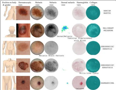
An innovative approach uses SIAscopy, a non-invasive multispectral scanning technique which gains micro-architectural information about the skin within seconds. The device shines near infrared and visible spectra light from a handset through the skin. The light remitted can then be calibrated for papillary dermis thickness, using information from the infrared wavebands. The amount of dermal blood is obtained by de-referencing a given colour location on the surface of normal skin coloura-tion. If melanin is present in the dermis, its presence can be detected from the fact that even after the papil-lary dermis thickness adjustment, the colours still do not lie on the surface of normal skin colouration. The amount of epidermal melanin is obtained by de-referen-cing skin colour locations on the surface of normal skin colouration. Within seconds all of this information is displayed graphically on the computer screen as SIAs-cans. SIAscans are therefore high-resolution images of the collagen and haemoglobin content of the papillary dermis, and melanin content of the epidermis and papil-lary dermis. Patterns within the SIAscans of pigmented skin lesions (such as the presence of dermal melanin
and blood displacement with erythematous blush) indicate the pathological changes consistent with mela-noma. Previous studies have demonstrated the diagnos-tic accuracy of SIAscopy for melanoma amongst patients referred to secondary care using the Moncrieff scoring system [16,17]. In that study the combination of the following features was found to be sensitive and spe-cific for the diagnosis of melanoma: presence of dermal melanin, collagen holes, erythematous blush and blood displacement (see Figure 1). However, the findings of diagnostic studies on referred populations cannot be applied to patients seen in primary care due to the potential for spectrum bias. The primary aim of this study therefore was to develop and validate a scoring system for SIAscopic diagnosis of pigmented skin lesions encountered in primary care. In addition, since all studies to date on SIAscopy have been conducted in the UK, we aimed to validate the technique in an Aus-tralian primary care setting to examine its generalisabil-ity to populations with greater sun-related skin damage.
Methods
This study was conducted in two consecutive settings in the UK and Australia and entailed the following three stages:
1. Development of the primary care scoring algo-rithm (PCSA) on a sub-set of lesions from the UK sample (UK Development lesion dataset);
2. Validation of the PCSA on a different sub-set of lesions from the same UK sample (UK Validation lesion dataset);
3. Validation of the PCSA on a new set of lesions from an Australian primary care population (Austra-lian Validation lesion dataset).
Ethical approval
Ethical approval for the UK component of the study was obtained from the Cambridge Research Ethics Commit-tee (REC Ref. 04/079) and research governance approval from Cambridge City and South Cambridgeshire Primary Care Trusts (Project number L00569). Ethical approval for the Australian component of the study was obtained from the University of Western Australia’s Human Research Ethics Committee (HREC Ref. RA/4/1/1739).
Settings UK setting
presented by the patients to their GP, they included lesions that were ultimately considered not clinically sus-picious. Participants were formally consented and data collected about their lesion by JH within two weeks of initial presentation to their GP. Data collection occurred between January 2005 and January 2006.
Australian setting
Three primary care skin cancer clinics operated by GPs were recruited from the metropolitan area of Perth, Western Australia. Adult patients aged over 18 years were recruited into the study by their GP if they pre-sented with concerns about a pigmented skin lesion: again, these included lesions that ultimately were con-sidered not clinically suspicious. Additional lesions were also included when a pigmented skin lesion was identi-fied as potentially suspicious during their clinical exami-nation. Participants were formally consented and data collected by AJW on the same day as they presented to
their GP. Data collection occurred between April 2008 and January 2009.
Data collection
The following data were collected by the medically qua-lified researchers (JH or AJW) for each skin lesion:
1. 7-point melanoma checklist11;
2. Macroscopic digital photograph (Canon EOS 400 D camera, Canon EF-S60 macro lens, Canon MR-14EX Macro Ring Lite flash, JPEG picture for-mat: 3888 × 2592 pixels);
3. Dermoscopic digital photograph (Canon EOS 400 D camera, Canon EF-S60 macro lens, Heine SLR Photadaptor, Heine Delta 20 dermatoscope, JPEG picture format: 3888 × 2592 pixels);
4. SIAscan (MoleMate™SIAscope V and Microsoft Windows™application).
SIAscan assessment
SIAscan images and data (including the location of the lesion and the age group and sex of the patient) were assessed by a SIAscopy expert, who was blinded to the 7-point melanoma checklist results and clinical photographs. The SIAscopy expert scored the presence or absence of each specific SIAscopic feature including those previously associated with melanoma [16]: size of lesion, age of patient, dermal melanin, collagen holes and blood displacement with erythematous blush. Addi-tional features that were also scored were: blood vessels, white dots on the collagen view, blood lacunes and a cerebriform melanin pattern (see Figure 1).
Diagnostic reference standards
Given that it would have been ethically unacceptable to obtain histological diagnosis on every recruited lesion, we applied the following hierarchical approach to refer-ence standard diagnosis:
1. Histopathology.
2. In-person clinical review of the lesion by one expert, including 7-point checklist and digital dermoscopy.
3. Clinical diagnosis made on the basis of the 7-point checklist, photographic and dermoscopy images.
The expert reviewers were blinded to the SIAscan images. For the reference standard diagnosis we cate-gorised lesions in two complementary ways relevant to primary care decision-making: (1)‘suspicious’or benign and (2) melanoma or other pigmented lesion. The defi-nition of‘suspicious’was a lesion that, if seen in general practice, would warrant referral, excision or short-term monitoring.
Analysis
This was undertaken in three stages:
(a)Development stage: a 66% sub-sample of lesions (UK Development lesion dataset) was scored using the Moncrieff scoring system. In order to account for the different prevalence of certain pigmented skin lesions seen in primary care, a Primary Care Scoring Algorithm (PCSA) was developed.
(b) Validation stage one: the PCSA was validated against the remaining 33% sub-sample of lesions (UK Validation lesion dataset).
(c) Validation stage two: the PCSA was validated against the lesions recruited in Australia (Australian Validation lesion dataset).
Data were recorded on a Microsoft Access database and analysed with Microsoft Excel and SPSS version 11.5 for Windows. Sensitivity, specificity, positive and negative
predictive values and their associated 95% confidence intervals were calculated using standard approaches including the Wilson method to account for small sample sizes in some of the cells [18]. Receiver operating charac-teristic (ROC) curves and associated area-under-the-curve (AUC) were created using standard functions within SPSS 11.5 to explore different cut-off scores for the PCSA. We compared the area under the curve for the PCSA and the 7-point checklist for all lesions for which we had obtained histology, using the manual method described by Hanley and McNeil [19].
Results
In the UK dataset (development and validation lesions) interpretable images were obtained on 630 lesions from 389 patients. The mean age of participants in the study was 44.9 years; 68.6% were female. In the Australian dataset (validation lesions) interpretable images were obtained on 581 lesions from 469 patients. Fifty two per cent of the subjects were male, and the mean age of participants was 50 years. Table 1 shows the types of lesion represented in the two datasets based on histo-pathology where known, or expert clinical diagnosis.
(a) Development stage
Table 2 presents the performance of the Moncrieff scor-ing system for the diagnosis of ‘suspicious’ using the Development Lesion dataset (n=422). In this subset there were 24 suspicious lesions and 3 melanomas, including 1 atypical melanoma with significant regres-sion. The Moncrieff scoring system did not perform that well for the diagnosis of‘suspicious’, predominantly due to misclassification of seborrhoeic keratoses and hae-mangiomas. In particular, of the 101 seborrhoeic kera-toses in the sample, 55 were misclassified as suspicious due to apparent ‘dermal melanin’ on the SIAscopic image. Specific SIAscopic features of seborrhoeic kera-toses were identified as: white dots on the collagen view, analogous to milia-like cysts seen on dermoscopy [20], and a cerebriform appearance on the total melanin view. Haemangiomas were identified by the presence of blood lacunes on the SIAscan‘blood’view.
improved if lesions classified as seborrhoeic keratoses or haemangiomas, based on SIAscopic features, were excluded from the dataset (Area Under Curve (AUC): MSS 0.732; MSS after exclusion of seborrhoeic keratoses and haemangiomas 0.759). For the diagnosis of mela-noma, performance was improved by scoring 2 points for the presence of blood vessels and by excluding lesions classified as seborrhoeic keratoses or haemangio-mas, based on SIAscopic features (AUC 0.916, see Figure 2). On this basis a new Primary Care Scoring Algorithm (PCSA) was developed that aims to identify lesions with features of seborrhoeic keratoses or hae-mangiomas first and then apply a scoring system based on the presence of other features associated with mela-noma (see Figure 3). In this way, seborrhoeic keratoses and haemangiomas are no longer misclassified.
(b) UK Validation stage
The new PCSA was tested against the 208 lesions in the Validation Lesion dataset, which included 6 suspicious lesions and two histopathologically confirmed melano-mas. The performance of the PCSA is presented in Table 2.
(c) Australian Validation stage
The PCSA was tested against the 581 lesions recruited in Australia. There were 52 suspicious lesions includ-ing 5 histopathologically confirmed melanomas and 2 lentigo malignas. The performance of the PCSA is pre-sented in Table 2. In this second validation stage, the sensitivity for the diagnosis of suspicious was similar to the UK findings (0.44; 95% CI 0.32-0.58). However, specificity was significantly better (0.95; 95% CI 0.93-0.97). Furthermore, due to the higher prevalence of suspicious lesions in the Australian dataset, the posi-tive predicposi-tive value for the diagnosis of suspicious was 0.52 (95% CI 0.38-0.66) while maintaining an accepta-bly high negative predictive value (0.95; 95% CI 0.92-0.96).
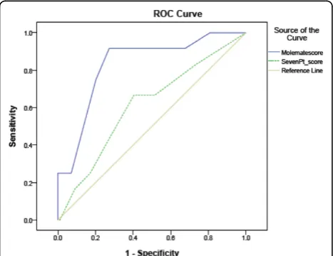
We compared the AUC for the PCSA and the 7-point checklist for the 111 lesions for which we had histologi-cal diagnosis (n = 42 UK dataset; n = 69 Australian dataset; included 10 melanomas and 2 lentigo maligna) (Table 3; Figure 4). The PCSA had a significantly greater AUC (0.83; 95% CI 0.71-0.95) than the 7-point checklist (AUC 0.61; 95% CI 0.44-0.78; p = 0.02 for difference).
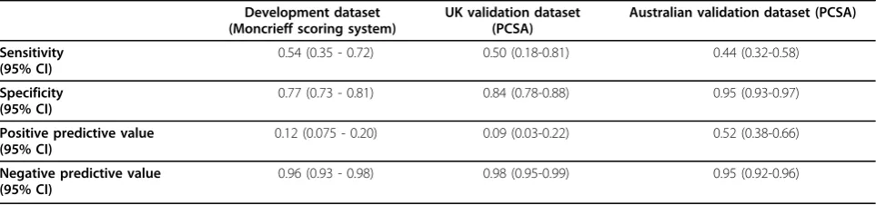
Table 2 Performance characteristics of SIAscopy for the diagnosis of‘suspicious’in the different datasets of lesions.
Development dataset (Moncrieff scoring system)
UK validation dataset (PCSA)
Australian validation dataset (PCSA)
Sensitivity (95% CI)
0.54 (0.35 - 0.72) 0.50 (0.18-0.81) 0.44 (0.32-0.58)
Specificity (95% CI)
0.77 (0.73 - 0.81) 0.84 (0.78-0.88) 0.95 (0.93-0.97)
Positive predictive value (95% CI)
0.12 (0.075 - 0.20) 0.09 (0.03-0.22) 0.52 (0.38-0.66)
Negative predictive value (95% CI)
0.96 (0.93 - 0.98) 0.98 (0.95-0.99) 0.95 (0.92-0.96)
Table 1 Distribution of lesions in Development and Validation datasets, based on expert clinical diagnosis or histology where available
Diagnosis Development dataset % UK validation dataset % Australian validation dataset %
Naevus 293 69. 4% 159 76. 4% 333 57. 3%
Seborrhoeic keratosis 101 23. 9% 39 18. 7% 128 22. 0%
Solar lentigo 0 0 0 0 67 11. 5%
Basal cell carcinoma 0 0 0 0 22 3. 8%
Melanoma 3 0.7% 2 1. 0% 7 1. 2%
Angiokeratoma 0 0.0% 0 0.0% 6 1. 0%
Dermatofibroma 14 3. 3% 6 2. 9% 5 0.9%
Lentigo maligna 0 0.0% 0 0.0% 4 0.7%
Haemangioma 11 2. 6% 2 1. 0% 0 0.0%
Lentigo simplex 0 0.0% 0 0.0% 5 0.9%
Ephilis 0 0.0% 0 0.0% 3 0.3%
Papilloma 0 0.0% 0 0.0% 1 0.2%
ROC curves for the diagnosis of melanoma after removal of lesions classified on
SIAscopy as haemangioma or seborrhoeic keratosis. Red = Moncrieff Scoring System
(MSS); green = MSS +1 point for blood vessels; blue = MSS + 2 points for blood vessels;
yellow = MSS +3 points for blood vessels.
Area under curve: Red = 0.892; green = 0.912; blue = 0.916; yellow = 0.887.
Figure 2ROC curves to show development of Primary Care Scoring Algorithm; ROC for diagnosis of melanoma after removal of lesions classified on SIAscopy as haemangioma or seborrhoeic keratosis.
Discussion
This is the first study to test the use of SIAscopy for lesions encountered in a primary care setting and also outside the UK. We have applied a systematic approach in which we tested the initial Moncrieff scoring system to see how it would function on lesions presented in a primary care setting. This is an important step in studies of new diagnostic techniques to reduce the effects of spectrum bias. In addition to developing a new diagnos-tic algorithm we have conducted a second validation study on a different primary care population. This sec-ond validation study was csec-onducted in an Australian primary care setting which, except for a higher preva-lence of solar lentigos, had a similar prevapreva-lence of lesions to the UK dataset. We accept that a limitation of this study is our inability to obtain histopathological diagnoses on all the lesions recruited, but this would have been ethically unacceptable. To inform the clinical-expert reference-standard diagnosis we deliberately chose to obtain maximum clinical data, including the
7-point checklist and dermoscopy. We chose to do this so we could be as accurate as possible with our refer-ence diagnosis where histology was not available. It is also theoretically possible that some amelanotic melano-mas were not recruited into the study on the basis of our inclusion criteria. Furthermore, we did not follow-up any lesions determined as benign so it is theoretically possible that some clinically significant lesions may have been missed by our reference standard diagnoses.
The Moncrieff scoring system was found to be less accurate than in the secondary care setting due to the different prevalence of lesions among the primary care population. In order to account for the higher preva-lence of non-melanocytic lesions, such as seborrhoeic keratoses and haemangiomas, we developed a new Primary Care Scoring Algorithm which was more speci-fic than the Moncrieff scoring system for ‘suspicious’ but no more sensitive. Higher specificity was particularly identified in the Australian dataset suggesting that sun-related skin damage does not adversely affect the diag-nostic accuracy of SIAscopy. The current algorithm accounts for size of lesion (> 6 mm) and age of patient. The mean age of participants from each studied popula-tion was 45 and 50 years respectively. It is not possible therefore to comment on the performance of the PCSA in elderly populations in which the algorithm may become less specific. It was reassuring that the PCSA’s moderate sensitivity for‘suspicious’does not appear to be reflected in its sensitivity for melanoma, but inevita-bly there were too few melanomas in this study to pro-vide robust estimates of diagnostic accuracy for melanoma in primary care. In subsequent research, simulation modelling of the PCSA in which a higher prevalence of melanomas was entered into the dataset suggests high sensitivity and specificity to detect mela-noma [21]. Ultimately, a primary care algorithm should be good at identifying ‘suspicious’pigmented lesions, including melanoma, as well as accurately ruling out lesions which are unlikely to be clinically significant. It is interesting that the PCSA appears to be more accu-rate than the 7-point checklist in diagnosing melanoma. The 7-point checklist was completed by the two medi-cally qualified and trained researchers including one who was a plastic surgeon (JH). The relatively poor per-formance of the 7-point checklist cannot be explained by inconsistent application of the items. There were sev-eral non-melanocytic lesions which were thought clini-cally to be pigmented. The 7-point checklist, and Siascopy, are intended for use only with melanocytic lesions, although in clinical primary care practice this distinction can sometimes be difficult. The inclusion of non-melanocytic lesions may partially explain the poorer performance of the 7-point checklist compared to pre-viously published data.
Table 3 Distribution of lesions for which histological diagnosis was available.
Diagnosis Number %
Naevus 62 55. 9
Seborrhoeic keratosis 16 14. 4
Melanoma* 12 10.8
Basal cell carcinoma 9 8. 1
Solar lentigo 5 4. 5
Dermatofibroma 5 4. 5
Lentigo simplex 2 1. 8
TOTAL 111
*includes 2 lentigo maligna
The analysis conducted assumes that there was no selection bias in the sampling of lesions chosen for biopsy. This is theoretically possible, for example if the 7-point score were used to inform the clinical decision to excise, and so our finding should be interpreted with some caution[22].
In this study we used experts in SIAscopy to interpret the SIAscans. This therefore reflects the best perfor-mance of SIAscopy in primary care and not how it would perform in the hands of general practitioners. As this is the first study of SIAscopy on lesions from primary care, we needed to determine the best possible performance of the technique in experienced hands. We are now con-ducting a randomised controlled trial of training English general practitioners in SIAscopy, including the applica-tion of the PCSA, to determine its effects on clinical practice (the MoleMate UK Trial). This trial will provide further evidence on the diagnostic accuracy of the PCSA when used by general practitioners as well as the cost-effectiveness of SIAscopy in English primary care.
We believe that the features of SIAscopy may be a great deal easier to learn than those of dermoscopy which can take a long period of training in which to become proficient. A recent study we have conducted suggests that SIAscopy features can be learnt by general practitioners using a CD-rom based tutorial in approxi-mately two hours [23]. In a recent trial in general prac-tice, dermoscopy had a sensitivity of 55% and specificity of 89% for malignant lesions which is comparable with our findings for SIAscopy, albeit in expert hands [14]. The MoleMate UK Trial will provide more comparable data in due course [24]. While there is no doubt that dermoscopy and digital monitoring can significantly improve the management of pigmented lesions in pri-mary care, we believe that SIAscopy could be simpler to learn and may therefore have greater utility for a wider group of primary care practitioners than dermoscopy.
Conclusions
The PCSA for SIAscopy could have an important role in improving the management of pigmented skin lesions in primary care. This study has confirmed the key diagnos-tic features of lesions commonly encountered in primary care. Further work is required to determine the impact of training GPs in SIAscopy on their clinical manage-ment of pigmanage-mented skin lesions, and the quality of their referrals to the secondary care skin cancer clinics.
Acknowledgements
We thank the BUPA Foundation who funded the UK component of this study, Tom Fanshawe, formerly of the Centre for Applied Medical Statistics, Department of Public Health & Primary Care, University of Cambridge, and Chris Brown from the Clinical Trials Centre, University of Sydney for their statistical input. We also thank the general practices, skin cancer clinics and patients who participated in this study.
Author details
1General Practice, School of Primary, Aboriginal and Rural Health Care,
University of Western Australia, 328 Stirling Highway, Claremont, WA 6010, Australia.2Dept of Plastic Surgery, Addenbrooke’s Hospital NHS Foundation Trust, Hills Road Cambridge, CB2 2QQ, UK.3Welsh Centre for Burns and Plastic Surgery, Morriston Hospital, Swansea, SA6 6NL, UK.4Dept of Plastic & Reconstructive Surgery, Norfolk & Norwich University Hospitals NHS Foundation Trust, Norwich, Norfolk NR4, UK.5General Practice & Primary Care Research Unit, Department of Public Health & Primary Care, Institute of Public Health. University of Cambridge, Cambridge CB2 0SR, UK.
Authors’contributions
JE, PH, and FW conceived the study and contributed to the overall design. JH conducted the UK Development and Validation studies as part of her MD thesis. All authors contributed to the conduct of the study and the final manuscript.
Competing interests
Part of the Australian study was funded by Astron Clinica who formerly produced devices based on SIAscopy. Astron Clinica had no input into the analyses or drafting the manuscript. From August 2009, MoleMate, SIAscopy, SIAscan and SIAscope are trademarks of Biocompatibles UK Limited, who have funded maintenance of the Australian dataset.
Received: 1 December 2009 Accepted: 25 September 2010 Published: 25 September 2010
References
1. UK skin cancer incidence statistics. CRUK.[http://info.cancerresearchuk. org/cancerstats/types/skin/incidence].
2. Lens MB, Dawes M:Global perspectives of contemporary epidemiological trends of cutaneous malignant melanoma.Br J Dermatol2004, 150(2):179-185.
3. Diffey BL:The future incidence of cutaneous melanoma within the UK.Br J Dermatol2004,151(4):868-872.
4. Murchie P, Campbell NC:Pigmented lesions, cutaneous melanoma, and future challenges for primary care.Eur J Gen Pract2007,13(3):151-154. 5. Chen SC, Pennie ML, Kolm P, Warshaw EM, Weisberg EL, Brown KM,et al:
Diagnosing and managing cutaneous pigmented lesions: primary care physicians versus dermatologists.J Gen Intern Med2006,21(7):678-682. 6. Morrison A, O’Loughlin S, Powell FC:Suspected skin malignancy: a
comparison of diagnoses of family practitioners and dermatologists in 493 patients.Int J Dermatol2001,40(2):104-107.
7. Brochez L, Verhaeghe E, Bleyen L, Naeyaert JM:Diagnostic ability of general practitioners and dermatologists in discriminating pigmented skin lesions.J Am Acad Dermatol2001,44(6):979-986.
8. NICE, Cancer service guidance:Improving Outcomes for People with Skin Tumours including Melanoma2006 [http://www.nice.org.uk/Guidance/ CSGSTIM].
9. Cox NH:Evaluation of the U.K. 2-week referral rule for skin cancer.Br J Dermatol2004,150(2):291-298.
10. Burton RC, Howe C, Adamson L, Reid AL, Hersey P, Watson A,et al:General practitioner screening for melanoma: sensitivity, specificity, and effect of training.J Med Screen1998,5(3):156-161.
11. Gerbert B, Bronstone A, Maurer T, Berger T, McPhee SJ, Caspers N:The effectiveness of an Internet-based tutorial in improving primary care physicians’skin cancer triage skills.J Cancer Educ2002,17(1):7-11. 12. Vestergaard ME, Macaskill P, Holt PE, Menzies SW:Dermoscopy compared
with naked eye examination for the diagnosis of primary melanoma: a meta-analysis of studies performed in a clinical setting.Br J Dermatol
2008,159(3):669-676.
13. Argenziano G, Puig S, Zalaudek I, Sera F, Corona R, Alsina M,et al: Dermoscopy improves accuracy of primary care physicians to triage lesions suggestive of skin cancer.J Clin Oncol2006,24(12):1877-1882. 14. Menzies SW, Emery J, Staples M, Davies S, McAvoy B, Fletcher J,et al:
Impact of dermoscopy and short-term sequential digital dermoscopy imaging for the management of pigmented lesions in primary care: a sequential intervention trial.Br J Dermatol2009.
16. Moncrieff M, Cotton S, Claridge E, Hall P:Spectrophotometric intracutaneous analysis: a new technique for imaging pigmented skin lesions.Br J Dermatol2002,146(3):448-457.
17. Govindan K, Smith J, Knowles L, Harvey A, Townsend P, Kenealy J: Assessment of nurse-led screening of pigmented lesions using SIAscope.
J Plast Reconstr Aesthet Surg2007,60(6):639-645.
18. Armitage P, Berry G:Statistical methods in medical research.Oxford; Boston: Blackwell Scientific Publications, 3 1994.
19. Hanley JA, McNeil BJ:A method of comparing the areas under receiver operating characteristic curves derived from the same cases.Radiology
1983,148(3):839-843.
20. Johr RH:Dermoscopy: alternative melanocytic algorithms-the ABCD rule of dermatoscopy, Menzies scoring method, and 7-point checklist.Clin Dermatol2002,20(3):240-247.
21. Hunter JE:Triaging suspicious pigmented skin lesions in primary care using the SIAscopeMD Thesis, University of Cambridge 2008.
22. Hanley JA, Hajian-Tilaki KO:Sampling variability of nonparametric estimates of the areas under receiver operating characteristic curves: an update.Acad Radiol1997,4(1):49-58.
23. Wood A, Morris H, Emery J, Hall PN, Cotton S, Prevost AT,et al:Evaluation of the MoleMate training program for assessment of suspicious pigmented lesions in primary care.Inform Prim Care2008,16(1):41-50. 24. Walter FM, Morris HC, Humphrys E, Hall PN, Kinmonth AL, Prevost AT,et al:
Protocol for the MoleMate UK Trial: a randomised controlled trial of the MoleMate system in the management of pigmented skin lesions in primary care [ISRCTN 79932379].BMC Fam Pract2010,11:36.
Pre-publication history
The pre-publication history for this paper can be accessed here: http://www.biomedcentral.com/1471-5945/10/9/prepub
doi:10.1186/1471-5945-10-9
Cite this article as:Emeryet al.:Accuracy of SIAscopy for pigmented skin lesions encountered in primary care: development and validation of a new diagnostic algorithm.BMC Dermatology201010:9.
Submit your next manuscript to BioMed Central and take full advantage of:
• Convenient online submission
• Thorough peer review
• No space constraints or color figure charges
• Immediate publication on acceptance
• Inclusion in PubMed, CAS, Scopus and Google Scholar
• Research which is freely available for redistribution