University of Pennsylvania
ScholarlyCommons
Publicly Accessible Penn Dissertations
2016
Role Of Maternal Sin3a In Reprogramming Gene
Expression During Mouse Preimplantation
Development
Richard A. JimenezUniversity of Pennsylvania, [email protected]
Follow this and additional works at:https://repository.upenn.edu/edissertations Part of theDevelopmental Biology Commons, and theGenetics Commons
This paper is posted at ScholarlyCommons.https://repository.upenn.edu/edissertations/2366
Recommended Citation
Jimenez, Richard A., "Role Of Maternal Sin3a In Reprogramming Gene Expression During Mouse Preimplantation Development" (2016).Publicly Accessible Penn Dissertations. 2366.
Role Of Maternal Sin3a In Reprogramming Gene Expression During
Mouse Preimplantation Development
Abstract
In mouse, the maternal-to-zygotic transition entails a dramatic reprogramming of gene expression during the course of zygotic genome activation, which is essential for continued development beyond the 2-cell stage. Superimposed on zygotic genome activation and reprogramming of gene expression is formation of a chromatin-mediated transcriptionally repressive state that promotes repression of genes at the 2-cell stage. Experimentally inducing global histone hyperacetylation relieves this repression and histone deacetylase 1 (HDAC1) is the major HDAC involved in the development of this transcriptionally repressive state. Because SIN3A is essential for mouse development and is part of a HDAC1/2-containing complex, I investigated the role of maternal SIN3A in the development of the global transcriptionally repressive state that develops during the course of genome activation and reprogramming. In addition, previous microarray data generated from our lab of oligo (dT) primed mouse oocyte and 1-cell embryo cDNA revealed an elevation in the relative abundance of the Sin3a transcript between the oocyte and 1-cell stages; the elevation in relative transcript abundance suggests that the Sin3a transcript undergoes translational recruitment during oocyte maturation because the elevation occurs during a period of transcriptional quiescence. Here I show that the Sin3a
transcript is recruited for translation during oocyte maturation and following fertilization. I demonstrated that maternal SIN3A is essential for preimplantation development and the reprogramming of genes expression, because inhibiting the maturation-associated increase in SIN3A leads to an arrest in mouse embryonic development and unfaithful reprogramming of gene expression in 2-cell mouse embryos. The mid 1-cell embryo contains the maximum level of maternal SIN3A protein and the protein then rapidly decreases to essentially an undetectable level by the mid 2-cell stage; the rapid loss of maternal SIN3A is likely mediated by the proteasome because a proteasome inhibitor substantially inhibits the loss of maternal SIN3A. Due to the restricted presence of the maturation-associated increase in SIN3A, the function of maternal SIN3A is likely constrained to the 1-cell stage of mouse development. However, the increase in maternal SIN3A does not play a role in the minor ZGA, as depleting maternal SIN3A had no effect on global transcription in 1-cell embryos, but surprisingly results in histone hypoacetylation in 1-cell mouse embryos. Maintaining the presence of maternal SIN3A beyond the 1-cell stage had no effect on pre- and postimplantation development. Collectively, these findings indicate that the maturation-associated increase in SIN3A regulates the reprogramming of gene expression and the oocyte may utilize the translational recruitment of transcripts encoding chromatin-modifying-related factors during oocyte maturation as a post-transcriptional mechanism to faithfully execute the reprogramming of gene expression through the utilization of a maternally-derived transcription machinery.
Degree Type
Dissertation
Degree Name
Doctor of Philosophy (PhD)
Graduate Group
First Advisor
Richard M. Schultz
Keywords
dormant maternal mRNA, maternal-to-zygotic transition, mouse preimplantation development, reprogramming, Sin3a, zygotic genome activation
Subject Categories
ROLE OF MATERNAL SIN3A IN REPROGRAMMING GENE EXPRESSION DURING MOUSE PREIMPLANTATION DEVELOPMENT
Richard A. Jiménez A DISSERTATION
in
Cell and Molecular Biology
Presented to the Faculties of the University of Pennsylvania in
Partial Fulfillment of the Requirements for the Degree of Doctor of Philosophy
2016
Supervisor of Dissertation _____________________ Richard M. Schultz, Ph.D.
Charles and William L. Day Distinguished Professor of Biology
Graduate Group Chairperson _____________________ Daniel S. Kessler, Ph.D.
Associate Professor of Cell and Developmental Biology
Dissertation Committee
Hua-Ying Fan, Ph.D., Assistant Professor of Biochemistry and Biophysics
George L. Gerton, Ph.D., Research Professor of Reproductive Biology in Obstetrics and Gynecology
Peter S. Klein, M.D., Ph.D., Professor of Medicine
ACKNOWLEGMENTS
I would first like to thank my mentor, Richard Schultz for his guidance, patience and intellect as all these qualities has kept me stimulated as a graduate student and has motivated me to continue my growth as a scientist and person. I am also tremendously grateful for connecting me to amazing researchers at the Smithsonian Conservation Biology Institute in Front Royal, Virginia. Several past and current members of the lab have provided invaluable advice, expertise and skills. I especially thank Eduardo Melo for helping me with the initial experiments and review of the literature when the project first began. Paula Stein has trained me on many of the methods used in this project and her tremendous patience and excellent proficiency was instrumental throughout my time in the Schulz lab. She also provided useful feedback on a number of scientific presentations and provided valuable intuition on politics, life and especially the World Cup. I especially thank Jun Ma, Sergey Medvedev, and Pengpeng Ma for providing technical advice and help throughout my graduate time in the lab.
I would also like to thank several people who have shared their expertise and time throughout my graduate career. I thank Chris Lengner, Peter Klein, George Gerton and Hua-Ying Fan for their support, constructive criticism, and honesty on my project and for providing an “out-of the box” perspective. They also were very helpful in keeping me focused throughout my graduate career.
I want to express my gratitude and acknowledge the VMD/PhD program. I particular want to thank Mike Atchison for his advice, and tremendous support over my veterinary and graduate studies!
I also want to thank the Predoctoral Training Grant in Genetics for their financial support through my graduate studies. I particularly want to thank Meera Sundaram for her strong dedication and time to keep this valuable training grant running strong!
processing the invaluable experimental samples for the microarray analysis. John Eppig for his guidance on the in vitro maturation and in vitro fertilization experiments that allowed the project to address questions that would have been impossible to tackle without his useful advise. Vedran Franke from the Kristian
Vlahoviček lab for all of his time and proficiency in bio-informatics that help with
our analysis of the microarray data.
ABSTRACT
ROLE OF MATERNAL SIN3A IN REPROGRAMMING GENE EXPRESSION
DURING MOUSE PREIMPLANTATION DEVELOPMENT
Richard A. Jiménez
Richard M. Schultz
In mouse, the maternal-to-zygotic transition entails a dramatic reprogramming of gene expression during the course of zygotic genome activation, which is
essential for continued development beyond the 2-cell stage. Superimposed on zygotic genome activation and reprogramming of gene expression is formation of a chromatin-mediated transcriptionally repressive state that promotes repression of genes at the 2-cell stage. Experimentally inducing global histone
hyperacetylation relieves this repression and histone deacetylase 1 (HDAC1) is the major HDAC involved in the development of this transcriptionally repressive state. Because SIN3A is essential for mouse development and is part of a HDAC1/2-containing complex, I investigated the role of maternal SIN3A in the development of the global transcriptionally repressive state that develops during the course of genome activation and reprogramming. In addition, previous microarray data generated from our lab of oligo (dT) primed mouse oocyte and 1-cell embryo cDNA revealed an elevation in the relative abundance of the Sin3a
transcript between the oocyte and 1-cell stages; the elevation in relative
transcript abundance suggests that the Sin3a transcript undergoes translational recruitment during oocyte maturation because the elevation occurs during a period of transcriptional quiescence. Here I show that the Sin3a transcript is recruited for translation during oocyte maturation and following fertilization. I demonstrated that maternal SIN3A is essential for preimplantation development and the reprogramming of genes expression, because inhibiting the maturation-associated increase in SIN3A leads to an arrest in mouse embryonic
embryos. The mid 1-cell embryo contains the maximum level of maternal SIN3A protein and the protein then rapidly decreases to essentially an undetectable level by the mid 2-cell stage; the rapid loss of maternal SIN3A is likely mediated by the proteasome because a proteasome inhibitor substantially inhibits the loss of maternal SIN3A. Due to the restricted presence of the maturation-associated increase in SIN3A, the function of maternal SIN3A is likely constrained to the 1-cell stage of mouse development. However, the increase in maternal SIN3A does not play a role in the minor ZGA, as depleting maternal SIN3A had no effect on global transcription in 1-cell embryos, but surprisingly results in histone
hypoacetylation in 1-cell mouse embryos. Maintaining the presence of maternal SIN3A beyond the 1-cell stage had no effect on pre- and postimplantation
TABLE OF CONTENTS i Title
ii Acknowledgements iv Abstract
vi Table of Contents viii List of Illustrations x List of Tables
Chapter 1 Introduction... 1
1.1 Overview of mouse oogenesis and preimplantation development... 1
1.2 Global transcriptional repression and large-scale changes to chromatin structure in mouse oocytes... 5
1.3 Zygotic gene activation... 7
1.4 Transcriptionally repressive state ... 8
1.5 Epigenetic reprogramming in the zygote... 10
1.6 SIN3A-co-repressor complex and HDAC1/2-containing complex... 13
Chapter 2 Materials and Methods... 17
Chapter 3 Inhibiting the maturation-association increase in maternal SIN3A impairs the reprogramming of gene expression during mouse preimplantation development... 28
3.1 Results... 29
3.1a Sin3a is a dormant maternal mRNA that is recruited for translation during oocyte maturation and following fertilization... 29
3.1bInhibiting the maturation-associated increase in SIN3A alters global H3 and H4 histone acetylation in 1-cell embryos... 31
3.1d Impairment of gene expression reprogramming in maternal SIN3A- depleted embryos... 33 3.1e Exogenously expressing SIN3A beyond the 1-cell stage does not impair preimplantation development... 35 3.2 Discussion... 67
LIST OF ILLUSTRATIONS
Figure 3.1. Developmental expression profile of Sin3a/SIN3A... 37 Figure 3.2. Time course for SIN3A protein loss ... 38 Figure 3.3. SIN3A protein loss is proteasome-dependent... 39 Figure 3.4. Developmental expression profile of SIN3A protein by
immunocytochemistry... 40 Figure 3.5. Similiar amount of chromatin-associated SIN3A protein between male
and female pronuclei……... 41
Figure 3.6. Sin3a 3’UTR contains elements that drive translational recruitment
during oocyte maturation and following activation... 42
Figure 3.7. Combined Sin3a morpholino and siRNA inhibit maturation-associated
increase in the amount of SIN3A protein... 43 Figure 3.8. Effect of inhibiting the maturation-associated increase in SIN3A on histone acetylation in 1-cell embryos... 44 Figure 3.9. Inhibiting the maturation-associated increase in SIN3A protein leads
to a developmental arrest at the 2-cell stage…... 46
Figure 3.10. DNA replication is not inhibited in 1- and 2-cell embryos when the
maturation-associated increase in SIN3A protein is inhibited………... 47
Figure 3.11. Transcription is reduced in 2-cell embryos depleted of
maternal-SIN3A………... 48
Figure 3.12. No change in global transcription at the 1-cell stage when the
maturation-associated increase in maternal SIN3A is inhibited... 49 Figure 3.13. Heat map of all samples from different treatment groups constructed using hierarchical clustering... 50 Figure 3.14 Karyogram of genes whose expression was elevated in 2-cell
embryos depleted of maternal SIN3A (left panel) and density of genes (right panel)... 51 Figure 3.15. Over-expressing SIN3A does not affect pre-implantation
Figure 3.16. Over-expressing SIN3A does not affect cell numbers in
blastocysts... 54 Figure 3.17. Over-expressing SIN3A does not affect pre-implantation
LIST OF TABLES
Table 3.1. Zygotically expressed genes whose expression is up-regulated
following inhibition of the maturation-associated increase in SIN3A………….... 56
Table 3.2. Genes not zygotically activated, but whose expression is up-regulated
following inhibition of the maturation-associated increase in SIN3A………….... 62
Table 3.3. qRT- PCR confirmation of four transcripts from each class whose expression was increased following inhibition of the maturation-associated
CHAPTER 1: INTRODUCTION
1.1 Overview of mouse oogenesis and preimplantation development
Mammalian oocytes embark on the path to fertilization and embryogenesis within the functional unit of the ovary, the ovarian follicle. Each follicle is
composed of a single oocyte arrested in prophase I of meiosis enclosed by one or more layers of specialized somatic cells that communicate with and support the oocyte during its growth and development via gap junctions. Starting with a resting or primordial follicle that comprises of an oocyte surrounded by a single layer of squamous, flattened, pre-granulosa cells, each primordial follicle’s prolonged resting phase is interrupted by factors that recruit the follicle for development into a primary follicle. The size of the initial primordial follicle pool in part dictates reproductive senescence in females.
Since the 1950s, the prevailing view was that mammalian females are provided with an extensive, but finite, nonrenewable ovarian reserve around the time of birth for mice or during mid-gestation in humans, which is diminished as the follicles are recruited to grow (Zuckerman, 1951). However, some
investigators challenge the 50-year dogma and contend that the ovarian reserve could be replenished by female germline stem cells referred to as oogonial stem cells. These paradigm-shifting studies claim that a population of cells isolated from both neonatal and adult mouse ovaries and adult human ovaries have stem cell characteristics and, specifically for the cells isolated from mouse ovaries, are capable of differentiating into oocytes that are able to mature, ovulate, and
fertilize to produce viable embryos and pups (Johnson et al., 2004; Pacchiarotti et al., 2010; Zou et al., 2009; White et al., 2012). The surface protein used to isolate the oogonial stem cells from the ovarian tissue, and the condition of the human ovarian tissues used to isolate the human stem cell-like cells are
questionable and need to be investigated more thoroughly in order to definitively determine if the ovarian reserve can be replenished.
follicles are locally produced (Kezele et al., 2002) and include epithelial growth factor kit ligand (KITL), leukemia inhibiting factor (LIF), basic fibroblast growth factor (BFGF), platelet derived growth factor (PDGF), keratinocyte growth factor (KGF) and connective tissue growth factor (CTGF) (Kezele et al., 2005; Nilsson et al., 2006; Nilsson and Skinner, 2004; Skinner, 2005; Schindler et al., 2010). KITL activates the ubiquitous phosphatidylinositol-3-kinase (PI3K) signaling pathway, which is important for the activation and survival of primordial follicles because removal of PTEN, a PI3K antagonist, leads to premature activation of follicles in mouse (Reddy et al., 2008; John et al., 2008). Once the primordial follicle is activated, the surrounding squamous pre-granulosa cells differentiate into cuboidal granulosa cells and begin to proliferate, while the oocyte grows
remarkably (increases in size from about 20 µm to 80 µm in diameter) (Eppig and
O’Brien, 1996). The growing primary follicles will subsequently develop into secondary and antral follicles.
After exposure to a preovulatory surge of luteinizing hormone (LH) from the pituitary gland, mammalian oocytes overcome meiotic arrest and proceed through meiotic maturation before ovulating (Russell et al., 2007). An increase in the activity of maturation-promoting factor (MPF), a cyclin B-CDK1 complex, drives meiotic progression of mammalian oocytes (Nurse, 1990). Mitogen-activated protein kinase (MAPK) cascade is another important kinase necessary for resumption of meiosis that interacts intimately with the MPF pathway in many species except for mice (Verlhac et al., 1993), where a normal pattern of MPF activity is seen when the MAPK cascade is disrupted in mice lacking c-MOS, a MAPK pathway activator (Araki et al., 1996).
Meiotic maturation entails nuclear membrane breakdown (germinal vesicle breakdown, GVBD), meiosis I spindle assembly, extrusion of the first polar body, and meiosis II spindle assembly. Also, a meiosis-specific deacetylation of
histones occurs during meiotic maturation in mouse, where all of the acetylated histones examined, with the exception of H4K8ac, are deacetylated to
shortly after meiotic resumption may facilitate chromosome condensation that occurs during oocyte maturation because hyperacetylation of H4K16 likely
contributes to defective chromosome condensation observed in histone
deacetylase 2 (Hdac2) null mouse oocytes (Ma and Schultz, 2013). Meiotic
maturation is complete after arrest at metaphase of meiosis II. Mouse oocytes acquire meiotic competence in a stepwise manner at the time of follicular antrum formation, where they first acquire the ability to reinitiate meiosis but are unable to complete meiosis I and, at a later time, acquire the capacity to complete meiosis I and progress to metaphase of meiosis II (Szybek, 1972; Sorensen and Wassarman, 1976; Wickramasinghe et al., 1991). However, meiotically
competent oocytes are unable to support preimplantation embryonic
development until additional metabolic and structural modifications are acquired during the periovulatory period (Eppig et al., 1994; Eppig, 1996). Competence to complete preimplantation development is also acquired by growing oocytes in a stepwise manner where they initially acquire the capacity to undergo fertilization and development to the 2-cell stage, and later acquire competence to develop from the 2-cell stage to the blastocyst stage (Eppig and Schroeder, 1989).
After ovulation, the metaphase II arrested mouse egg is fertilized within the oviduct, which triggers the completion of meiosis and formation of a 1-cell embryo. The newly formed 1-cell embryo enters the first mitotic cell cycle that begins with a prolonged Gap 1 (G1) phase during which two spatially separated maternal and paternal haploid pronuclei form, with the larger paternal pronucleus forming first between 4 and 8 hours post-fertilization (hpf) and the maternal pronucleus forming between 5 and 9.5 hpf (Howlett and Bolton, 1985). Each pronucleus undergoes DNA replication before entering the first mitosis to produce a 2-cell embryo, which continues to undergo successive reductive cleavage divisions without a significant increase in cell volume to form the 4-cell embryo, 8-cell embryo, and later the blastocyst (Lehtonen, 1980).
or zygotic gene activation (ZGA), has occurred. ZGA occurs in two phases: (i) minor ZGA occurs at the late one-cell stage, which results in a low-level of global transcriptional activation and generation of non-functional transcripts because they are inefficiently spliced and polyadenylated (Latham et al., 1991; Park et al., 2013; Abe et al., 2015), and (ii) major ZGA occurs at the late two-cell stage, which generates distinct functional transcripts that are not expressed in the germ cells, thus promoting a dramatic reprogramming of gene expression (Zeng et al., 2005; Hamatani et al., 2004). Interestingly, when transcription in mice was analyzed at the 1-cell stage either using a luciferase plasmid-born reporter gene or BrUTP incorporation to assess endogenous gene transcription, enhanced luciferase expression or BrUTP incorporation was observed from the male pronucleus (Ram and Schultz, 1993; Wiekowski et al., 1993; Aoki et al., 1997). This transcription is about four to five times greater in the male pronucleus than that of the female pronucleus, which is consistent with a greater concentration of transcription factors present in the male pronucleus, such as the TATA-box binding protein (TBP) and SP1 transcription factor (Ram and Schultz, 1993; Wiekowski et al., 1993; Worrad et al., 1994). The difference in the transcriptional activity and transcription factor concentration between the male and female pronucleus may likely reflect the difference in chromatin organization between the two pronculei. As discussed below, the male pronucleus exchanges the sperm-derived protamines that densely package the sperm DNA for maternally-derived histones during pronuclear formation (Nonchev and Tsanev, 1990), thereby providing a more open chromatin structure for transcription factors to associate preferentially with and likely enhancing the transcriptional activity of the male pronucleus.
calcium-dependent compaction process because E-cadherin null embryos fail to maintain compaction, in addition to failing to form an intact trophectodermal epithelium or a blastocoele cavity (Larue et al., 1994). Following compaction, further cleavage divisions and blastocoele cavity formation, a blastocyst is formed. The blastocyst stage represents the first cellular differentiation event during development where two distinct cellular populations are formed: an outer layer of trophectoderm (TE) cells that give rise to extraembryonic tissues and allow for embryo implantation, and an inner layer of inner cell mass (ICM) cells, which gives rise to the embryo proper. One day after the formation of the blastocyst, the mouse embryo
implants into the uterine wall, which marks the completion of preimplantation development.
1.2 Global transcriptional repression and large-scale changes to chromatin structure in mouse oocytes
Towards the end of the oocyte growth phase, a dramatic change in the transcriptional activity and chromatin organization occurs (Bouniol-Bay et al., 1999). Global transcription within the full-grown oocyte rapidly ceases and the oocyte becomes transcriptionally inactive and remains largely inactive until the 2-cell stage is reached in mouse (Moore and Lintern-Moore, 1978). The chromatin in mouse oocytes progressively transforms from a decondensed configuration (referred to as non-surrounded nucleolus, NSN) to a condensed configuration (surrounded nucleolus, SN) during the final stages of oocyte growth (Debey et al., 1993). This alteration in chromatin organization is temporally correlated with but not required for global transcriptional repression in mammalian oocytes
because global transcription is repressed in nucleoplasmin 2 (Npm2) null oocytes
despite failing to remodel theirchromatin into the SN configuration (Bouniol-Bay
et al., 1999; Burns et al., 2003; De La Fuente et al., 2004). It has been
suggested that the condensed chromatin configuration and silencing of global transcription is not only needed for effective resumption and completion of
egg (Liu et al., 2002).
The mechanism responsible for this large-scale chromatin structure alteration and global silencing of transcription in mouse oocytes remains to be determined. However, histone deacetylases (HDACs) may participate in the maintenance of the SN chromatin configuration in mouse oocytes (De La Fuente et al., 2004); exposing transcriptionally quiescent, full-grown mouse oocytes exhibiting the SN configuration to trichostatin A (TSA), an inhibitor of HDACs (Yoshida et al., 1995), induced large-scale chromatin structure decondensation without restoring the transcriptional activity of the oocyte. However, TSA exposure had no effect on the immunostaining patterns of histone H3 and H4 acetylation. The observed failure to restore transcriptional activity in oocytes that underwent chromatin condensation after TSA treatment is consistent with the
observation that Npm2 null mouse oocytes are still able to undergo
transcriptional silencing despite failing to undergo chromatin condensation (Burns et al., 2003; De La Fuente et al., 2004).
Changes in the nuclear availability or expression levels of several
RPB1 was investigated after permeabilizing the nuclear membrane before
fixation, RPB1 was absent from the nuclei of full-grown mouse oocytes, although it remained in the nuclei of growing oocytes. This suggests that Pol II is
dissociated from DNA in full-grown oocytes and may be the cause of global repression of transcription in mouse oocytes. Phosphorylation may destabilize the RNA polymerase II holoenzyme and lead to the dissociation of RNA
polymerase II from the DNA. However, how CTD phosphorylation contributes to the abolishment of global transcription in mouse oocytes is not known and needs to be investigated further.
1.3 Zygotic gene activation
ZGA serves two functions for embryonic development. The first function is to replace maternal transcripts with embryonic transcripts that are common to the oocyte and embryo like tubulin (Schultz, 1993; Davis et al., 1996). Expression of these transcripts is essential for further development but does not contribute to the reprogramming of gene expression, although they are part of the gene expression program. The second function of ZGA is to promote a dramatic reprogramming of gene expression by transcribing a new set of mRNAs that are not expressed in the sperm or oocyte. Support for the second function of ZGA was initially shown by analysis of high-resolution, two-dimensional gels, which revealed that a dramatic reprogramming in the pattern of protein expression occurred during the late 1-cell and mid 2-cell stages (Latham et al., 1991). However, only the change in the pattern of protein synthesis that occurred at the 2-cell stage was dependent on de novo RNA transcription because the synthesis of several polypeptides at the 2-cell stage was inhibited by an RNA polymerase II
inhibitor (i.e., α-amanitin), whereas the pattern of protein synthesis at the 1-cell
stage was unaffected by α-amanitin (Flach et al., 1982; Bolton et al., 1984).
Although an observable change in the pattern of protein synthesis by α-amanitin
Interestingly, cleavage of 2-cell embryos to the 4-cell stage was inhibited by α
-amanitin, whereas cleavage of the 1-cell to 2-cell stage was unaffected (Flach et al., 1982; Bolton et al., 1984). This result suggests that the expression of these new sets of genes at the 2-cell stage is essential for continued development beyond the 2-cell stage because perturbing their expression with an RNA
polymerase II inhibitor (i.e., α-amanitin) results in an early embryonic arrest. The
reprogramming process is likely essential for transforming a highly differentiated oocyte and sperm into a totipotent blastomere, and if this crucial step is not successfully performed, it is plausible that a developmental checkpoint prevents further development of the embryo.
1.4 Transcriptionally repressive state
Superimposed on zygotic genome activation (ZGA) is the development of a transcriptionally repressive state. Several lines of evidence support the
developmental acquisition of a repressive state in the 2-cell embryo. Studies using luciferase plasmid-born reporter genes driven by the thymidine kinase (tk)-promoter demonstrated that by the 2-cell stage efficient luciferase expression requires an enhancer, e.g., the embryo-responsive polyomavirus F101 enhancer (Wiekowski et al., 1991; Majumder et al., 1993). Enhancers are regulatory DNA elements that confer transcriptional activation by recruiting RNA polymerase, histone modifying enzymes or chromatin remodelers to promoters in a distance- and orientation- independent manner (Johnson and Bresnick, 2002). The requirement for an enhancer following genome activation during the 2-cell stage suggests formation of a transcriptionally repressive state that is relieved by an enhancer. Moreover, the strength of the transcriptionally repressive state
al., 1991; Henery et al., 1995).
Inducing histone acetylation by treating mouse embryos with butyrate, which is another inhibitor of histone deacetylases, also relieves the repression in 2-cell embryos. Histone hyperacetylation lead to an 18-fold stimulation of a promoter lacking an enhancer and reduced the enhancer stimulation of a promoter to only 2-fold in 2-cell embryos (Wiekowski et al., 1991).
Experimentally induced histone hyperacetylation also relieves the repression observed for the expression of endogenous genes that transiently increases between the 1-cell and mid 2-cell stage, and then decreases by the late
2-cell/4-cell stage. A normal decrease in expression of endogenous genes like Eif1a is
seen with formation of the transcriptionally repressive state, which is prevented when histone hyperacetylation is induced (Davis et al., 1996). The repression of endogenous genes is believed to be global because inducing histone
hyperacetylation results in a 2-fold increase in the extent of global BrUTP
incorporation during the 2-cell stage (Aoki et al., 1997). The increase in the total amount of BrUTP in the presence of a histone deacetylase inhibitor indicates an increase in global transcription due to a global relief of transcriptional repression. Therefore, formation of an enhancer and histone hyperacetylation responsive transcriptionally repressive state by the 2-cell stage of development is likely mediated by changes in histone acetylation and thus global changes in chromatin structure.
Histone hyperacetylation also inhibited development of mouse embryos beyond the cell stage. Experimentally inducing histone hyperacetylation in 2-cell embryos using TSA prevented cleavage to the 4-2-cell stage, whereas
treatment of 1-cell embryos with TSA did not inhibit cleavage to the 2-cell stage (Ma et al., 2001). Formation of the chromatin-mediated transcriptionally
not HDAC2 in preimplantation embryos leads to hyperacetylation of histone H4 and prevented the normal decrease in expression of some endogenous genes
including Eif1a (Ma and Schultz, 2008). These results are consistent with the
results observed following treatment of embryos with TSA. However, induction of histone hyperacetylation following depletion of HDAC1 in 2-cell embryos did not stimulate global transcription as observed following treatment with TSA. A likely explanation for this difference is that TSA treatment induces a more profound increase in histone acetylation than that observed following depletion of HDAC1.
The role of the transcriptionally repressive state during the 2-cell stage of development needs to be further investigated. It is likely that the development of the repressive stage is needed to sculpt and refine the global ZGA process that is occurring at around the same time that the repressive state is formed. A foreseen consequence of a global process such as ZGA may be the
inappropriate expression of many genes that are not conducive to the continued development of the embryo. Formation of a transcriptionally repressive state may decrease or terminate expression of the inappropriately activated genes, but allow the continued expression of genes that are regulated by strong promoters or enhancers that are able to relieve the newly formed transcriptionally
repressive state.
1.5 Epigenetic reprograming in the zygote
Chromatin organization and epigenetic modifications of the male and female genomes are distinct at fertilization. The male and female genomes are also in different stages of the cell cycle at insemination; the male genome has completed meiosis, whereas the female genome is arrested at metaphase II and needs to complete the second meiotic division. Two different types of epigenetic modifications occur during zygotic development. One type occurs at the
protamines are rapidly exchanged with maternally-derived histones, whereas the maternal genome essentially retains a chromatin structure present at fertilization (Nonchev and Tsanev, 1990). The exchange and assembly of the new
nucleosomes on the paternal genome requires histone chaperones. Specifically, histone variant H3.3-containing nucleosomes are assembled onto the sperm DNA and this process requires the H3.3-specific histone chaperone protein HIRA. Loss of maternal HIRA in mouse zygotes leads to a paternal genome devoid of acetylated-H4, H2A and H3.3 (Lin et al., 2014; Loppin et al., 2005).
Histone replacement on the paternal genome affords the newly formed embryo a unique window of opportunity to dramatically remodel its chromatin. Once the histones are assembled and incorporated into the nucleosomes, changes in the acetylation and methylation pattern can occur. Histones of both the maternal and paternal genome become acetylated soon after fertilization, with the paternal chromatin appearing more acetylated when compared to the
maternal chromatin (Adenot et al., 1997; Santos et al., 2002). Soon after histone
acquisition of the paternal genome, the methylation status of the histones in the newly assembled nucleosomes changes also. The paternal chromatin initially lacks H3K4me1 and H3K4me3 (active marks), H3K9me1, H3K9me2 and
H3K9me3, H3K27me1, H3K27me2 and H3K27me3, and H4K20me3 (repressive marks), whereas the maternal genome maintains all of these histone
modifications. However, the paternal chromatin gains new histone
post-translational modifications such as H3K9me1 and H3K27me1 immediately after the protamine-histone exchange (Santos et al., 2005), and gains H3K4me1 and H3K4me3, H3K9me2, H3K27me2 and H3K27me3 modifications later during
pronuclear development (Erhardt et al., 2003; Lepikhov and Walter, 2004; Liu et
al., 2004). The de novo appearance of histone methylation modifications within the paternal pronucleus at histone residues that can sustain alternative histone modifications indicates that a rapid deacetylation and subsequent
monomethylation, G9a and ESET methyltransferases are needed for K3K9me1, and the monomethyltransferases, EZH1/EED and EZH2/EED are responsible for H3K27me1 (Wang et al., 2001; Tachibana et al., 2002; Shen et al., 2008). The new histone modifications accumulating in the paternal pronucleus conceivably shapes the newly formed paternal chromatin to a state equivalent to the mature maternal chromatin.
At the time of fertilization, the epigenetic modifications at the DNA level are diverse and change dramatically during zygotic development. The two genomes contain sex-specific 5-methlycytosine (5mC) patterns, which are acquired during development of the gametes. The global DNA methylation level in the paternal haploid genome is high, with 80%-90% of all CpG dinucleotides being methylated (Mayer et al., 2000; Santos et al., 2002; Peat et al., 2014). The female haploid genome is less heavily methylated in the oocyte, where about 40% of all CpG dinucleotides are methylated (Howlett and Reik, 1991;
Smallwood et al., 2011; Peat et al., 2014). Interestingly, over a decade ago, two pivotal studies observed by different methods that fertilization triggers rapid
global and active loss of DNA methylation within the paternal genome, but not its
maternal counterpart (Mayer et al., 2000; Oswald et al., 2000). The initiation of this active DNA demethylation occurs shortly after histone acquisition of the
paternal genome as evaluated by indirect immunofluorescence (Mayer et al.,
2000; Dean et al., 2001; Santos et al., 2002; Beaujean et al., 2004; Fulka et al.,
2004) and by bisulphite sequencing at specific loci (Oswald et al., 2000; Lane et
al., 2003).
Recently, a vital enzyme responsible for mediating active DNA
TET2 (Ito et al., 2010, Thiliani et al., 2009). TET3 mediates the oxidation of the paternal 5mC to hydroxymethylcytosine (5hmC), formylcytosine (5fC) and 5-carboxylcytosine (5caC), which are gradually lost during each successive
reductive cleavage division along with the maternal 5mC (Inoue et al., 2011; Inoue and Zhang, 2011; Rougier et al., 1998). Deletion of maternal TET3 leads to a retention of 5mC in the paternal pronucleus and reduced fecundity (Gu et al.,
2011). It is unclear what role the demethylation process has on development of
the embryo, but the erasure of the specialized germ cell epigenetic memory may be essential to generate a clean, baseline state on which to build upon to create a new DNA landscape for the viability of the embryo since perturbing one of the demehtylation mechanisms leads to a reduction of embryo viability.
1.6 SIN3A-co-represssor complex and HDAC1/2-containing complexes
The SIN3A co-repressor complex, composed of SWI-independent-3 homolog A (SIN3A), histone deacetylase 1/2 (HDAC1/2), suppressor of defective silencing protein 3 (SDS3), retinoblastoma binding protein 4/7 (RBBP4/7),
SIN3A-associated protein 30/130/180 (SAP30/130/180), SIN3A associated
protein 18 (SAP18), retinoblastoma-binding protein 1 (RBP1), inhibitor of growth
family, member 1/2 (ING1/2), breast cancer metastasis suppressor 1 (BRMS1),
family with sequence similarity 60, member A (FAM60A), PHD finger protein 12 (PHF12), and mortality factor 4 like 1 (MORF4L1), interacts with transcription factors like TP53, SOX2, E2F4 and Krüppel-like factor 11 (KLF11) (Smith et al., 2012; Kadamb et al., 2013; Bansal et al., 2015). With no enzymatic or
recognizable DNA-binding activity, the highly conserved SIN3A protein acts as a scaffold upon which a diverse set of proteins dock (Silverstein and Ekwall, 2005). This scaffolding role of SIN3A makes it an essential component of the
multi-subunit SIN3A co-repressor complex that has been described in many organisms from plants to humans (Hill et al., 2008; Hassig et al., 1997). Due to its
chromatin. The SIN3A co-repressor complex is recruited to promoters of several genes via transcription factors resulting in localized histone deacetylation and gene silencing (Ayer et al., 1995; Kadosh and Struhl, 1997; Knoepfler and Eisenman, 1999). Although the SIN3A co-repressor complex can extend its enzymatic function by interacting with other enzymes such as the ESET histone methyltransferase (Yang et al., 2003), the complex is commonly referred to as a co-repressor complex primarily due to its HDAC activity that leads to
transcriptional repression (Bansal et al., 2016).
Interestingly, SIN3A is now being appreciated as a dual regulator of transcription because SIN3A both activates and represses transcription. For
example, in mouse embryonic stem (ES) cells, Sin3a stimulates Nanog
expression though SOX2 under proliferating conditions (Baltus et al., 2009a).
However, at the same locus, when TP53 recruits SIN3A to the Nanog promoter
during mouse ES cell differentiation, expression of Nanog is suppressed (Lin et
al., 2005). These studies suggest that the SIN3A-co-repressor complex may differentially regulate a common set of SIN3A target genes depending on the cellular state (i.e. a proliferative or differentiative state). However, the molecular mechanisms explaining SIN3A mediated gene activation is so far unknown. Whether the mechanism is HDAC-dependent or independent is not known. At
the Nanog locus, there is evidence that activation of transcription may be
HDAC-dependent, but the target of the HDAC activity may not involve histones but rather the transcription factor SOX2. Because the nuclear export and
proteasomal degradation of SOX2 is mediated by acetylation in mouse ES cells (Baltus et al., 2009b), it is conceivable that the SIN3A-HDAC complex maintains SOX2 in the deacetylated state in order to retain SOX2 in the nucleus and
sustain the expression of Nanog.
The SIN3A co-repressor complex is not the sole HDAC1/2-containing complex. Several other HDAC1/2-containing complexes other than the SIN3A co-repressor have been characterized in mammals. These include the
2007), the ES cell specific NANOG and Oct4 (POU5F1) associated deacetylase (NODE) complex (Liang et al., 2008), the CoREST complex (You et al., 2001), and the SHIP1 containing complex, which is a testis-specific complex (Choi et al., 2008). HDAC1/2 are present in large multiprotein complexes mainly because HDAC1/2 do not bind DNA directly and are likely inactive when they not
incorporated into a complex. The necessity of HDAC1/2 to be integrated into a complex in order to function was shown for the NuRD complex. In the absence of MTA2, an interacting member of the NuRD complex, the HDAC enzymatic activity of the complex was severely compromised (Zhang et al., 1999). This finding suggests that MTA2 may promote the formation of the catalytic active histone deacetylase center of the NuRD complex.
With the involvement of HDACs in the establishment of the
transcriptionally repressive state during early embryonic development and the existence of several HDAC-containing complexes, there is clearly an emerging question as to which HDAC-containing complex is mediating the development of the repressive state. This question can be addressed, in part, by assessing each HDAC-containing complexes’ role in early development and the formation of the repressive state.
The following work focuses on the SIN3A-co-repressor complex because
SIN3A is essential for mouse development. In this system, zygotic Sin3a
deletion leads to embryonic lethality shortly after implantation (around Embryonic Day 6.5) (Cowley et al., 2005; Dannenberg et al., 2005). Because crossing
Sin3a heterozygous mice generated the Sin3a null embryos, the experimental
design limited the scope of their observations to zygotic SIN3A and did not address the role of maternal SIN3A, specifically, the role of SIN3A in the development of the transcriptionally repressive state.
maternal SIN3A in a live embryo, and to determine if maternal SIN3A is essential prior to Embryonic Day 6.5. Because SIN3A has been strongly associated with the transcriptional regulation of several genes, specifically gene repression, I hypothesized that depleting mouse embryos of maternal SIN3A would result in a failure to form the transcriptional repressive state, thereby, affecting the fidelity of the reprogramming of gene expression during the two-cell stage of mouse
CHAPTER 2: MATERIALS AND METHODS
Oocyte and Embryo Collection, and Embryo Culture and Transfer
Germinal vesicle (GV)-intact oocytes were collected from 6-week-old CF-1 female mice that received an intraperitoneal (IP) injection of 5 IU pregnant mare serum gonadotropin (eCG, Sigma). Following 44 h of eCG injection, the mice
were killed by CO2 asphyxiation, the ovaries excised and placed in collection
medium. The collection medium used was minimal essential medium (Earle’s salts) containing gentamicin (10 ug/mL), polyvinylpyrrolidone (3 mg/mL),
pyruvate (100 ug/mL), and 10 mM Hepes, pH 7.21 (MEM/PVP). 10 uM milrinone was present in the collection medium to maintain meiotic arrest (Tsafriri et al., 1996). After puncturing the ovaries with 30.5-gauge needles, large preovulatory follicles were released and collected. The cumulus cells were gently stripped from cumulus-cell enclosed oocytes (CEOs) using a mouth-operated pipette (Schultz et al., 1983). Metaphase I (MI) oocytes were collected 7 h after transferring full-grown oocytes to milrinone-free Chatot Ziomek Brinster (CZB) medium (Chatot et al., 1989). In vivo metaphase II (MII) eggs were collected from eCG-primed 6-week old CF-1 female mice 13-16 h following hCG administration as previously described (Endo et al., 1987). In brief,
superovulated female mice were given an IP injection of 5 IU of eCG, followed 48 h later by 5 IU human chorionic gonadotropin (hCG, Sigma). The superovulated
mice were killed by CO2 asphyxiation 13-16 h after hCG injection, the oviducts
excised, and the eggs obtained by tearing the oviducts with 27.5-gauge needles in MEM/PVP containing 3 mg/mL hyaluronidase (Sigma). As soon as the
cumulus cells detach from the eggs, the eggs were washed in several drops of
MEM/PVP. In vitro MII eggs were also obtained following maturation in vitro for
16-18 h after transferring full-grown oocytes to milrinone-free CZB medium. Mid 1-cell, late 1-cell, early 2-cell, mid 2-cell, 8-cell and blastocyst stage embryos were collected from eCG-primed 6-week old CF-1 female mice mated to
20-21, 30-32, 36, 44, 68, 94-96 h post hCG as previously described (Manejwala et al., 1986). The superovulated females were placed overnight in a cage with a single male and examined for the presence of a vaginal plug the next morning.
For the mouse embryo transfer experiments, Gfp-/- virgin CF-1 female
mice were mated to Gfp+/- males. The resulting Gfp+/- embryos were collected 18
h after fertilization, and embryos with two distinct pronuclei were microinjected at
19–21 h after fertilization with T7 C-terminal-tagged Sin3a cRNA; controls were
injected with buffer. The embryos were cultured for 96 h (to E4.5) in KSOM medium as described above at which time the number of blastocysts was scored and GFP expression assessed. To determine the incidence of implantation of
Sin3a cRNA-injected embryos compared with control embryos, blastocyst stage
embryos were transferred to pseudopregnant female mice on Postcoital Day 3.5 using the Non-Surgical Embryo Transfer Device (Paratechs) according to the manufacturer’s protocol. Each female received 8-10 embryos, half of which were
injected with Sin3a (Gfp-/- or Gfp+/-) and the other half injected with buffer (Gfp
+/-or Gfp-/-) to serve as controls. Thus, each female received 4-5 GFP-positive (or
negative) Sin3a cRNA-injected embryos and 4-5 GFP-negative (or positive)
control embryos. The females were killed by CO2 asphyxiation 7 days after
embryo transfer (E10.5), and the presence of GFP expression in the implanted embryos was assessed.
All animal experiments were approved by the Institutional Animal Use and Care Committee and were consistent with the National Institutes of Health
guidelines.
Microinjection
Full-grown GV-intact oocytes isolated from CEOs were microinjected with
5 pl of a solution containing 5 µM short interfering RNA (siRNA) and 1 mM
morpholino while being cultured in MEM supplemented with 10 uM milrinone and
20% fetal bovine serum (Kurasawa et al., 1989). The cRNA for Sin3a-T7 and
Luciferase siRNA (D-001100-01-05, Dharmacon) were both injected at a
concentration of 5 µM. The concentration of the Sin3a
(5’-CCTGGTCATCCAAACGTCGCTTCAT-3’, Gene Tools) and standard control morpholinos (Gene Tools) was 1 mM.
IVM and IVF
For in vitro maturation (IVM) and in vitro fertilization (IVF), CEOs from 24-day-old B6SJLF1/J females primed with eCG for 44 h before isolation were collected in MEM supplemented with 10 uM milrinone and 20% fetal bovine serum as previously described (Downs et al., 1986). The cumulus cells were gently stripped from the CEOs using a mouth-operated pipette. The medium used for oocyte maturation was MEM supplemented with 0.23 mM pyruvate and 20% fetal bovine serum without milrinone and FSH. The denuded oocytes were
cultured in this medium under drops of mineral oil at 37 °C in 5% O2, 5% CO2
and 90% N2 for 13 -14 h in the presence of 10-15 CEOs as previously described
(Schroeder and Eppig, 1984).
Fertilization of eggs in vitro was performed as previously described (Hoppe & Pitts, 1973; Schroeder and Eppig, 1984). Sperm suspensions were obtained from the cauda epididymis of B6SJL males that were at least 4 months old, and housed individually for at least 3 days. The cauda epididymis were excised and minced into 0.9 mL of warm equilibrated TYH fertilization medium supplemented with 4 mg/mL of BSA (Toyoda et al., 1971; Tateno and Kamiguchi, 2007) overlaid with mineral oil. The sperm were allowed to disperse from the minced epididymis for 5-10 min. The suspension was examined qualitatively for vigor, and a dilute sample was used to quantify the sperm concentration using a hemocytometer. The in vitro MII eggs were washed in 4 drops of fertilization
medium and placed into 50 µl drops overlaid with mineral oil. The in vitro MII
eggs were then inseminated using capacitated sperm (5 x 10⁵ sperm/mL)
capacitated for 1.5 h at 37 °C in 5% O2, 5% CO2 and 90% N2. After 3 h of
mouth-operated pipette, and the inseminated eggs were then transferred to and cultured in KSOM medium (Erbach et al., 1994; Ho et al., 1995) under mineral oil
at 37°C in 5% O2, 5% CO2 and 90% N2.
Synchronization of 1-cell and 2-cell Embryos
One-cell embryos generated from IVF were cultured in KSOM medium and examined for the appearance of pronuclei at 1 h intervals starting at 3 h
post-insemination or following the first cleavage at 2 h intervals starting at 19 h
post-insemination. Embryos that formed pronuclei or underwent the first
cleavage within the previous hour were collected and cultured separately. One-cell embryos used for global histone modification immunofluorescenceanalysis were fixed 6 h after pronucleus formation. One-cell or 2-cell embryos used for global transcriptional analysis were added to 2 mM 5-ethynyl uridine (EU) in KSOM medium from the time the first pronucleus formed until 17 h
post-insemination or 12 h post-cleavage for 1 h. Two-cell embryos used for
microarray analysis were collected and frozen 12 h post-cleavage.
Immunofluorescence
Oocyte, MI, egg, or embryo samples were all collected and fixed in 2.5% paraformaldehyde for 20 min at room temperature within 2 days. The samples were permeabilized for 15 min in PBS containing 0.1% Triton X-100, washed and blocked for 30 min with PBS containing 0.1% BSA, 0.01% Tween-20 at room temperature. Then the cells were incubated with the SIN3A primary antibody
(MBL International) at 1:100 in blocking buffer overnight at 4 °C, followed by
three 15-min washes in blocking solution. After the washes, the samples were incubated for 1 h with the anti-rabbit cy5-conjugated secondary antibody (Jackson ImmunoResearch) diluted 1:100 in blocking solution.
In some experiments, 1-cell embryos were first permeabilized for 15 min in PBS containing 0.1% Triton X-100, washed and then fixed in 2.5%
processed as described above.
Polyclonal antibodies against histone H3 acetylated on K18 (39756, Active Motif, 1:100), histone H4 acetylated on K5 (06-759, Millipore, 1:50), histone H4 acetylated on K8 760, Millipore, 1:100), histone H4 acetylated on K12 (06-761, Millipore, 1:100), histone H4 acetylated on K16 (06-762, Millipore, 1:100) were used to assess histone modifications. Trichostatin A (TSA, Sigma), a histone deacetylase inhibitor, was used to induce histone hyperacetylation of early 1-cell embryos by treating the cells with 50 nM TSA for 5 h. Polyclonal antibody against NANOG (ab80892, Abcam, 1:200), and monoclonal antibodies against POU5F1/OCT4 (sc-5279, Santa Cruz Biotechnology, 1:100) and CDX2 (MU392A-UC, BioGenex, 1:100) were used to assess these proteins in the blastocyst. After three 15-min washes with blocking solution, the samples were
incubated with 1 µM SYTOX Green (Molecular Probes) to stain DNA. The cells
were mounted under a coverslip in VECTASHIELD medium (Vector
Laboratories). A Leica TCS SP laser-scanning confocal microscope captured the images and detected the fluorescence intensity of the samples. For each
experiment, all samples were processed in parallel. For SIN3A, the laser power was adjusted so that the signal intensity was below saturation for the
developmental stage that showed the highest signal intensity and all images were then scanned at that laser power. With all the images being scanned at the same laser power in a developmental series, the signal intensity for SIN3A can be compared to different developmental stages. The images were processed and the fluorescence intensity was quantified using ImageJ software (National Institutes of Health).
Immunoblot Analysis
1 h and incubated at 4 °C overnight with the primary antibody in blocking
solution. The membrane was then washed four times with PBST (phosphate-buffered saline with 0.1% Tween-20), incubated with a secondary antibody conjugated with horseradish peroxidase for 1 h in blocking solution and washed four times with PBST. The signal was detected with the Amersham ECL Select Western blotting detection reagent (GE Healthcare Life Sciences) according to the manufacturer’s instructions. The rabbit SIN3A primary antibody (BMP004, MBL International) was diluted 1:2,000 in blocking solution. The TUBA primary antibody (T6074, Sigma) was diluted 1:5,000 in blocking solution. The
Amersham ECL secondary antibody (NA934V and NA931V, GE Healthcare Life Sciences) was diluted 1:75,000 in blocking solution.
Proteasome Inhibition
Mid 1-cell embryos were collected as described above and cultured in
KSOM medium containing either 20 µM MG132 (Sigma), a reversible
proteasome inhibitor (Palombella et al., 1994), or DMSO (Sigma) at 37 °C in 5%
O2, 5% CO2 and 90% N2. After 5 and 10 h of culture, 40 1-cell embryos were
collected from the MG132 group and the DMSO control group at each of the two time points for immunoblot analysis.
RNA Extraction, RT-PCR and Real-Time PCR
Total RNA from 20 embryos was extracted using the Arcturus PicoPure RNA Isolation Kit (Life Technologies) according to the manufacturer’s
instructions. However, before the cell extract was added to the purification
column, Gfp cRNA was added to the samples as an external standard. Reverse
transcription reactions were performed using Superscript II reverse transcriptase (Invitrogen) and random hexamers in a 20 ul reaction volume (Ma and Schultz, 2008). The cDNA was then quantified by quantitative real-time PCR (qRT-PCR)
using the ABI Taqman Assay-on-demand probe/primer sets for Sin3a and GFP
equivalent of cDNA was used with a minimum of three replicates as well as a minus RT and minus template control. Quantification was normalized to GFP.
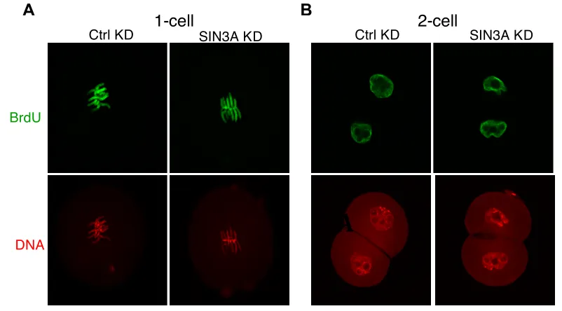
DNA Replication Assay by BrdUTP Incorporation
Inseminated MII eggs were cultured in KSOM containing 10 µM BrdUTP
either 3 h post-insemination or soon after the first cleavage. One-cell embryos were fixed 19 h insemination and the 2-cell embryos were fixed 34 h post-insemination. The embryos were then processed using the immunofluorescence protocol described above with the addition of a denaturing step and neutralizing step after permeabilization. The samples were denatured for 30 min in 2N HCl and neutralized for 20 min in 100 mM Tris-HCl, pH 8.5 at room temperature. The samples were incubated with the mouse monoclonal anti-BrdUTP antibody
(11170376001, Roche) at 1:50 in blocking solution overnight at 4 °C. The
samples were incubated with the secondary FITC anti-mouse IgG1 antibody (1144-02, Southern Biotech) at 1:100 in blocking solution for 1 h. The samples
were mounted in VECTASHIELD medium (Vector Laboratories) containing 2 µM
TO-PRO-3 (Life Technologies).
Global Transcriptional Assay
Click-iT RNA Imaging kit (Invitrogen) was used to assay global
transcription following the manufacturer’s instructions. Briefly, synchronized 2-cell embryos were cultured with 2 mM 5-ethynyl uridine (EU) in KSOM medium for 1 h before fixation in 2.5% paraformaldehyde for 20 min at room temperature. To assay global transcription in 1-cell embryos, the synchronized embryos were incubated in KSOM medium containing 2 mM EU from the time the first
pronucleus formed until 17 h post-insemination. After washing and membrane permeabilization, incorporated EU was detected using the Click-iT detection molecule. The samples were mounted in VECTASHIELD medium (Vector
Laboratories) containing 2 µM TO-PRO-3 (Life Technologies) to visualize the
confocal microscopy. The intensity of the fluorescence was quantified using ImageJ software (National Institutes of Health) as previously described (Aoki et al., 1997).
Plasmid DNA Constructs
To generate a T7 C-terminal-tagged Sin3a cRNA, mouse Sin3a coding
sequence was amplified from mouse cDNA clone 6837144 (Thermo Scientific Open Biosystems) by PCR using the forward primer 5’-
gaggGCATGCcATGAAGaGgaGacTGGAcGACCA -3’ and the reverse primer 5’- gcgGTCGACCCGGCCACGCGTAGGGGCTTTGAATACTGTGCCGTA -3’. After
enzymatic digestion by SphI and SalI-HF, the amplified Sin3a coding sequence
was subcloned into the pIVT-T7 vector to generate pIVT-Sin3a-T7.
Firefly luciferase reporter constructs under the control of the Sin3a 3’
untranslated region (3’ UTR) were generated as previously described (Ma et al.,
2013). Briefly, The entire 1-kb Sin3a 3’ UTR with a ploy(A) site was amplified
using the forward primer 5’-GATATCTAGACTGCAGAGCCAGAGCAGGTAGC-3’ and reverse primer 5’-GCGCCGAATTCACTTATTTCCTTAAGAATCAAGCT-3’.
Amplified Sin3a 3’ UTR were digested by XbaI and EcoRI and subcloned
downstream of the coding sequence of the pIVT-Luc vector.
In Vitro Transcription
The DNA sequence-verified pIVT-Sin3a-T7 construct was linearized by
SfoI digestion. Capped cRNAs were made using in vitro transcription with T7
mMESSAGE mMachine (Ambion) according to the manufacturer’s instructions. Following in vitro transcription, template plasmid DNAs were digested by adding RNase-free DNase and the synthesized cRNA were purified by MEGAclear Kit (Ambion), precipitated and redissolved in RNase-free water.
The DNA sequence verified pIVT-firefly Luc/Sin3a 3’ UTR construct was
linearized by EcoRI digestion. The linearized product was then processed as
was observed for each cRNA sample following electrophoresis in an 1% formaldehyde denaturing agarose gel. Synthesized cRNA was aliquoted and
stored at -80 °C. For microinjection controls, polyadenylated Renillaluciferase
cRNA was generated by using a NotI linearized Renilla luciferase based vector
phRL-SV40 (Promega). The lineralized vector was in vitro transcribed by T7, and then polyadenylated by Poly(A) Tailing Kit (Ambion) according to the manufacturer’s instructions. After polyadenylation and electrophoresis on 1% formaldehyde denaturing agarose gel, it was estimated that approximately a 150
bp poly(A) tail was added to the 3’ terminus of the Renilla Luc cRNA (Ma et al.,
2013).
Luciferase Reporter Assay
Full-grown GV-intact oocytes were microinjected with 5 pl of a solution
containing pIVT-firefly Luc/Sin3a 3’ UTR cRNA (0.365 ug/µl) and control Renilla
Luc cRNA (0.075 ug/µl) (Ma et al., 2013). Injected oocytes were transferred to
milrinone-free CZB medium and matured in vitro for 18 h. Injected GV oocytes cultured for 18 h in CZB supplemented with 10 uM milrinone served as controls. In addition to injecting GV full-grown oocytes, MII eggs were microinjected with 5 pl of the same cRNA mixture. Injected MII eggs were activated with 5 mM
strontium chloride in modified CZB medium that is free of Ca2+ and Mg2+ for 6 h
at 37 °C under an atmosphere of 5% CO2 in air. Injected MII eggs cultured in
Ca2+- and Mg2+ -free CZB medium without SrCl
2 for 6 h served as controls.
Luciferase activity was assayed by lysing oocytes/eggs/embryos in 1x passive lysis buffer and analyzed using a dual-luciferase reporter assay system
(Promega) according to the manufacturer’s instructions. For signal
normalization, the background firefly/Renilla luciferase activity readout from
noninjected oocytes/eggs was subtracted, and the firefly luciferase activity was
Microarrays Analysis
Total RNA was extracted from 20 oocytes/synchronized 2-cell embryos as described above and amplified with the Ovation Pico WTA system V2 (NUGen). The product was then fragmented and labeled with the Encore Biotin Module V2 (NuGen). Four independent biological replicates were hybridized to GeneChip Mouse 2.0 ST microarrays (Affymetrix, Sata Clara, CA, USA).
The microarray datasets from the MoGene-2_0-st (GPL16570) platform were processed using the Oligo package (Carvalho and Irizarry, 2010) from the Bioconductor framework (Gentleman et al., 2004). Raw data was background corrected, normalized and summarized using the robust multi-array average procedure.
Differential expression analysis was done using a robust linear model with empirical Bayes from the limma package (Ritchie et al., 2015). We compared the
following conditions: α-amanitin vs. control 2-cell embryo; Sin3a KD (knockdown)
2- cell embryo vs. control 2-cell embryo; control 2-cell embryo vs. GV oocyte; and
Sin3a KD 2-cell embryo vs. GV oocyte. The P-values were calculated using a
moderate t-statistic. The calculated P-values were adjusted for multiple testing using the false discovery rate procedure. Genes were marked as differentially expressed if they had a minimal absolute fold-change >1.5 and a calculated P-value <0.05.
Platform annotations were downloaded from the Gene Expression Omnibus repository, and all genes that mapped to differentially expressed Affymetrix probesets (defined as false discovery rate <0.05 and absolute
Statistical Analysis
CHAPTER 3: INHIBITING THE MATURATION-ASSOCIATED INCREASE IN MATERNAL SIN3A IMPAIRS THE REPORGRAMMING OF GENE EXPRESSION DURING MOUSE PREIMPLANTATION DEVELOPMENT
Research presented in this chapter was carried out in collaboration with the
laboratory of Monica Mainigi and Kristian Vlahoviček/Petr Svoboda. Total RNA
extraction, amplification, fragmentation and labeling for microarray analysis were performed by Jun Ma, a postdoctoral fellow in the Schultz laboratory. Microarray analysis was performed by Vedran Franke, a doctoral student in the Kristian
Vlahoviček laboratory. Olga Davydenko, a postdoctoral fellow in the Mainigi
laboratory, performed the embryo transfer experiments.
This work was originally published in Biology of Reproduction. Richard Jimenez, Eduardo O. Melo, Olga Davydenko, Jun Ma, Monica Maingi, Vedran Franke, and Richard M. Schultz. Maternal SIN3A regulates reprogramming of gene
3.1 Results
3.1a Sin3a is a dormant maternal mRNA that is recruited for translation
during oocyte maturation and following fertilization
To gain a sense of when the Sin3a gene could be functioning, we analyzed the temporal pattern of Sin3a abundance by qRT-PCR using random hexamers in mouse oocytes and embryos. The observed differences in relative abundance between the different stages of development will unlikely be
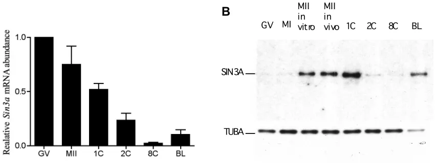
attributed to changes in the length of the poly (A) tail of Sin3a because random hexamers were used for cDNA synthesis. As with many maternal mRNAs that are degraded during oocyte maturation (Su et al., 2007), Sin3a was highly expressed in the full-grown GV intact-oocyte and was degraded upon oocyte maturation as evidenced by the decrease in relative abundance. Sin3a
abundance reached its lowest level by the 8-cell stage, and between the 8-cell and blastocyst stage,its abundance increased, presumably due to zygotic
transcription (Fig. 3.1A). Interestingly, immunoblot analysis of the SIN3A protein revealed a small amount of SIN3A protein present in the full-grown GV-intact
oocyte, even though there was an abundant amount of Sin3a transcript present
at this stage. The amount of SIN3A protein dramatically increased between MI and MII and a further increase was observed following fertilization before a dramatic and rapid loss of SIN3A protein by the 2-cell stage (Fig. 3.1B). The lowest relative abundance of SIN3A protein was observed at the 8-cell stage and
then increased by the blastocyst stage, which is consistent with Sin3a mRNA
relative abundance. It is also noteworthy that this increase in the amount of
Sin3a transcript and protein by the blastocyst stage nearly coincides with the
stage at which Sin3a embryonic null embryos (born from Sin3a+/- intercrosses)
perish.
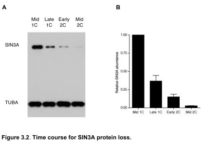
reduction in the amount of SIN3A protein occurred as early as the late 1-cell stage (~63% SIN3A loss compared to the mid 1-cell), and continued to the early 2-cell stage (~ 87% SIN3A loss compared to the late 1-cell) and late 2-cell stage, where SIN3A was faintly detected by immunoblot analysis (Fig. 3.2). Exposure of the embryos to MG132, a proteasome inhibitor, significantly inhibited the reduction in the amount of SIN3A protein, suggesting that the loss of maternal SIN3A protein is potentially mediated by the proteasome (Fig. 3.3).
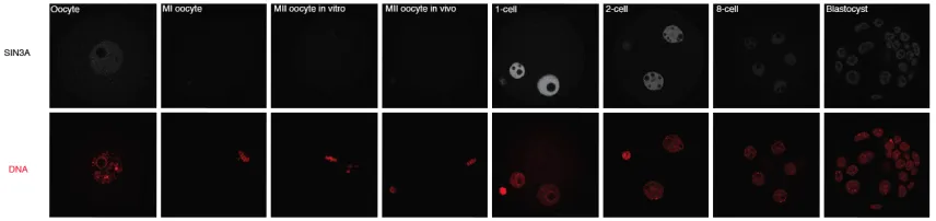
Immunocytochemical detection of SIN3A protein revealed that the SIN3A protein was nuclear, present in both pronuclei at the 1-cell stage, present in the nuclei of both the TE and ICM cells at the blastocyst stage, and had a similar pattern of expression as revealed by immunoblot analysis (Fig. 3.4). The localization of the SIN3A protein remained nuclear in the male and female pronuclei even after the 1-cell mouse embryos were permeabilized prior to fixation (Fig. 3.5), indicating that the SIN3A protein is likely associated with the chromatin in both pronuclei.
The results described above indicate that Sin3a is a dormant maternal
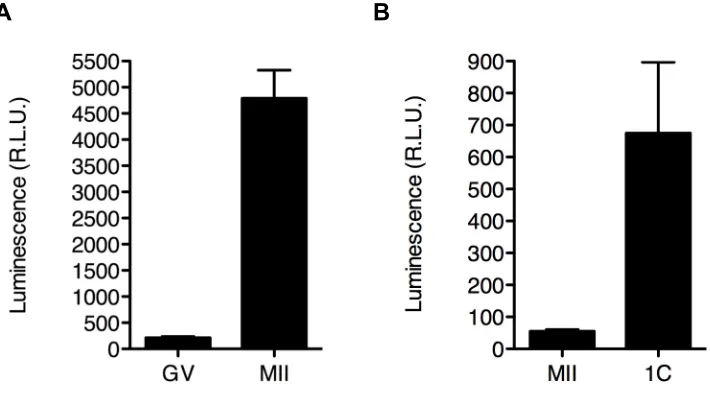
mRNA that is recruited for translation during oocyte maturation and following fertilization. In mouse, recruitment of transcripts for translation during oocyte maturation is driven by sequences within the 3’ untranslated region (3’ UTR) of their respective transcripts, e.g., cytoplasmic polyadenlyation elements (CPEs) (Oh et al., 2000). Given the increase in the relative abundance of SIN3A protein between the full-grown GV-intact oocyte and the MII egg and the presence of
CPEs within the 3’ UTR of the Sin3a transcript, we measured the luciferase
activity of lysed MII eggs following maturation of full-grown GV-intact oocyte
microinjected with firefly luciferase reporter cRNA under the control of the Sin3a
3’ UTR. This experiment revealed a dramatic increase in luciferase activity following maturation of oocytes microinjected with the luciferase reporter cRNA (Fig. 3.6A). An increase in luciferase activity following microinjection of the cRNA and later activation of MII eggs was also observed (Fig. 3.6B), which is
protein following fertilization.
3.1b Inhibiting the maturation-associated increase in SIN3A alters global H3 and H4 histone acetylation in 1-cell embryos
Because SIN3A is a part of the SIN3A-co-repressor complex and the results described above suggesting that the function of SIN3A is restricted mainly to the 1-cell stage, we wanted to assess whether the loss of maternally recruited SIN3A had an effect on histone acetylation at the 1-cell stage. We utilized a combined morpholino/siRNA approach to inhibit the oocyte maturation-associated increase in SIN3A. Following oocyte maturation of full-grown GV-intact oocytes microinjected with the morpholino/siRNA sample targeted against
the Sin3a transcript, no increase in the amount of SIN3A protein was seen (Fig.
3.7). As a control, when a scramble siRNA and a standard morpholino were injected into the full-grown oocyte, a normal maturation-associated increase in SIN3A was observed (Fig. 3.7). These results show that this approach can effectively inhibit the maturation-associated increase in SIN3A.
Because the epigenetic modifications of the male and female genomes are distinct at fertilization and during the first cell cycle of early embryo
development as described above (Santos et al., 2005; Erhardt et al., 2003;
Lepikhov and Walter, 2004; Liu et al., 2004) and the results above suggest that SIN3A is chromatin-associated in both male and female pronuclei (Fig. 3.5), it is plausible that SIN3A may have differential effects on the chromatin of each pronuclei. To determine if the inhibition of the maturation-associated increase in SIN3A affected global histone acetylation in 1-cell embryos and to determine if the effects were dependent on the parental origin of the chromatin, we performed immunocytochemistry analysis of 1-cell embryos generated by in vitro
dependent on the stage of pronuclear development, highly synchronized 1-cell embryos were used for immunocytochemistry analysis to minimize cell-cycle differences.
We assessed global H3K18ac, H4K5ac, H4K8ac, H4K12ac and H4K16ac in highly synchronized 1-cell embryos because many of these marks are affected in mouse embryos when HDAC function is perturbed (Ma and Schultz, 2008) and because these histone modifications represent three distinct regions of genes; H3K18ac is enriched in the region surrounding the transcriptional start site, whereas the others are enriched in the promoter and transcribed regions of active genes (Wang et al., 2008). Interestingly, the enrichment of H4K12ac in transcribed regions is associated with transcriptional elongation (Cho et al., 1998). Although a moderate hyperactylation of all these marks globally was observed when 1-cell embryos were incubated in the presence of Trichostatin A, an HDAC inhibitor, such was not the case when the maturation-associated increase in SIN3A was inhibited (Fig. 3.8). Surprisingly, we observed a modest hypoacetylation for global H3K18ac, H4K8ac and H4K12ac.
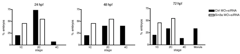
3.1c Inhibiting the maturation-associated increase in SIN3A impairs development beyond the 2-cell stage
Next, we assessed the effect of inhibiting the maturation-associated increase in SIN3A on preimplantation development. After blocking the
maturation associated-increase in SIN3A, the embryos depleted of maternally recruited SIN3A developed poorly beyond the 2-cell stage when compared to the control-injected embryos (Fig. 3.9). The deleterious effects on development of subjecting denuded, microinjected oocytes to in vitro maturation and