1535-9778/05/$08.00⫹0 doi:10.1128/EC.4.1.166–177.2005
Copyright © 2005, American Society for Microbiology. All Rights Reserved.
Mcl1p Is a Polymerase
␣
Replication Accessory Factor Important for
S-Phase DNA Damage Survival
Dewight R. Williams* and J. R. McIntosh
Department of Molecular, Cellular, and Developmental Biology, University of Colorado, Boulder, Colorado
Received 15 June 2004/Accepted 26 October 2004
Mcl1p is an essential fission yeast chromatin-binding protein that belongs to a family of highly conserved eukaryotic proteins important for sister chromatid cohesion. The essential function is believed to result from
its role as a Pol1p (polymerase ␣) accessory protein, a conclusion based primarily on analogy to Ctf4p’s
interaction with Pol1p. In this study, we show that Mcl1p also binds to Pol1p with high affinity for the N
terminus of Pol1p during S phase and DNA damage. Characterization of an inducible allele of mcl1ⴙ,
nmt41
mcl1-MH, shows that altered expression levels of Mcl1p lead to sensitivity to DNA-damaging agents and
synthetic lethality with the replication checkpoint mutationsrad3⌬,rqh1⌬, andhsk1-1312. Further, we find
that the overexpression of the S-phase checkpoint kinase, Cds1, or the loss of Hsk1 kinase activity can disrupt Mcl1p’s interaction with chromatin and Pol1p during replication arrest with hydroxyurea. We take these data
to mean that Mcl1p is a dynamic component of the polymerase␣complex during replication and is important
for the replication stress response in fission yeast.
Pol1 is the largest subunit of the heterotetrameric polymerase ␣holoenzyme (pol-prim), where it serves as the deoxyribonucle-otidyl transferase (5, 39). pol-prim is essential for DNA replica-tion initiareplica-tion because it synthesizes the nascent DNA on a single-strand DNA template (57). Due to the inherent 5⬘ to 3⬘ directionality of DNA synthesis, it is required to initiate the dis-continuous synthesis of DNA on the lagging strand every 100 to 300 bp (63). The synthesis of the short, initiator primer by pol-prim establishes the S-phase checkpoint in fission yeast and Xe-nopus(15, 37, 68). This conserved signal transduction pathway ensures that S phase is complete prior to chromosome segrega-tion (23). In addisegrega-tion, pol-prim funcsegrega-tion is important for telomere length regulation, DNA recombination, and site-specific hetero-chromatin formation (1, 3, 9, 11, 17, 20, 36, 41, 51, 52, 66).
Mcl1p and Ctf4p are yeast members of a eukaryotic family of WD (tryptophan and aspartic acid) repeat proteins that are important for genome stability. This important function is be-lieved to result from a regulatory role for Pol1p. The strongest support for this conclusion comes from Ctf4p’s being the prin-cipal budding yeast protein that bound to a Pol1p affinity column (38). All fungal mutants of this protein family are dependent on components of the S-phase checkpoint for sur-vival, and they show strong genetic interactions with replica-tion factors important for lagging strand synthesis, such as
dna2helicase andfen1nuclease (20, 22, 32, 66). Characteriza-tions of these mutants have revealed chromosome loss result-ing from both decreased sister chromatid cohesion, which pro-motes missegregation of chromosomes during mitosis, and decreased fidelity of DNA replication, which promotes mitotic chromosomal rearrangements (21, 32, 38, 58, 66). In Schizo-saccharomyces pombe, both of these mechanisms for chromo-some loss can be attributed to Pol1p function, since Pol1
mu-tants have both sister chromatid cohesion defects and replication defects (1, 3, 41). Additional support for the idea that Ctf4p and Mcl1p regulate Pol1p during replication comes from their effect on telomere length homeostasis, which has a distinct Pol1p-dependent component.
CTF4 and mcl1 mutants are exquisitely sensitive to the DNA-methylating agent methyl methanesulfonate (MMS). Detection of DNA alkylation occurs primarily during DNA replication and results in slower DNA replication, which oc-curs due to the S-phase checkpoint and through modified bases forming a blockade to replication (48). The checkpoint re-sponses in S. pombeand Saccharomyces cerevisiae are com-pletely dependent on the checkpoint kinases Rad3 (S. pombe) and Mec1 (S. cerevisiae) and their downstream effectors Cds1 (S. pombe) and Rad53 (S. cerevisiae). Their activation inhibits DNA replication initiation from “late” initiating origins (35, 53), and both act to maintain ongoing replication forks that stall at sites of DNA damage (45, 56, 61, 62). Recent work on twomec1alleles has shown that the delay of late origin firing by the checkpoint has only limited importance to yeast survival in MMS compared to the maintenance of replication forks. This conclusion is based on the analysis of mec1-100 and
mec1⌬yeast, which are both defective in blocking late origin firing in MMS, but onlymec1⌬is sensitive to MMS (45). The difference observed between these twomec1alleles is a 10-fold increase in stalled replication forks inmec1⌬cells in compar-ison to themec1-100cells (62), suggesting that fork stability and return to replication play crucial roles in the S-phase checkpoint.
DNA replication complexes established at origins require the Dfp1 (S. pombe)- or Dbf4 (S. cerevisiae) dependent kinase (DDK) Hsk1 (S. pombe) or Cdc7 (S. cerevisiae) for activation (28). Cds1 (S. pombe) and Rad53 (S. cerevisiae) negatively regulate the DDK, and this may delay late origin firing (13, 29, 55, 60). However, mutations in the DDK components that retain their essential function of DNA replication initiation and checkpoint delay still exhibit hypersensitivity to MMS and
* Corresponding author. Present address: Molecular Physiology and Biophysics, 710 Light Hall, Vanderbilt University-Medical Center, Nashville, TN 37232-8725. Phone: (615) 322-7898. Fax: (615) 322-7236. E-mail: [email protected].
166
on September 8, 2020 by guest
http://ec.asm.org/
hydroxyurea (HU) (25, 59), suggesting an additional role for this kinase complex in DNA damage tolerance. The activity of Rad53 (S. cerevisiae) kinase is necessary for pol-prim phos-phorylation in response to checkpoint activation (18). This phosphorylation may be mediated through activity of the DDK, since it can phosphorylate Pol1p in budding yeast (65), and Rad53 is competent to recruit the DDK to replication origins in budding yeast (14). Themcl1-1mutant has a strong genetic interaction with the fission yeast DDK mutant (66). In addition to sharing sensitivity to MMS and decreased sister chromatid cohesion (2), both DDK and mcl1-1 mutants are partially rescued by deletion of thecds1 gene but are lethal when Cds1 is overexpressed orrad3is deleted. The tempera-ture-sensitive DDK mutant, the hsk1-1312 mutant, initiates DNA replication inefficiently at permissive temperatures (55), and this phenotype can be exacerbated by the overexpression of Mcl1p (66). In sum, these results hint that Mcl1p is part of the Cds1-Hsk1 regulatory loop, possibly regulating Pol1p func-tions.
The phenotypes found in the mcl1-1 and thectf4 mutants suggest that Mcl1 and Ctf4 regulate Pol1p. To test whether this interaction between members of the Ctf4 family and Pol1p is conserved in S. pombe, we constructed two epitope-tagged versions of Mcl1p for the study of Pol1p interactions. Our data suggest that Mcl1p interacts directly with Pol1p through its N terminus and that this region of Pol1p is sufficient for high-affinity interaction with Mcl1p in vivo during S phase or during DNA replication stress. Furthermore, the mcl1-1 mutant is acutely sensitive to DNA damage during DNA replication. We also examined the effect that the overexpression of Cds1p or mutations inrad3,cds1, andhsk1would have on Pol1p- and Mcl1p-containing complexes arrested in S phase and interpret our data to mean that the Mcl1p-Pol1p interaction is affected by the Cds1-Hsk1 checkpoint regulatory loop.
MATERIALS AND METHODS
Yeast strain construction.Genotypes of yeast strains are listed in Table 1. The
mcl1⫹gene was tagged with a hexahistidine and a dual myc epitope by subclon-ing a SalI-BglII fragment (66) from pTOPO4Blunt (Invitrogen, Carlsbad, Calif.), which containedmcl1⫹, into SalI-BamHI sites of the multiple cloning site in pREP41x MH vector (8). This dual-epitope-tagged allele was integrated into the chromosomal locus under the control of the no message in thiamine 41x (nmt41x) promoter by first subcloning the nmt41x promoter plus 1 kb ofmcl1⫹, contained within a PstI-NsiI fragment, into pSport-ura4⫹flanked by 1.2 kbp of the endogenous promoter region ofmcl1⫹, contained in a SphI-SpeI genomic fragment. Primers 5⬘-GAATCAGTGGACGAAACCAC-3⬘and 5⬘-ATGTTTG TTTACATTAAATAG-3⬘were used to amplify the integration cassette. PCR products were gel purified and transformed into haploid 99 cells. Homologous integrants were identified by PCR screening and linkage tomcl1-1(data not shown). Construction of an Mcl1-green fluorescent protein (GFP) strain was previously described (66). Strain phenotypes were confirmed by using standard fission yeast procedures (40).
Cell synchronizations and chemical treatments.The reversible, temperature-sensitive arrest of thecdc10-129mutation in either anmcl1⫹,mcl1-1, orrad3⌬
background were used to synchronize early-log-phase cultures (A595⫽0.3) in G1.
These cells were arrested by a shift to 36°C for 3 h, either treated with 12 mM HU or 0.02% MMS or mock treated 30 min prior to a temperature shift down to the permissive growth conditions (25°C), which releases the cells into G1,
re-moved at the times stated, and washed three times. Serial dilution assays were performed by patch plating onto yeast extract medium plus supplements (YES) followed by 3 days of growth at 25°C. Flow cytometry was performed as described previously (4). For an assessment of UV sensitivity, patched cells were dried onto the agar surface and then immediately exposed to 100 J of UVB light/m2
in a Stratalinker (Stratagene, La Jolla, Calif.).
Two-dimensional DNA gels.Two hundred milliliters of G1synchronized
cul-tures was treated with 5 mM HU 30 min prior to release from arrest. Replicating genomic DNA was isolated as described previously (30). A Molecular Dynamics Storm PhosphorImager was used for signal collection, and their ImageQuant 5.1 software was used to quantify total hybridization signal by selection of regions of interest that contained the 1 to 2 N DNA range.
Yeast cellular disruption.Cells (1⫻1010) were harvested and resuspended in
a lysis buffer containing 50 mM Na2HPO4, pH 8.0, 150 mM NaCl, 10 mM
imidazole, 5 mM NaF, 1 mM NaVO4, 15 mM-glycerolphosphate, 2 mM MgCl2,
0.1% Triton X-100, 1 mM 4-(2-aminoethyl)benzenesulfonylfluoride HCl, 1 mM phenylmethylsulfonyl fluoride, 10M pepstatin, 100 M leupeptin, 10 U of aprotinin/ml, 10 U of soybean trypsin inhibitor/ml, and 5 mMN-ethylmaleimide. Cells were disrupted by a 30-min burst of agitation at 4°C in a Ribolyser (Bio101, Carlsbad, Calif.) using setting 6.5 in the presence of 0.45-m-diameter glass beads. This process achieved⬎90% lysis of cells. Clarification of lysates was performed by centrifugation at 15,000⫻gin a Sorvall SS-34 rotor (Dupont, Newtown, Conn.) at 4°C for 30 min.
Chromatin extraction.The procedure used for chromatin extraction was pre-viously described (27).
Pulldown assays.pGT-Pol1p118–634
(46), pGT-Pol1p1180–1405
, and pGEX2T (empty) plasmids were expressed inEscherichia coliDH5␣cells. Bacterial lysates were batch bound to glutathione-Sepharose 4B (GSH-Sepharose) resin (Phar-macia, Uppsala, Sweden), washed with 50 column volumes of phosphate-buff-ered saline with 0.1 mM phenylmethylsulfonyl fluoride, and stored at 4°C. Clar-ified yeast lysates containing 2 mg of total protein were mixed with 100l of settled glutathioneS-transferase (GST) or GST-Pol1p affinity matrices for 1 h at 4°C and then placed into a disposable polycarbonate column, washed with 300 bed volumes of yeast lysis buffer, and then eluted with 1 bed volume of lysis buffer plus 20 mM glutathione.
Strains 592 to 597 were generated from strains 598 to 603 by transformation with pTB19, which contains the first 180 amino acids of Pol1p fused in frame with GFP under its endogenous promoter (12). Fifty-milliliter cultures were grown to mid-log phase in selective medium and harvested. Three milligrams of total protein in clarified lysates was mixed with 25 to 50l of a cobalt-immobilized affinity matrix (Talon resin; Clontech, Palo Alto, Calif.) for 1 h at 4°C to bind all available Mcl1-MHp. DNase I treatment of Talon resin-bound Mcl1-MHp com-plexes was performed at 4°C using 100 U of DNase I for 1 h in the presence of 2 mM MgCl2. Talon resin was washed with 30 column volumes of lysis buffer, and
the bound proteins were eluted with 250 mM imidazole in lysis buffer. Sucrose gradients.Three milligrams of total protein was layered atop 5 to 15% (wt/vol) sucrose gradients. Sedimentation by centrifugation at 155,000⫻gin a SW40 rotor at 4°C for 20 h was done in a Beckman L5 preparative ultracentri-fuge. Fractions (22 by 0.5 ml) were collected from the bottom of gradients, with fraction 1 being the bottom of the gradient. Sedimentation velocity for different yeast backgrounds was determined by comparison of the unknown to a standard line, generated by linear regression from the sedimentation velocities of known standards. Protein sedimentation peaks were determined by quantifying band intensities of scanned images of blots with the image analysis software Meta-morph (Universal Imaging Corporation, Downington, Pa.) to determine the integrated intensity of each band. The mean for the area under each peak was calculated by generating a curve by nonlinear regression with a 95% confidence interval to best fit the data.
Antibodies.Proteins were visualized following sodium dodecyl sulfate-polyac-rylamide gel electrophoresis separation by Western blotting and immunodetec-tion. Primary antibodies were detected with horseradish peroxidase secondary antibodies (Sigma, St. Louis, Mo.) and SuperSignal chemiluminescent substrate (Pierce, Rockford, Ill.). Immunoblotting and immunoprecipitations were carried out as described previously (33). 9E10 (monoclonal anti-myc) was used for immunostaining (16), an affinity-purified rabbit anti-myc from Covance (Prince-ton, N.J.) was used for immunoprecipitation, an affinity-purified chicken anti-Pol1p was used to detect p180 (47), an affinity-purified rabbit antibody raised against the green fluorescent protein was used for immunostaining and immu-noprecipitation of GFP fusion proteins (a gift from the Pam Silver lab, Harvard Medical School, Boston, Mass.), and a monoclonal antibody with affinity for GST from BabCo (Richmond, Calif.) was used for immunostaining of GST conju-gates.
RESULTS
Repression ofmcl1ⴙexpression in yeast has genetic
inter-actions and chemical sensitivities like those of mcl1-1. The
screen that identifiedmcl1-1demonstrated a high incidence of
on September 8, 2020 by guest
http://ec.asm.org/
chromosome loss and synthetic lethality with bub1⌬ but not another spindle assembly checkpoint mutant,mad2⌬. In addi-tion, we found that mcl1-1 was synthetic lethal with rad3⌬,
rad26⌬,hsk1-1312, andrqh1⌬(rad12), indicating that the via-bility of mcl1-1 strains depended on DNA damage check-points. However, themcl1-1mutant was partially rescued for its slow growth whencds1or bothchk1andcds1genes were deleted, demonstrating that the downstream effectors of the checkpoint were dispensable and, in the case of Cds1p, dele-terious (66). Sequencing of themcl1-1allele revealed a single lesion changing the 124th amino acid from a W to an ochre stop. To ensure that the phenotypes associated withmcl1-1
were due to decreased protein product from translational read-through as opposed to neomorphic effects arising from the production of the short 124-amino-acid fragment, we examined the growth, genetic interactions, and chemical sensitivities of the mutant with the repressible promoter,nmt41mcl1-MH (Ta-ble 1). This new allele ofmcl1produced a protein of predicted size and had no overt growth phenotypes in conditions where the promoter was induced (Fig. 1A), but overexpression of Mcl1 did have genetic interactions with DNA damage check-point genes (Table 2), suggesting that the overexpression of Mcl1 is also deleterious. Under promoter-repressed condi-tions, thenmt41mcl1-MHstrains had similar growth character-istics and genetic interactions asmcl1-1(Fig. 1A and Table 2). After several generations at 25°C, cell cycle-arrested cells were evident (Fig. 1A and C). This phenotype was more pronounced at 36°C, possibly due to the more rapid dilution of excess
Mcl1p upon promoter shutdown through the increased cell division rate at 36°C. Deletion ofmcl1is lethal to fission yeast (66), so not surprisingly, the repression ofnmt41mcl1-MH ex-pression for greater than 50 division cycles led to no detectable cell viability in cultures (Fig. 1A).
Thenmt41mcl1-MHmutant had genetic interactions similar to those of the mcl1-1 mutant. For instance, the
bub1⌬nmt41mcl1-MH mutant was unable to grow with nmt41mcl1-MHrepressed, whereas themad2⌬nmt41mcl1-MH
mu-tant grew like thenmt41mcl1-MHmutant alone (Fig. 1B and Table 2).nmt41mcl1-MH was also synthetic lethal withrad3⌬,rad26⌬,
rqh1⌬, andhsk1-1312under promoter-repressed conditions (Ta-ble 2). In attempts to recover via(Ta-ble dou(Ta-ble mutant spores from these various genetic backgrounds, we performed crosses and plated spores onto selective medium lacking thiamine in the hope that the expression of Mcl1-MHp would allow recovery of the DNA damage checkpoint double mutants. Still, no double mutant strains were recovered from these crosses, demonstrating that these mutations are synthetic lethal withnmt41mcl1-MH. In par-ticular, the hsk1-1312 mutant crossed to nmt41mcl1-MH strains consistently produced asci with five to eight spores, suggestive of meiotic segregation failures. In contrast,mcl1-GFPwhich is ex-pressed from the endogenous promoter showed no genetic inter-actions or growth defect with any of these mutant combinations. Thenmt41mcl1-MHstrain also showed a sensitivity to growth in the continual presence of 0.0025% MMS, 5 mM HU, and 10 mg of thiabendazole (TBZ)/ml (Fig. 1C). nmt41mcl1-MH ex-pression was repressed by thiamine addition to cultures 18 h
TABLE 1. Yeast strains
Strain Source Genotype
53 cdc10-129 leu1-32
99 h⫺ade6-M210 leu1-32 ura4-D18 his3-D1 546 h⫺mcl1-1 ade6-M210 leu1-32 ura4-D18 his3-D1 547 S. Sazar h⫺mad2⌬::ura4⫹ade6-M216 leu1-32 ura4-D18 549 J. P. Javarzet h⫺bub1⌬::ura4⫹ade6-M216 leu1-32 ura4-D18
551 h⫺mcl1GFP::ura4⫹ade6-M210 leu1-32 ura4-D18 his3-D1
553 h⫺mcl1GFP::ura4⫹cds1⌬::ura4⫹ade6-M210 leu1-32 ura4-D18 his3-D1 554 h⫹mcl1GFP::ura4⫹nmt1::GSTcds1 LEU2 ade6-704 leu1-32 ura4-D18 555 h⫺mcl1GFP::ura4⫹hsk1-1312 ade6-M210 leu1-32 ura4-D18 his3-D1 558 h⫺mcl1GFP::ura4⫹cdc25-22 ade6-M210 leu1-32 ura4-D18 his3-D1 561 h⫺mcl1GFP::ura4⫹cdc22-M45 ade6-M210 leu1-32 ura4-D18 his3-D1 560 h⫺mcl1GFP::ura4⫹cdc10-129 ade6-M210 leu1-32 ura4-D18 his3-D1 562 h⫺mcl1GFP::ura4⫹rad3⌬::ura4⫹ade6-M210 leu1-32 ura4-D18 his3-D1
578 h⫺mcl1-1 cdc10-129 leu1-32
590 G. Freyer h⫺rqh1⌬::ura4⫹ade6-M210 lueu1-32 ura4-D18 591 h⫺rad3⌬::ura4⫹cdc10-129 leu1-32
592 h⫺nmt41x-mcl1-MH::ura4⫹ade6-M210 leu1-32 ura4-D18 his3-D1 593 h⫺nmt41x-mcl1-MH::ura4⫹ade6-M210 leu1-32 ura4-D18 his3-D1 cdc25-22 594 h⫺nmt41x-mcl1-MH::ura4⫹ade6-M210 leu1-32 ura4-D18 his3-D1 cdc22-M45 595 h⫺nmt41x-mcl1-MH::ura4⫹ade6-M210 leu1-32 ura4-D18 his3-D1 cdc21-M68 596 h⫺nmt41x-mcl1-MH::ura4⫹ade6-M210 leu1-32 ura4-D18 his3-D1 cdc10-129 597 h⫺nmt41x-mcl1-MH::ura4⫹ade6-M210 leu1-32 ura4-D18 his3-D1 orp1-4 598 h⫺nmt41x-mcl1-MH::ura4⫹ade6-M210 leu1-32 ura4-D18 his3-D1/pTB19 599 h⫺nmt41x-mcl1-MH::ura4⫹ade6-M210 leu1-32 ura4-D18 his3-D1 cdc25-22/pTB19 600 h⫺nmt41x-mcl1-MH::ura4⫹ade6-M210 leu1-32 ura4-D18 his3-D1 cdc22-M45/pTB19 601 h⫺nmt41x-mcl1-MH::ura4⫹ade6-M210 leu1-32 ura4-D18 his3-D1 cdc21-M68/pTB19 602 h⫺nmt41x-mcl1-MH::ura4⫹ade6-M210 leu1-32 ura4-D18 his3-D1 orp1-4/pTB19 603 h⫺nmt41x-mcl1-MH::ura4⫹ade6-M210 leu1-32 ura4-D18 his3-D1 cd1⌬::ura4⫹/pTB19 1123 A. Carr h⫺rad26⌬::ura4⫹ade6-704 leu1-32 ura4-D18
FY865 S. L. Forsburg h⫺cds1⌬::ura4⫹leu1-32 ura4-D18 FY945 S. L. Forsburg h⫺hsk1-1312 ade6-M210 leu1-32 ura4-D18 FY1105 S. L. Forsburg h⫹rad3⌬::ura4⫹ade6-M216 leu1-32 ura4-D18
on September 8, 2020 by guest
http://ec.asm.org/
prior to plating (Fig. 1C, “off” rows). Promoter repression of this duration depletes Mcl1-MHp to levels undetectable by immunoblotting of cellular lysates (Fig. 2D). Under these pro-moter-repressed conditions,nmt41mcl1-MHmutant drug sensi-tivities were similar to those of themcl1-1mutant (Fig. 1C). Deletion ofcds1⫹in thenmt41mcl1-MHcell line rescued the strain’s sensitivity to TBZ and the slower growth induced by the repression ofmcl1gene expression. Induction of this pro-moter did not, however, produce a truly wild phenotype. When Mcl1-MHp was expressed, thenmt41mcl1-MHmutant showed a 125-fold greater sensitivity to MMS and 25-fold more
sensitiv-ity to HU and TBZ than themcl1-GFPmutant, which grows with a wild-type phenotype.
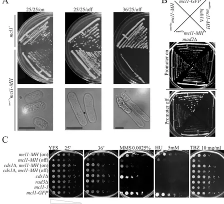
Mcl1p interacts with the N terminus of Pol1p both in vitro
and in vivo.Two epitope-tagged Mcl1 proteins (diagrammed
in Fig. 2A) were highly enriched in material recovered from yeast cellular lysates bound to an affinity matrix composed of an N-terminal Pol1 protein fragment fused to GST (GST-NT in Fig. 2B and C). This protein fragment contains amino acids 118 to 634 of Pol1p (47). None of these tagged alleles of Mcl1p interacted strongly with the C-terminal GST-Pol1p protein chimera (GST-CT in Fig. 2B and C) or GST (Fig. 2C). Such
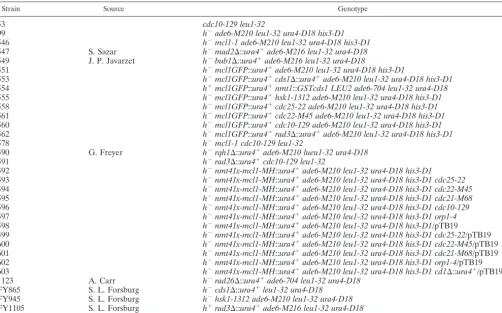
FIG. 1. Thenmt41xmcl1-MHmutant is phenotypically similar to themcl1-1mutant. (A) Growth of thenmt41xmcl1-MH55strain and the parental
mcl1⫹ strain at 25°C for 25 generations in selective medium containing no thiamine (25/25/on), with 10 mM thiamine to repressnmtgene expression (25/25/off), or at 36°C (36/25/off). The lower panel is a phase-contrast light microscope image ofnmt41xmcl1-MH55cells from the liquid
culture prior to plating (bar in phase-contrast images represent 5m). (B) Recovered double mutantnmt41xmcl1-MH bub1⌬andnmt41xmcl1-MH
mad2⌬strains were struck onto selective media containing 0 (on) or 10 (off) mM thiamine to examine the differences in the genetic interactions betweennmt41xmcl1-MHand the two spindle checkpoint mutations at permissive temperature of 25°C. (C) Cultures ofnmt41xmcl1-MH,cds1⌬ nmt41xmcl1-MH(603),cds1⌬(FY865),rad3⌬(591),mcl1-1(546), andmcl1-GFP(551) cells were grown for 25 generations in selective media and
then split into medium without (on) or with (off) 10 mM thiamine for 6 generations before plating on YES agar plates (a thiamine-enriched medium) containing 0.0025% MMS, 5 mM HU, or 10 mg of TBZ/ml. Cells were serially diluted 1:5 from 106to 102cells/ml.
on September 8, 2020 by guest
http://ec.asm.org/
GST pulldown assays from whole-cell lysates demonstrate a high-affinity interaction between Mcl1p and the N terminus of Pol1p.
An in vivo interaction between the N terminus of Pol1p and Mcl1-MHp was tested by expression of the first 180 amino acids of Pol1p C-terminally fused to GFP (Fig. 2B, bottom) and expressed under the regulation of its endogenous pro-moter from a plasmid (pTB19) innmt41mcl1-MHcells. Purifi-cation of Mcl1-MHp with Talon resin recovered only a fraction of the GFP-NT chimera (Fig. 2B) from cell lysates when cells were grown in the absence of 10 mM thiamine (Fig. 2D, ON) but did not recover any detectable GFP-NT when the expres-sion of Mcl1-MHp was repressed by 10 mM thiamine (Fig. 2D, OFF). Because DNA might have mediated this weak interac-tion, we digested the purified material while it was still on the Talon beads with DNase I, followed by extensive washes (Fig. 2D). This treatment was not sufficient to disrupt the weak Mcl1-MHp interaction with the GFP-NT protein fragment. Expression of GFP with Mcl1-MHp does not recover GFP with Mcl1-MHp, demonstrating that the Pol1p fragment interaction was not induced by nonspecific interaction between GFP and Mcl1-MHp.
The contrast between strong in vitro and weak-appearing in vivo interactions between Mcl1p and the N-terminal Pol1p fragments led us to hypothesize either that these different protein fragments had different affinities for Mcl1-MHp or that the in vivo interaction was regulated, such that only a fraction of cells contained Pol1-NTp/Mcl1-MHp complexes. Since a similar observation had been reported for reciprocal immuno-precipitation of Ctf4p and Pol1p from budding yeast cell ly-sates, we looked for cell cycle dependence for this interaction. Cell lysates arrested by the mutationscdc25-22(G2),cdc22 -M45(S phase),cdc21-M68(S), ororp1-4(pre-S) were used in this analysis (Fig. 2E). In G2and pre-S-phase arrests (cdc25-22
and orp1-4, respectively), the majority of the GFP-NT re-mained unbound to Mcl1-MHp recovered by cobalt-immobi-lized resin, but during S phase nearly all of the GFP-NT in the cellular lysate was recovered with the Mcl1-MHp. Surprisingly, a similar analysis of the N-terminally-tagged Mcl1-MHp failed to detect any direct interaction with endogenous Pol1p (data not shown), but immunoprecipitation of the
C-terminally-tagged Mcl1-GFP does recover endogenous Pol1p (see Fig. 6B).
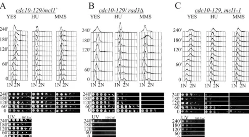
Themcl1-1mutant is sensitive to DNA damage specifically
during G1/S phase.To assess themcl1-1mutant’s response to
perturbed DNA replication, we used thecdc10-129mutation and a temperature shift to the restrictive conditions to arrest cells in G1, followed by release into permissive conditions to
observe synchronous DNA replication in the presence of HU or MMS or exposure to UV irradiation. Since the mcl1-1
mutant is also temperature sensitive, we tested for any effects of our temperature shift regimen on DNA replication and viability in the absence of treatment in thecdc10-129 back-grounds. Like mcl1⫹and rad3⌬ mutants, themcl1-1mutant had increased DNA fluorescence in flow cytometry at 60 min and reached 2C DNA content by 120 min with no detectable loss in relative viability upon return to the permissive temper-ature (Fig. 3, panels labeled “YES”). This result demonstrates that thecdc10-129arrest point precedes the essential point of Mcl1 function for this experiment, so our double mutant can be taken as synchronized at thecdc10-129restriction point. In the presence of either HU or MMS, the cdc10-129 mcl1⫹
mutant showed no significant DNA content increase (Fig. 3A, two right histograms) and retained high viability (Fig. 3A, panels below histograms), demonstrating that DNA replication was strongly inhibited in these cells by both compounds. In four independent experiments, however, thecdc10-129 mcl1-1
andcdc10-129 rad3⌬mutants consistently showed a dramatic decrease in relative cellular viability in the presence of HU and MMS. This decrease in viability was consistently coincident with the normal timing of DNA replication onset (Fig. 3B and C). Examination of thecdc10-129 mcl1-1andcdc10-129 rad3⌬
strain cytology demonstrated that thecdc10-129 mcl1-1mutant does not progress into mitosis in HU and MMS, unlike the
cdc10-129 rad3⌬mutant (data not shown), so themcl1-1 mu-tation is not defective in checkpoint cell cycle arrest. It follows that the increased sensitivity is most likely due to an abnormal intra-S-phase response to DNA replication perturbation. Close examination of the MMS DNA content in themcl1-1strain showed a slight but significant and consistent increase in DNA content when compared to themcl1⫹controls. Further sup-port of this S-phase-specific sensitivity was found upon
expos-TABLE 2. Genetic interactionsa
Mutation Function
Interaction with
mcl1-1 mcl1-GFP
nmt41mcl1-MH
On Off
mad2⌬ Spindle assembly checkpoint NI NI NI NI
bub1⌬ Spindle assembly checkpoint kinase SL NI S SL
rad3⌬ DNA damage checkpoint kinase SL NI SL SL
rad26⌬ Rad3 interaction protein SL NI SL SL
rad9⌬ DNA damage checkpoint (PCNA like) S NI NI S
hsk1-1312 S-phase initiation kinase SL NI ML ML
cds1⌬ Transducer kinase for S-phase PR NI NI PR
rqh1⌬ RecQ helicase homolog SL NI SL SL
pol1-1 DNA polymerase-␣ NI NI NI NI
rad22⌬ Rad52 homologue NI NI
rph51⌬ Rad51 homologue ML NI
rph54⌬ Rad54 homologue ML NI
aNI, no interaction; S, synthetic; SL, synthetic lethal; PR, partial rescue; and ML, meiotic lethal.
on September 8, 2020 by guest
http://ec.asm.org/
ing the cdc10-129 mcl1-1 mutant to 100 J of UVB light/m2
prior to or during DNA replication. This step leads to a sig-nificant loss of viability, whereas when it is done following DNA replication the strain appears far less affected (Fig. 3C, panel labeled “UV”). This result differs from our original ob-servation, wheremcl1-1cells exposed in log phase showed little sensitivity to this dosage of UVB irradiation compared to the wild type (66). This sensitivity may have been previously over-looked because two-thirds of the fission yeast cell cycle is spent in G2.
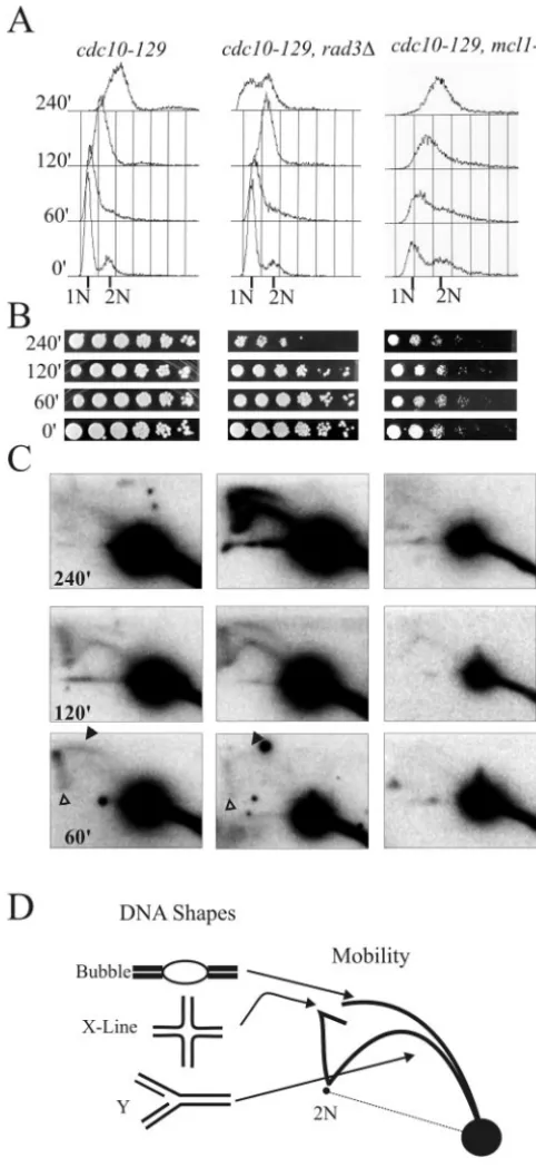
Replication forks appear stable and persistent in mcl1-1
cells under conditions of mild replication stress.Our results
suggest that mcl1⫹is important for replication stress. Given the recent evidence that replication fork stability is a major factor in yeast survival of replication stress, we examined rep-lication fork structure by two-dimensional gel analysis using the same three strains as described above and identical syn-chronization procedures but with release into 5 mM HU. This concentration of the ribonucleotide reductase inhibitor slows rather than completely blocks DNA replication, but it still leads to appreciable cell death in themcl1-1mutant cell line (Fig. 4A). We examined replication at thears2-1chromosomal locus, an early firing origin that remained active during this treatment.
DNA replication intermediates in cdc10-129 mcl1⫹ cells were resolved by two-dimensional gel analysis as the X line and bubble arc at 60 min after G1release, and a Y arc becomes
prominent by 120 min after release (Fig. 4C, left panels, and D). In similarly treatedcdc10-129 rad3⌬cells, similar replica-tion forks were detected at ars2-1 at 60 and 120 min but appeared to accumulate at the 240-min time point. Therad3⌬
strain reached a near 2C DNA content more rapidly than
cdc10-129 mcl1⫹orcdc10-129 mcl1-1cells (Fig. 4A), and cells in the culture began entering mitosis with replication forks still present at 240 min.
An X-DNA line that is indicative of four-way or hemicat-enae structures was present in thecdc10-129 rad3⌬andcdc10
-FIG. 2. Mcl1p interacts with N-terminal fragments of Pol1p. (A) Schematic representation of the two epitope-tagged alleles ofmcl1 (strains 551 and 589) used in this study. (B) Schematic representation of the polymerase␣holoenzyme showing the three conserved domains of the eukaryotic Pol1 (p180): an acidic N terminus, a central de-oxynucleotide transferase catalytic core composed of seven B-polymer-ase as well as five polymerB-polymer-ase␣ conserved sequences, and the
C-terminal Zn finger domain with B subunit and primase association presented. In the lower panel, the two bacterially expressed GST-Pol1p fragments used for in vitro interaction studies presented in panel C and a fission yeast-expressed Pol1-GFP protein fragment used for in vivo interaction studies presented in panels D and E are shown. (C) Western blot analysis of GST pulldowns from yeast lysates con-taining Mcl1-GFP (left) or Mcl1-MHp (center) with the various GST proteins (right). The epitope-tagged Mcl1 proteins strongly interact with the GST-NT fragment (center lanes) but not the GST or GST-CT proteins (right and left lanes). (D) Mcl1-MHp interacts in vivo with a small fragment of Pol1p tagged with GFP (GFP-NT). Yeasts were grown in conditions where thenmtpromoter ofmcl1-MHwas either induced (ON) or repressed with thiamine (OFF). The six-His-tagged Mcl1-MHp was recovered with cobalt-immobolized agarose resin (Talon). Mcl1-MHp interaction with GFP-NT was not disrupted by DNase I digestion of Talon-bound material. Additionally, no interac-tion was seen between Mcl1-MH and GFP alone. (E) Cell cycle re-striction points for the temperature-sensitivecdc25-22(G2arrest of
strain 596),orp1-4(pre-S arrest of strain 599),cdc22-M45 (early S arrest of strain 597), orcdc21-M68(mid-S arrest of strain 598) mutants were used to test for a cell cycle-dependent interaction between Mcl1-MHp and GFP-NT. Only those cells arrested in S phase (flow cytom-etry not shown) had enrichment of GFP-NT in the Mcl1-MHp pull-down with Talon resin.
on September 8, 2020 by guest
http://ec.asm.org/
129backgrounds at 60 and 120 min (Fig. 4C, open arrowhead). Such an X line is a natural feature of normal origin activation in yeast (30, 49, 50, 56). InS.pombe, it is dependent on DNA recombination (54). Both this feature and a bubble arc are not detected in thecdc10-129 mcl1-1two-dimensional gel analysis. Although the apparent rates of bulk DNA content increase, as seen by flow cytometry, were similar incdc10-129 mcl1⫹and
cdc10-129 mcl1-1cells, the total signal obtained at thears2-1
locus remained nearly constant in themcl1-1strain, whereas it doubled in both control strains. It is possible that replication failed to initiate from this region inmcl1-1cells but progressed slowly into this locus and stalled in some cells. Unfortunately, analysis of a number of other autonomously replicating se-quences (ars-727,ars2-2, centromeres, telomeres, andars3001) also failed to detect active origins in mcl1-1 under similar conditions. Since a similar poor origin activation phenotype is found in the recombination mutants rad22⌬, rph51⌬, and
rph54⌬(54), we tested themcl1-1mutant’s genetic interactions with these two null mutants. We found thatmcl1-1was lethal with both rph51⌬ and rph54⌬. However, mcl1-1 showed no overt interaction withrad22⌬, such as poor growth or depres-sion in restrictive temperature (Table 2).
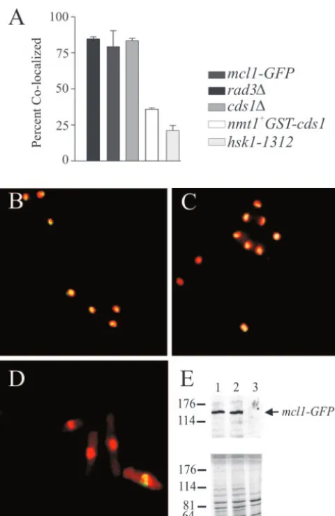
Binding of Mcl1-GFP to chromatin during HU arrest is
dependent on Hsk1 kinase activity. Mcl1-GFP is chromatin
bound during G1, but it is progressively released as cells move
from S phase into G2(66). Upon HU arrest, a large fraction of
Mcl1-GFP is retained in the nucleus following cell wall disrup-tion and detergent extracdisrup-tion, suggesting that it is tightly bound
to chromatin (Fig. 5A, B, and E, lane 1). Using a previously described chromatin extraction method, we looked at Mcl1-GFP nuclear retention in HU-treated cells lacking Cds1, Rad3, or Hsk1 kinase activities and when Cds1p was overproduced from a chromosomalnmt1GST-cds1gene (6). The loss of either Rad3p or Cds1p from the Mcl1-GFP cells had no effect on nuclear retention of GFP fluorescence during HU arrest (Fig. 5A, C, and E, lane 2). Retention was lost, however, inhsk1
-1312cells shifted to the restrictive temperature for 3 h after HU arrest (Fig. 5A, D, and E, lane 3). Retention of Mcl1-GFP in the nucleus during HU arrest was also lost by a high level of Cds1 kinase expression (Fig. 5A).
S-phase checkpoint kinases can affect Mcl1-GFP
endoge-nous complexes.The sedimentation velocity of Mcl1-GFP
dur-ing HU arrest, a condition when the above-mentioned mutant kinases are normally activated, was measured to assess the effect of these enzymes on Mcl1-GFP protein complexes (Fig. 6A). Loss of Cds1p from HU-arrested Mcl1-GFP cells pro-duced a slight sedimentation velocity change (a mean sedimen-tation velocity of 14.1 S compared to 14.4 S incds1⫹cells). In contrast, loss of either Rad3 (rad3⌬) or Hsk1 (hsk1-1312 at 36°C), as well as overexpression of Cds1 (nmtGST-cds1), caused Mcl1-GFP to sediment significantly more slowly (13, 13.6, and 11 S, respectively). These sedimentation velocities were calcu-lated from the sedimentation profiles assuming a Gaussian distribution of material, but in thehsk1-1312andrad3⌬mutant backgrounds the sedimentation profiles included either an ex-aggerated trailing fraction or a secondary peak. Thus, the
ob-FIG. 3.mcl1-1mutants are sensitive to DNA damage specifically in S phase. (A) Cultures ofcdc10-129cells (strain 53) were arrested for 3 h at 36°C and then released into either YES, YES with 12 mM HU, or YES with 0.02% MMS. Cells were collected to determine DNA content by flow cytometry (top row of three panels) or relative viability, as determined by serial dilution plating (middle row of panels). Cells plated from untreated cultures were also irradiated with 100 J of UV light/m2at given time points (bottom panels). (B and C)cdc10-129 rad3⌬cells (strain
591) andcdc10-129 mcl1-1cells (strain 578), respectively, were treated as described above. Data presented is representative of four independent experiments.
on September 8, 2020 by guest
http://ec.asm.org/
served changes in sedimentation velocities probably represent the real change in Mcl1-GFP sedimentation to only a limited extent. Most likely, the sedimenting material breaks into two distinct populations.
To determine whether the observed changes in mobility represented a loss of Pol1p from these Mcl1p complexes, we used a high-affinity antibody to GFP to precipitate Mcl1-GFP from the sucrose gradient fractions. Immunoprecipitation of Mcl1-GFP from fraction 9 coprecipitates Pol1p from all ge-netic backgrounds exceptnmt1GST-cds1, which contained no Mcl1-GFP in fraction 9 (Fig. 6B, top panels). Pol1p also pre-cipitated with Mcl1-GFP from the trailing fractions ofrad3⌬
andcds1⌬, indicating that the mobility differences observed in these mutants were not due to a loss of Pol1p interaction. In contrast, the slower-sedimenting material in hsk1-1312 and nmt1GST-cds1 fractions contained no detectable Pol1p (Fig.
6B, bottom panels), suggesting that in these backgrounds the association between Pol1p and Mcl1-GFP is weaker than in the other conditions. To test this result in a different way, we used the GST-Pol1NT118–634 bound to GSH-Sepharose to collect
Mcl1-GFP from 3 mg of total protein in whole-cell extracts from the above arrested strains. This technique also showed that Mcl1-GFP interacts with Pol1p in themcl1-GFP, cds1⌬, andhsk1-1312backgrounds. It was not, however, present when GST-Cds1p was overexpressed. This result does not appear to be due to the displacement of GST-Pol1NT from the GSH-Sepharose, since the protein is readily detected in the recov-ered resin along with Mcl1-GFP.
DISCUSSION
Mcl1p belongs to a family of conserved Pol1p accessory
factors.Both Ctf4p from budding yeast and Mcl1p from fission
yeast interact with Pol1p, suggesting that this is an evolution-arily conserved interaction. Similar proteins have been studied inAspergillus nidulansandNeurospora crassa(sepB), humans, and Xenopus laevis (AND-1 proteins), and possible homo-logues have been identified by genome sequencing projects for
Drosophila melanogaster,Caenorhabditis elegans, and Arabidop-sis thaliana(22, 31, 38, 66), suggesting that a family of eukary-otic Pol1 accessory proteins has been identified. This family is distinguished from the myriad of other WD domain-containing proteins by a highly conserved series of three novel sequence domains (66). WD domains are common proteprotein in-teraction modules that provide a platform for protein complex formation, suggesting that this family may play a role in regu-lating polymerase␣protein complexes. In the vertebrate ho-mologues, this family also contains a high-mobility group pro-tein type B motif that has been shown to have affinity for X-DNA structures (31).
FIG. 4. DNA replication forks atars2-1inmcl1-1mutants exam-ined during replication stress are stable and persistent. The three cdc10-129strains shown in Fig. 3 were arrested at 36°C and released into 5 mM HU to induce replication stress. Genotypes of strains are given at the top of each column, and each row is labeled from the corresponding time points. (A) Flow cytometric analysis of DNA con-tent in cells released into 5 mM HU. Cytology taken through the experiment showed that most cells entered mitosis by 240 min, except formcl1-1cells, which appeared cell cycle arrested at this time point (data not shown). (B) Relative viability was assessed in serial plating of cultures at each time point. (C) Southern blot analysis of 10g of total DNA by two-dimensional gels probed forars2-1sequences (an early firing origin). Quantification of total signal with Molecular Dynamic ImageQuant 5.1 shows that the hybridization signal increases twofold
in thecdc10-129(3⫻105to 6⫻105counts) andrad3⌬(3⫻105to 5.5
⫻105counts) strains but not themcl1-1strain (2⫻105to 2.4⫻105
counts). Filled arrowheads mark the bubble arc, and open arrowheads mark the X line. (D) Diagrammatic representation of two-dimensional DNA gel patterns showing how autonomously replicating sequence DNA structure relates to mobility in two-dimensional gel electro-phoresis, which is adapted from http://fangman-brewer.genetics .washington.edu/2Dgel.html.
on September 8, 2020 by guest
http://ec.asm.org/
Direct interaction between Mcl1p and Pol1p. We have shown that the N terminus of Pol1p exhibits high-affinity bind-ing to two epitope-tagged alleles of Mcl1p. Tandem affinity purification of the Mcl1-MHp with immobilized metal affinity chromatography followed by affinity for the Pol1118-634-GST
recovered a single Mcl1-MHp band on silver-stained gels, demonstrating that this interaction can be direct (our
unpub-lished observations). Although we have not detected a direct interaction between Mcl1-MHp and endogenous Pol1p, this may have been due either to a masking of the relevant epitope and/or to overexpression of Mcl1-MHp. Relevant interactions were readily detected between Mcl1-GFP and endogenous Pol1p as well as Pol1NT protein fragments and Mcl1-MHp. These fragments that interact with Mcl1p all contain a con-served acidic domain of low complexity (Fig. 2B). Studies of mice and yeast have mapped binding sites for the two primase and the regulatory B subunits to the conserved C-terminal Zn finger domain of Pol1 (mouse p180) (5, 39). The C-terminal Zn finger domain had only very weak to no interaction with Mcl1-GFP and no interaction with Mcl1-MHp (Fig. 1C). Based on this and a lack of S-M checkpoint defects inmcl1mutants, we believe that the association of Mcl1p with Pol1p is probably not regulating primase and B subunit association or activities, al-though we have not assayed for differences in Pol1p association with other members of the polymerase␣-primase tetramer.
The acidic, N-terminal domain of Pol1p that binds to Mcl1p is homologous to the region of mammalian p180 that binds to the simian virus 40 viral replication initiator protein, T antigen, which acts as a dodecameric replicative helicase for viral rep-lication (10). Thus, Mcl1p may regulate the association of replication fork complexes with Pol1p. In budding yeast, the binding of Ctf4p to Pol1p is mutually exclusive with the Pob3/ Cdc68 heterodimeric protein complex (67). The Pob3/Cdc68 protein complex in budding yeast, as well as a homologous complex inX. laevisand humans, is important for DNA un-winding and chromatin remodeling. Such activity appears to be essential for polymerase access to the DNA template, since loss or inhibition of this complex blocks DNA replication ini-tiation and alters transcription (19, 42). In light of themcl1-1
and ctf4⌬ defects in chromatid cohesion and the ability of Mcl1p to exacerbate DNA replication initiation when overex-pressed, it appears that this family could regulate the associa-tion of important chromatin modifiers with Pol1p. Swi6 is a heterochromatin protein 1 equivalent that has recently been shown to bind to Pol1p in fission yeast (1). This interaction is essential for heterochromatin formation at telomeres and cen-tromeres and for mating type information silencing, as well as for recombination and chromatid cohesion. It is very interest-ing, then, that Mcl1p, Pol1p, and DDK affect sister chromatid cohesion (1–3). Likewise, Mcl1p, Pol1p, and checkpoint pro-teins affect telomere length regulation in fission yeast (9, 36). These phenotypes taken together suggest that the Mcl1p-Pol1p interaction may affect locus-specific alterations of chromatin and may be affected by the functions of S-phase regulatory kinases.
Possible regulation of the interaction between Mcl1p and
Pol1p.Mcl1p interaction with chromatin and Pol1p-containing
complexes can be partially disrupted by loss of Hsk1 kinase or overexpression of Cds1 kinase. The Hsk1/Cdc7 family of ki-nases phosphorylates a number of replication proteins, includ-ing Pol1p and Swi6p, in vitro (34, 65). It is active only durinclud-ing S phase because of its dependence on the cycling cofactor Dfp1/ Dbf4p (7, 43). Data from both budding and fission yeast support a role for the DDK in three pathways: replication initiation, DNA damage tolerance, and meiotic DNA recom-bination (26, 44, 59). We previously found that overexpression of Mcl1-GFP can lead to an increased accumulation of cells
FIG. 5. Hsk1 and Cds1 affect the localization of Mcl1p to chroma-tin during HU arrest. (A) The percentage of cells that retained Mcl1-GFP in association with chromatin following chromatin extraction procedures is plotted in the histogram, with error bars representing standard error of the mean from four experiments. Cells were at 36°C for 3 h, and cell cycle arrests were confirmed by flow cytometric quantification of DNA content for each experiment (data not shown). (B, C, and D) Representative deconvolved fluorescence images of mcl1-GFP(strain 551; panel B),cds1⌬mcl1-GFP(strain 553; panel C), andhsk1-1312 mcl1-GFP(strain 555; panel D) cells are shown after chromatin extraction. DNA stained with Hoecsht 33258 is false colored red, Mcl1-GFP is in green, and colocalized signal appears yellow. (E) To quantify the remaining Mcl1-GFP in the chromatin fraction, the postextraction chromatin-containing material was separated by sodium dodecyl sulfate–8% polyacrylamide gel electrophoresis and transferred by Western blotting for immunodetection of Mcl1-GFP: lane 1,mcl1-GFP; lane 2,cds1⌬; and lane 3,hsk1-1312. The bottom panel is Coomassie-stained gel loaded identically for a loading control, since protein expression appears altered in the hsk1mutant back-ground.
on September 8, 2020 by guest
http://ec.asm.org/
with a G1- to mid-S-phase DNA content, especially in the hsk1-1312mutant background (66), but the exact nature of this arrest remains enigmatic. Both DDK and Mcl1 mutants show similar sensitivities to HU and MMS, as well as being defective in centromeric cohesion (60). Finally, a cross between the nmtmcl1-MHandhsk1-1312mutants appeared to be meiotically
lethal, since asci produced from the mating of these two strains contained five to eight spores, instead of the usual four spores (our unpublished observations), suggesting that thishsk1 mu-tant is particularly sensitive to altered Mcl1p expression levels. Together, these data imply that DDK and Mcl1p may share a regulatory pathway.
Hsk1 is a target of Cds1 phosphorylation, as is the cycling subunit, Dfp1. Phosphorylation of either DDK component down-regulates Hsk1/Dfp1 kinase activity (29, 55, 59). This may suggest that the overexpression of Cds1p is equivalent to inactivation of Hsk1 in our experiments. One surprising result was the ability of the Cds1 null mutation to rescue the TBZ sensitivity of the thiamine-repressednmtmcl1-MHstrain, which would suggest that Cds1 exacerbates the cohesion defects of
mcl1mutants, possibly through down-regulation of Hsk1. It is interesting that recent work with budding yeast shows a con-nection between Rad53 and cohesion that was uncovered by genetic interaction with Ctf4 mutants (64). Although intrigu-ing, we believe that the interaction between Cds1p, Hsk1p, and Mcl1p is more complex, because the overexpression of even a kinase dead version of Cds1p is toxic to mcl1-1 strains (66), suggesting that Cds1 is a molecular poison in themcl1-1 mu-tant. Thus, Cds1p may have multiple intersecting roles that exacerbate replication defects in Mcl1 mutants. Certainly, the overexpression of Cds1p creates a condition where it is difficult to detect interaction between Pol1p from Mcl1p.
Mcl1p is important for S-phase response to DNA damage.
Themcl1-1mutant has a higher sensitivity to UV DNA dam-age during S phase than had previously been found in cycling cells (Fig. 1 and 3). In light of this strain’s increased sensitivity to HU and MMS, we interpret our observation as a result of a defect in an S-phase response to DNA damage that is down-stream from checkpoint activity. Normally, inhibition of DNA replication occurs when conditions are unfavorable to replica-tion. This behavior appears less robust by flow cytometry anal-ysis of bulk DNA replication inmcl1-1 cells than in control cells (Fig. 3A and C). Our previous work has shown that this diminished block to DNA replication does not result from a checkpoint defect, becausemcl1-1mutants are not defective in
FIG. 6. Checkpoint kinases affect Mcl1p complexes. (A) Sucrose gradient sedimentation of Mcl1-GFP from nonmutant (mcl1-GFP), cds1⌬,rad3⌬, andhsk1-1312cells arrested with 12 mM HU and cells
where Cds1p was overexpressed (nmt1GST-cds1) prior to 12 mM HU
arrest. Gradients were fractioned into 22 0.5-ml fractions from the bottom (fraction 1) of the gradient. For reference, sedimentation of known standards with their relative molecular weights and sedimenta-tion values are shown at the bottom of the ␣-GFP Western blots. (B) Western blot analysis of␣-GFP immunoprecipitations from su-crose gradient fractions containing Mcl1-GFP show altered association with Pol1p inhsk1-1312andnmt1GST-cds1backgrounds but notrad3⌬
andcds1⌬backgrounds. (C) GST-Pol1118–634-GSH-Sepharose
copre-cipitates Pol1p (top panel) and Mcl1GFP (middle panel) from mcl1-GFP,hsk1-1312, andcds1⌬but not fromnmtGST-cds1whole-cell
ex-tracts. Immunodetection of GST molecules shows that this result is not due to the displacement of the GST-Pol1118–634from the
GSH-Sepha-rose by the coexpression of GST-Cds1p.
on September 8, 2020 by guest
http://ec.asm.org/
Cds1 kinase activation or cell cycle arrest in response to HU treatment (66).
The accumulation of replication intermediates in therad3⌬
mutant was extensive under mild replication stress, whereas we observed only a slight persistence of replication forks inmcl1-1
mutants (Fig. 4). This result suggests that Mcl1 does not affect replication fork stability but affects some other replication function that is important for surviving replication arrest or DNA damage. One possibility, based on analogy with studies of Ctf4, is that polymerases are inappropriately allowed access to the DNA template inmcl1 mutants, through unregulated Pol1 interaction with Pob3/Cdc68-like complexes. This would account for several properties of themcl1-1cell line: the slight but apparent increase in DNA content associated with repli-cation arrest in HU and MMS, the synergy between thecds1⌬
andmcl1-1mutants in surviving HU, the strong mutator phe-notype, and the synthetic lethality between the polymerase switch mutant cdc24-M38 and mcl1-1. Alternatively, Mcl1p may be important for or affect DNA recombination. This could explain the poor recovery of replicating DNA, since a recent study with fission yeast suggests that recombination interme-diates may be necessary for efficient origin activity (54) and the lethality betweenrph51andrph54withmcl1-1mutants.
Three pieces of evidence suggest that mcl1-1, sepB3, and
ctf4⌬mutants have a high incidence of unresolved replication or recombination intermediates being carried into mitosis. First, these mutants have high rates of mitotic recombination and chromosomal rearrangement (32, 66). Second, these mu-tants have increased incidences of mitotic chromatin bridges, especially after HU treatment (20, 22, 66), which suggests that DNA cross-links between sister chromatids are not resolved prior to chromosome segregation. Third, these mutants appear to have a strong checkpoint-dependent delay to mitotic entry, and they depend on the RecQ helicase family for mitotic sur-vival at permissive temperatures (24, 38, 66). Thus, the repli-cation defects observed here for themcl1mutants may result in increased mitotic recombination via break-induced replication following mitosis. This, in turn, would lead to dependence on Rad3, but not Cds1, since repair would need to occur prior to S phase inmcl1mutants.
One main unresolved question is the relationship between the increased recombination, replication defects, chromatin structure, and decreased sister chromatid cohesion seen in mutants of this family (58, 66). Our present hypothesis is that Mcl1 affects these multiple pathways through its role as Pol1p regulator, since Pol1 has distinct roles in each of these path-ways. Alternatively, it is becoming increasingly evident that these pathways have intersecting roles that could be driven through a primary defect that gives rise to the multiple phe-notypes, such that loss of chromatin structure precipitates the cohesion, replication, and recombination phenotypes.
ACKNOWLEDGMENTS
We thank G. Freyer, S. Sazar, T. S.-F. Wang, A. M. Carr, M. J. O’Connell, J. P. Javerzat, I. Hagan, A. Yamamoto, P. Russell, Susan Forsburg, and D. Q. Ding for yeast strains and reagents.
This work was supported by National Institutes of Health grant GM-33787 to J.R.M., who is a Research Professor of the American Cancer Society.
REFERENCES
1.Ahmed, S., S. Saini, S. Arora, and J. Singh.2001. Chromodomain protein Swi6-mediated role of DNA polymerase alpha in establishment of silencing in fission yeast. J. Biol. Chem.276:47814–47821.
2.Bailis, J. M., P. Bernard, R. Antonelli, R. C. Allshire, and S. L. Forsburg. 2003. Hsk1-Dfp1 is required for heterochromatin-mediated cohesion at cen-tromeres. Nat. Cell Biol.5:1111–1116.
3.Bernard, P., J.-F. Maure, J. F. Partridge, S. Genier, J.-P. Javerzat, and R. C. Allshire. 2001. Requirement of heterochromatin for cohesion at centro-meres. Science294:2539–2542.
4.Birkenbihl, R. P., and S. Subramani.1992. Cloning and characterization of rad21 an essential gene ofSchizosaccharomyces pombeinvolved in DNA double-strand-break repair. Nucleic Acids Res.20:6605–6611.
5.Biswas, S. B., S. M. Khopde, F. X. Zhu, and E. E. Biswas.2003. Subunit interactions in the assembly ofSaccharomyces cerevisiaeDNA polymerase␣. Nucleic Acids Res.31:2056–2065.
6.Boddy, M. N., B. Furnari, O. Mondesert, and P. Russell.1998. Replication checkpoint enforced by kinases Cds1 and Chk1. Science280:909–912. 7.Brown, G. W., and T. J. Kelly.1998. Purification of Hsk1, a minichromosome
maintenance protein kinase from fission yeast. J. Biol. Chem.273:22083– 22090.
8.Craven, R. A., D. J. Griffiths, K. S. Sheldrick, R. E. Randall, I. M. Hagan, and A. M. Carr.1998. Vectors for the expression of tagged proteins in
Schizosaccharomyces pombe. Gene221:59–68.
9.Dahlen, M., T. Olsson, G. Kanter-Smoler, A. Ramne, and P. Sunnerhagen. 1998. Regulation of telomere length by checkpoint genes in Schizosaccha-romyces pombe. Mol. Biol. Cell9:611–621.
10.Dehde, S., O. Schub, G. Rohaly, H.-P. Nasheuer, W. Bohn, J. Chemnitz, W. Deppert, and I. Dornreiter.2001. Two immunologically distinct human DNA polymerase␣-primase subpopulations are involved in cellular DNA replica-tion. Mol. Cell. Biol.21:2581–2593.
11.Diede, S. J., and D. E. Gottschling.1999. Telomerase-mediated telomere addition in vivo requires DNA primase and DNA polymerases alpha and delta. Cell99:723–733.
12.Ding, D. Q., Y. Tomita, A. Yamamoto, Y. Chikashige, T. Haraguchi, and Y. Hiraoka.2000. Large-scale screening of intracellular protein localization in living fission yeast cells by the use of a GFP-fusion genomic DNA library. Genes Cells5:169–190.
13.Dohrmann, P. R., G. Oshiro, M. Tecklenburg, and R. A. Sclafani.1999. RAD53 regulates DBF4 independently of checkpoint function in Saccharo-myces cerevisiae. Genetics151:965–977.
14.Duncker, B. P., K. Shimada, M. Tsai-Pflugfelder, P. Pasero, and S. M. Gasser.2002. An N-terminal domain of Dbf4p mediates interaction with both origin recognition complex (ORC) and Rad53p and can deregulate late origin firing. Proc. Natl. Acad. Sci. USA99:16087–16092.
15.D’Urso, G., B. Grallert, and P. Nurse.1995. DNA polymerase alpha, a component of the replication initiation complex, is essential for the check-point coupling S phase to mitosis in fission yeast. J. Cell Sci.108:3109–3118. 16.Evan, G. I., G. K. Lewis, G. Ramsay, and J. M. Bishop.1985. Isolation of monoclonal antibodies specific for human c-mycproto-oncogene product. Mol. Cell. Biol.5:3610–3616.
17.Fan, X., and C. M. Price.1997. Coordinate regulation of G- and C strand length during new telomere synthesis. Mol. Biol. Cell8:2145–2155. 18.Ferrari, M., G. Lucchini, P. Plevani, and M. Foiani.1996. Phosphorylation
of the DNA polymerase alpha-primase B subunit is dependent on its asso-ciation with the p180 polypeptide. J. Biol. Chem.271:8661–8666. 19.Formosa, T., P. Eriksson, J. Wittmeyer, J. Ginn, Y. Yu, and D. J. Stillman.
2001. Spt16-Pob3 and the HMG protein Nhp6 combine to form the nucleo-some-binding factor SPN. EMBO J.20:3506–3517.
20.Formosa, T., and T. Nittis.1999. Dna2 mutants reveal interactions with DNA polymerase alpha and Ctf4, a Pol alpha accessory factor, and show that full Dna2 helicase activity is not essential for growth. Genetics151:1459– 1470.
21.Hanna, J. S., E. S. Kroll, V. Lundblad, and F. A. Spencer.2001. Saccharo-myces cerevisiaeCTF18 and CTF4 are required for sister chromatid cohe-sion. Mol. Cell. Biol.21:3144–3158.
22.Harris, S. D., and J. E. Hamer.1995.sepB: anAspergillus nidulansgene involved in chromosome segregation and the initiation of cytokinesis. EMBO J.14:5244–5257.
23.Hartwell, L. H., and T. A. Weinert.1989. Checkpoints: controls that ensure the order of cell cycle events. Science246:629–634.
24.Hofmann, A. F., and S. D. Harris.2000. TheAspergillus nidulans uvsBgene encodes an ATM-related kinase required for multiple facets of the DNA damage response. Genetics154:1577–1586.
25.Hollingsworth, R. E., Jr., R. M. Ostroff, M. B. Klein, L. A. Niswander, and R. A. Sclafani.1992. Molecular genetic studies of the Cdc7 protein kinase and induced mutagenesis in yeast. Genetics132:53–62.
26.Jiang, W., and T. Hunter.1997. Identification and characterization of a human protein kinase related to budding yeast Cdc7p. Proc. Natl. Acad. Sci. USA94:14320–14325.
27.Kearsey, S. E., S. Montgomery, K. Labib, and K. Lindner.2000. Chromatin
on September 8, 2020 by guest
http://ec.asm.org/
binding of the fission yeast replication factor mcm4 occurs during anaphase and requires ORC and cdc18. EMBO J.19:1681–1690.
28.Kelly, T. J., and G. W. Brown.2000. Regulation of chromosome replication. Annu. Rev. Biochem.69:829–880.
29.Kihara, M., W. Nakai, S. Asano, A. Suzuki, K. Kitada, Y. Kawasaki, L. H. Johnston, and A. Sugino.2000. Characterization of the yeast Cdc7p/Dbf4p complex purified from insect cells. Its protein kinase activity is regulated by Rad53p. J. Biol. Chem.275:35051–35062.
30.Kim, S. M., and J. A. Huberman.2001. Regulation of replication timing in fission yeast. EMBO J.20:6115–6126.
31.Kohler, A., M. S. Schmidt-Zachmann, and W. W. Franke.1997. AND-1, a natural chimeric DNA-binding protein, combines an HMG-box with regu-latory WD-repeats. J. Cell Sci.110:1051–1062.
32.Kouprina, N., E. Kroll, V. Bannikov, V. Bliskovsky, R. Gizatullin, A. Kirillov, B. Shestopalov, V. Zakharyev, P. Hieter, F. Spencer, et al.1992. CTF4 (CHL15) mutants exhibit defective DNA metabolism in the yeast Saccharo-myces cerevisiae. Mol. Cell. Biol.12:5736–5747.
33.Lane, E. H. A. D.1998. Using antibodies: a laboratory manual, 2nd ed. Cold Spring Harbor Laboratory Press, Cold Spring Harbor, N.Y.
34.Lei, M., Y. Kawasaki, M. R. Young, M. Kihara, A. Sugino, and B. K. Tye. 1997. Mcm2 is a target of regulation by Cdc7-Dbf4 during the initiation of DNA synthesis. Genes Dev.11:3365–3374.
35.Marchetti, M. A., S. Kumar, E. Hartsuiker, M. Maftahi, A. M. Carr, G. A. Freyer, W. C. Burhans, and J. A. Huberman.2002. A single unbranched S-phase DNA damage and replication fork blockage checkpoint pathway. Proc. Natl. Acad. Sci. USA99:7472–7477.
36.Martin, A. A., I. Dionne, R. J. Wellinger, and C. Holm.2000. The function of DNA polymerase␣at telomeric G tails is important for telomere ho-meostasis. Mol. Cell. Biol.20:786–796.
37.Michael, W. M., R. Ott, E. Fanning, and J. Newport.2000. Activation of the DNA replication checkpoint through RNA synthesis by primase. Science 289:2133–2137.
38.Miles, J., and T. Formosa. 1992. Evidence that POB1, aSaccharomyces cerevisiaeprotein that binds to DNA polymerase alpha, acts in DNA metab-olism in vivo. Mol. Cell. Biol.12:5724–5735.
39.Mizuno, T., K. Yamagishi, H. Miyazawa, and F. Hanaoka.1999. Molecular architecture of the mouse DNA polymerase␣-primase complex. Mol. Cell. Biol.19:7886–7896.
40.Moreno, S., A. Klar, and P. Nurse.1991. Molecular genetic analysis of fission yeastSchizosaccharomyces pombe. Methods Enzymol.194:795–823. 41.Nakayama, J., R. C. Allshire, A. J. Klar, and S. I. Grewal.2001. A role for
DNA polymerase alpha in epigenetic control of transcriptional silencing in fission yeast. EMBO J.20:2857–2866.
42.Okuhara, K., K. Ohta, H. Seo, M. Shioda, T. Yamada, Y. Tanaka, N. Dohmae, Y. Seyama, T. Shibata, and H. Murofushi.1999. A DNA unwinding factor involved in DNA replication in cell-free extracts ofXenopuseggs. Curr. Biol.9:341–350.
43.Oshiro, G., J. C. Owens, Y. Shellman, R. A. Sclafani, and J. J. Li.1999. Cell cycle control of Cdc7p kinase activity through regulation of Dbf4p stability. Mol. Cell. Biol.19:4888–4896.
44.Ostroff, R. M., and R. A. Sclafani.1995. Cell cycle regulation of induced mutagenesis in yeast. Mutat. Res.329:143–152.
45.Paciotti, V., M. Clerici, M. Scotti, G. Lucchini, and M. P. Longhese.2001. Characterization ofmec1kinase-deficient mutants and of new hypomorphic
mec1alleles impairing subsets of the DNA damage response pathway. Mol. Cell. Biol.21:3913–3925.
46.Park, H., R. Davis, and T. S. Wang.1995. Studies ofSchizosaccharomyces pombeDNA polymerase alpha at different stages of the cell cycle. Nucleic Acids Res.23:4337–4344.
47.Park, H., S. Francesconi, and T. S. Wang.1993. Cell cycle expression of two replicative DNA polymerases alpha and delta fromSchizosaccharomyces pombe. Mol. Biol. Cell4:145–157.
48.Paulovich, A. G., R. U. Margulies, B. M. Garvik, and L. H. Hartwell.1997. RAD9, RAD17, and RAD24 are required for S phase regulation in Saccha-romyces cerevisiae in response to DNA damage. Genetics145:45–62.
49.Postow, L., N. J. Crisona, B. J. Peter, C. D. Hardy, and N. R. Cozzarelli. 2001. Topological challenges to DNA replication: conformations at the fork. Proc. Natl. Acad. Sci. USA98:8219–8226.
50.Postow, L., C. Ullsperger, R. W. Keller, C. Bustamante, A. V. Vologodskii, and N. R. Cozzarelli.2001. Positive torsional strain causes the formation of a four-way junction at replication forks. J. Biol. Chem.276:2790–2796. 51.Qi, H., and V. A. Zakian.2000. TheSaccharomycestelomere-binding protein
Cdc13p interacts with both the catalytic subunit of DNA polymerase alpha and the telomerase-associated Est1 protein. Genes Dev.14:1777–1788. 52.Ray, S., Z. Karamysheva, L. Wang, D. E. Shippen, and C. M. Price.2002.
Interactions between telomerase and primase physically link the telomere and chromosome replication machinery. Mol. Cell. Biol.22:5859–5868. 53.Santocanale, C., and J. F. Diffley.1998. A Mec1- and Rad53-dependent
checkpoint controls late-firing origins of DNA replication. Nature395:615– 618.
54.Segurado, M., M. F. Gomez, and F. Antequera.2002. Increased recombina-tion intermediates and homologous integrarecombina-tion hot spots at DNA replicarecombina-tion origins. Mol. Cell10:907–916.
55.Snaith, H. A., G. W. Brown, and S. L. Forsburg.2000.Schizosaccharomyces pombeHsk1p is a potential Cds1p target required for genome integrity. Mol. Cell. Biol.20:7922–7932.
56.Sogo, J. M., M. Lopes, and M. Foiani. 2002. Fork reversal and ssDNA accumulation at stalled replication forks owing to checkpoint defects. Sci-ence297:599–602.
57.Stillman, B.1989. Initiation of eukaryotic DNA replication in vitro. Annu. Rev. Cell Biol.5:197–245.
58.Suter, B., A. Tong, M. Chang, L. Yu, G. W. Brown, C. Boone, and J. Rine. 2004. The origin recognition complex links replication, sister chromatid cohesion and transcriptional silencing in Saccharomyces cerevisiae. Genetics 167:579–591.
59.Takeda, T., K. Ogino, E. Matsui, M. K. Cho, H. Kumagai, T. Miyake, K. Arai, and H. Masai.1999. A fission yeast gene, him1⫹/dfp1⫹, encoding a regulatory subunit for Hsk1 kinase, plays essential roles in S-phase initiation as well as in S-phase checkpoint control and recovery from DNA damage. Mol. Cell. Biol.19:5535–5547.
60.Takeda, T., K. Ogino, K. Tatebayashi, H. Ikeda, K. Arai, and H. Masai.2001. Regulation of initiation of S phase, replication checkpoint signaling, and maintenance of mitotic chromosome structures during S phase by Hsk1 kinase in the fission yeast. Mol. Biol. Cell12:1257–1274.
61.Tercero, J. A., and J. F. Diffley.2001. Regulation of DNA replication fork progression through damaged DNA by the Mec1/Rad53 checkpoint. Nature 412:553–557.
62.Tercero, J. A., M. P. Longhese, and J. F. Diffley.2003. A central role for DNA replication forks in checkpoint activation and response. Mol. Cell 11:1323–1336.
63.Waga, S., and B. Stillman.1998. The DNA replication fork in eukaryotic cells. Annu. Rev. Biochem.67:721–751.
64.Warren, C. D., D. M. Eckley, M. S. Lee, J. S. Hanna, A. Hughes, B. Peyser, C. Jie, R. Irizarry, and F. A. Spencer.2004. S-phase checkpoint genes safeguard high fidelity sister chromatid cohesion. Mol. Biol. Cell15:1724– 1735.
65.Weinreich, M., and B. Stillman.1999. Cdc7p-Dbf4p kinase binds to chro-matin during S phase and is regulated by both the APC and the RAD53 checkpoint pathway. EMBO J.18:5334–5346.
66.Williams, D. R., and J. R. McIntosh.2002. mcl1⫹, theSchizosaccharomyces pombehomologue ofCTF4, is important for chromosome replication, cohe-sion, and segregation. Eukaryot. Cell1:758–773.
67.Wittmeyer, J., and T. Formosa.1997. TheSaccharomyces cerevisiaeDNA polymerase␣catalytic subunit interacts with Cdc68/Spt16 and with Pob3, a protein similar to an HMG1-like protein. Mol. Cell. Biol.17:4178–4190. 68.You, Z., L. Kong, and J. Newport.2002. The role of single-stranded DNA
and polymerase alpha in establishing the ATR, Hus1 DNA replication checkpoint. J. Biol. Chem.277:27088–27093.