Article 1
Characterization of
Two Undescribed Mucoralean
2
Species with Specific Habitats in Korea
3Seo Hee Lee, Thuong T. T. Nguyen and Hyang Burm Lee* 4
Division of Food Technology, Biotechnology and Agrochemistry, College of Agriculture and Life Sciences, 5
Chonnam National University, Gwangju 61186, Korea; [email protected] (S.H.L.); 6
[email protected] (T.T.T.N.) 7
* Correspondence: [email protected]; Tel.: +82-(0)62-530-2136 8
9
Abstract: The order Mucorales, the largest in number of species within the Mucoromycotina, 10
comprises typically fast-growing saprotrophic fungi. During a study of the fungal diversity of 11
undiscovered taxa in Korea, two mucoralean strains, CNUFC-GWD3-9 and CNUFC-EGF1-4, were 12
isolated from specific habitats including freshwater and fecal samples, respectively, in Korea. The 13
strains were analyzed both for morphology and phylogeny based on the internal transcribed 14
spacer (ITS) and large subunit (LSU) of 28S ribosomal DNA regions. On the basis of their 15
morphological characteristics and sequence analyses, isolates GWD3-9 and CNUFC-16
EGF1-4 were confirmed to be Gilbertella persicaria and Piloboluscrystallinus, respectively.To the 17
best of our knowledge, there are no published literature records of these two genera in Korea. 18
Keywords: Gilbertella persicaria; Piloboluscrystallinus; mucoralean fungi; phylogeny; morphology; 19
undiscovered taxa 20
21
1. Introduction 22
Previously, taxa of the former phylum Zygomycota were distributed among the phylum 23
Glomeromycota and four subphyla incertae sedis, including Mucoromycotina, Kickxellomycotina, 24
Zoopagomycotina, and Entomophthoromycotina [1]. Recently, Spatafora et al. [2] proposed two new 25
phyla, Mucoromycota and Zoopagomycota, on the basis of phylogenetic analyses of a genome-scale 26
data set for 46 taxa, including 25 zygomycetes and 192 proteins. According to these results, 27
Mucoromycota and Zoopagomycota were newly formalized phyla of fungi and comprised six 28
subphyla. The phylum Mucoromycota comprises the subphyla Mucoromycotina, 29
Mortierellomycotina, and Glomeromycotina, whereas the phylum Zoopagomycota comprises the 30
subphyla Entomophthoromycotina, Zoopagomycotina, and Kickxellomycotina. 31
Mucorales is the largest order within the Mucoromycotina and comprises 15 families, 57 genera, 32
and approximately 334 species [3]. Most mucoralean species are saprotrophic and grow on different 33
organic substrates, such as fruits, soil, dung, and plants [4, 5]. Several species are parasites or 34
pathogens of animals, plants, and fungi [4, 5]. Among these, a few species cause human and animal 35
diseases called mucormycosis, as well as allergic reactions [6]. The traditional classification of 36
Mucorales has been determined on the basis of morphological characteristics, such as the size and 37
shape of the sporangium, sporangiophore, sporangiospore (asexual reproduction), and zygospore 38
(sexual reproduction) [4, 5]. Recently, several molecular studies evaluating mucoralean species had 39
indicated that some of the genera may be polyphyletic [4, 5]. 40
The genus Gilbertella belongs to the subphylum Mucoromycotina, order Mucorales, family 41
Choanephoraceae. It was named Choanephorapersicaria by E.D. Eddy in 1925 [7] and then renamed 42
as the genus Gilbertella by C.W. Hesseltine in 1960 [8]. Species of this genus are characterized as 43
having sporangia with a persistent wall dehiscing via a longitudinal suture; sporangiospores with 44
apical, hyaline appendages; and Mucor-type zygospores [9]. Previously, the genus Gilbertella was 45
assigned within the Choanephoraceae because it had not been seen since its original description [7]. 1
Hesseltine placed the genus within the Mucoraceae because the zygospores are of Mucor-type [8]. 2
Later, Gilbertella was confirmed through studies of DNA sequence data as in fact belonging to the 3
family Choanephoraceae [10]. 4
Gilbertella persicaria, which is heterothallic, has a sporangial wall that splits into hemispheres at 5
maturity, and sporangiospores that bear long filamentous appendages on the ends. This species has 6
been reported as a plant pathogen of peach, pear, tomato, and some tropical fruits [7, 11-15]. In Index 7
Fungorum (2018; http://www.indexfungorum.org), the genus Gilbertella contains only one species, 8
named Gilbertellapersicaria (E.D. Eddy) Hesselt. 9
Genus Pilobolus Tode (Pilobolaceae, Mucorales) is characterized by positive phototropism and 10
its method of spore dispersal; that is, through the ballistic discharge caused by the elevated pressure 11
generated by subsporangial swelling of the sporangiophore [16, 17]. Pilobolus species are attached to 12
the substrate by an absorptive structure, the swollen trophocyst, which is semi-immersed in the 13
substrate [17]. The trophocysts are generally ovoid to globose, whereas the rhizoidal extension is long 14
and cylindrical [17]. The sporangiophores are straight, unbranched, and positively phototropic, with 15
two rings of orange pigment at the base and near the subsporangial vesicle [17]. The sporangia are 16
hemispherical and contain the spores, which are globose or ellipsoidal depending on the species [17]. 17
Zygospores are formed in the substrate and have apposed suspensors [18]. 18
Pilobolus species are coprophilous and have typically been detected on herbivore dung and are 19
frequently observed sporulating on this substrate [19, 20]. Coprophilous fungi are an important part 20
of the ecosystem, participating in the recycling of nutrients in animal dung [21]. In Index Fungorum 21
2018, the genus Pilobolus contains 15 species; namely, P. crystallinus (F.H. Wigg.) Tode, P. hallierii 22
Rivolta, P. kleinii Tiegh., P. lentiger Corda, P. longipes Tiegh., P. microsporus Bref., P. minutus Speg., P. 23
nanus Tiegh., P. oedipus Mont., P. pestis-bovinae Hallier, P. pirottianus Morini, P. proliferens McVickar, 24
P. pullus Massee, P. roridus (Bolton) Pers., and P. umbonatus Buller. 25
Until now, 8 new species from Korea have been registered in Index Fungorum and dozens of 26
unreported species have been discovered in Korea, but information about the species diversity of 27
mucoralean fungi is still lacking. In Korea, within the Choanephoraceae, only 3 species have been 28
described, whereas species belonging to the Piloboloceae have not yet been described. 29
The aim of the present study was to perform molecular and morphological analyses to 30
characterize two unrecorded mucoralean species from specific habitats such as freshwater and animal 31
feces in Korea: Gilbertellapersicaria and Piloboluscrystallinus. 32
2. Results 33
2.1. Molecular Phylogenetic Status 34
A molecular phylogenetic analysis was generated using two sequence datasets (ITS and 28S 35
rDNA). Phylogenetic analyses showed that the strains CNUFC-GWD3-9 and CNUFC-EGF1-4 were 36
placed within the same clade with species of Gilbertella and Pilobolus (Figure 1, 2 and 3). A BLASTn 37
search showed that the ITS rDNA sequences of CNUFC-GWD3-9 and CNUFC-EGF1-4 have high 38
sequence identities of 99.7% (490/491 bp) and 99.3% (572/576 bp) with G. persicaria (GenBank 39
Accession No. NR111692) and P. crystallinus (GenBank Accession No. FJ160958), respectively. In the 40
BLASTn analysis of the 28S rDNA sequences, CNUFC- GWD3-9 and CNUFC-EGF1-4 strains 41
revealed 100% (653/653 bp) and 100% (538/538 bp) identity values with G. persicaria (GenBank 42
1
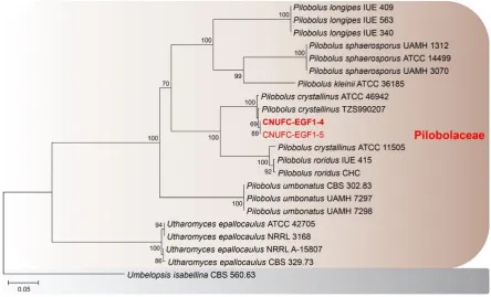
Figure 1. Phylogenetic tree based on neighbor-joining analysis of internal transcribed spacer rDNA 2
sequences for Gilbertella persicaria CNUFC-GWD3-9 and G. persicaria CNUFC-GWD3-10. Hyphomucor 3
assamensis was used as an outgroup. Bootstrap support values of ≥50% are indicated at the nodes. The 4
bar indicates the number of substitutions per position. 5
6
Figure 2. Phylogenetic tree based on neighbor-joining analysis of internal transcribed spacer rDNA 7
sequences for Pilobolus crystallinus CNUFC-EGF1-4 and P. crystallinus CNUFC-EGF1-5. Umbelopsis 8
isabellina was used as an outgroup. Bootstrap support values of ≥50% are indicated at the nodes. The 9
1
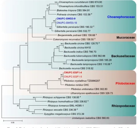
Figure 3. Phylogenetic tree based on neighbor-joining analysis of 28S rDNA sequences for Gilbertella 2
persicaria CNUFC-GWD3-9, G. persicaria CNUFC-GWD3-10, Pilobolus crystallinus CNUFC-EGF1-4, 3
and P. crystallinus CNUFC-EGF1-5. Umbelopsis isabellina was used as an outgroup. Bootstrap support 4
values of ≥50% are indicated at the nodes. The bar indicates the number of substitutions per position. 5
2.2. Morphological Characterization 6
Taxonomic descriptions of the morphological structures for the two species (G. persicaria 7
CNUFC-GWD3-9 and P. crystallinus CNUFC-EGF1-4) are shown in details below. 8
2.2.1. CNUFC-GWD3-9 Gilbertella persicaria 9
Colonies grew rapidly at 25°C on SMA, filling the Petri dish after 2 days of incubation. The 10
colony color was initially white and later grayish yellow. The colony reverse side was white and later 11
pale yellow. Sporangiophores were 10.5‒50.0 μm wide, variable in length, hyaline, light brown to 12
grayish, sometimes branched, and uncommonly had a septum under the sporangia. The sporangia 13
separated longitudinally into two halves, were globose to subglobose, many-spored, initially white-14
yellowish and then turning brown or black at maturity, and measured 36.5‒250.5 × 37.2‒253.5 μm. 15
Columellae were variable in shape, ovoid to pyriform, subglobose, and measured 20.5‒110.7 × 25.2‒ 16
139.0 μm. Sporangiospores were irregular in shape, mainly ellipsoidal, and measured 5.9‒15.5 × 4.5‒ 17
8.9 μm. Chlamydospore formations were well defined on the medium. Zygospores were not observed. 18
Subsidiarily, colonies grew slowly on SMA, PDA, and MEA at 5°C. Among these, the best mycelial 19
1
Figure 4. Morphology of Gilbertella persicaria CNUFC-GWD3-9. A, D: Colony on synthetic mucor agar; 2
B, E: Colony on potato dextrose agar; C, F: Colony on malt extract agar (A–C, top view; D–F, reverse 3
view); G‒I, N, O: Immature and mature sporangia and sporangiophores; J, K: Wall suturing in two 4
equal halves; L, M: Columellae with collarette; P: Sporangiospores with appendages (red arrow) (scale 5
bars H, I = 200 μm; J‒M = 50 μm; N, O = 20 μm; P = 10 μm). 6
7
2.2.2. Distinguishing Characters 1
The CNUFC-GWD3-9 isolate was similar to the description of G. persicaria as detailed by 2
Hesseltine [8], in terms of the shape, size of the sporangiospores (5.9‒15.5 × 4.5‒8.9 μm). However, 3
some morphological features differed. The size of columellae described by Hesseltine [8] was larger 4
(40–119 × 20–170 μm) than that (20.5–110.7 × 25.2–139.0 μm) observed in our isolate. Our G. persicaria 5
isolate presented sporangiophores that were sometimes branched, which was not described by 6
Hesseltine [8]. Moreover, our G. persicaria isolate had a septum under the sporangia. In conclusion, 7
comparing the morphology of the isolate with previous descriptions [8], our present isolate was 8
similar to G. persicaria, with some exceptions (Table 1). 9
Table 1. Morphological characteristics of CNUFC-GWD3-9 and the reference species Gilbertella 10
persicaria grown on synthetic mucor agar medium at 25‒26°C. 11
Characteristic CNUFC-GWD3-9 Gilbertella persicaria a
Colony color Rapid-growing, first white and then grayish yellow
Rapid-growing, first white and then grayish olive
Sporangiophores 10.5‒50.0 μm in width, variable in length
Up to 40‒50 μm in width, up to 3‒4 mm in height
Sporangia
Many-spored, globose to
subglobose, first white-yellowish and then brown or black when mature, 36.5‒250.5 × 37.2‒253.5 μm
Many-spored, globose to irregularly globose, first white and then yellow and then black and glistening when mature, 40‒260 μm in diameter
Columellae
Variable in shape, ovoid to pyriform, subglobose, 20.5‒110.7 × 25.2‒139.0 μm
Variable in shape depending on size, 40‒119 × 20‒170 μm
Sporangiospores Irregular in shape, mainly ellipsoidal, 5.9‒15.5 × 4.5‒8.9 μm
Short oval and rather irregular in shape, 5‒13 × 4.5‒11 μm, up to 8.6 × 17 μm
Chlamydospores Present Present
Zygospores Not observed Present
aFrom the description by Hesseltine [8]. 12
2.2.3. CNUFC-EGF1-4 Pilobolus crystallinus 13
Trophocysts were subglobose to ellipsoidal, and measured 199.0‒409.8 × 147.5‒186.9 µ m. 14
Sporangiophores were 57.0‒100.5 µ m wide, variable in length, erect, nonseptate, and unbranched. 15
Subsporangial vesicles were ovoid, with an orange ring at the base, and measured 298.0‒551.5 × 16
175.0‒416.1 µ m. Sporangia were hemispherical, umbonate, yellow or brown when young and turning 17
black at maturity, and measured 169.5‒288.5 × 151.5‒256.5 µ m. Columellae were ellipsoidal to conical, 18
and measured 110.3‒186.7 × 122.1‒230.5 µ m. Sporangiospores were elliptical, hyaline, yellowish, and 19
Int. J. Mol. Sci. 2018, 19, x FOR PEER REVIEW 7 of 13
1
Figure 5. Morphology of Pilobolus crystallinus CNUFC-EGF1-4. A: Young sporangia and 2
sporangiophores on dung agar medium; B‒K: Yellow and black sporangia, subsporangial 3
vesicles, and sporangiophores (B–G, on water deer dung); L‒N: Substrate mycelia with 4
trophocysts and rhizoidal extensions; O: Sporangiospores (scale bars E‒N = 200 μm; O = 10 5
μm). 6
2.2.4. Distinguishing Characters 7
The sporangiospores of isolate CNUFC-EGF1-4 were morphologically similar to the description 8
for those of P. crystallinus by Boedijn [32], some differences in other morphological characteristics 9
were found (Table 2). 10
Int. J. Mol. Sci. 2018, 19, x FOR PEER REVIEW 8 of 13
Table 2. Morphological characteristics of CNUFC-EGF1-4 and the reference species Pilobolus 1
crystallinus. 2
Characteristic CNUFC-EGF1-4 Piloboluscrystallinusa
Trophocysts Subglobose to ellipsoidal,
199.0‒409.8 × 147.5‒186.9 µ m Oblong, 500‒575 × 200‒230 µ m
Sporangiophores Variable in length, 57.0‒100.5 μm wide
5‒15 mm long, 115‒160 µ m wide
Subsporangial vesicles
Ovoid, orange ring at the base, 298.0‒551.5 × 175.0‒416.1 µ m
Oviform, colorless except for an orange ring at the base,
400‒920 × 350‒720 µ m
Sporangia
Hemispherical, umbonate, first yellow or brown and then black when mature, 169.5‒288.5 μm × 151.5‒242.5 μm
Semiglobose, black, 237‒529 µ m wide near the base,
138‒345 µ m high
Columellae Ellipsoidal to conical,
110.3‒186.7 μm × 122.1‒230.5 μm
Broadly conical, 92‒287 µ m high, 172‒345 µ m wide below
Sporangiospores Elliptical, hyaline, yellowish, 6.0‒8.5 × 4.0‒5.5 μm
Elliptical, hyaline, dark yellow in mass, 7‒10 × 4‒6 µ m
Zygospores Not observed Unknown
aFrom the description by Boedijn [32]. 3
3. Discussion 4
The finding of such species belonging to Piloboloceae has not yet been described in Korea. Thus, 5
it is of great scientific significance not only in fungal diversity belonging to undiscovered taxa but 6
also in ecology. 7
Order Mucorales has been distinguished on the basis of morphological characteristics [4, 5]. 8
Similarly, Gilbertella and Pilobolus have been identified on the basis of morphology in the past. 9
However, many of the morphological features used to classify species of Pilobolus are problematic [28, 10
29]. 11
According to Foos et al. [28], although the size and shape of the sporangiospores have been 12
detected to be stable within species [30], species descriptions typically give a large range of 13
sporangiospore sizes. Moreover, the sizes of many structures used for species identification vary 14
greatly depending on changes in the environmental conditions [28, 31]. In 2011, Foos et al. [28] 15
conducted sequence analysis of the ITS region of rRNA, small subunit of 18S rRNA, and LSU (23S) 16
of mitochondrial rRNA, and showed that the genus Pilobolus is polyphyletic. The results revealed 17
that molecular phylogenetic identification of Pilobolus species based on sequence analysis of pure 18
culture isolates was more reliable than the traditional method of identification [28, 29]. 19
In the phylogenetic trees for species within the Choanephoraceae based on ITS and LSU 20
sequences, our strains CNUFC-GWD3-9 and CNUFC-GWD3-10 were clustered with other G. 21
persicaria species in a well-supported clade with high bootstrap values (100% and 99%). In the trees 22
for species within the Piloboloceae, the two investigated strains 4 and CNUFC-EGF1-23
5 were clustered with other P. crystallinus species in a well-supported clade. 24
Despite the wide intraspecific variation found among some taxa, the ITS and D1/D2 regions have 25
been used as appropriate barcode markers for identifying mucoralean fungi at the species level [4, 5]. 26
Int. J. Mol. Sci. 2018, 19, x FOR PEER REVIEW 9 of 13
required to reconcile the molecular and morphological conceptions of families and genera. In the 1
present study, we also used the molecular strategy for fungal identification at the level of species, 2
specifically utilizing of ITS rDNA gene sequence and phylogenetic analysis. 3
4
4. Materials and Methods 5
4.1. Sampling and Isolation of Fungal Strain 6
Water deer dung samples were collected on Eulsukdo Island (35°6'17.92" N, 128°56'24.52" E; 7
located in Busan, Korea) in June 2017. The samples were transferred to sterile 50-mL conical tubes 8
(SPL Life Sciences Co., Pocheon, Korea), and stored at 4°C until examination. The fecal samples were 9
placed onto sterile moist Whatman’s filter paper in a Petri dish using sterile forceps, and incubated 10
in a moist chamber at 25°C for 6–9 days. 11
Freshwater samples were collected from the Geum River (36°27'47.32" N, 127°6'3.24" E; located 12
in Gongju, Korea) in August 2017. These samples were transported in sterile 50-mL conical tubes, 13
and stored at 4°C until examination. Fungi were isolated by the direct plating method. In brief, plant 14
debris in the freshwater samples was placed onto synthetic mucor agar (SMA; 40 g of dextrose, 2 g of 15
asparagine, 0.5 g of KH2PO4, 0.25 g of MgSO4·7H2O, 0.5 g of thiamine chloride, and 15 g of agar in 1 L
16
of deionized water) using sterile forceps and incubated at 25°C for 1–3 days. To isolate pure cultures, 17
individual colonies of varied morphologies were picked up, transferred to potato dextrose agar (39 g 18
of PDA in 1 L of deionized water; Becton, Dickinson and Co., Sparks, MD, USA) plates, and 19
subcultured until pure mycelia were obtained. All pure isolates, including those of G. persicaria and 20
P. crystallinus,were stored in 20% glycerol at −80°C at the Environmental Microbiology Laboratory 21
Fungarium (Chonnam National University, Gwangju, Korea), as GWD3-9 and CNUFC-22
EGF1-4, respectively. Strain CNUFC-EGF1-4 was also deposited at the Culture Collection of the 23
National Institute of Biological Resources (NIBR, Incheon, Korea), whereas strain CNUFC-GWD3-9 24
was also deposited at the Culture Collection of the Nakdonggang National Institute of Biological 25
Resources (NNIBR, Sangju, Korea). 26
4.2. DNA Extraction, PCR, and Sequencing 27
Genomic DNA was extracted directly from mycelia and spores of the fungal isolates, using the 28
Solg Genomic DNA Prep Kit for fungi (SolGent Co. Ltd., Daejeon, Korea). The internal transcribed 29
spacer (ITS) region and large subunit (LSU) of 28S rDNA were amplified with the primer pairs ITS1 30
and ITS4 [22], and LROR and LR5F [23, 24], respectively (Table 3). The PCR amplification mixture 31
(total volume, 20 μL) contained fungal DNA template, 5 pmol/μL of each primer, and Accupower 32
PCR Premix (Taq DNA polymerase, dNTPs, buffer, and a tracking dye; Bioneer Corp., Daejeon, 33
Korea). The PCR products were purified using the Accuprep PCR Purification Kit (Bioneer Corp.) 34
according to the manufacturer’s instructions. DNA sequencing was performed on an ABI 3700 35
Automated DNA sequencer (Applied Biosystems Inc., Foster City, CA, USA). 36
Table 3. Primers used in this study, with sequences and sources. 37
Gene Product Name Primer Direction Sequence (5’-3’) Reference
ITS
Internal transcribed
spacer
ITS1 Forward TCCGTAGGTGAACCTGCG G
[22]
ITS4 Reverse TCCTCCGCTTATTGATAT GC
LSU Large subunit (28S)
LROR Forward ACCCGCTGAACTTAAGC
[23, 24]
LR5F Reverse GCTATCCTGAGGGAAAC
Int. J. Mol. Sci. 2018, 19, x FOR PEER REVIEW 10 of 13
4.3. Phylogenetic Analysis 1
Fungal sequences (Table 1) were used for phylogenetic analysis through alignment with Clustal_X 2
v.2.0 [25] and editing using Bioedit v.7.2.5 software [26]. Phylogenetic trees based on the ITS rDNA 3
and D1/D2 sequences were constructed using the neighbor-joining method in MEGA 6 [27]. The 4
reliability of the internal branches was assessed using the p-distance substitution model, with 1,000 5
bootstrap replications. The GWD3-9, GWD3-10, EGF1-4, and CNUFC-6
EGF1-5 sequences were deposited in the NCBI database under the accession numbers shown in Table 7
4. 8
Table 4. Taxa, collection numbers, sequences, and GenBank accession numbers used in this study. 9
Taxon name Collection No.
(Isolate No.)
GenBank accession No.
ITS LSU
Backusella circina CBS 128.70 (T) - JN206529
B. circina KH10 - JX644493
B. indica CBS 786.70 - JN206526
B. lamprospora CBS 195.28 - JN206530
B. lamprospora CBS 118.08 (T) - JN206531
B. recurva CBS 318.52 - JN206522
B. tuberculispora CBS 562.66 - JN206525
Benjaminiella poitrasii CBS 158.68 (T) - JN206411
Blakeslea trispora CBS 130.59 JN206227 -
Bl. trispora EML-PUKI88 KY047144 -
Bl. trispora CBS 564.91 JN206230 JN206515
Choanephora cucurbitarum CBS 120.25 JN206231 -
C. cucurbitarum CBS 674.93 JN206233 JN206514
C. infundibulifera CBS 153.51 JN206236 JN206513
C. infundibulifera CBS 155.51 JN206237 -
Cokeromyces recurvatus CBS 158.50 (T) - HM849699
Gilbertella persicaria CBS 785.97 JN206218 -
G. persicaria CBS 190.32 (T) HM999958 HM849691
G. persicaria CBS 532.77 JN206224 JN206517
G. persicaria CNUFC-GWD3-9 MG906872 MG906876
G. persicaria CNUFC-GWD3-10 MG906873 MG906877
Hyphomucorassamensis CBS415.77 JN206211
-Pilobolus crystallinus ATCC 11505 FJ160947 -
P. crystallinus ATCC 46942 FJ160958 -
P. crystallinus TZS990207 JN942689 JN982939
P. crystallinus CNUFC-EGF1-4 MG906874 MG906878
P. crystallinus CNUFC-EGF1-5 MG906875 MG906879
P. kleinii ATCC 36185 FJ160957 -
P. longipes IUE 340 FJ160950 -
P. longipes IUE 409 FJ160951 -
P. longipes IUE 563 FJ160952 -
P. roridus IUE 415 FJ160948 -
P. roridus CHC JN942692 JN982944
P. sphaerosporus ATCC 22499 DQ059382 -
P. sphaerosporus UAMH 1312 FJ160953 -
P. sphaerosporus ATCC 14499 FJ160954 -
P. umbonatus NRRL 6349 FJ160955 -
P. umbonatus UAMH 7297 FJ160956 -
Int. J. Mol. Sci. 2018, 19, x FOR PEER REVIEW 11 of 13
Poitrasia circinans CBS 153.58 (T) JN206239 JN206516
Pt. circinans CBS 647.70 JN206240 -
Rhizopus homothallicus CBS 336.62 (T) - HM849670
R. koreanus EML-HO95-1 - KU058196
R. schipperae CBS 138.95 (T) - HM849672
R. sexualis CBS 336.39 (T) - HM849673
Syzygites megalocarpus CBS 372.39 - JN206401
Umbelopsis isabellina CBS 560.63 JN206400 JN206573
Utharomyces epallocaulus ATCC 42705 FJ160943 -
U. epallocaulus NRRL A-15807 FJ160945 -
U. epallocaulus NRRL A-21843 FJ160946 -
U. epallocaulus CBS 329.73 JN206276 HM849660
Bold letters indicate the isolates and accession numbers determined in our study. ITS, internal 1
transcribed spacer; ATCC, American Type Culture Collection (Manassas, VA, USA); CBS, 2
Centraalbureau voor Schimmelcultures (Utrecht, The Netherlands); CNUFC, Chonnam National 3
University Fungal Collection (Gwangju, South Korea); EML, Environmental Microbiology Laboratory 4
(Fungarium, Chonnam National University, Gwangju, South Korea); NRRL (Agricultural Research 5
Service Culture Collection, Peoria, IL, USA); T, ex-type strain. 6
4.4. Morphological Studies 7
For detailed morphological studies, strain CNUFC-GWD3-9 was cultured on SMA, PDA, and 8
malt extract agar (33.6 g of MEA in 1 L of deionized water; Becton, Dickinson and Co.). The plates 9
were incubated at 5°C, 15°C, 25°C, 35°C, and 40°C in the dark for 2–3 days. Samples were mounted 10
in distilled water and observed using an Olympus BX51 microscope with differential interference 11
contrast (DIC) optics (Olympus, Tokyo, Japan). CNUFC-EGF1-4 strain was cultured on dung agar 12
medium (2 g of water deer dung and 2 g of agar in 100 mL of deionized water) and the plates were 13
incubated at 20°C, 25°C, and 35°C in the dark for 7–14 days. Additionally, fungal spores of strain 14
CNUFC-EGF1-4 were inoculated on surface-sterilized pieces of water deer dung by touching with a 15
sterile needle, and the plates were then incubated at 25°C in the dark for 7–14 days. Samples were 16
observed under an Olympus BX51 microscope with DIC optics. 17
5. Conclusions 18
In present work, by using morphological and phylogenetic analyses (ITS and LSU rDNA), the 19
two mucoralean strains CNUFC-GWD3-9 and CNUFC-EGF1-4 were identified as G. persicaria and P. 20
crystallinus, respectively. There is no species of G. persicaria and P. crystallinus described in Korea, 21
therefore, they can be considered as new record. 22
Our findings contribute to the current knowledge of diversity of the order Mucorales in Korea. 23
However, data regarding the diversity of the order Mucorales in Korea are still lacking, further 24
studies on the classification of different orders and families within the Mucoromycotina are required 25
to expand our knowledge of undiscovered taxa with specific habitats in Korea. 26
Acknowledgments: This work was supported by the Graduate Program for the Undiscovered Taxa of Korea,
27
and by the Project on Survey and Discovery of Indigenous Fungal Species of Korea funded by the NIBR, and in 28
part by the Project on Discovery of Fungi from Freshwater and Collection of Fungarium funded by the NNIBR 29
of the Ministry of Environment (MOE). We are grateful to Dr. Paul M. Kirk for his kindness in reviewing the 30
paper. 31
Author Contributions: Hyang Burm Lee, Seo Hee Lee and Thuong T. T. Nguyen conceived the study. Seo Hee
32
Lee performed the experiments and wrote the paper. Thuong T. T. Nguyen reviewed and edited the paper. 33
Hyang Burm Lee designed and edited the paper, and supervised the study. All authors read and approved the 34
final manuscript. 35
Conflicts of Interest: The authors declare no conflict of interest. 36
Int. J. Mol. Sci. 2018, 19, x FOR PEER REVIEW 12 of 13
References 1
1. Hibbett, D.S.; Binder, M.; Bischoff, J.F.; Blackwell, M.; Cannon, P.F.; Eriksson, O.E.; Huhndorf, S.; James, T.; 2
Kirk, P.M.; Lucking, R.; Thorsten Lumbsch, H.; Lutzoni, F.; Matheny, P.B.; McLaughlin, D.J.; Powell, M.J.; 3
Redhead, S.; Schoch, C.L.; Spatafora, J.W.; Stalpers, J.A.; Vilgalys, R.; Aime, M.C.; Aptroot, A.; Bauer, R.; 4
Begerow, D.; Benny, G.L.; Castlebury, L.A.; Crous, P.W.; Dai, Y.C.; Gams, W.; Geiser, D.M.; Griffith, G.W.; 5
Gueidan, C.; Hawksworth, D.L.; Hestmark, G.; Hosaka, K.; Humber, R.A.; Hyde, K.D.; Ironside, J.E.; 6
Koljalg, U.; Kurtzman, C.P.; Larsson, K.H.; Lichtwardt, R.; Longcore, J.; Miadlikowska, J.; Miller, A.; 7
Moncalvo, J.M.; Mozley-Standridge, S.; Oberwinkler, F.; Parmasto, E.; Reeb, V.; Rogers, J.D.; Roux, C.; 8
Ryvarden, L.; Sampaio, J.P.; Schussler, A.; Sugiyama, J.; Thorn, R.G.; Tibell, L.; Untereiner, W.A.; Walker, 9
C.; Wang, Z.; Weir, A.; Weiss, M.; White, M.M.; Winka, K.; Yao, Y.J.; Zhang, N. A higher-level phylogenetic 10
classification of the fungi. Mycol. Res. 2007, 111, 509–547. http://dx.doi.org/10.1016/j.mycres.2007.03.004 11
2. Spatafora, J.W.; Chang, Y.; Benny, G.L.; Lazarus, K.; Smith, M.E.; Berbee, M.L.; Bonito, G.; Corradi, N.; 12
Grigoriev, I.; Gryganskyi, A.; James, T.Y.; O'Donnell, K.; Roberson, R.W.; Taylor, T.N.; Uehling, J.; Vilgalys, 13
R.; White, M.M.; Stajich, J.E. A phylum-level phylogenetic classification of zygomycete fungi based on 14
genome-scale data. Mycologia 2016, 108, 1028–1046. http://dx.doi.org/10.3852/16-042 15
3. Benny, G.L.; Humber, R.A.; Voigt, K. The zygomycetous fungi: the phylum Entomophthoromycota and 16
subphyla Kickxellomycotina, Mortierellomycotina, Mucoromycotina, and Zoopagomycotina. In 17
Systematics of fungi, 2nd ed.; McLaughlin, D.J., Blackwell, M., Spatafora, J.W., Eds.; Springer Verlag: NY, US, 18
2014; The Mycota. Vol. VII, part A, pp. 209–250, 978-3-642-55317-2. 19
4. Walther, G.; Pawlowska, J.; Alastruey-Izquierdo, A.; Wrzosek, M.; Rodriguez-Tudela, J.L.; Dolatabadi, S.; 20
Chakrabarti, A.; de Hoog, G.S. DNA barcoding in Mucorales: an inventory of biodiversity. Persoonia 2013, 21
30, 11–47. http://dx.doi.org/10.3767/003158513X665070 22
5. Hoffmann, K.; Pawlowska, J.; Walther, G.; Wrzosek, M.; de Hoog, G.S.; Benny, G.L.; Kirk, P.M.; Voigt, K. 23
The family structure of the Mucorales: a synoptic revision based on comprehensive multigene-genealogies. 24
Persoonia 2013, 30, 57–76. http://dx.doi.org/10.3767/003158513X666259 25
6. Nguyen, T.T.; Lee, S.H.; Bae, S.; Jeon, S.J.; Mun, H.Y.; Lee, H.B. Characterization of two new records of 26
zygomycete species belonging to undiscovered taxa in Korea. Mycobiology 2016, 44, 29–37. 27
http://dx.doi.org/10.5941/MYCO.2016.44.1.29 28
7. Eddy, E.D. A storage rot of peaches caused by a new species of Choanephora. Phytopathology 1925, 15, 607– 29
610. 30
8. Hesseltine, C.W. Gilbertella gen. nov. (Mucorales). Bull. Torrey Bot. Club 1960, 87, 21–30. 31
http://dx.doi.org/10.2307/2483058 32
9. Benny, G.L. Gilbertellaceae, a new family of the Mucorales (Zygomycetes). Mycologia1991, 83, 150–157. 33
http://dx.doi.org/10.2307/3759930 34
10. Voigt, K.; Olsson, L. Molecular phylogenetic and scanning electron microscopical analyses places the 35
Choanephoraceae and Gilbertellaceae in a monophyletic group within the Mucorales (Zygomycetes, Fungi). 36
Acta Biol. Hung. 2008, 59, 365–383. http://dx.doi.org/10.1556/ABiol.59.2008.3.10 37
11. Mehrotra, M.D. Fruit rot of tomato caused by Gilbertella persicaria. Sydowia 1964, 17, 17–19. 38
12. Mehrotra, M.D. Fruit rot of pear caused by Gilbertella persicaria var. indica. Sydowia 1964, 17, 124–125. 39
13. Mehrotra, M.D. Fruit rot of peach by Gilbertella persicaria var. indica from India. Mycopathol. Mycol. Appl. 40
1966, 29, 151–154. https://doi.org/10.1007/bf02055072 41
14. Guo, L.W.; Wu, Y.X.; Mao, Z.C.; Ho, H.H.; He, Y.Q. Storage rot of dragon fruit caused by Gilbertellapersicaria. 42
Plant Dis. 2012, 96, 1826. https://doi.org/10.1094/PDIS-07-12-0635-PDN 43
15. Pinho, D.B.; Pereira, O.L.; Soares, D.J. First report of Gilbertellapersicaria as the cause of soft rot of fruit of 44
Syzygiumcumini. Australasian Plant Dis. Notes 2014, 9, 143–146. http://dx.doi.org/10.1007/s13314-014-0143-0 45
16. Page, R.M. Light and the asexual reproduction of Pilobolus. Science 1962, 138, 1238–1245. 46
http://dx.doi.org/10.1126/science.138.3546.1238 47
17. Viriato, A. Pilobolus species found on herbivore dung from the São Paulo Zoological Park, Brazil. Acta Bot. 48
Bras. 2008, 22, 614–620. http://dx.doi.org/10.1590/S0102-33062008000300002 49
18. Zygomycota. Available online: http://www.zygomycetes.org (accessed on 19 March 2018). 50
19. Krug, J.C.; Benny, G.L.; Keller, H.W. Coprophilous fungi. In Biodiversity of fungi, 1st ed.; Mueller G.M., Bills 51
G.F., Foster M.S., Eds.; Elsevier Academic Press: MA, US, 2004; pp. 468–499, 9780125095518. 52
20. Souza, C.A.F.; Lima, D.X.; Gurgel, L.M.S.; Santiago, A.L.C.M. Coprophilous Mucorales (ex Zygomycota) 53
from three areas in the semi-arid of Pernambuco, Brazil. Braz. J. Microbiol. 2017, 48, 79–86. 54
http://dx.doi.org/10.1016/j.bjm.2016.09.008 55
21. Richardson, M.J. Records of coprophilous fungi from the Lesser Antilles and Puerto Rico. Caribb. J. Sci. 2008, 56
44, 206–214. http://dx.doi.org/10.18475/cjos.v44i2.a8 57
Int. J. Mol. Sci. 2018, 19, x FOR PEER REVIEW 13 of 13
for phylogenetics. In PCR protocols: a guide to methods and applications, Innis M.A., Gelfand D.H., Sninsky J.J., 1
White T.J., Eds.; Academic Press, NY, US, 1990; pp. 315–322. 2
23. Vilgalys, R.; Hester, M. Rapid genetic identification and mapping of enzymatically amplified ribosomal 3
DNA from several Crytococcus species. J. Bacteriol. 1990, 172, 4238–4246. 4
http://dx.doi.org/10.1128/jb.172.8.4238-4246.1990 5
24. Lee, H.B. Molecular phylogenetic status of korean strain of Podosphaera xanthii, a causal pathogen of 6
powdery mildew on japanese thistle (Cirsium japonicum) in Korea. J. Microbiol. 2012, 50, 1075–1080. 7
http://dx.doi.org/10.1007/s12275-012-2618-z 8
25. Thompson, J.D.; Gibson, T.J.; Plewniak, F.; Jeanmougin, F.; Higgins, D.G. The CLUSTAL_X windows 9
interface: flexible strategies for multiple sequence alignment aided by quality analysis tools. Nucleic Acids 10
Res. 1997, 25, 4876–4882. https://doi.org/10.1093/nar/25.24.4876 11
26. Hall, T.A. BioEdit: a user-friendly biological sequence alignment editor and analysis program for Windows 12
95/98/NT. Nucleic Acids Symp. Ser. 1999, 41, 95–98. 13
27. Tamura, K.; Stecher, G.; Peterson, D.; Filipski, A.; Kumar, S. MEGA6: Molecular Evolutionary Genetics 14
Analysis version 6.0. Mol. Biol. Evol. 2013, 30, 2725–2729. http://dx.doi.org/10.1093/molbev/mst197 15
28. Foos, K.M.; May, N.L.; Beach, D.L.; Pomper, M.; Sheehan, K.B.; Ruch, D.G. Phylogeny of Pilobolaceae. 16
Mycologia 2011, 103, 36–44. http://dx.doi.org/10.3852/09-314 17
29. Foos, K.M.; Sheehan, K.B. Molecular identification of Pilobolus species from Yellowstone National Park. 18
Mycologia 2011, 103, 1208–1215. http://dx.doi.org/10.3852/11-107 19
30. Foos, K.M.; Jeffries, B.S. Sporangiospore variability in Pilobolus. Proc. Indiana Acad. Sci. 1988, 98, 105–108. 20
31. Hu, F.M.; Zheng, R.Y.; Chen, G.Q. A redelimitation of the species of Pilobolus. Mycosystema 1989, 2, 111–133. 21