Title: Moraxella nonliquefaciens and M. osloensis are Important Moraxella Species that
1
Cause Ocular Infections
2
Running Title: Moraxella Ocular Pathogens
3
4
Authors: Samantha LaCroce, BA, MD1, Mollie N. Wilson, MS, MLS(ASCP)2, John E. 5
Romanowski3, Jeffrey D. Newman, PhD4, Vishal Jhanji3, MD, Robert M.Q. Shanks, PhD3, Regis
6
P. Kowalski, MS, M(ASCP)3
7
8
Affiliation:
9
1-Wake Forest University School of Medicine, Winston-Salem, NC 27157
10
2- University of Pittsburgh Medical Center, Clinical Laboratory – Microbiology, Pittsburgh, PA
11
3- The Charles T. Campbell Ophthalmic Microbiology Laboratory, Department of
12
Ophthalmology, University of Pittsburgh School of Medicine, Pittsburgh, PA, USA.
13
(http://eyemicrobiology.upmc.com)
14
4-Department of Biology, Lycoming College, Williamsport PA 17701, USA.
15
16
Corresponding Author: Regis P. Kowalski, MS, M(ASCP), The Eye and Ear Institute Bldg.,
17
Ophthalmic Microbiology, Room 642, 203 Lothrop Street, Pittsburgh, PA 15213.
18
Phone #: (412) 647-7211, FAX (412) 647-5331, Email: [email protected]
19
20
Keywords: Moraxella, keratitis, conjunctivitis, endophthalmitis, eye infections, DNA
21
sequencing, MALDI-TOF MS, Biolog
22
23
Abstract
24
Purpose. Moraxella is an ocular bacterial pathogen isolated in cases of keratitis, conjunctivitis,
25
and endophthalmitis. Gram-negative brick-shaped diplobacilli from ocular specimens, and slow
26
growth in culture, are early indications of Moraxella ocular infection; however, identifying
27
Moraxella to species can be complex and inconsistent.
28
Methods. In this study, bacteria consistent with Moraxella were identified to species using: 1)
29
DNA sequencing coupled with vancomycin susceptibility, 2) MALDI-TOF Mass Spectrometry,
30
and 3) Biolog ID System. Study samples consisted of 9 ATCC Moraxella controls, 82 isolates
31
from keratitis, 21 isolates from conjunctivitis, and 4 isolates from endophthalmitis.
32
Results. The ATCC controls were correctly identified. For keratitis, 66 (80.5%) were identified
33
as M.nonliquefaciens, 7 (9.0%) as M. lacunata, 5 (6%) as M. osloensis, 2 (2.5%) as
34
Acinetobacter lwoffi, 1 (1.0%) as M.bovis/nonliquefaciens, and 1 (1.0%) as M. 35
osloensis/nonliquefaciens. For conjunctivitis, 9 (43.0%) were identified as M.osloensis, 6
36
(29.0%) as M. nonliquefaciens, 3 (14.3%) as Roseomonas, 2 (9.5%) as Acinetobacter (parvus, 37
junii), and 1 (4.5%) as M. catarrhalis/M. nonliquefaciens. From endophthalmitis, 3 of 4 of the
38
isolates were M.nonliquefaciens. Overall, M. nonliquefaciens and M.osloensis were identified
39
in 70% (75 of 107) and 13% (14 of 107) of cases, respectively, totaling 83% (89 of 107).
40
Conclusions. M. nonliquefaciens and M.osloensis are important bacterial pathogens of the eye
41
as determined by DNA sequencing, MALDI-TOF MS, and Biolog. Although Moraxella 42
catarrhalis is a clinical pathogen, other species of Moraxella appear to have a prominent role in
43
eye infections.
44
45
Introduction
47
As first described by Morax1, the genus, Moraxella, appears to have a specific tropism as
48
an ocular pathogen for keratitis2-5, conjunctivitis6, and endophthalmitis7. Moraxella keratitis
49
appears to be on the rise based on recent reports.2-5,8 Bacterial conjunctivitis is generally
self-50
limiting, but Moraxella conjunctivitis can persist for weeks and presents as a follicular
51
conjunctivitis which can be misdiagnosed as inclusion conjunctivitis due to Chlamydia.6,9-11 52
Moraxella has been reported as an ocular pathogen in Japan2,4,12, the United States5,6,7,9,10, and
53
Pakistan13. Although quite susceptible to ophthalmic topical antibiotics as highlighted on our 54
frequently updated website (http://eyemicrobiology.upmc.com/Antibiotic.htm), Moraxella 55
keratitis can persist and induce severe inflammation resulting in hypopyon formation. Like most
56
bacteria, with entrance to the inner eye, Moraxella can also cause endophthalmitis.7 57
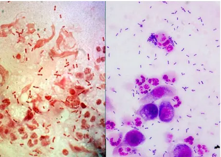
In the clinical laboratory, Moraxella is easily identified to genus by its classic appearance
58
as a negative diplobacilli (brick shaped) which is easily distinguish from other
Gram-59
negative bacteria (Figure 1), and this may be the only indication of Moraxella infections due to
60
antibiotic pretreatment or the fastidious nature of Moraxella. Moraxella can retain crystal violet
61
during Gram staining and it is classified as oxidase-positive, non-glucose fermenting bacteria.
62
Clinically, ocular Moraxella isolates are generally only identified to the genus as Moraxella,
63
because it is difficult and complex to classify Moraxella to species using phenotypic testing.
64
There are multiple species of Moraxella, but the most noted, M. catarrhalis, formerly known as
65
Branhamella catarrhalis, is not the only species implicated in ocular infection.
66
Matrix Assisted Laser Desorption/Ionization Time of Flight Mass Spectrometry
67
(MALDI-TOF MS) and molecular techniques such as DNA sequencing can identify bacteria to
68
genus and species with more consistent results.14,15 A commercial system, Biolog GenIII
(Hayward, CA), utilizing 96 substrates and controls, can also be used to identify bacteria to
70
genus and species.
71
The objective of the present study is to more precisely identify ocular isolates of
72
Moraxella to include species level differentiation. It is important to recognize these bacteria as
73
pathogens.
74
75
76
Methods
77
Laboratory Diagnosis of Ocular Moraxella Infection
78
The ophthalmic microbiology laboratory at the University of Pittsburgh Medical Center,
79
Pittsburgh, PA, is a fully certified clinical laboratory dedicated for the diagnosis and treatment of
80
ocular infection. (http://eyemicrobiology.upmc.com) Samples for keratitis (cornea) are collected
81
using soft-tipped applicators, spatula, surgical blades, and jeweler’s forceps. Conjunctival
82
samples are generally obtained with soft-tipped applicators, and samples to detect
83
endophthalmitis are obtained by tapping the vitreous humor with a needle and syringe. The
84
initial diagnostic test to detect infection from ocular samples are the Gram and Giemsa stain
85
which can provide rapid and definitive identification of an etiologic pathogen.
86
All corneal ulcers that are either central or are over 3 mm in size of infiltration are
87
scraped for microscopic examination and isolation of pathogens. The decision to perform
88
laboratory studies is a judgment call by the attending ophthalmologist.
89
Moraxella can be isolated in culture on trypticase soy agar supplemented with 5% sheep
90
blood (5%SB) and chocolate agar. Our laboratory recommends that ocular collection be placed
91
directly to the media. Growth is better on 5% SB, but growth is generally slow with colonies not
appearing until 48 hours of incubation at 37O C in 6% CO
2. The initial appearance can be 93
pinpoint colonies with larger colonies (2-3 mm) appearing after 2 days. Most Moraxella (not M.
94
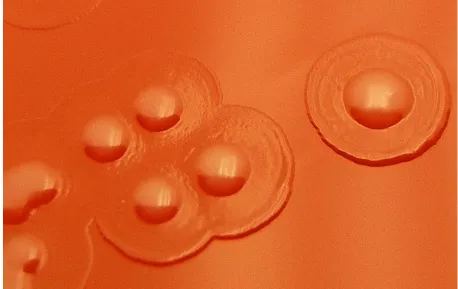
catarrhalis) isolated from ocular specimens will appear as grey to white colonies, often with a
95
clearer beach and umbonate colony morphology, giving a fried egg appearance (Figure 2).
96
97
Antibiotic Susceptibility of Ocular Moraxella
98
In vitro antibiotic susceptibility for ocular isolates are performed using Kirby Bauer Disk
99
Diffusion16 on Mueller Hinton Agar supplemented with 5% sheep blood. Moraxella 100
susceptibility cannot be determined on regular Mueller Hinton agar because of the need for red
101
blood cells to grow. It is important to note that ocular Moraxella infections are not treated
102
systemically (i.e. orally or intravenously). Ocular Moraxella and other bacterial pathogens of
103
keratitis are treated topically with antibiotics or intravitreally by direct injection in cases of
104
endophthalmitis. There are no standards for susceptibility interpretation of topical or intravitreal
105
treatment, but the CLSI (Clinical and Laboratory Standards Institute) standards can be used to
106
guide therapy if the concentration of antibiotics in the ocular tissue is assumed to be equal or
107
greater than in the serum.16 A routine keratitis testing battery of antibiotics for patient topical
108
treatment consists of bacitracin, vancomycin, gentamicin, ciprofloxacin, ofloxacin, polymyxin B,
109
cefazolin, tobramycin, sulfacetamide, moxifloxacin, and cefoxitin. In general, Moraxella isolates
110
test susceptible to these antibiotics
111
(http://eyemicrobiology.upmc.com/AntibioticSusceptibilities/Keratitis.htm). A zone greater of
112
18mm denotes susceptibility to vancomycin. Streptococcus pneumoniae ATCC 49619 was the
113
control strain for assuring the antibiotic disk concentrations.
114
Moraxella Study Isolates 116
Since 1993, all bacterial pathogens isolated at UPMC from keratitis, conjunctivitis, and
117
endophthalmitis have been stored at -80O C with broth containing 15% glycerol for validation of
118
new testing and patient treatment. These isolates constitute an Ocular Clinical Tissue Bank in
119
which the isolates were de-identified to comply with IRB protection of patient identity. Clinical
120
presentation data, patient identity and demographics were not tabulated for any of the bacterial
121
isolates.
122
The collection was reviewed for the retrieval of Moraxella isolates from keratitis,
123
conjunctivitis, and endophthalmitis. No patient contact was involved in this study (University of
124
Pittsburgh, Institutional Review Board, IRB# PRO17050362). None of these isolates (except M. 125
catarrhalis) had been identified to species.
126
Our identifications also included nine American Type Culture Collection (ATCC)
127
controls: M. bovis ATCCT 10900, M.caviae ATCCT 14659, M.cuniculi ATCCT 14688, M. 128
nonliquefaciens ATCCT 19975, M. osloensis ATCCT 19976, M. atlantae ATCCT 29525, M. 129
lincolnii ATCCT 51388, M. lacunata ATCCT 17967, and Moraxella (Branhamella) catarrhalis 130
ATCCT 24250.
131
132
DNA Sequencing
133
Presumptive Moraxella isolates were retrieved from -80O C and streaked onto 5%SB.
134
and incubated at 37 O C in a 6% CO2 incubator for 48 hours. The isolates were Gram stained to 135
presumptively identify as Gram-negative diplobacilli. If identified as Gram negative diplobacilli,
136
the isolates were passed onto new 5% SB and incubated at 37 O C in a 6% CO
2 incubator for 48 137
hours. The DNA from the isolates were extracted using Epicentre DNA Extraction solution
Quick Extract (Madison, WI). PCR was performed with Taq DNA polymerase (New England
139
Biolabs, Ipswich, MA) and the 16S rRNA gene sequence was amplified (95O C for 5 minutes, 33
140
cycles of 95O C for 30 seconds, 50O C for 15 seconds, 72O C for 1 minute, followed by a 10
141
minute extension at 72O C) using Primers 27F and 1492R.18 If a single band was visualized by
142
gel electrophoresis of the PCR products, the amplicons were purified using Qiagen QIAquick
143
PCR Purification kit (Hilden, Germany). The PCR products were each analyzed with a single
144
Sanger sequencing reaction using Primer 330F at the University of Pittsburgh Genomics
145
Research Core.17 DNA sequences were initially compared to the NCBI non-redundant 146
nucleotide database using BLASTN.18-20 After initial screening, sequences were aligned with 147
type strain sequences using ClustalW and a neighbor joining tree21 was constructed using the
148
Kimura 2 parameter model22 and 1000 bootstrap replicates.23 Due to differing lengths of 149
sequence obtained, all positions with less than 50% site coverage were eliminated. Evolutionary
150
analyses were conducted in MEGA7.24
151
152
MALDI-TOF MS
153
As with DNA sequencing, presumptive Moraxella isolates were retrieved from -80O C
154
and streaked onto 5%SB. The isolates were incubated at 37 O C in a CO2 incubator for 48 hours. 155
The isolates were Gram stained to presumptively identify as Gram-negative diplobacilli, passed
156
to another 5% SB for 24 hours, and delivered for MALDI-TOF MS testing (UPMC, Clinical
157
Microbiology).
158
Colony material from each isolate was transferred to a polished steel target (Bruker
159
Daltonik, Bremen, Germany) using a clean toothpick. One microliter of Matrix (Bruker HCCA
160
[α-cyano-4-hydroxycinnamic acid in 50% acetonitrile with 2.5% trifluoroacetic acid]) was
applied within an hour and air dried for 10 minutes. The target was analyzed using the Bruker
162
Microflex LT/SH MALDI-TOF instrument and Bruker Biotyper software version 4.1 with the
163
MBT BDAL library containing 7854 mass spectra. The library contained 22 species of
164
Moraxella. Spectra were obtained after 240 laser shots yielding spectra with mass/charge (m/z)
165
ratios between 2k and 20k Da. Measurements meeting the quality criteria (log score ≥1.8) and a
166
log scopre > 0.2 between different species were deemed acceptable identifications. Samples
167
with scores below this cutoff or with < 0.2 log between different species were retested using the
168
formic acid tube extraction method as previously described.25 A control organism (E. coli ATCC 169
25922 or P. aeruginosa ATCC 27853) was extracted and analyzed once each day to ensure the
170
extraction procedure yielded successful identification.
171
172
Biolog Gen III Plate
173
The Biolog identification system was performed according to manufacturer’s protocol.
174
(Biolog, GEN III MicroPlate ™, Instructions for Use, www.biolog.com). As with MALDI-TOF
175
MS and DNA sequencing, presumptive Moraxella isolates were retrieved from -80O C and 176
streaked onto 5%SB. The streaks were passed to 5%SB and testing was performed on 24 hour
177
growth. Biolog testing was performed on a GEN III MicroPlate™ which contained 94
178
biochemicals consisting of 71 carbon source utilization assays, 23 chemical sensitivity assays, a
179
negative control, and a positive control. These provide a phenotypic fingerprint for species
180
identification by utilizing tetrazolium redox dyes to colorimetrically indicate carbon utilization
181
or resistance to inhibitory chemicals.
182
In brief, a medium for fastidious bacteria (IF-C, Biolog) was inoculated with a Moraxella 183
isolate to a turbidity of 65% transmittance measured by a Turbidimeter (Biolog). The inoculum
was aliquoted to the microplate using a reservoir and multipipetor to 96 wells at a volume of
185
0.1ml per well. The plate was incubated at 34O C and read manually for color changes at 4 hours,
186
8 hours, and 20 hours. The tabulated data at each time point was entered into the Biolog’s
187
Identification Systems Software (OOP 188rG Gen III Database v2.8). The database contained 14
188
species of Moraxella. Species identification was determined as the most probable as indicated by
189
the software. It must be noted that there is an automated system for reading the plates for color
190
changes. Costs dictated the manual approach for this study.
191
192
Identification of Moraxella to Species
193
Isolates of M.catarrhalis were not of prime interest in this study because these isolates
194
can be identified to genus and species using standard laboratory methods. M.catarrhalis are
195
Gram-negative diplococci (not diplobacilli) closely resembling Neisseria but are
oxidase-196
positive, fast growing, form friable movable tan-like colonies, and are resistant to
197
vancomycin.26,27 The API NH (bioMérieux, La Balme-les-Grottes, France) system can accurately
198
confirm identification.28 199
In this study, all presumptive Moraxella isolates were observed to be Gram-negative
200
diplobacilli. There was some variability in the size of the bacilli, whereas most were
brick-201
shaped, boxcar bacilli, but some were thinner bacilli and diplococcobacilli. Some isolates
202
retained crystal violet staining which is a characteristic of Moraxella. As noted previously, the
203
colonies initially appeared as pinpoint colonies with larger colonies (2-3 mm) appearing after 2
204
days. These colonies appeared as grey to white, often with a clearer beach that give a fried egg
205
appearance (Figure 2). Susceptibility to vancomycin indicated a Moraxella species other than M. 206
Results
208
The laboratory records (1993-2017) indicated that there were 9 cases of keratitis, 18 cases
209
of conjunctivitis, and 0 cases of endophthalmitis caused by M.catarrhalis. These Moraxella 210
isolates and ATCC controls of M. caviae, M. cuniculi, M. catarrhalis (all once part of
211
Branhamella), and most M osloensis were vancomycin resistant.
212
All control ATCC isolates were identified correctly by DNA sequencing coupled with
213
vancomycin susceptibility, MALDI-TOF MS, and Biolog Gen III plates. The vancomycin zone
214
of inhibition for Matlantae was 16mm, and zones of inhibition were not clear for M. lincolnii 215
and M. lacunata.
216
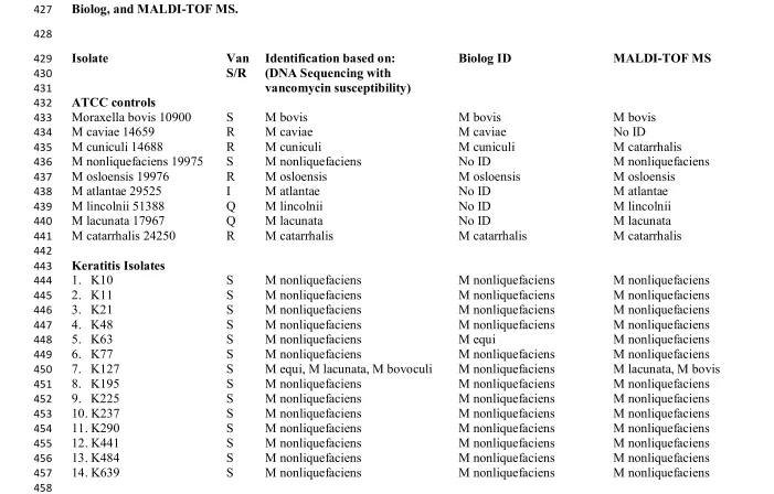
Table 1 summarizes the identification of Moraxella from keratitis, conjunctivitis, and
217
endophthalmitis using DNA sequencing with vancomycin susceptibility, MALDI-TOF MS, and
218
Biolog Gen III plates. Identification was reported for 82 cases of keratitis, 21 cases for
219
conjunctivitis, and 4 cases of endophthalmitis. The identification of DNA sequencing was more
220
closely associated with MALDI-TOF MS (106 of 116) than with the identification of DNA
221
sequencing with Biolog (78 of 116) (p=0.001, Fisher’s Exact) and MALDI-TOF MS with Biolog
222
(87 of 116) (p=0.005, Fisher’s Exact).
223
Table 2 details the incidence of Moraxella species for keratitis, conjunctivitis, and
224
endophthalmitis. Identification of Moraxella to species was based on DNA sequencing coupled
225
with vancomycin susceptibility and MALDI-TOF MS.
226
Many isolates could not be identified by 16S rRNA sequencing alone. In the segment of
227
the 16S rRNA gene sequenced in this study, the M. catarrhalis and M. nonliquefaciens type
228
strains are identical (Figure 3), but can be distinguished based on vancomycin resistance in M. 229
catarrhalis and susceptibility in M. nonliquefaciens. The sequences from strains K127, K1630,
K1664, K2450, K2695, and K2757 cluster together, but are essentially equidistant from M. 231
bovis, M. bovoculi, M. caprae, M. equi,, and M. lacunata, suggesting that they may be members
232
of a new species. Sequence analysis could differentiate M. atlantae, M. caviae, M. lincolnii, and
233
M. osloensis. DNA sequencing was complemented with vancomycin susceptibility to identify
234
most Moraxella species. Vancomycin resistant isolates were associated with M. catarrhalis and 235
M. osloensis.
236
For keratitis, 66 (80.5%) were identified as M.nonliquefaciens, 7(9.0) as M.lacunata, 5
237
(6.0%) as M. osloensis, 2 (2.5%) as Acinetobacter lwoffi, 1 (1.0%) as M. bovis/ nonliquefaciens,
238
and 1 (1.0%) as M.osloensis/ nonliquefaciens. All of the M. nonliquefaciens were susceptible to
239
vancomycin, while 3 of the M. osloensis were susceptible to vancomycin and 2 were resistant.
240
Although M. osloensis can be resistant to vancomycin, our study indicates that this may not be a
241
consistent characteristic. On the closer laboratory examination of the two Acinetobacter isolates,
242
both were initially classified as Moraxella based on the observation of diplococcobacilli on
243
Gram stain.
244
For conjunctivitis, 9 (43.0%) were identified as M. osloensis, 6 (29%) as M. 245
nonliquefaciens, 3 (14.0%) as Roseomonas mucosa, 2 (9.5%) as Acinetobacter, and 1 (4.5%) as
246
M. catarrhalis/ nonliquefaciens. Seven of the M.osloensis isolates were resistant to vancomycin
247
and two were susceptible, while vancomycin resistance was noted for one M. nonliquefaciens 248
isolates with 5 susceptible. The three Roseomonas mucosa isolates were re-examined and all
249
three were Gram-negative diplobacilli consistent with Moraxella, but all three presented as
250
pinkish highly mucoid colonies slightly different from colonies observed with Moraxella. Once
251
again, on closer examination, both Acinetobacter isolates were diplococcobacilli on Gram stain.
From endophthalmitis, 3 of 4 (75%) of the isolates were M. nonliquefaciens with all three
253
isolates susceptible to vancomycin. The lone Neisseria shayeganii isolate (identified by DNA
254
sequencing only) from endophthalmitis was observed to be Gram-negative diplococcoid,
255
oxidase-positive, and vancomycin resistant.
256
Overall, M. nonliquefaciens and M. osloensis were identified in 70% (75 of 107) and
257
13% (14 of 107) of cases, respectively, totaling 83% (89 of 107).
258
259
260
Discussion
261
DNA sequencing coupled with vancomycin susceptibility, MALDI-TOF MS, and Biolog
262
GenIII plates have given us more diagnostic options to identify Moraxella to species when
263
combined with established laboratory tests such as Gram stain, culture isolation, and
264
susceptibility testing. Gram stain and culture provided us with classical laboratory
265
characteristics. Vancomycin provided us an additional test for separation and identification of
266
Moraxella to species. M. liquefaciens was generally found to be susceptible to vancomycin and
267
M. osloensis was found to be more resistant.
268
Biolog using GenIII plates was more problematic in confidently identifying Moraxella to
269
species based on manual interpretation. We were required to repeat testing for many isolates,
270
because the controls were not positive or negative as expected and identification was not
271
conclusive. It may be that the automated Biolog system without human interpretation would be
272
more definitive for identification. We had more confidence with DNA sequencing coupled with
273
vancomycin susceptibility and MALDI-TOF MS for identifying the Moraxella isolates to
274
species.
It may be unusual to some that we are testing the susceptibility of Gram-negative bacteria
276
to vancomycin, but vancomycin is part of a broad-spectrum battery of antibiotics used in our
277
laboratory to ‘guide’ the topical treatment of bacterial keratitis. In general, the topical empiric
278
treatment of Moraxella keratitis is fortified tobramycin (14 mg/ml) and cefazolin (25 mg/ml).
279
Some ophthalmologists may use topical vancomycin (25 mg/ml) instead of cefazolin or use
280
topical monotherapy with a commercially available fluoroquinolone (moxifloxacin,
281
ciprofloxacin). From a previous study, we suggested topical tobramycin (3mg/ml) to be used as
282
treatment for conjunctivitis, but other commercial topical antibiotics can be used (i.e.
283
fluoroquinolones).6 Endophthalmitis is treated by direct intravitreal injection with vancomycin (1 284
mg) and amikacin (0.4 mg) or ceftazidime (2 mg).
285
Our study determined that M nonliquefaciens is the predominant species isolated from
286
keratitis and M osloensis was a frequent species implicated in conjunctivitis. A small sample (3
287
of 4) demonstrated that M nonliqufaciens was implicated in endophthalmitis, and this has been
288
reported previously.7 Our study also indicated Roseomonas mucosa has characteristics similar to
289
Moraxella and there needs to be close examination of cultures and diagnostic testing to
290
definitively distinguish these two genera. We have designated Roseomonas mucosa as a
291
conjunctivitis pathogen, and it has been previously reported to be a causative agent in keratitis29
292
and endophthalmitis.30,31 293
The literature has sparse reports of ocular Moraxella infections, but there is no definitive
294
consistent method to precisely identify Moraxella ocular isolates to genus and species. A recent
295
report of 17 cases of keratitis accurately identified 4 cases of M. catarrhalis, 1 case of M. 296
osloensis, and 12 cases of Moraxella species with the VITEK 2 system using Gram-negative
297
cards (SYSMEX bioMѐrieux, Tokyo, Japan) with ID-test HN20 Rapid Kit (Nissui
Pharmaceutical, Tokyo, Japan).2 Public Health England has provided an algorithm for 299
identification of Moraxella, not specifically for ocular isolates, that included a battery of
300
laboratory tests and the possible introduction of mass spectrometry and nucleic acid
301
amplification testing.32 Automated systems and test kits can be accurate methods to identify
302
ocular isolates of Moraxella to genus for expedient patient care. Advances in technology now
303
allow for a more precise and consistent methods to correlate specific ocular clinical presentations
304
to species of Moraxella.
305
In conclusion, our study has identified M. liquefaciens, M. osloensis, and other Moraxella 306
species as ocular pathogens. DNA sequencing coupled with vancomycin susceptibility and
307
MALDI-TOF MS are reliable methods for the identification of Moraxella to species, but added
308
investigation with automation may be required to validate Biolog.
309
310
311
Acknowledgments
312
This study was supported by the Department of Pathology at the University of Pittsburgh
313
Medical Center (UPMC), Pittsburgh, PA. The Charles T. Campbell Ophthalmic Microbiology
314
Laboratory, Pittsburgh, PA, has received indirect support: NIH Core grant P30-EY08098 to the
315
Department of Ophthalmology; NIH grant EY027331 (RMQS); the Eye and Ear Foundation of
316
Pittsburgh, Pittsburgh, PA; unrestricted funds from Research to Prevent Blindness Inc., New
317
York, NY; and PA Lions Sight Conservation & Eye Research Foundation.
318
319
References
321
1. Morax V. Note sur un diplobacille pathogéne pour la conjunctivite humaine. Ann Inst 322
Pasteur 1896;10:337-345.
323
2. Tobimatsu Y, Inada N, Shoji J, Yamagami S. Clinical characteristics of 17 patients with
324
Moraxella keratitis. Sem Ophthalmol 2018;pp 1-7.
325
3. Zafar H, Tan SZ, Walkden A, Fullwood C, Au L, Brahma A, Carley F. Clinical
326
characteristics and outcomes of Moraxella keratitis. Cornea 2018; doi: 10.1097/ICO
327
0000000000001749 PMID: 30222715
328
4. Inoue H, Suzuki T, Inoue T, Hattori T, Nejima R, Todokoro D, Hoshi S, Eguchi H,
329
Miyamoto H, Ohashi Y. Clinical characteristics and bacteriological profile of Moraxella
330
keratitis. Cornea 2015;34:1105-1109.
331
5. Durrani A, Faith SC, Kowalski RP, Yu M, Romanowski EG, Shanks RM, Dhaliwal DK,
332
Jhanji V. Moraxella keratitis: Analysis of risk factors, clinical characteristics,
333
management and treatment outcomes. Am J Ophthalmol 2018; Sept 7,
piiS0002-334
9394(18)30517-8. doi:10.1016/j.ajo.2018.08.055.
335
6. Kowalski RP, Harwick JC. Incidence of Moraxella conjunctival infection. Am J 336
Ophthalmology 1986;101:437-440.
337
7. Ebright JR, Lentino JR, Juni E. Endophthalmitis caused by Moraxellanonliquefaciens.
338
Am J Clin Pathol 1982;77:362-363.
339
8. Tan SZ, Walkden A, Au L, et al. Twelve-year analysis of microbial keratitis at a UK
340
tertiary hospital Eye (Lond) 2017;31:1229-1236. doi:10.1038/eye.2017.55 PMID:
341
28452995
9. Thygeson P, Kimura S. Chronic conjunctivitis. Trans Am Acad Ophthalomol 343
Otolaryngol 1963;63:494-517.
344
10.Dawson C. Follicular conjunctivitis. Conjunctivitis. In Duane TD (ed). Clinical 345
Ophthalmology. External Disease. Philadelphia, Harper and Row,1983:1-19.
346
11.Balliart P, Tillé H. Lésions cutanées tenaces des paupiéres et de la face, et lesions des
347
conjonctives dues au diplobacille de Morax. Bull Soc Ophtalmol 1935;157-159.
348
12.Mitsui Y, Hinokuma S, Tanaka C. Etiology of angular conjunctivitis. Am J Ophthalmol 349
1951;34:1579-1586.
350
13.Van Bijsterveld OP. Acute conjunctivitis and Moraxella. Am J Ophthalmol 351
1967;63:1702-1705.
352
14.Murray PR. What is new in clinical microbiology-Microbial identification by
MALDI-353
TOF Mass Spectrometry. J Mol Diagn 2012;14:419-423.
354
http://dx.doi.org/10.1016/j.jmoldx.2012.03.007.
355
15.Enright MC, Carter PE, MacLean IA, McKenzie H. Phylogenetic relationships between
356
some members of the genera Neisseria, Acinetobacter, Moraxella, and Kingella based
357
on partial 16S ribosomal DNA sequence analysis. Int J Syst Bacteriol
1994;44(3):387-358
391.
359
16.Clinical and Laboratory Standards: Performance standards for antimicrobial disk 360
susceptibility tests, ed. 10. Approved standard. Wayne, Pennsylvania, Clinical and
361
Laboratory Standards Institute, 2009, document M02-A10, vol.29, No.1.
362
17.Lane DJ. 16S/23S rRNA Sequencing. In Stackebrandt E and Goodfellow M, Eds.,
363
Nucleic Acid Techniques in Bacterial Systematic, John Wiley and Sons, New York,
364
1991;115-175.
18.Frank JA, Reich CI, Sharma S, Weisbaum JS, Wilson BA, Olsen GJ. Critical evaluation
366
of two primers commonly used for amplification of bacterial 16S rRNA genes. Appl 367
Environ Microbiol 2008;74(8):2461-2470.
368
19.Altschul SF, Gish W, Miller W, Myers EW, Lipman DJ. Basic local alignment search
369
tool. J Mol Biol 1990;215:403-410.
370
20.Nucleotide Blast [Internet]. Bethesda (MD): National Library of Medicine (US),
371
National Center for Biotechnology Information; 2004 – [cited 2018 January 22].
372
Available from: https:// blast.ncbi.nlm.nih.gov
373
21.Saitou N. and Nei M. The neighbor-joining method: A new method for reconstructing
374
phylogenetic trees. Molecular Biology and Evolution 1987;4:406-425.
375
22.Felsenstein J. Confidence limits on phylogenies: An approach using the bootstrap.
376
Evolution 1985;39:783-791.
377
23.Kimura M. A simple method for estimating evolutionary rate of base substitutions
378
through comparative studies of nucleotide sequences. Journal of Molecular Evolution 379
1980;16:111-120.
380
24.Kumar S., Stecher G., and Tamura K. MEGA7: Molecular Evolutionary Genetics
381
Analysis version 7.0 for bigger datasets. Molecular Biology and Evolution 382
2016;33:1870-1874.
383
25.P.D. Khot, M.R. Couturier, A. Wilson, A. Croft, M.A. Fisher. Optimization of
matrix-384
assisted laser desorpotion ionization-time of flight mass spectrometry analysis for
385
bacterial identification. J Clin Microbiol 2012;50(12): 3845-3852.
386
26.Bailey and Scott’s – Diagnostic Microbiology, 12 edition, Forbes BA, Sahm DF,
387
Weissfeld AS, eds. Neisseria and Moraxellacatarrhalis, Mosby Elsevier, St Louis, MO.
2007;447-454.
389
27.Wallace RJ, Nash DR, Steingrube VA. Antibiotic susceptibilities and drug resistance in
390
Moraxella (Branhamella) catarrhalis. Am J Med 1990;88(5a);46S-50S.
391
28.Barbé G, Babolat M, Boeufgras JM, Monget D, Frene J. Evaluation of API NH, a new
392
2-hour system for identification of Neisseria and Haemophilus species and Moraxella 393
catarrhalis in routine clinical labotatory. J Clin Microbiol 1994;32(1):187-189.
394
29.Goyal S, Warner DB. Roseomonas keratitis after remote penetrating keratoplasty. Arch 395
Clin Exp Ophthalmol 2013;251:1025-1027.
396
30.Bhende M, Karpe A, Sukanya A, Therese KL, Biswas J. Endogenous endophthalmitis
397
due to Roseomonas mucosa presenting as a subretinal abscess. J Ophthalmic Inflamm 398
Inf 2017;7:5-8.
399
31.Chen KJ, Chi-Chun L, Ya-Hui K, We-Chi W, Tun-Lu C. 2009. Chronic postoperative
400
Roseomonas endophthalmitis. J Clin Microbiol 47(1):266-267.
401
32.Public Health England. Identification of Moraxella species and morphologically similar
402
organisms. UK standards for microbiology investigations. ID 11 issue 3. 2015.
403
https:www.gov.uk/uk-standards-for-microbiology-investigations-smi-quality-and-404
consistency-in-clinical laboratories
405
406
407
Legend
409
Figure 1. The presence of Moraxella diplobacilli from corneal scrapings using Gram stain
410
(left picture) and Giemsa (right picture).
411
412
Figure 2. Fried egg appearance of Moraxella growing on trypticase soy agar supplemented
414
with 5% sheep blood.
415
416
Figure 3. A diagram of the neighbor joining tree of 16S rRNA sequences for the
418
Moraxella isolates in the study. Panel A details the neighbor joining tree (25) constructed with
419
all sequences from this study and relevant type strains. Clades corresponding to M. 420
nonliquefaciens/M.catarrhalis and M. osloensis are condensed. Panel B depicts clade
421
corresponding to M. nonliquefaciens/M.catarrhalis. Panel C corresponds to M. osloensis. Red
422
indicates strains from our clinical collection and black indicates select type strains. The
423
vancomycin susceptibility status is indicated as S for susceptible and R for resistant.
428
Isolate Van Identification based on: Biolog ID MALDI-TOF MS
429
S/R (DNA Sequencing with
430
vancomycin susceptibility)
431
ATCC controls
432
Moraxella bovis 10900 S M bovis M bovis M bovis
433
M caviae 14659 R M caviae M caviae No ID
434
M cuniculi 14688 R M cuniculi M cuniculi M catarrhalis
435
M nonliquefaciens 19975 S M nonliquefaciens No ID M nonliquefaciens
436
M osloensis 19976 R M osloensis M osloensis M osloensis
437
M atlantae 29525 I M atlantae No ID M atlantae
438
M lincolnii 51388 Q M lincolnii No ID M lincolnii
439
M lacunata 17967 Q M lacunata No ID M lacunata
440
M catarrhalis 24250 R M catarrhalis M catarrhalis M catarrhalis
441 442
Keratitis Isolates
443
1. K10 S M nonliquefaciens M nonliquefaciens M nonliquefaciens
444
2. K11 S M nonliquefaciens M nonliquefaciens M nonliquefaciens
445
3. K21 S M nonliquefaciens M nonliquefaciens M nonliquefaciens
446
4. K48 S M nonliquefaciens M nonliquefaciens M nonliquefaciens
447
5. K63 S M nonliquefaciens M equi M nonliquefaciens
448
6. K77 S M nonliquefaciens M nonliquefaciens M nonliquefaciens
449
7. K127 S M equi, M lacunata, M bovoculi M nonliquefaciens M lacunata, M bovis
450
8. K195 S M nonliquefaciens M nonliquefaciens M nonliquefaciens
451
9. K225 S M nonliquefaciens M nonliquefaciens M nonliquefaciens
452
10. K237 S M nonliquefaciens M nonliquefaciens M nonliquefaciens
453
11. K290 S M nonliquefaciens M nonliquefaciens M nonliquefaciens
454
12. K441 S M nonliquefaciens M nonliquefaciens M nonliquefaciens
455
13. K484 S M nonliquefaciens M nonliquefaciens M nonliquefaciens
456
14. K639 S M nonliquefaciens M nonliquefaciens M nonliquefaciens
vancomycin susceptibility)
461
15. K660 S M nonliquefaciens M nonliquefaciens M nonliquefaciens
462
16. K678 S M nonliquefaciens M nonliquefaciens M nonliquefaciens
463
17. K720 S M nonliquefaciens M nonliquefaciens M nonliquefaciens
464
18. K840 S M nonliquefaciens M equi M nonliquefaciens
465
19. K885 S M osloensis M canis M osloensis
466
20. K892 S M osloensis M osloensis M osloensis
467
21. K913 S M nonliquefaciens M nonliquefaciens M nonliquefaciens
468
22. K942 S M nonliquefaciens M nonliquefaciens M nonliquefaciens
469
23. K1038 S M nonliquefaciens M equi M nonliquefaciens
470
24. K1059 S M nonliquefaciens M bovis M nonliquefaciens
471
25. K1109 S M nonliquefaciens M nonliquefaciens M nonliquefaciens
472
26. K1124 S M nonliquefaciens M nonliquefaciens M nonliquefaciens
473
27. K1128 S M osloensis M osloensis M osloensis
474
28. K1135 R M osloensis M osloensis M osloensis
475
29. K1184 S M osloensis M nonliquefaciens M nonliquefaciens
476
30. K1211 S M nonliquefaciens M nonliquefaciens M nonliquefaciens
477
31. K1219 S M nonliquefaciens No ID No ID
478
32. K1244 S M nonliquefaciens M nonliquefaciens M nonliquefaciens
479
33. K1248 S M nonliquefaciens M nonliquefaciens M nonliquefaciens
480
34. K1250 S M nonliquefaciens M nonliquefaciens M nonliquefaciens
481
35. K1259 S M nonliquefaciens M nonliquefaciens M nonliquefaciens
482
36. K1350 S M nonliquefaciens M nonliquefaciens M nonliquefaciens
483
37. K1361B R Acinetobacter lwoffi M osloensis Acinetobacter lwoffi
484
38. K1373 S M nonliquefaciens M catarrhalis M nonliquefaciens
485
39. K1377 S M nonliquefaciens No ID No ID
486
40. K1442 S M nonliquefaciens M nonliquefaciens M nonliquefaciens
487
41. K1449 S M nonliquefaciens No ID No ID
488
42. K1454 S M nonliquefaciens M nonliquefaciens M nonliquefaciens
489
43. K1524 S M nonliquefaciens M equi M nonliquefaciens
490
44. K1586 S M nonliquefaciens M equi M nonliquefaciens
vancomycin susceptibility)
495
45. K1630 S M equi, M bovoculi, M lacunata M equi M lacunata
496
46. K1643 S M nonliquefaciens M nonliquefaciens M nonliquefaciens
497
47. K1650 S M nonliquefaciens M nonliquefaciens M nonliquefaciens
498
48. K1661 S M nonliquefaciens M nonliquefaciens M nonliquefaciens
499
49. K1664 S M equi, M bovoculi, M lacunata No ID No ID
500
50. K1773 S M nonliquefaciens M nonliquefaciens M nonliquefaciens
501
51. K1784 S M equi, M bovoculi, M lacunata M ovis No ID
502
52. K1661 S M nonliquefaciens M nonliquefaciens M nonliquefaciens
503
53. K1854 S M nonliquefaciens M nonliquefaciens M nonliquefaciens
504
54. K1855 R M osloensis M osloensis M osloensis
505
55. K1916 S M nonliquefaciens M nonliquefaciens M nonliquefaciens
506
56. K1932 S M nonliquefaciens M caprae M nonliquefaciens
507
57. K1947 R Acinetobacter lwoffi Acinetobacter lwoffi Acinetobacter lwoffi
508
58. K2169 S M nonliquefaciens M nonliquefaciens M nonliquefaciens
509
59. K2231 S M nonliquefaciens M nonliquefaciens M nonliquefaciens
510
60. K2265 S M nonliquefaciens M nonliquefaciens M nonliquefaciens
511
61. K2275 S M nonliquefaciens M nonliquefaciens M nonliquefaciens
512
62. K2294 S M nonliquefaciens M nonliquefaciens M nonliquefaciens
513
63. K2301 S M nonliquefaciens M nonliquefaciens M nonliquefaciens
514
64. K2359 S M nonliquefaciens M nonliquefaciens M nonliquefaciens
515
65. K2380 S M nonliquefaciens M nonliquefaciens M nonliquefaciens
516
66. K2419 S M nonliquefaciens M nonliquefaciens M nonliquefaciens
517
67. K2436 S M nonliquefaciens M equi M nonliquefaciens
518
68. K2450 S M equi, M bovoculi, M lacunata M nonliquefaciens M lacunata
519
69. K2451 S M nonliquefaciens M nonliquefaciens M nonliquefaciens
520
70. K2519 S M nonliquefaciens M nonliquefaciens M nonliquefaciens
521
71. K2565 S M nonliquefaciens M nonliquefaciens M nonliquefaciens
522
72. K2622 S M nonliquefaciens M nonliquefaciens M nonliquefaciens
523
73. K2625 S M nonliquefaciens M nonliquefaciens M nonliquefaciens
524
74. K2695 S M equi, M bovoculi, M lacunata M nonliquefaciens M lacunata
vancomycin susceptibility)
529
75. K2711 S M nonliquefaciens M nonliquefaciens M nonliquefaciens
530
76. K2717 S M nonliquefaciens M nonliquefaciens M nonliquefaciens
531
77. K2718 S M nonliquefaciens M bovis M bovis
532
78. K2734 S M nonliquefaciens M nonliquefaciens M nonliquefaciens
533
79. K2757 S M equi, M bovoculi, M lacunata No ID M lacunata
534
80. K2774 S M nonliquefaciens M nonliquefaciens M nonliquefaciens
535
81. K2880 S M nonliquefaciens M nonliquefaciens M nonliquefaciens
536
82. K2891 S M nonliquefaciens M nonliquefaciens M nonliquefaciens
537 538
Conjunctivitis Isolates
539
1. B186 R M osloensis M osloensis M osloensis
540
2. B353 R M osloensis M osloensis M osloensis
541
3. B364 R Acinetobacter parvus No ID Acinetobacter parvus
542
4. B455 S M nonliquefaciens M nonliquefaciens M nonliquefaciens
543
5. B470 R M osloensis M osloensis M osloensis
544
6. B590 R M osloensis M osloensis No ID
545
7. B601 R Acinetobacter junii Acinetobacter junii Acinetobacter junii
546
8. B662 S M nonliquefaciens M nonliquefaciens M nonliquefaciens
547
9. B694 R Roseomonas mucosa No ID Roseomonas mucosa
548
10. B695 S M osloensis M osloensis M osloensis
549
11. B795 R M osloensis M canis M osloensis
550
12. B803 R M osloensis No ID M osloensis
551
13. B825 S M nonliquefaciens No ID No ID
552
14. B841 R Roseomonas mucosa No ID Roseomonas mucosa
553
15. B911 S M nonliquefaciens M nonliquefaciens M nonliquefaciens
554
16. B971 R Roseomonas mucosa No ID Roseomonas mucosa
555
17. B1221 S M nonliquefaciens M nonliquefaciens M nonliquefaciens
556
18. B1225 S M osloensis M bovis M osloensis
vancomycin susceptibility)
562
19. B1418 R M nonliquefaciens M equi M nonliquefaciens
563
20. B1431 R M osloensis M osloensis M osloensis
564
21. B1659 R M nonliquefaciens M catarrhalis M catarrhalis
565 566
Endophthalmitis Isolates
567
1. E141 R Neisseria shayeganii No ID No ID
568
2. E542 S M nonliquefaciens M nonliquefaciens M nonliquefaciens
569
3. E614 S M nonliquefaciens M bovis M nonliquefaciens
570
4. E812 S M nonliquefaciens M nonliquefaciens M nonliquefaciens
571 572
Vancomycin susceptibility was determined using the standard Kirby-Bauer disk diffusion method. Zones greater than 18mm were
573
interpreted as susceptible (S) and less were resistant (R). M atlantae had a zone of 16mm. “I” is intermediate and “Q” is questionable.
574
“No ID” indicated that no identification was made. Biolog is an identification system that uses 94 biochemicals to phenotypically
575
fingerprint bacterial isolates. MALDI-TOF MS is matrix-assisted laser desorption ionization time-of-flight mass spectrometry
576
Table 2 - The Identification of Moraxella to species as Isolated from Keratitis, Conjunctivitis, and
578
Endophthalmitis (1993-2016)
579 580
Incidence (Percent)
581
Keratitis (n=82)
582
Moraxella nonliquefaciens 66 (80.5%)
583
Moraxella lacunata 7 (9.0%)
584
Moraxella osloensis 5 (6.0%)
585
Acinetobacter lwoffi 2 (2.5%)
586
Moraxella bovis/ nonliquefaciens 1 (1.0%)
587
Moraxella osloensis/ nonliquefaciens 1 (1.0%)
588 589
Conjunctivitis (n=21)
590
Moraxella osloensis 9 (43.0%)
591
Moraxella nonliquefaciens 6 (29.0%)
592
Roseomonas mucosa 3 (14.0%)
593
Acinetobacter (parvus, junii) 2 (9.5%)
594
Moraxella catarrhalis/nonliquefaciens 1 (4.5%)
595 596
Endophthalmitis (n=4)
597
Moraxella nonliquefaciens 3 (75%)
598
Neisseria shayeganii 1 (25%)
599 600
All Infections
601
Moraxella nonliquefaciens 70% (75 of 107)
602
Moraxella osloensis 13% (14 of 107)