ISSN 2077-0464 http://journal-ecobiotechnology.com/
High Throughput DNA-DNA Microarray Chip Strategy for
Detection and Identification of Enteropathogenic bacteria
R. Elangomathavan*, S. Ramesh, S. Sankar Samipillai
Centre for Research and Development, PRIST University, Thanjavur - 613 403, Tamil Nadu, India *Corresponding Author, Email: [email protected]
Keywords
DNA Microarray Escherichia coli Pathogenic bacteria
Oligonucleotide Probe Multiplex PCR
Abstract
The usability of the DNA microarray system for the specific detection of bacteria based on their unique genes was systematically evaluated with a model system composed of two pathogenic strains and two species specific oligonucleotide probes. Escherichia coli O157: H7 and Salmonella enterica are pathogens which have very low infectious doses (as low as 10 cells), and these bacteria often exist within complex biological matrixes. Detection and identification of these pathogenic bacteria in less number was achieved. Bacteria was subjected to whole genome multiplication and labeled while amplifying the specific partial target gene sequence itself. Microarry chips were printed by free hand method and used for hybridization. This culture independent detection method could be fastening the diagnosis term for the swift food material quality control and therapeutic purpose too.
1. Introduction
Bacteria have existed long before mammalian evolution and infectious diseases have been present as long as there have been humans. Bacterial species have adapted to different niches and cause different diseases. The spread of infectious disease has been attributed to the spread of humans, i.e., between gatherings of people, via trade routes, by animal and food materials carriers etc. [1]. Food monitoring and quality control play an important role in human health care. Besides pesticides, pharmaceuticals and toxins, pathogenic microorganisms are the most dangerous food contaminants, which have to be detected and identified quickly in order to prevent an outbreak of food-borne diseases [2]. Food control is even of greater interest for military defense since pathogenic bacteria can be considered as possible biological warfare agents [3]. Emerging and known Food-borne pathogenic bacteria are, among others,
Escherichia coli O157:H7 and Salmonellaenterica [4, 5]. The advent of molecular techniques has been one of the most important developments of food microbiology. Molecular techniques have allowed the discovery of some totally new perspectives about
food microbial diversity, distribution, function and adaptation that would otherwise have been undermined by the biases and insensitivity of cultivation methods. However, a wide variety of molecular techniques have become available, ranging from the conventional PCR-based fingerprinting and
in situ detection of target nucleic acids to the emerging microarray and microfluidic platforms [6] as well as different ‘omics’ techniques [7,8]. These techniques altogether offer different levels of sensitivity, resolution and throughput suitable for different investigation objectives [9, 10].
However, applications of this technology to the identification of microbial communities are still limited, mainly because of the complex composition of food samples [15, 16]. Many pathogens have very low infectious doses, e.g., the doses for E. coli O157: H7 and Salmonella are as low as 10 cells, and these bacteria often exist within complex biological matrixes. In this way false-negative test results may arise [3]. Until now, mainly microbiological methods have been applied for routine detection of bacteria, which include selective pre-enrichment steps through cultivation and a number of serological and biochemical tests for identification [17,18]. These tests usually provide for reliable and robust results (no false-positives) and only viable cells are detected. However, they are very time consuming (e.g 18hr for
E. coli; Salmonella) and labor intensive. Above all Sensitivity, selectivity, reliability, and assay time are major limitations for most detection methods. Sustainable utilization of available new methods or strategies to overcome the limitations at all levels is required now. For example instead of pre-enrichment of bacteria from sample we can utilize the method of whole genome amplification. This yield sound amount of gDNA even from a single cell with good quality to use for microarray hybridization and also reduces the processing time into one fourth. Perusal of the literature revealed that there are very limited attempts have been made for detection of food borne pathogens E. coli and S. enterica from fish sample using microarray strategy. In this article we have revealed a method to detect and distinguish these closely related two Enterobacteriaceae bacteria.
2. Materials and Methods
Bacterial culture
Authentic E. coli and S. enterica ATCC cultures were grown on LB broth overnight at 37°C in incubator shaker with 150 rpm.
DNA preparation and Sensitivity assay
Tissue sample (25g) harvested from chilled fish purchased from local market was resuspended in 225ml of 0.9% saline solution taken in a stomacher propylene bag. Sample was homogenated for 10 minutes in Stomacher and filtered serially through sterile Whatman No1. Paper followed by 5.0µm pore size filter paper. One ml of filtered fish homogenate
was added with 1.0 ml aliquotes of serially diluted (10 -1 to 10-12) pure respective bacterial culture in saline solution (0.9% w/v Sodium chloride) taken in 2.0ml tube. Bacterial numbers in the form of CFU per ml was calculated by viable plate count method in LB solid medium in parallel. Sample mixture was centrifuged at high speed for 5 min to get bacteria as pellet. Bacterial pellet was resuspended in 50µl 0.1% v/v Triton X-100 in sterile water and boiled for 4min [19] and cooled in ice water for 5 min. Finally sample was centrifuged at high speed for 10min to collect dissolved DNA in supernatant. DNA sample was processed by using Zymo clean and concentratorTM5 kit and finally eluted with 10µl of sterile nuclease and nucleus free water and quantified in NanoDrop-1000 UV-Vis spectrophotometer (NanoDrop Products, USA). Pure DNA was used for whole genome library construction and further amplification by using the Genomeplex® Single cell whole Genome Amplification Kit (Sigma, USA). Prepared genome library was analyzed for the presence of 16S rDNA gene with universal primer set (678F and 888R).
Probes and primers
PCR primer sets and internal probe sequences were designed by using the AlleleID5.0 program (Premierbiosoft, USA). PCR products ranged from 100 to 200 bp in length. Two specific loci from chromosomal DNA (ybgD, invA) were selected for the probe and primer targets. All oligonucleotides were purchased from Metabion (Metabion International AG, Germany) and were desalted without further modification.
Microarray Chip Construction
Corning epoxy coated microarray chips without barcode (Corning, USA.) were used. Oligonucleotide probes were diluted in printing buffer (Sodium phosphate buffer pH 8.5, 150mM) to a final concentration of 50µM and spotted (1.0µl) onto the slides manually free hand printing. Printed arrays were incubated at 70% relative humidity (i.e. in Humidity chamber) kept at 24C for 12 hrs., followed by baking for 60min at 80°C in a hybridization oven and stored in desiccators at room temperature.
Multiplex PCR and Labeling
genomic DNA, 200µM each deoxynucleoside triphosphate, 50µM of Cy5-dCTP (wherever required), 400nM each primer, 25µl of 2X reaction buffer (ABgene, UK). Thermal cycling was performed with a Mastercycler (Eppendorf, Hamburg, Germany) and included an initial incubation at 95°C for 3min followed by 42 amplification cycles. Cycling was included denaturation for 30s at 94°C followed by annealing for 30s at 46°C. Extension was done for 30s at 72°C, and cycling was concluded with a final elongation for 5min at 72°C. The labeled PCR products were purified and concentrated by using Zymo DNA Clean & Concentrator-5™ kit (DCC™ Zymo Research Corp. USA) finally eluted with Nuclease free water (30µl) and stored at -20°C in dark.
Hybridization and Detection
We used a Cy5 fluorescence dye (Cy5dCTP from Amersham Bioscience, UK) to detect hybridized targets. Array slide were pre-hybridized with blocking reagent (5% SSC, 0.1 % SDS and 0.1% BSA) at 42C for 60 min. Followed by slides were washed four times in 0.1% SSC buffer for 5 min at ambient temperature. Finally washed in pure crystal clear double distilled water and dried by spinning at 1600g for 3 min. Labeled target DNA (100ng) were taken in 40µl of hybridization solution (6X SSC, 20% Formaldehyde, 0.1% SDS) and heat denatured (2min at 95C) and chilled at 4C rapidly. After 5 min the chilled hybridization solution was dropped on the cover slip and then the array side was slowly slide down on the cover slip without any air bubble inside. This set up was incubated in hybridization oven at 50C for about 12 hrs. Post hybridization washing was done by immersing the array in 2X SSC and 0.1% SDS buffer at 50C for 30 min. And then slides were transferred to 1X SSC without SDS at ambient temperature for 5 min, followed by washed in 0.1X SSC for 4 times with 5 min interval. Finally the slides were dried by spinning at 1600g for 3 min. processed arrays were scanned for the fluorescence emission using Agilent Microarray scanner (Agilent Technologies, USA) at Cy5 Red channel detector with PMT 100% set up. Image analysis software ScanAlyze2 (free software downloaded from Eisen Lab, California University, USA) was used to view and qualify hybridization signals.
3. Results
Serial dilution of bacterial culture were used for viable counting to know exact CFU per ml. Based on the viable counting mean CFU was nX109 (where ‘n’ is 2 to 4) in most of the bacterial culture used in this study. Genomic DNA was isolated from all the aliquots and checked the sensitivity by amplifying the partial 16S rDNA gene (PCR product size 210bp) with universal primers (678F+888R). It was obvious that the detection ability of regular PCR for the amplified product on gel was only up to the bacterial dilution 103 CFU per ml. Aliquots of bacterial dilution below the 103 CFU/ml were used up for Whole Genome Amplification strategy. Figure 1 shows the amplified genome as smear obtained from less number of bacterial cells (101 CFU/ml). Whole genome amplification of bacterial cells yields 250bp to 500bp size products as smear. Lane 2 is the control DNA supplied with the kit and lane 3 & 4 are the genomic DNA library of E.coli (3X101) and S. enterica (2X101) respectively.
Table 1. Details of Primers and probes used for the amplification of internal control Sequence
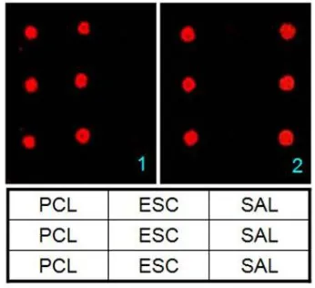
Based on the basic experiments we have tested the specific and sensitive detection and identification of E. coli and S. enterica in the background of Fish tissue homogenate. As explained in the methodology parts we have designed probes and prepared array Chips for hybridization. Microarray scan images (Fig. 2) were given along with the legend of microarray probe printing pattern.
hybridization signal and also served as left border for the array.
Figure 1. Whole genome amplification obtained from less number of bacterial cells (101 CFU/ml) (Lane 1- DNA ladder 250bp; Lane 2 – Control DNA supplied with kit; Lane 3- E.coli DNA from 101 CFU/ml; lane 4- S. enterica
DNA from 101 CFU/ml).
Figure 2. Microarray scan image showing the specific hybridization signal to detect E.coli and S. enterica bacteria. 1. E. coli partial ybgD gene as target. 2.
S. enterica partial invA gene as target. [Table: Printing pattern of Probe: PCL – Positive control; ESC – Escherichia coli probe; SAL – Salmonella enterica]
4. Discussion
Nucleic acid-based diagnostics of infectious diseases involves detection and characterization of both bacterial and viral infection using DNA/RNA methods. The four major techniques that initiated the field and constitute a platform for the development of new technology are enzymatic DNA restriction, nucleic acid hybridization, polymerase chain reaction (PCR), and fluorescence-based detection methods. There has been an enormous development within nucleic acid-based diagnostics during the last decade,
build on the increasing number of published genome sequences from pathogenic bacteria. Today the major driving forces for developing new diagnostic techniques are reduced hands on-time and faster methods, as well as increased sensitivity. These goals are reached mainly by automating processes and refining detection methods.
Detection of diseases causing bacterial pathogens from food sample is very important in the sense that to avoid completely or reduce the casualty because of food poisoning. This emphasis that there is a need of an assay protocol to sort out this problem. Perusal of literature revealed that there are many methods for different kind of pathogens and only very few methods are available for the detection and identification of E. coli and Salmonella spp. because of the taxonomically close (Enterobacteriaceae) and genetically more similar features reduce the percentage of reaction specificity rather than sensitivity. Another bottleneck is the bacterial detection or sensitivity limit of the methods. It is obvious that the protocol developed in this study has the ability to detect even low number of pathogens (101CFU/ml) in the food sample.
In the case of PCR or multiplex PCR assays the amplicon size should be distinct to tell apart the various genes target products which are not essential in microarray based detection. Because detection is based on hybridization to specific complementary sequences of probes rather than product length, time-consuming sequencing or blot-and-probe techniques are not necessary to confirm product identity. So the fragment size of the target gene part could be equal or very close in length with one and the same amplification efficiency. In this study also we have two very close in length amplicon and we did not encounter any problem in its amplification. In addition, Products of various lengths also present a challenge for developing optimal PCR conditions (primer annealing temperatures and similar MgCl2 concentrations) [20, 21]. But, the current assay is sufficient for simultaneous screening for these two pathogenic bacterial markers even though they have unequal PCR products.
actual microarray hybridization for instances like initial species identification and error in process. As far as this present study is concerned there was not even a single event of getting of nonspecific hybridization and indirectly added higher certainty value for the probes used here. In general high degree of specificity of an assay format is not prone to generating false-positives; a larger problem is that of false-negatives. False-negatives can arise due to naturally occurring sequence polymorphisms in PCR primer or probe hybridization sequenced. This is not a significant issue if all polymorphisms are known and can be included on the microarray or if relatively conserved genes are selected [22, 23]. In order to avoid completely or reduce the error level we have designed the species specific primer and probe sequences in the unique and conserved region of genes ybgD and invA.
During the execution of any hybridization assay, false-negatives can also result when hybridization buffer components or hybridization conditions is not suitable for the sample or matrix under study. In order to monitor the hybridization performance we have added an internal control DNA fragment in hybridization solution along with the sample target DNA. It hybridized perfectly with the respective positive control probe printed in both the edges of the array. These internal positive control spots guide as being the margin of the array and also exhibit the degree of hybridization process. In this study we have used another partial 16S rDNA gene fragment (173bp) as target for positive internal control. Since the primers and probes were designed in the conserved region (1353bp to 1525bp based on E. coli 16S rRNA gene) we can use the same primer sets as one of the components of multiplex PCR wherever applicable in future. This is the first microarray technique depicted for the detection of pathogenic bacteria E. coli and S. enterica on the same array for fish sample. The sensitivity and specificity of the described method and the simultaneous detection of two bacterial species make it suitable for detection of potential human pathogens in fish as well as other related food products.
References
1. Barken, K. B., Haagensen, J.,Tolker-Nielsen, T., 2007, Advances in nucleic acid-based
diagnostics of bacterial infections. Clinica Chemica Acta 384, 1–11.
2. Richardson, S. D., 2007, Anal. Chem., 79, 4295–4323.
3. Ivnitski, D., Abdel-Hamid, I., Atanasov, P., Wilkins, E., 1999, Biosens. Bioelectron., 14, 599–624.
4. Sharma, S., Sachdeva, P., Virdi, J. S., 2003, Appl. Microbiol. Biotechnol., 61, 424–428. 5. Gerba, C. P., 1996, In: Pollution Science;
Pepper, I. L., Gerba, C. P., Brusseau, M. L., Ed., Academic Press: New York, pp 279-299. 6. Weibel, D. B., DiLuzio, W. R., Whitesides
G.M., 2007. Microfabrication meets microbiology. Nat Rev Microbiol 5, 209–218. 7. Valenzuela, L., Chi, A., Beard, S., Orell, A.,
Guiliani, N., Shabanowitz, J., Hunt, D. F., Jerez, C. A., 2006, Genomics, metagenomics and proteomics in biomining microorganisms. Biotechnol Adv 24, 197–211.
8. Mashego, M. R., Rumbold, K., Mey, M.D., Vandamme, E., Soetaert, W., Heijnen, J. J., 2007 Microbial metabolomics: past, present and future methodologies. Biotechnol Lett 29, 1–16.
9. Depardieu, F., Perichon, B., Courvalin, P., 2004, Detection of the van alphabet and identification of enterococci and staphylococci at the species level by multiplex PCR. J. Clin. Microbiol. 42, 5857–5860.
10. Chamberlain, J. S., R. A. Gibbs, J. E., Ranier, P. N., Nguyen, C. T., Caskey, 1988, Deletion screening of the Duchenne muscular dystrophy locus via multiplex DNA amplification. Nucleic Acids Res. 16, 11141– 11156.
rRNA gene polymorphism to target diversity of cyanobacteria. Appl. Environ. Microbiol. 70, 7161–7172.
12. Liu, W.T., Mirzabekov, A.D., Stahl, D.A., 2001. Optimization of an oligonucleotide microchip for microbial identification studies: a nonequilibrium dissociation approach. Environ. Microbiol. 3, 619–629.
13. Loy, A., Bodrossy, L., 2006. Highly parallel microbial diagnostics using oligonucleotide microarrays. Clin. Chim. Acta 363, 106–119. 14. Sanguin, H., Herrera, A., Oger-Desfeux, C.,
Dechesne, A., Simonet, P., Navarro, E., Vogel, T.M., Moenne-Loccoz, Y., Nesme, X., Grundmann, G.L., 2006. Development and validation of a prototype 16S rRNA-based taxonomic microarray for Alphaproteobacteria. Environ. Microbiol. 8, 289–307.
15. Call, D.R., Borucki, M.K., Loge, F.J., 2003. Detection of bacterial pathogens in environmental samples using DNA microarrays. J. Microbiol. Methods 53, 235– 243.
16. Zhou, J., 2003. Microarrays for bacterial detection and microbial community analysis. Curr. Opin. Microbiol. 6, 288–294.
17. Hobson, N. S., Tothill, I., Turner, A. P. F., 1996. Microbial detection. Biosens. Bioelectron. 11, 455–477.
18. Kaspar, C. W., Tartera, C., 1990. Methods Microbiol. 22, 497–530.
19. Kohrer, K., Emr, S. D., 1993. The yeast VPS17 gene encodes a membrane-associated protein required for the sorting of soluble vacuolar hydrolases. J. Biol. Chem., 268, 559 – 569. 20. Volokhov, D., Rasooly, A., Chumakov, K.,
Chizhikov, V., 2002. Identification of Listeria species by microarray-based assay. J. Clin. Microbiol. 4, 4720 – 4728.
21. Rasooly, A., Herold, K. E., 2008. Food Microbial Pathogen Detection and Analysis Using DNA Microarray Technologies.
Foodborne Pathogens and Disease. August. 5, 531-550.
22. Chizhikov, V., Rasooly, A., Chumakov, K., Levy, D., 2001. Microarray analysis of microbial virulence factors. Appl. Environ. Microbiol. 67, 3258 – 3263.