Original Research Article
Ascertainment of risk factors and clinical course for neonates with early
onset sepsis in a tertiary care hospital, rural area, Punjab
Rashmi Kashyap
1*, Surinder Kaur
2, Aarti Sareen
3INTRODUCTION
Neonatal sepsis is the most common cause for neonatal mortality and morbidity.1 Early onset neonatal sepsis manifests during the first week of life. Three-fourth of neonatal deaths occurs in the first week and more than one-fourth occur in the first 24 hours. The aetiology is the acquisition of pathogens from the maternal genital tract in early onset neonatal sepsis, whereas late onset sepsis manifests after first week till 28 days of life has its basis either from the community or from hospital.2 Early onset
sepsis can manifest as asymptomatic bacteraemia, generalized sepsis, pneumonia or meningitis. Even the presence of one or two signs or the presence of one risk factor in the mother is given due consideration in arriving at an appropriate diagnosis and institution of appropriate and adequate treatment to the neonate. Since the presentation is varied and subtle signs are present, an efficient and vigilant action is required so as to reduce the morbidity and mortality. A sepsis screen is usually performed in the neonate with no signs or symptoms of sepsis but only the presence of risk factors. Hence, risk
ABSTRACT
Background: Neonatal sepsis is the most common cause for neonatal mortality and morbidity. Despite advances in neonatal care, the impact of neonatal sepsis remains marked in developing countries. Thus identifying the risk factors is crucial for optimizing neonatal care.
Methods: A prospective study was conducted among inborn and outborn neonates with risk factors or clinical features of Early Onset Neonatal Sepsis admitted in nursery and Neonatal intensive care unit, GSMCH, Banur, Distt Patiala, Punjab during the period from August 2014 to January 2015. Outcome variables were the association of neonatal, maternal and environmental factors with Early Onset Neonatal Sepsis (EONS). Overall clinical course in terms of survival/death, Short term outcome of those who survived based on clinical improvement and culture sensitivity report. Analysis was done using percentage, range, mean, standard deviation. Chi square test and multivariate regression analysis was done for comparison between various risk factors and EONS.
Results: A total of 85 neonates were enrolled in the study. There were 71 (83.42%) inborn neonates and 14 (16.47%) neonates were outborn. Based on Haematological System Score of >3, sepsis among the inborn neonates was present in 29(40.84%), while in outborn babies sepsis was present in 13 (92.85%) neonates which was statistically significant (p=0.000). Comparison of risk factors like birth asphyxia and unclean vaginal examination showed statistically significant difference (p=0.002; 0.002) between neonates with sepsis and those without sepsis.
Conclusions: Neonatal sepsis is a major cause of mortality and morbidity. The study concludes that birth asphyxia and unclean vaginal examination are strong risk factors for early onset neonatal sepsis.
Keywords: Newborns, Risk factors, Sepsis
1
Department of Community Medicine, Dr. Y.S Parmar Government Medical College, Nahan, Sirmour, H.P., India Department of Paediatrics, 2
GMC Patiala, Punjab, 3MMU, Mullana, Ambala, Haryana, India
Received: 17 January 2018
Accepted: 10 February 2018
*Correspondence:
Dr. Rashmi Kashyap,
E-mail: [email protected]
Copyright: © the author(s), publisher and licensee Medip Academy. This is an open-access article distributed under the terms of the Creative Commons Attribution Non-Commercial License, which permits unrestricted non-commercial use, distribution, and reproduction in any medium, provided the original work is properly cited.
factor assessment in the mother is of utmost concern as it has significant impact on the early detection of signs of sepsis. Despite recent advances in neonatal care, the impact of neonatal sepsis remain marked in developing countries. Thus identifying the risk factors is crucial for optimizing neonatal care. With this background, the present study was planned to ascertain the risk factors and clinical course of neonates with early onset sepsis in a tertiary care hospital, rural area, Punjab.
METHODS
A prospective study was conducted among inborn and outborn neonates with risk factors or clinical features of Early Onset Neonatal Sepsis admitted in nursery & Neonatal intensive care unit, GSMCH, Banur, Patiala, Punjab during the period from August 2014 to January 2015. Inclusion criteria were the inborn/outborn neonates with risk factors in mother as assessed by Risk scoring and those with clinical features suggestive of early onset neonatal sepsis. Healthy neonates with absence of risk factors in mother, neonates with age >7 days, newborn, whose parents did not give consent and newborn, whose parents not willing for follow up visits in OPD were excluded from the study. Outcome variables were the association of neonatal, maternal and environmental factors with Early Onset Neonatal Sepsis (EONS). Overall clinical course in terms of survival/death, Short term outcome of those who survived based on clinical improvement and culture sensitivity report. Approval was obtained from Ethical committee, Gian Sagar Medical College and Hospital, Banur, Patiala, Punjab. Written informed consent was obtained from the legal guardians of all the newborns. Detailed history, clinical examination and laboratory investigations were recorded in the Proforma. Maternal history was taken and risk factors were noted. Electronic weighing scale was used for measuring the weight of the baby at birth. Clinical signs and symptoms were observed and documented by the treating paediatrician. Modified Ballard’s assessment scale was used for the assessment of gestational age. All these babies were assessed as per the risk score for early onset of sepsis. Net score was assessed and the babies with presence of even one risk factor in the mother were included. For the inborn babies, if the mother was discharged early, then the baby was followed up in the OPD alternate day for a period of 7 days. Newborns who did not turn up for follow up, phone reminder was sent. Those who did not respond to three phone calls were excluded from the study. Haematological scoring was done for diagnosing risk of sepsis in the babies presenting with signs and symptoms of sepsis. Baby was admitted in the Neonatal Intensive Care Unit. Hematological scoring was done based on hematological scoring system. Babies having score ≤2 will be categorized as sepsis unlikely, score 3-4, Sepsis possible and Score ≥4, Sepsis or infection very likely.
Babies with criteria, abnormality score ≤2 (sepsis unlikely) were kept in the nursery or mother side and
observed closely for development of any signs of sepsis till 7 days of life. Newborns with hematogical score of ≤2, but strong clinical suspicion of EONS were further investigated. Blood culture and sensitivity testing was done for babies with score >3. Neonates were given injectable antibiotics till blood culture sensitivity report was available. The treatment plan was changed based upon the report of blood C/S. In case of negative blood culture report but strong clinical suspicion of sepsis, antibiotics were continued as per protocol. Newborns were assessed round the clock for the clinical course (improvement/same status/deterioration). Analysis was done using percentage, range, mean, standard deviation. Chi square test was used for comparison of various parameters and multivariate regression analysis was done for determining the association between various risk factors and EONS. P<0.05 was considered statistically significant. SPSS 22 package was used for data entry and analysis.
RESULTS
A total of 85 neonates were enrolled in the study. There were 71 (83.42%) inborn neonates and 14 (16.47%) neonates were outborn. Out of these 40 (47.05%) were males and 45 (52.94%) were females. Age wise distribution showed that 69 (81.17%) neonates were <24 hrs and 16 (18.82%) neonates were between 24 hrs to 7 days. In neonates aged <24 hrs, females constituted 44 (63.76%) and males constituted 25 (36.23%). However, in neonates aged 24 hrs to 7 days, majority 15 (93.75%) were males. Mean age of the mother was 24.80±3.53, range 20-36 years. Majority 56 (65.91%) of the mothers were in the age group of 20-35 years, 26-31 years age group constituted 24 (28.23%) and 32 years- 36 years age group constituted 5 (5.91%) of the mothers. Educational level assessment showed that 31 (36.4%) of the mothers were educated till matric and 23 (27.05%) mothers were educated till higher secondary. Illiterate women constituted 6 (7.05%) of the women. Majority 54 (63.5%) mothers were primigravida. There were seven twins 14 (16.41%) in the study population. All the twins were premature and delivered in the hospital.
Table 1: Comparison of background characteristics between between neonates with sepsis and those without sepsis.
Parameter Sepsis (n=40) No sepsis (n=45) P value
Age of mother (yrs)
20-25 23 33
0.176
26-31 13 11
32-36 4 1
Education
Illiterate 4 2
0.070
Primary 4 6
Middle 2 1
Matric 13 18
Secondary 13 10
Graduate 3 8
Postgraduate 1 0
Occupation
0.171
Professional 1 2
Skilled 1 0
Unskilled 1 0
Unemployed 37 43
Mode of delivery
Normal vaginal delivery 20 23
0.990
Forceps 2 2
LSCS 18 20
Place of delivery
Hospital 35 45
0.014
Home 5 0
Gestational age
27-30 weeks 5 2
0.062
31-34 weeks 13 22
35-38 weeks 16 20
39-42 weeks 6 1
Weight of the baby
0.771
< 1000 gms 0 1
1000-1499 gms 10 13
1500-2499 gms 22 23
> 2500 gms 8 8
H/O PIH 8 3 0.068
H/O Diabetes Mellitus 2 2 0.904
H/O UTI - 2 0.177
Anemia 15 5 0.004
Mean risk score (SD) 4.10 (1.336) 1.58 (0.782) 0.000
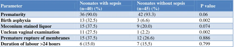
Table 2: Comparison of risk factors between neonates with sepsis and those without sepsis.*
Parameter Neonates with sepsis
(n=40) (%)
Neonates without sepsis
(n=45) (%) P value
Prematurity 36 (90.0) 42 (93.3) 0.06
Birth asphyxia 13 (32.5) 3 (6.6) 0.002
Meconium stained liquor 15 (37.5) 9 (20.0) 0.074
Unclean vaginal examination 11 (27.5) 1 (2.2) 0.002
Premature rupture of membranes 15 (37.5) 12 (26.6) 0.886
Duration of labour >24 hours 6 (15.0) 7 (15,5) 0.799
Table 3:Predictors of risk for early onset neonatal sepsis.
Regression model Predictors P value
1 Duration of labour, Unclean vaginal examination, Premature rupture of
membranes, Prematurity, Birth asphyxia 0.006
2 Duration of labour, Unclean vaginal examination, Prematurity, Birth asphyxia 0.002
3 Unclean vaginal examination, Prematurity, Birth asphyxia 0.001
4 Unclean vaginal examination, Birth asphyxia 0.000
Table 4: Comparison of clinical presentation between groups.
Clinical presentation Neonates with sepsis* (n=40) (%)
Neonates without sepsis**
(n=45) (%) P value
Decreased acceptance of feeds 5 (12.5) 3 (6.6) 0.376
Refusal for feeds 6 (15.0) 1 (2.2) 0.035
Poor weak cry 9 (22.5) 5 (11.1) 0.171
Excessive crying 1 (2.5) - 0.291
Lethargy 9 (22.5) 3 (6.6) 0.040
Yellowish discolouration of body 3 (7.5) 2 (4.4) 0.568
Vomitting - 3 (6.6) 0.097
Increased respiratory rate 28 (70.0) 12 (26.6) 0.001
Abnormal body movements 2 (5.0) - 0.133
Pustules 1 (2.5) - 0.291
Abdominal distension 1 (2.5) - 0.291
Shrill cry 2 (5.0) - 0.133
Passage of less urine 1 (2.5) 1 (2.2) 0.946
*Combination of more than one symptom was present in 37 (92.5%) of the neonates with sepsis; **No symptom was present in 15 (33.3%) of the neonates without sepsis
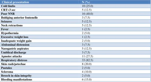
Table 5: Clinical presentation in neonates with sepsis (n=40).
Clinical presentation N (%)
Cold limbs 10 (25.0)
CRT>3 sec 5 (12.5)
Poor NNR 16 (40.0)
Buldging anterior fontenelle 3 (7.5)
Seizures 5 (12.5)
Neck retractions 5 (12.5)
Fever 1 (2.5)
Hypothermia 2 (5.0)
Excessive weight loss 1 (2.5)
Inadequate weight gain 2 (5.0)
Abdominal distension 3 (7.5)
Nasogastric aspirates 5 (12.5)
Umblical discharge 3 (7.5)
Apnoiec attacks 11 (27.5)
Respiratory distress 33 (82.5)
Skin rash/petechae 8 (20.0)
Pustules 2 (5.0)
Sclerema 4 (10.0)
Breech in skin integrity 2 (5.0)
Table 6: Outcome of the neonates in the study population.
Outcome N (%)
Improved with treatment and
discharged 35 (41.1) Observed for 7 days and discharged 42 (49.4)
Lost to follow up 3 (3.5)
Death 5 (5.8)
Male neonates constituted 23 (57.5%) while female neonates constituted 17 (42.5%) of the neonates with sepsis. Mean risk score in neonates with sepsis was 4.05 (SD 1.324) and those without sepsis was 3.23(SD 0.782) which was statistically significant (p=0.001). Based on Haematological System Score of >3, sepsis among the inborn neonates was present in 29 (40.84%), while in outborn babies sepsis was present in 13 (92.85%) neonates which was statistically significant (p=0.000). Comparison of risk factors like birth asphyxia and unclean vaginal examination showed statistically significant difference (p=0.002; 0.002) between neonates with sepsis and those without sepsis (Table 2). Backward regression model also showed birth asphyxia and unclean vaginal examination as the significant predictors for Early Onset Neonatal Sepsis (p<0.001) (Table 3). Increased respiratory rare was the commonest presentation amongst neonates; however 28 (70.0%) of the neonates with sepsis presented with increased respiratory rate as compared to 12 (26.6%) of the neonates without sepsis (p=0.001). The other two common presentations were lethargy and refusal for feeds, 9 (22.5%); 6 (15%) in neonates with sepsis and 3 (6.6%); 1 (2.2%) in neonates without sepsis (p=0.040; 0.035). Other clinical presentations were decreased acceptance of feeds, poor weak cry, excessive crying, yellowish discolouration of body, vomiting, abnormal body movements pustules, abdominal distension, shrill cry and passage of less urine. Combination of more than one symptom was present in 37 (92.5%) of the neonates with sepsis (Table 4). Among the neonates with sepsis, the most common finding on clinical examination was respiratory distress in 33 (82.5%), followed by poor neonatal reflexes, apnoeic attacks and cold limbs in 16 (40%); 12 (27.5%); 10 (25.0%) neonates with sepsis (Table 5). Among the neonates with sepsis, first line antibiotics, Ampicillin + Gentamicin were started in 30 (70%), second line antibiotics, vancomycin + piperacillin/tazobactam started in 8 (20.0%) and third line antibiotics meropenem, ciprofloxacin were administered to 2 (5%) neonates with sepsis. Antibiotics were changed from first line to second line in 8 neonates. The antibiotics were changed after 48 hours of the primary treatment. The criteria for change in treatment was the clinical deterioration or based on the blood culture sensitivity report. Overall culture sensitivity report was positive in 6 (15%) neonates; blood culture sensitivity report was positive in 4 (10%) neonates with sepsis which revealed Klebseilla pneumonia as the causative organism., umbilical stump swab culture sensitivity
showed E. coli in 2 (5%) neonates. Outcome of the neonates in the study population showed that 35 (41.1%)of the neonates were treated for sepsis and discharged with duration of the treatment ranging from 7 days to 21 days depending upon the protocol for treatment.42 (49.4%) neonates were observed for 7 days and discharged. Mortality was present in 5(5.8%) of the neonates in the study population and 3 (3.5%) neonates were lost to follow up (Table 6).
DISCUSSION
Among a total of 85 neonates, with an underlying potential maternal risk factor for sepsis 40.05% developed EOS, the findings are approximately double of the study conducted by Chacko et al which showed that 20.6% of the newborns with maternal risk factors developed EOS.1 Overall case fatality rate was 5 (5.8%). However, mortality rate in culture proved sepsis cases was 3 (60.0%). The findings of our study are comparable to study in west which reported case fatality rate of 7.6%.3 This emphasizes the provision of quality care in our institute.
Other studies from India have reported higher rates of 37-47.5%.4,5 The most common clinical features were tachypnoea in 28 (70.0%), lethargy in 9 (22.5%)and refusal to feeds in 9 (22.5%). The findings of our study are contrary to study by Sudhir et al which reported that the most common features were refusal of feeds (70%), lethargy (36%), jaundice (28%) and tachypnea (32%) with tachypnea only was significantly associated with early onset sepsis.6 Study by Jajoo et al showed common clinical presentations as lethargy/refusal to feed (77%), hypothermia (47.5%), and respiratory distress in (44%) neonates.7
Our study showed no difference in sex distribution to EONS (p=0.086).
clinical sepsis.11 The EONS occurrence had no significant difference between the infants to the gestation of <37 weeks. A study by Kilbride HW showed that the incidence of culture-proven sepsis in combination with prematurity is 4%-6%; in highly suspected sepsis, the rate is 7%-11%.12 Culture sensitivity report was positive in 6 (15%) neonates which revealed Klebsiella pneumonae as the causative organism in 4 (10%) neonates with EONS. This is similar to the report from NNPD 2000 which revealed K. pneumoniae and S. aureus to be the most frequent causative organism in India.13 In addition to bacterial blood cultures, various laboratory markers have been used for the early recognition of neonatal sepsis in clinically septic neonates.14 In our study Haematological scoring system was used for early recognition of neonatal sepsis which showed high mean score by Haematological Scoring System in neonates with early Onset Neonatal Sepsis. A study by Ghosh also concluded that higher the score by Haematological scoring system, greater the certainty of neonatal sepsis.15 Hematological scoring system is an effectual score for early diagnosis and hence management of early onset neonatal sepsis. In the current study, analysis of the different maternal and neonatal risk factors showed that these factors have an influence on the acquisition of early onset neonatal sepsis.
CONCLUSION
Neonatal sepsis is a major cause of mortality and morbidity. The study concludes that birth asphyxia and unclean vaginal examination are strong risk factors for early onset neonatal sepsis. Screening and observation of the neonates for early onset neonatal sepsis should be done in neonates presenting with such risk factors and considered for early institution of antibiotics so as to reduce the mortality and morbidity in neonates.
ACKNOWLEDGEMENTS
We are earnestly grateful to neonates and their parents for their extreme cooperation.
Funding: No funding sources Conflict of interest: None declared
Ethical approval: The study was approved by the Institutional Ethics Committee
REFERENCES
1. Chacko B, Sohi I. Early Onset Neonatal Sepsis. Indian J Pediatr. 2005;72(1):23-6.
2. Stoll BJ. Infections of the neonatal infant. In: Behrman RE, Kleigman RM, Jenson HB (eds.) Nelson textbook ofpediatrics.17th ed. Philadelphia Saunders; 2004: 623-640.
3. Clemente Yago F, Tapio collados C, Escriva Tomas P, Rubio Coriano A, Garcia Martinez R, limenez
Cobo B. Neonatal septicemia: Incidence and risk factors. An Esp Pediatr. 1992;37:481-3.
4. Tallur SS, Kasturi AV, Nadgir SD, Krishna BVS. Clinico-Bacteriological study of Neonatal Septicaemia in Hubli. Indian J Pediatr. 2000;67:169-74.
5. Leibovitz E, Flidel-Rimono O, Juster-Reicher A, Amitay M, Miskin A, Barak Y, et al. Sepsis at a Neonatal Intensive Unit. Lsr J Med Sci. 1997;33:734-8.
6. Sudhir D, Riyaz A, Reddy L, Ramesh K. Profile of Neonatal sepsis ina tertiary care hospital: A Descriptive study. Int J Curr Res Aca Rev. 2014;2(10):197-202.
7. Jajoo M, Kapoor K, Garg L, Manchanda V, Mittal SK. To study the incidence and risk factors of early onset neonatal sepsis in an outborn neonatal intensive care unit of India. J Clin Neonatol. 2015;4:91-5.
8. Shah GS, Budhathoki S, Das BK, Mandal RN. Risk factors in early neonatal sepsis. Kathmandu Univ Med J. 2006;4(2):187-91.
9. Nawshaduddin Ahmed ASM, Azad chowdhury MAK, Hoque M, Dannstadt GL. Clinical and Bactericological profile of neonatal septicemia in a tertiary level pediatric hospital in Bangladesh. Indian Pediatr. 2002;392:1034-9.
10. Shah N, Upadhyay C, Sahota R. Neonatal outcome in anemic mothers: a prospective study. J Evolution Med Dental Sci. 2013;43:8324-8.
11. Muthusami A, Devi C, Kanungo S, Shashikala RP, Srinivasan, Murmu UC, et al. Vaginal colonization as a risk factor for the development of neonatal sepsis. Biomed. 2007;27:186–8.
12. Kilbride HW, Thibeault DW. Neonatal complications of preterm premature rupture of membranes. Pathophysiology and management. Clin Perinatol. 2001;28:761.
13. Report of the National Neonatal Perinatal Database (National Neonatology Forum). 2000.
14. Ahmed Z, Ghafoor T, Waqar T, Ali S, Aziz S, Mahmud S. (2005) Diagnostic value of C-reactive protein and haematological parameters in neonatal sepsis. J Coll Physicians Surg Pak. 2005;15:152-6. 15. Ghosh S, Mittal M, Jaganathan G. Early diagnosis
of neonatal sepsis using a hematological scoring system. Indian J Med Sci. 2001;55(9):495-500.