Available Online at www.ijpret.com
555
INTERNATIONAL JOURNAL OF PURE AND
APPLIED RESEARCH IN ENGINEERING AND
TECHNOLOGY
A PATH FOR HORIZING YOUR INNOVATIVE WORK
A REVIEW ON URINE ANALYSIS USING DIGITAL IMAGE PROCESSING
SUNIL V. MUKHARE1, PROF. S. M. AGRAWAL2, PROF. M. A. KHAN3
1. M.E. (Digital Electronics), BNCOE, Pusad (M.S.) India.
2. Associate Professor, H.O.D. Department of Electrical Engineering, BNCOE, Pusad (M.S.) India.
3. Associate Professor, Department of Electronics and telecommunication, BNCOE, Pusad (M.S.) India.
Accepted Date: 15/02/2014 ; Published Date: 01/04/2014
\
Abstract: Digital image Processing is a vast field in industrial world. But today’s world is
getting highly automated and technologically more advanced, so the analysis is one of the important factor for getting the things highly protected. So we need more sophisticated and reliable methods that will be easily operated. This paper describes the different methods related to the analysis of urine for diagnosis of medical diseases using digital image processing. The images of the urine are analyzed with the help of Digital Image Processing software. Generally the urine has different attributes. The usual methods adopted by the pathologist require time, money and frequent visits of patients. Sometime’s patient may not be in position to go there and get tested, particularly in villages. Keeping that in view, this paper is thought of develop.
Keywords: Urine Analysis, Test Strip
Corresponding Author: MR. SUNIL V. MUKHARE
Access Online On:
www.ijpret.com
How to Cite This Article:
Available Online at www.ijpret.com
556 INTRODUCTION
Image Processing involves techniques and algorithms for processing the digital images. Image processing provides greater contribution to science and technology as the digital images have a greater impact on modern society. Image processing includes many techniques like segmentation, morphological operations, the different shapes, size and colors or urine constituents are obtained. Based on that the disease can be recognized and also by counting percentage changes in these attributes[3].
URINE AND UEINALYSIS:
Urine is a waste product made by the kidneys. The kidneys take waste material, minerals, fluids, and other substances from the blood to be passed in the urine.
Urine test checks different components of urine. A regular urine test may be done to find the cause of symptoms. The test can give information about health and problems.
Biochemical analysis of urine is called ‘urinalysis’ and is commonly used to diagnose a wide range of diseases. High levels of urinary glucose are symptoms of diabetics, and high levels of urinary bodies ketone is of ketonuria[5].
Physical Characteristics of Normal Urine
1. Color -Pale yellow.
Vitamin B supplements can turn urine bright yellow. Some medicines, blackberries, beets, or blood in the urine can turn red-brown.
2. Clarity
Urine is normally clear. Bacteria, blood, sperm, crystals or mucus can make urine look cloudy.
3. Odor
Urine has slightly ‘nutty’ odor.
Some diseases cause a change in the odor of urine. Diabetes or starvation can cause a sweet, fruity odor.
4. Specific gravity
Available Online at www.ijpret.com
557 5. PH
The normal range is 4.8-7.5. The PH is a measure of acidic or alkaline nature of the urine.
6. Protein
Protein is normally not found in the urine. Fever, hard exercise, pregnancy, and some diseases, especially kidney disease, may cause protein to be in the urine.
7. Glucose.
Glucose found in urine when the kidneys are damaged or diseased.
8. Nitrites.
Nitrites in urine show urinary tract infection is present.
9. Leukocyte esterase (WBC esterase).
Leukocyte esterase shows leukocytes in the urine. WBCs in the urine may mean a UTI present.
10. Ketones.
Available Online at www.ijpret.com
558 URINE TEST STRIP:
The urine test strip consist of a ribbon made of plastic or paper of about 5mm wide, plastic strips have pads impregnated with chemicals which react with the compounds present in urine producing a characteristic color. On paper strips, the reactants are directly absorbed. Paper strips are specific to a single reaction[1].
The strips with pads allow several determinations simultaneously .qualitative strips checks the sample if it is positive or negative, and quantitative strip gives quanitative result, the color reactions are approximately proportional to the concentration of substance in the sample.
The results are obtained by comparing the colors with the manufacturer’s color scale. Semi quantitative values are expressed as trace, eg 1+, 2+, 3+, 4+. These test also provides an estimate of constituent in milligrams per deciliter.
Leukocyt ee
Nitrite
Protein
PH
Spgravit y
Ketone
Available Online at www.ijpret.com
559 Diseases Identified With A Urine Test Strip:
a. Diseases of the kidneys and urinary tract :
Chronic kidney disease, Glomerulonephritis.
b. Carbohydrate Metabolism Disorders:
Glucose – Identified as Glycosuria
Ketones –Identified as Ketonuria
c. Specific Carbohydrate Metabolism Disorders:
Able to be identified Diabetes, Mellitu
d. Liver diseases and Haemolutic Disorders:
Urobilinogen- Identified as Urobilinogenuria
Bilirubin-Identified as Bilirubinuria
LITERATURE SURVEY
A. A new strategy for urinary sediment segmentation based on wavelet, morphology and
combination method
Yong-Ming Li, Xiao-Ping, presents a strategy for segmenting urinary sediment based on wavele morphology and combination method. Firstly, the wavelet transforms and morphology are used to get rid of the effect of the defocusing and we get the sub images that include the particles. Then based on the characteristics of the sub images, edge detection and adaptive thresholding are employed adaptively. Finally, a simplified watershed for the overlapping particles is used. The experiment results show that the method can segment the defocusing urinary sediment images effectively and precisely.
B. Identification by Proteomic Analysis of Calreticulin as a Marker for Bladder Cancer and
Evaluation of the Diagnostic Accuracy of Its Detection in Urine
Available Online at www.ijpret.com
560 C. Urine Cytologic Analysis: Special Techniques for Bladder Cancer Detection
Anirban P. Mitra, suggest method for Detection of bladder cancer using morphologic molecular tests. This method allows diagnosing tumors of the aggressive phenotype earlier and thus improve the prognosis of patients. In cases of less aggressive tumors, it identifies early disease on set and recurrences.
PROPOSED SYSTEM
We propose a system in which the procedure for the analysis of urine as follows.
Proposed work:
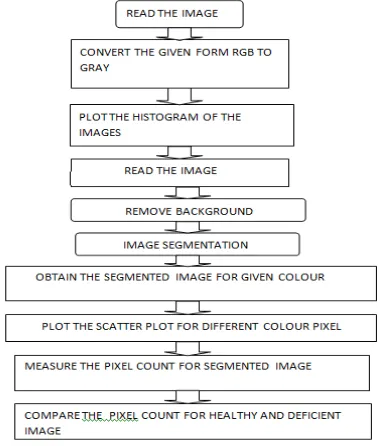
Figure 1 shows the Proposed System Architecture. It is propose to take urine image of healthy person as a reference for the comparison purpose. Then convert RGB urine image under test to gray scale, a histogram is plotted for the segmentation purpose. Then use the threshold technique to segment the region of interest (ROI) for the background of the image.
Then pixel count is measured and compared with healthy image. This count is correlated with medical reports and relation is obtained.
Available Online at www.ijpret.com
561 CONCLUSION:
The existing work is based on microscopic image of the urine. It is expected to get the results from comparing the pixel count of plain images of urine. The computer aided diagnosis of urine contents helps to the physician as well as patients. Its save the time, does not require the chemicals for analysis.
REFERENCES:
1. U. Satyanarayana, Dr. U. chakrapani, “Biochemistry” Publisher Uppala
2. A new strategy for urinary sediment segmentation based on wavelet morphology and combination method ‘Yong-Ming Li, Xiao_Ping.
3. Susmu Kageyama, Takhiro Isono et al: “Identification by Proteomic Analysis of Calreticulin as a Marker for Bladder Cancer and Evalulation of the Diagnostic Accuracy of Its Detection in Urine”,
4. Anirban P. Mitra: “Urine Cytologic Analysis: Special Techniques for Bladder Cancer Detection”,